herpes simplex virus membrane proteins ge/gi and us9 act
TRANSCRIPT

Herpes Simplex Virus Membrane Proteins gE/gI and US9 ActCooperatively To Promote Transport of Capsids and Glycoproteinsfrom Neuron Cell Bodies into Initial Axon Segments
Paul W. Howard, Tiffani L. Howard, David C. Johnson
Department of Molecular Microbiology and Immunology, Oregon Health & Science University, Portland, Oregon, USA
Herpes simplex virus (HSV) and other alphaherpesviruses must move from sites of latency in ganglia to peripheral epithelialcells. How HSV navigates in neuronal axons is not well understood. Two HSV membrane proteins, gE/gI and US9, are key to un-derstanding the processes by which viral glycoproteins, unenveloped capsids, and enveloped virions are transported towardaxon tips. Whether gE/gI and US9 function to promote the loading of viral proteins onto microtubule motors in neuron cell bod-ies or to tether viral proteins onto microtubule motors within axons is not clear. One impediment to understanding how HSVgE/gI and US9 function in axonal transport relates to observations that gE�, gI�, or US9� mutants are not absolutely blocked inaxonal transport. Mutants are significantly reduced in numbers of capsids and glycoproteins in distal axons, but there are lessextensive effects in proximal axons. We constructed HSV recombinants lacking both gE and US9 that transported no detectablecapsids and glycoproteins to distal axons and failed to spread from axon tips to adjacent cells. Live-cell imaging of a gE�/US9�
double mutant that expressed fluorescent capsids and gB demonstrated >90% diminished capsids and gB in medial axons andno evidence for decreased rates of transport, stalling, or increased retrograde transport. Instead, capsids, gB, and enveloped viri-ons failed to enter proximal axons. We concluded that gE/gI and US9 function in neuron cell bodies, in a cooperative fashion, topromote the loading of HSV capsids and vesicles containing glycoproteins and enveloped virions onto microtubule motors ortheir transport into proximal axons.
Alphaherpesviruses depend upon highly evolved mechanismsto move from mucosal epithelial tissues within neuronal ax-
ons to ganglia where latency is established. Following reactivationfrom latency, virus particles move from ganglia back to peripheraltissues for spread to other hosts. This anterograde transport in-volves fast axon transport involving microtubules and kinesinmotors that propel viral particles from neuron cell bodies (in gan-glia) over large distances to axon tips.
Depending upon the strain of alphaherpesvirus and the type ofneuron, anterograde transport can apparently involve either fullyassembled virions or unenveloped capsids (reviewed in refer-ences1, 2, and3). Fully assembled, enveloped virions or “Married”particles (4) are produced by capsid envelopment in the cytoplasmof neuron cell bodies, while “Separate” (4) unenveloped capsids(lacking viral glycoproteins) become enveloped at or near axontips. Early electron microscopy (EM) studies produced evidencefor Separate herpes simplex virus (HSV) capsids in human and ratneuronal axons (5–7). Other, more recent EM studies observed amixture of Separate capsids (25%) and Married particles for twoHSV strains (8), but this ratio was reversed, so that 70% of theparticles in axons were Separate particles with another HSV strain(T. Mettenleiter, personal communication). Our antibody stain-ing of HSV-infected human neuroblastoma cells produced evi-dence for mainly Separate capsids and distinct glycoprotein-con-taining vesicles (4, 9, 10). EM and fluorescent protein analyses ofpig pseudorabies virus (PRV) strongly support only Marriedtransport (11–14). A study involving a “two-color” HSV recom-binant expressing a fluorescent glycoprotein and capsids con-cluded that most HSV anterograde transport involved Marriedparticles (15). Using another “two-color” HSV recombinant ex-pressing fluorescent capsids and glycoproteins gB, we concludedthat a majority of capsids moving in rat superior cervical ganglion
(SCG) neurons were Separate particles (60%) (16). Thus, we be-lieve that both modes of transport are possible and, in fact, occur.
HSV and PRV express two membrane proteins, gE/gI and US9,which are key to the understanding of anterograde transport inneuronal axons (reviewed in references 2 and3). gE/gI is a het-erodimer, with both gE and gI required for function, and possessesboth substantial extracellular domains and �100-amino-acid (aa)cytoplasmic domains with acidic clusters, dileucine, and tyrosinemotifs that cause the protein to extensively localize to the trans-Golgi network (TGN) (17–20). HSV and PRV US9 proteins aretype II membrane proteins, tail anchored, with no significant ex-tracellular domains and cytoplasmic domains that also containTGN localization motifs (21–24).
We previously demonstrated that HSV gE/gI and US9 promotethe anterograde transport of both viral glycoproteins (gB and gD)and Separate capsids (4). Given that gE/gI and US9 are membraneproteins yet influence the transport of unenveloped capsids (ap-parently without membranes), we proposed the “loading hypoth-esis” (4). In this model, gE/gI and US9 localize to TGN mem-branes and promote the accumulation of other viral membraneand tegument proteins in the TGN (depicted in Fig. 1A). By thisaccumulation, gE/gI and US9 together collect viral proteins into
Received 11 September 2012 Accepted 13 October 2012
Published ahead of print 17 October 2012
Address correspondence to David C. Johnson, [email protected].
Supplemental material for this article may be found at http://dx.doi.org/10.1128/JVI.02465-12.
Copyright © 2013, American Society for Microbiology. All Rights Reserved.
doi:10.1128/JVI.02465-12
January 2013 Volume 87 Number 1 Journal of Virology p. 403– 414 jvi.asm.org 403
on February 5, 2018 by guest
http://jvi.asm.org/
Dow
nloaded from

vesicles that are subsequently loaded onto kinesin motors(Fig. 1B). In this model, the TGN serves as a platform and sortingmachinery for the assembly of transport vesicles onto kinesin mo-tors that subsequently transport in axons. To explain how gE/gIand US9, membrane proteins, might increase the axonal transportof Separate capsids, we note that gE/gI interacts extensively with
tegument proteins that are encrusted on the surfaces of capsids (2,25–28). An alternative hypothesis, which might explain how gE/gIand US9 promote the transport of HSV structural proteins, wastermed the “adaptor hypothesis.” In this model, the relativelylarge cytoplasmic domains of gE/gI and US9 promote the tether-ing of HSV glycoprotein-laden vesicles onto adaptors or kinesins,maintaining the transport along axons (depicted in Fig. 1C) (re-viewed in reference 2).
One problem in testing these two hypotheses relates to obser-vations that HSV gE-, gI-, or US9-null mutants do not exhibit atotal block in anterograde transport of capsids and glycoprotein-containing vesicles. When the numbers of capsids and glycopro-teins were quantified in distal axons, gE�, gI�, and US9� mutantsoften exhibited 5- to 10-fold reductions in the numbers of capsidsand glycoproteins in distal axons, but only infrequently were thereno distal puncta (4). Moreover, in proximal axons, the reductionsin numbers puncta associated with the loss of gE, gI, or US9 weremuch lower. Similar observations have been made with PRV gE�,gI�, and US9� mutants (29, 30). Observations of reduced trans-port of alphaherpesvirus proteins, especially in more distal axons,support the adaptor hypothesis. However, there were also de-creased numbers of capsids and glycoproteins in the most proxi-mal regions of axons, supporting the loading hypothesis.
The understanding of how HSV gE/gI and US9 function inneurons has, in some cases, been blurred because some investiga-tions have not considered that gE/gI and US9 can potentially playtwo roles in virus spread: (i) anterograde transport of virus parti-cles from neuron cell bodies to axon tips and (ii) extracellularspread of virus from the surfaces of neuronal axons to adjacentcells. For example, McGraw et al. (31) concluded that HSV US9 isdispensable for anterograde spread, while gE/gI is essential forspread. In these studies, HSV spread was measured from neuroncell bodies isolated in Campenot chambers to axon tips, followedby infection of adjacent Vero cells and measuring virus producedby these Vero cells. Two HSV US9� mutants produced 10- to100-fold less infectious virus in Vero cells after 24 h than repairedviruses, although after 48 h, the differences were less. Early timepoints better represent HSV transport in axons and spread, be-cause virus is massively amplified in Vero cells. To us, these resultsshowed that US9 is important for HSV anterograde transport andspread. Important to consider is that the results did not specifi-cally measure anterograde transport but instead represented thesum total of anterograde transport, extracellular spread from neu-rons to Vero cells, infection of Vero cells, and then spread betweenVero cells.
To try to better understand how HSV gE/gI and US9 function,whether by the adaptor versus loading model, we used two ap-proaches. First, we constructed two different HSV recombinantslacking both gE and US9 in order to test whether these proteinshad overlapping or redundant functions in axonal transport. Oneof these mutants expressed fluorescent capsids and glycoproteins.Second, we used neurons growing in microfluidic chambers (32),which allowed us to perform live-cell imaging of fluorescent cap-sids and glycoproteins produced by gE�, US9�, and gE�/US9�
mutant viruses in medial and distal axons that were isolated fromneuron cell bodies. As before, HSV mutants lacking just gE or US9transported quantitatively reduced numbers of both capsids andglycoprotein gB to distal axons. However, gE�/US9� double mu-tants transported no detectable capsids and gB puncta to distalaxons, and there was no infection of adjacent nonneuronal cells.
FIG 1 Models for how HSV gE/gI and US9 might promote anterograde axonaltransport of capsids and glycoproteins. (A) The loading hypothesis suggeststhat gE/gI and US9 accumulate in the TGN of neuron cell bodies and causeother HSV membrane proteins to accumulate there. Transport vesicles thatbud from these specific TGN membranes are loaded onto kinesins for trans-port into proximal axons. Similar vesicles containing enveloped HSV (Mar-ried) particles might also be loaded in this way. (B) The loading of capsids ontokinesin motors may similarly be affected by gE/gI and US9 accumulation in theTGN. gE/gI is known to extensively interact with tegument proteins that, inturn, interact with capsids. Thus, by causing the accumulation of capsids inTGN loading compartments, gE/gI and US9 may promote the axonal trans-port of unenveloped capsids. (C) The adaptor hypothesis functions more ex-tensively in axons rather than cell bodies. In this model, the cytoplasmic do-mains of gE/gI and US9 interact with kinesin adaptors or directly with kinesinsto tether vesicles containing other HSV glycoproteins (gD and gB) and cellularcargo (synaptophysin) onto motors during transport.
Howard et al.
404 jvi.asm.org Journal of Virology
on February 5, 2018 by guest
http://jvi.asm.org/
Dow
nloaded from
Live-cell imaging of fluorescent capsids and gB in medial andproximal axons showed that the loss of both gE and US9 substan-tially (�90%) reduced the numbers of capsids and glycoproteinsin axons. The results largely supported the loading hypothesis,although there was also some evidence supporting the adaptorhypothesis.
MATERIALS AND METHODSCells and viruses. Vero cells were grown in Dulbecco’s modified Eagle’smedium (D-MEM) containing 10% fetal bovine serum and used to prop-agate and determine the titer of HSV. F-gE/GFP and F-gE/GFP-R (a re-paired virus) (33) as well as F-US9/GFP and FUS9/GFP-R (repaired) (24)were all derived from HSV-1 strain F.
Neuronal cell cultures. Superior cervical ganglia (SCG) were dis-sected from day 18 embryos removed from pregnant Sprague-Dawley ratsand dissociated by incubation in 0.25% trypsin in Hibernate A medium(lacking calcium; Brainbits) at 37°C for 10 min. Ganglia were then incu-bated in 1% soy bean trypsin inhibitor in Hibernate A medium lacking Cafor 5 min at 37°C and then transferred into Neurobasal medium supple-mented with 2% B27 containing 50 ng/ml murine nerve growth factor(NGF) 2.5S subunit (all from Invitrogen). Ganglia were then mechani-cally dissociated by repeated passage through a fire-polished Pasteurpipette. Cells were counted and plated in microfluidic devices (Xona Mi-crofluidics) that were mounted onto glass coverslips or glass-bottomed35-mm dishes (32, 34). Glass surfaces were prepared by overnight incu-bation in poly-D-lysine (1 mg/ml) in 0.1 M Na-borate (pH 8.5). The glasswas washed twice in water, dried, and then incubated for 2 h in poly-D-lysine (30 �g/ml)–laminin (2 �g/ml) in phosphate-buffered saline (PBS).Glass was then washed twice in water and air dried. SND450 (Xona) mi-crofluidic devices were used for studies involving spread into axonal com-partments and spread assays, and �40,000 SCG neurons were plated inthe somal compartments. RD450 microfluidic devices were used for livecell-imaging in somal compartments and involved �15,000 neurons.Two days after neurons were plated into microfluidic devices, cytosinearabinoside (AraC) (2 �M) was added to the medium in somal compart-ments to kill non-nonneuronal cells, and after 2 days, the medium waschanged. Neurons were infected by adding HSV to somal compartmentsafter 6 or 7 days in culture. For experiments measuring HSV spread tononneuronal cells, 40,000 Vero cells were plated in the axonal compart-ments of microfluidic devices 24 h before infection. Human gamma glob-ulin (0.1%) (a source of HSV-neutralizing antibodies) was added to theaxonal compartments at the time of virus addition to the somal compart-ments.
Construction of HSV lacking both gE and US9 genes. An HSV-1mutant, denoted F-gE/US9/GFP, in which both the gE (US8) and US9genes were replaced with enhanced green fluorescent protein (EGFP) se-quences, was constructed. Homology arms were constructed by PCR en-compassing �1,000 bp 5= of the gE initiation codon and �1,125 bp 3= ofthe US9 stop codon. These homology arms were inserted upstream anddownstream of EGFP sequences in plasmid pEGFP-C1 (Clontech). Thisplasmid was cotransfected with DNA derived from HSV-1 F-infected cellsinto Vero cells by using the calcium phosphate technique (35). Virusesthat expressed EGFP were selected and plaque purified three times, viralDNA was sequenced, and a virus (F-gE/US9/GFP) was used in all studiesdescribed below. A repaired form of F-gE/US9/GFP was constructed bycotransfecting DNA derived from F-gE/US9/GFP viral DNA with a plas-mid (pUC18-7,8,9) containing the wild-type sequence for gI, gE, and US9.Viruses which did not express EGFP were selected and plaque purifiedthree times, and this repaired virus was denoted F-gE/US9/GFP-R.
Construction of HSV recombinants that express fluorescent capsidsand glycoprotein gB and with deletions in gE, US9, or gE and US9.HSV-1 strain F DNA in the form of a bacterial artificial chromosome(BAC) and encoding monomeric red fluorescent protein (mRFP1) fusedto the N terminus of the small capsid protein VP26 and EGFP fused afterthe signal peptide of gB was described previously (36). This BAC was a
kind gift of Greg Smith (Northwestern University Feinberg School ofMedicine, Chicago, IL) and had been used to produce an HSV recombi-nant virus, denoted GS2843, that expresses mRFP-VP26 capsids andEGFR-labeled gB (36). We inserted a kanamycin resistance gene cassette(Kanr) flanked by FLP recombination target (FRT) sites into the gE, US9,or gE and US9 coding sequences of this BAC using standard recombineer-ing techniques in bacteria, as previously described (37). Homology armswere constructed using PCR so that sequences upstream and downstreamof the gE or US9 open reading frames were fused onto Kanr sequencesusing the following primers: GGGTTGGTGCGGTGCTGTTTGTTGGGCTCCCATTTTACCCGAAGATCGGCTGCTATCCCC and AACAGGGAGGGGGCGTCGACAGCCTGGAGGGCCATCGGGGAGACAACGGCCGTGTAGCCC for �gE and ATTAAAAATCGTGAGTCACTGCGACCGCAACTTCCCACCCGGAGCTTTCTTCCGGCCTCG and TTGCGGGGTGATGGGGGGGAAGAGAGACGACAAGAAGGACGCGCGTGTCGATGCGGTCTT for �US9.
To construct the �gE �US9 double mutant, the 5= gE homology armdescribed above was used in conjunction with the 3= US9 homology arm.The resulting PCR products (containing Kanr sequences inserted into gEor US9 or both gE and US9 sequences) were electroporated into EL250bacteria harboring the GS2843 BAC. Recombinant bacteria were selectedby plating onto agar plates containing chloramphenicol and kanamycin,and DNA sequences in multiple clones were PCR amplified and se-quenced. The kanamycin cassette was collapsed in these BACs by inducingFLP recombinase into bacteria and growing bacteria on medium contain-ing arabinose, producing BACs with the gE, US9, or both gE and US9genes replaced with �50 bp of DNA containing one FRT site. Sequencesaround the gE and US9 genes were amplified by PCR and sequenced.BACs were transfected into Vero cells by using Lipofectamine 2000 (In-vitrogen). Two independent BAC clones for each virus were used to pro-duce virus stocks, denoted GS2843, GS2843�gE, GS2843�US9, andGS2843�gE�US9, and each stock was plaque purified 3 times and thencharacterized for anterograde transport.
Antibodies. Rabbit anti-VP26 antibody was kindly provided byPrashant Desai (Johns Hopkins University, Baltimore, MD). Mousemonoclonal antibody (MAb) specific for ICP4 (58S) was kindly providedby Roger Everett (University of Glasgow Centre for Virus Research, Scot-land, United Kingdom). Mouse anti-gB MAb (SS10) and anti-gD MAb(DL6) were kindly provided by Gary Cohen (University of Pennsylvania,Philadelphia, PA). Rabbit anti-US9 antipeptide polyclonal serum was pre-viously described (4). Mouse anti-gE MAb (3114) and anti-gI MAb (3104)were gifts from Howard Marsden (MRC Virology Unit, Glasgow, Scot-land, United Kingdom) and were described previously (38). Mouse anti-VP5 MAb was obtained from Virusys (North Berwick, ME). A guinea piganti-tau antibody was obtained from Synaptic Systems. Rabbit anti-extra-cellular signal-regulated kinase 1 (ERK-1) antibody was obtained fromSanta Cruz Biotechnology. DyLight fluorescent secondary antibodieswere obtained from Jackson ImmunoResearch.
Immunofluorescence microscopy of HSV-infected neurons in ax-onal chambers and counts of viral capsids and gB puncta. Microfluidicchambers containing SCG neurons and, in spread experiments, Vero cellswere disassembled and fixed in 4% paraformaldehyde for 10 min at 20°C,permeabilized in 0.1% Triton X-100 for 10 min, and incubated with pri-mary antibodies in PBS containing 0.1% Tween 20 and 2% normal don-key serum. The cells were washed in PBS containing 0.1% Tween 20 andincubated with secondary fluorescent antibodies in PBS containing 0.1%Tween 20 and 2% normal donkey serum. Microscopy was performed atthe Oregon Health and Sciences University Advanced Light MicroscopyCore using a Deltavision CoreDV Widefield Deconvolution system. Im-ages were captured with a 60� (numerical aperture, 1.42) Plan Apo Nobjective in three channels, 488 nm, 549 nm, and 649 nm. For the quan-tification of VP26 and gB puncta, 10 10,551-�m2 images per slide werecaptured, with a minimum of six 0.3-�m z sections. After deconvolution,the images were processed with the ImageJ program. For VP26 and gB, anintensity threshold of 1,500 and a minimum punctum area of 7 square
HSV gE/gI and US9 Cooperate in Axonal Transport
January 2013 Volume 87 Number 1 jvi.asm.org 405
on February 5, 2018 by guest
http://jvi.asm.org/
Dow
nloaded from
pixels (6.4 � 10�3 �m2) were chosen by comparing manual counts of gBand capsid puncta in several representative images using the softWoRxsExplorer program from Applied Precision and comparing these to theImageJ software counts (using the Analyze Particles function). Counts ofcapsid and glycoprotein puncta produced by using ImageJ software werecorrected based on the total area of axons in each axonal compartmentmeasured using tau-specific antibodies to stain axons. In other experi-ments, SCG neurons were infected with GS2843 or mutants derived fromthis recombinant virus, and fluorescent capsids were manually counted inaxonal chambers without fixation or following fixation with cold 50%acetone–50% methanol for 15 min and staining with anti-VP5 MAb.
Live-cell imaging of rat SCG axons in microfluidic chambers. Ratneurons in microfluidic chambers were infected with HSV (8 PFU/cell)for 20 to 24 h and were subjected to live-cell imaging at 37°C under 6%CO2 in an enclosed chamber using an inverted objective and the decon-volution system described previously (16). Images were acquired as opti-cal axis images in a 512-by-512 binning-of-2 format with a 60� (numer-ical aperture, 1.42) Plan Apo N objective in 2 colors, green fluorescentprotein (GFP) and mRFP. Axons or cell bodies were imaged for a total of2 min, taking exposures in both channels every 2 s. Images were decon-volved using softWoRxs Explorer software from Applied Precision.
Analyses of HSV replication in SCG neurons. SCG neurons wereprepared as described above and plated into 24-well tissue culture dishes(80,000 cells/well) coated with poly-D-lysine and laminin and then withAraC, as described above. Six days after plating, the cells were infectedwith HSV at 5 PFU/cell. After 2 h, the cells were washed once in Neuro-basal medium, and some cells and media were immediately harvested (2-htime point), while other dishes were incubated for 16 to 32 h. In each case,cells were scraped off the plastic dishes into cell culture medium, thismixture was frozen and sonicated, and virus titers were determined usingVero cells. These experiments were performed in triplicate.
Immunoblotting. For Western blot analysis, Vero cells were infectedwith HSV at 10 PFU/cell for 16 h, the cells were washed once with PBS andcollected by scraping into PBS, cells were pelleted, and pellets were sus-pended in lysis buffer containing 50 mM Tris (pH 8.0), 150 mM NaCl, 1%NP-40, and 1 mM EDTA for 10 min on ice. Insoluble debris was removedby centrifugation at 2,000 � g for 5 min; proteins were then denaturedusing 2% sodium dodecyl sulfate (SDS)–2% �-mercaptoethanol; and theproteins were separated using polyacrylamide gels and then transferredonto polyvinylidene difluoride (PVDF) membranes (Millipore). Mem-branes were blocked in a solution containing 50 mM Tris-HCl (pH 7.5),150 mM NaCl, 0.1% Tween 20, and 5% nonfat milk; washed; incubatedwith primary antibody blots; and then washed and incubated with horse-radish peroxidase-conjugated secondary antibody (Santa Cruz). A chemi-luminescent reagent was then added, as recommended by the supplier(PerkinElmer).
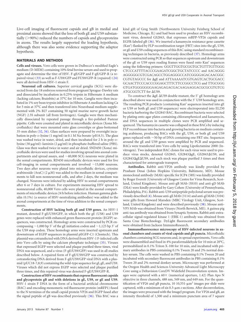
RESULTSConstruction of an HSV-1 recombinant unable to express bothgE and US9. We previously described HSV-1 strain F recombi-nants in which gE, gI, or US9 sequences were replaced with GFP(4, 24, 33). There were no major defects in the replication of theserecombinants in neurons, but the loss of gE, gI, or US9 quantita-tively reduced anterograde transport of both capsids and glyco-proteins, especially in distal axons (4). These previous studies in-volved a human neuroblastoma cell line differentiated to produceaxons or neurites of 20 to 30 �m. To further investigate the modelsdescribed in Fig. 1, we constructed a double mutant, denotedF-gE/US9/GFP, by replacing both the gE (US8) and US9 geneswith a GFP expression cassette. Note that this mutant was alsomissing the US8.5 gene (39), which has unknown functions and isnot normally considered in these types of studies. To characterizethe expression of gE/gI, gD, and US9, Vero cells were infected withmutant viruses, and cell extracts were subjected to Western blot
analysis. As expected, F-gE/GFP failed to express gE, and F-US9/GFP failed to express US9 (Fig. 2). F-gE/US9/GFP did not expressboth gE and US9. Importantly, the expression levels of both gI(encoded by the adjacent US7 gene) and gD (encoded by the US6gene) were normal in the double mutant. A repaired virus, F-gE/US9/GFP-R, was constructed by cotransfecting Vero cells withF-gE/US9/GFP viral DNA and plasmid DNA containing wild-typeUS7, US8, and US9 sequences, followed by the selection of non-fluorescent plaques. Sequencing of the US8 and US9 genes of thisrepaired virus showed the acquisition of gE and US9 sequences(not shown), and this repaired virus expressed gE and US9(Fig. 2).
We characterized the replication of these mutants in rat SCGneurons derived from day 18 embryos, as described previously(16, 30). Importantly, these neurons produce much longer axonsthan SK-N-SH neurons, and the derivation of these axons is ro-bust; i.e., the axons grow up into microfluidic chambers for amore accurate study of transport (see below). Previous studies byus (4) and others (31, 40) have indicated that HSV mutants lack-ing gE, gI, or US9 produced relatively normal quantities of infec-tious virus in neurons, although there have been small decreases inyields of virus or kinetics of virus growth. The defects did notaccount for the marked defects in anterograde transport. Figure 3shows that F-gE/GFP, F-US9/GFP, F-gE/US9/GFP, and F-gE/US9/GFP-R all produced 8 � 103 to 20 � 103 PFU/ml in thecombined cell and cell culture supernatants. It should be notedthat we visually confirmed that our neuronal cultures containedno detectable nonneuronal cells after treatment of the SCG neu-rons for 2 days with 2 �M cytosine arabinoside (AraC), conditionsthat effectively kill all or most dividing cells. In contrast, otherstudies showed higher titers of HSV (105 PFU/ml) (31), a differ-ence that appears to be related to higher proportions of nonneu-ronal cells and to the use of lower concentrations of AraC forshorter times. Nonneuronal cells produce much more virus thando neurons (data not shown). We also observed that smaller num-bers of nonneuronal cells in our cultures allowed us to administer
gB
gD
gI
gE
US9
Erk-137 -
50 -
75 -
50 -
150 -75 -
15 -
20 -
F F-gE/G
FP
F-gE/G
FP-R
F-US9/G
FP
F-gE/U
S9/GFP
unife
ct
F-gE/U
S9/GFP-R
F-US9/G
FP-R
FIG 2 Expression of gE and US9 in cells infected with HSV mutant viruses.Vero cells were infected with wild-type HSV strain F; the gE�, US9�, or gE�/US9� mutant; or repaired versions of these viruses for 16 h, and SDS-contain-ing cell extracts were then produced. These extracts were resolved on poly-acrylamide gels, proteins were transferred onto PVDF membranes, and themembranes were incubated with antibodies specific for gB, gD, gI, gE, US9, orthe cellular protein ERK-1. Membranes were washed and incubated withhorseradish peroxidase-conjugated secondary antibodies and a chemilumi-nescent reagent. Numbers indicate molecular mass markers in kilodaltons.
Howard et al.
406 jvi.asm.org Journal of Virology
on February 5, 2018 by guest
http://jvi.asm.org/
Dow
nloaded from

higher doses of HSV (10 PFU/cell), and neurons survived longer,i.e., several days (data not shown). Cultures containing more non-neuronal cells exhibited more cell death with these doses of virus.
HSV gE� and US9� mutants exhibited reduced anterogradetransport, while a gE�/US9� double mutant showed no trans-port. To assess HSV anterograde transport, we used SCG neuronscultured in microfluidic chambers, as described previously (32,34). These neuron cell bodies growing in somal compartmentsextend axons through the 450-�m microchannels connecting toaxonal compartments, where distal axons and axon tips reside.Hydrostatic pressure prevents diffusion of virus and viral antigensfrom somal chambers into axonal chambers. In every experiment,Vero cells were plated in the axonal compartments, with no neu-rons plated in somal compartments, in order to detect any possi-ble virus leakage into axonal compartments. Imaging of axons inmicrochannels was not compromised by input viral antigens, andthere was little or no chance of reinfection of axons, producingretrograde transport of viral proteins. Moreover, the structures ofthese axons extending through microchannels were highly ame-nable to live-cell imaging, allowing more accurate assessments ofHSV anterograde transport (32).
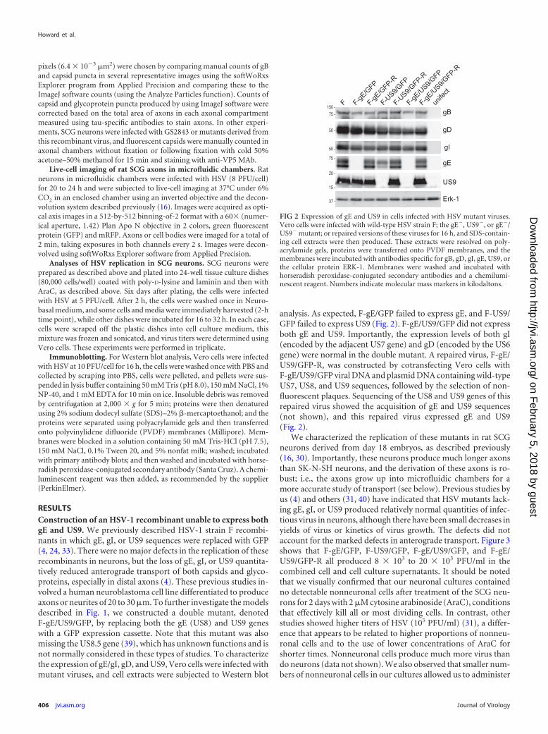
In order to characterize the culmination of the anterogradetransport process, we first enumerated the numbers of capsids andglycoproteins in distal axons found in axonal compartments. SCGneurons were infected in the somal compartments with wild-typeor mutant HSV (8 PFU/cell) and then incubated for 18 h. Distalaxons present in axonal compartments were simultaneouslystained with antibodies specific for VP26 (small capsid protein),gB, and tau (a cellular axon protein). Wild-type HSV-infectedneurons displayed numerous capsid puncta (green) and gBpuncta (red) within tau-stained axons (Fig. 4A). Axons of neuronsinfected with either the gE-null virus, F-gE/GFP, or the US9-nullmutant, F-US9/GFP, displayed reduced numbers of both capsidsand gB puncta. Note that these viruses expressed GFP (from the
gene inserted in place of the gE or US9 gene), but this GFP wasfaint in cell bodies and not detected in axons (not shown). ImageJsoftware was used to count puncta in axons, using tau staining tocontrol for the numbers of axons present. Variations in the num-bers of capsids and gB puncta were relatively large when differentmicrofluidic chambers were compared. However, the results ofthree experiments showed quantitative reductions in the numbersof capsids and gB puncta in distal axons with gE� and US9� vi-ruses, as in our previous report (4). F-gE/GFP exhibited 95%-reduced numbers of capsids and 73%-reduced numbers of gBpuncta, compared with wild-type HSV (Fig. 4A and B). F-US9/GFP exhibited 70%-reduced numbers of capsids and 46%-re-duced numbers of gB puncta. The repaired viruses F-gE/GFP-Rand F-US9/GFP-R transported 102 to 104% of the number of gBpuncta and 85 to 103% of the number of capsid puncta (Fig. 4Aand B). With wild-type HSV and repaired viruses, there was al-ways 4 to 5 times more gB puncta in terminal axons than capsids.Interestingly, the loss of gE disproportionally affected capsids, sothe ratio of gB/capsid puncta with F-gE/GFP was 20 to 25 (Fig. 4Aand B).
102
103
104
105
F F-gE/GFP
2 hr18 hr26 hr
F-US9/GFP F-gE/US9/GFP
F-gE/US9/GFP-R
titer
(pfu
/ m
l)
FIG 3 Production of infectious progeny in neurons infected with gE�, US9�,and gE�/US9� mutants. SCG neurons in 24-well dishes (�40,000 neurons perwell) were infected with wild-type HSV strain F; the gE�, US9�, or gE�/US9�
mutant; or repaired viruses at 5 PFU/cell for 2 h, and the cells were then washedonce with medium. At this time (2 h) and after 18 and 26 h, the cells werescraped into the medium and then frozen and sonicated, and viral titers weredetermined using plaque assays involving Vero cells. Each time point wasassayed in triplicate, and the data are presented with standard deviations.
10µm
F
F-gE/GFP
F-US9/GFP
F-gE/US9/GFP
A
01020304050607080 VP26
050
100150200250300350 gB
FF-
gE/G
FPF-
gE/G
FP-R
F-U
S9/
GFP
F-gE
/US
9/G
FPF-
gE/U
S9/
GFP
-R
F-U
S9/
GFP
-R
B
C
punc
ta /
field
VP26 (capsid)gB
FIG 4 Numbers of capsids and gB puncta in distal axons of SCG neuronsinfected with the gE�, US9�, or gE�/US9� mutant. SCG neurons were platedin the somal compartment of microfluidic chambers, and axons were allowedto grow into the axonal side. HSV-1 (8 PFU/cell) was introduced into somalchambers, and 18 h later, the devices were disassembled, and axons in theaxonal chambers were fixed with paraformaldehyde and simultaneously im-munostained with antibodies specific for VP26 (capsids) (one per panelmarked with a green arrow), gB (red) (one per panel marked with a red arrow),and the microtubule-associated protein tau (blue) and then with secondaryfluorescent antibodies. (A) Representative images of axons in the axonal com-partment. VP26 is stained in green, gB puncta are red, and tau is blue. Thepuncta were small, and one gB punctum (red) and one VP26 punctum areindicated by arrows. Puncta containing both VP26 and gB (Married particles)appear yellow. (B and C) The ImageJ software program was used to countcapsid and gB puncta in 10 distinct 10,551-�m2 fields of the axonal compart-ments from three separate infections. Puncta with an intensity of �1,500 anda size of 7 square pixels or greater were counted. The total area of tau stainingwas also measured and was used to correct the data for the quantities of axonspresent.
HSV gE/gI and US9 Cooperate in Axonal Transport
January 2013 Volume 87 Number 1 jvi.asm.org 407
on February 5, 2018 by guest
http://jvi.asm.org/
Dow
nloaded from
In contrast to the quantitative reductions in numbers of capsidand gB puncta with the gE- and US9-null viruses, the F-gE/US9/GFP double mutant exhibited not a single VP26 or gB punctum indistal axons in any of the three experiments described above (Fig.4A and B) or in a fourth experiment comparing just wild-typeHSV with F-gE/US9/GFP (not shown). The repaired version ofF-gE/US9/GFP, F-gE/US9/GFP-R, transported 89% of gB and80% of capsid puncta into distal axons. We concluded that gE/gIand US9 each contribute to the axonal transport of both capsidsand glycoproteins, but the loss of both gE and US9 abolishedtransport into distal axons.
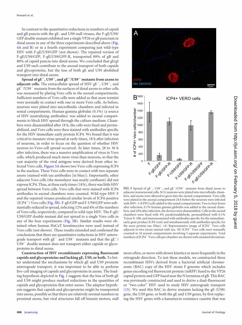
Spread of gE�, US9�, and gE�/US9� mutants from axons toadjacent cells. The extracellular spread of HSV gE�, US9�, andgE�/US9� mutants from the surfaces of distal axons to other cellswas measured by plating Vero cells in the axonal compartments.Sufficient numbers of Vero cells were added so that axon terminiwere normally in contact with one or more Vero cells. As before,neurons were plated into microfluidic chambers and infected insomal compartments. Human gamma globulin (0.1%) (a sourceof HSV-neutralizing antibodies) was added to axonal compart-ments to block HSV spread through the culture medium. Cham-bers were disassembled after 18 h, the cells were fixed and perme-abilized, and Vero cells were then stained with antibodies specificfor the HSV immediate-early protein ICP4. We found that it wascritical to measure virus spread at early times, 18 h after infectionof neurons, in order to focus on the question of whether HSVneuron-to-Vero-cell spread occurred. At later times, 28 to 36 hafter infection, there was a massive amplification of virus in Verocells, which produced much more virus than neurons, so that thevast majority of the viral antigens were derived from other in-fected Vero cells. Figure 5A shows two Vero cells expressing ICP4in the nucleus. These Vero cells were in contact with two separateaxons (stained with tau antibodies [in blue]). Importantly, otheradjacent Vero cells (the monolayer was nearly confluent) did notexpress ICP4. Thus, at these early times (18 h), there was little HSVspread between Vero cells. Vero cells that were stained with ICP4antibodies in axonal chambers were counted. Wild-type HSV Fand the repaired viruses produced similar levels of ICP4-positive(ICP4�) Vero cells (Fig. 5B). F-gE/GFP and F-US9/GFP were sub-stantially reduced in spread, infecting 12% and 4% of the numbersof Vero cells, respectively, compared to wild-type HSV. The F-gE/US9/GFP double mutant did not spread to a single Vero cells inany of the four experiments (Fig. 5B). Similar results were ob-tained when human HaCaT keratinocytes were used instead ofVero cells (not shown). These results extended and confirmed theconclusions that there are quantitative reductions in HSV antero-grade transport with gE� and US9� mutants and that the gE�/US9� double mutant does not transport either capsids or glyco-proteins to distal axons.
Construction of HSV recombinants expressing fluorescentcapsids and glycoproteins and lacking gE, US9, or both. To bet-ter understand the mechanisms by which gE and US9 promoteanterograde transport, it was important to be able to performlive-cell imaging of capsids and glycoproteins in axons. The load-ing hypothesis depicted in Fig. 1 suggests that the loss of both gEand US9 might produce marked reductions in the quantities ofcapsids and glycoproteins that enter axons. The adaptor hypoth-esis suggests that capsids and glycoproteins might be transportedinto axons, possibly so that there are relatively normal numbers inproximal axons, but viral structures fall off kinesin motors, stall
more often, or move with slower kinetics or more frequently in theretrograde direction. To test these models, we constructed threerecombinant HSVs derived from a bacterial artificial chromo-some (BAC) copy of the HSV strain F genome which includesgenes encoding red fluorescent protein (mRFP) fused to the VP26capsid protein and GFP fused near the N terminus of gB. This BACwas previously constructed and used to derive a dual-fluorescentor “two-color” HSV used to study HSV anterograde transport(15). We used this BAC to derive mutants lacking the gE (US8)gene, the US9 gene, or both the gE and US9 genes, by first replac-ing the HSV genes with a kanamycin resistance cassette that was
0
200
400
600
800
1000
1200
1400
1600
1800 ICP4+ VERO cells
F
F-gE/G
FP
F-gE/G
FP-R
F-US9/G
FP
F-gE/U
S9/GFP
F-gE/U
S9/GFP-R
F-US9/G
FP-R
# of
ICP
4+ V
ero
cells
/cha
mbe
r
B
10µm
A
ICP4tau (axons)
FIG 5 Spread of gE�, US9�, and gE�/US9� mutants from distal axons toadjacent nonneuronal cells. SCG neurons were plated into microfluidic cham-bers, and axons were allowed to grow into the axonal compartment. Vero cellswere plated in the axonal compartment 24 h before the neurons were infectedwith HSV-1 (8 PFU/cell) added to the somal compartments. Two to four hoursafter infection, 0.1% human gamma globulin was added to the axonal cham-bers, and 18 h after infection, the devices were disassembled. Cells in the axonalchambers were fixed with 4% paraformaldehyde, permeabilized with 0.1%Triton X-100, and immunostained with antibodies specific for the immediate-early gene product ICP4 (red) and simultaneously with antibodies specific forthe axon protein tau (blue). (A) Representative image of ICP4� Vero cellsadjacent to two axons stained with tau. (B) ICP4� Vero cells were manuallycounted in 10 axonal compartments involving 3 separate experiments. Totalnumbers of ICP4� Vero cells per chamber are shown with standard deviations.
Howard et al.
408 jvi.asm.org Journal of Virology
on February 5, 2018 by guest
http://jvi.asm.org/
Dow
nloaded from
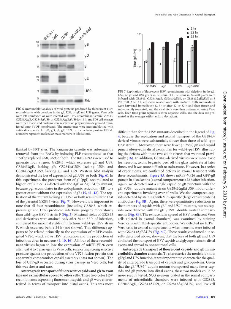
flanked by FRT sites. The kanamycin cassette was subsequentlyremoved from the BACs by inducing FLP recombinase so that�50 bp replaced US8, US9, or both. The BAC DNAs were used togenerate four viruses: GS2843, which expresses gE and US9;GS2843�gE, lacking gE; GS2843�US9, lacking US9; andGS2843�gE�US9, lacking gE and US9. Western blot analysisdemonstrated the loss of expression of gE, US9, or both (Fig. 6). Inthis experiment, the precursor form of gI (pgI) accumulated tohigher levels in cells infected with the �gE or �gE �US9 mutant,because pgI accumulates in the endoplasmic reticulum (ER) to agreater extent without the expression of gE (19, 41, 42). The rep-lication of the mutant lacking gE, US9, or both was similar to thatof the parental GS2843 virus (Fig. 7). However, it is important tonote that all four recombinants (including GS2843, which ex-presses gE and US9) produced infectious progeny more slowlythan wild-type HSV-1 strain F (Fig. 3). Maximal yields of GS2843and derivatives were attained only after 30 to 32 h of infection,compared the maximal yields of infectious wild-type HSV strainF, which occurred before 24 h (not shown). This difference ap-pears to be related primarily to the expression of mRFP-conju-gated VP26, which slows HSV replication and the production ofinfectious virus in neurons (4, 10, 16). All four of these recombi-nant viruses began to lose the expression of mRFP-VP26 evenafter just 4 to 5 passages in Vero cells, supporting strong selectivepressure against the production of the VP26 fusion protein thatapparently compromises capsid assembly (data not shown). Theloss of GFP-gB occurred during virus passage in Vero cells, butthis was slower and rare.
Anterograde transport of fluorescent capsids and gB to axontips and extracellular spread to other cells. These two-color HSVrecombinants expressing fluorescent capsids and gB were charac-terized in terms of transport into distal axons. This was more
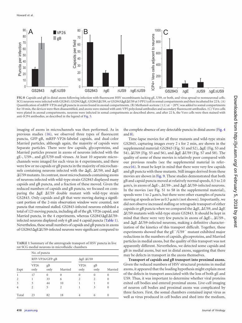
difficult than for the HSV mutants described in the legend of Fig.4, because the replication and axonal transport of the GS2843-derived viruses were substantially slower than those of wild-typeHSV strain F. Moreover, there were fewer (�25%) gB and capsidpuncta observed in distal axons than for wild-type HSV, illustrat-ing the defects with these two-color viruses that we noted previ-ously (16). In addition, GS2843-derived viruses were more toxicfor neurons, axons began to peel off the glass substrate at latertimes, and it was more difficult to stain axons. However, in a seriesof experiments, we confirmed defects in axonal transport withthese recombinants. Figure 8A shows mRFP-VP26 and GFP-gBpuncta in distal axons (axonal compartments) 22 h after infection.Again, we detected not a single capsid or gB punctum with thegE�/US9� double mutant strain GS2843�gE�US9 in four differ-ent experiments involving over 40 wells. We also compared cap-sids detected by staining with VP5-specific (large capsid protein)antibodies (Fig. 8B). Again, there were quantitative reductions inthe numbers of capsids with gE� and US9� mutants, but no cap-sids were detected with the gE�/US9� double mutant compart-ments (Fig. 8B). The extracellular spread of HSV to adjacent Verocells (plated in axonal chambers) was examined by stainingVero cells with ICP4-specific antibodies. We detected no ICP4�
Vero cells in axonal compartments when neurons were infectedwith GS2843�gE�US9 (Fig. 8C). These results confirmed our re-sults described above, showing that the loss of both gE and US9abolished the transport of HSV capsids and glycoproteins to distalaxons and spread to nonneuronal cells.
Anterograde transport of fluorescent capsids and gB in mi-crofluidic chamber channels. To characterize the models for howgE/gI and US9 function, it was important to characterize the qual-ity of anterograde transport of capsids and glycoproteins. Giventhat the gE�/US9� double mutant transported many fewer cap-sids and gB puncta into distal axons, these two models could bemore readily tested. SCG neurons plated in the somal compart-ments of microfluidic chambers were infected with GS2843,GS2843�gE, GS2843�US9, or GS2843�gE�US9, and live-cell
gB
gD
gI
gE
US9
Erk-1
GS2843
uninf
ected
50 -37 -
50 -
100 -
50 -
15 -
100 -75 -
50 -
FIG 6 Immunoblot analyses of viral proteins produced by fluorescent HSVrecombinants with deletions in the gE, US9, or gE and US9 genes. Vero cellswere left uninfected or were infected with HSV recombinant strain GS2843,GS2843�gE, GS2843�US9, or GS2843�gE�US9 for 16 h, and SDS cell extractswere then made, and proteins were resolved on polyacrylamide gels and trans-ferred onto PVDF membranes. The membranes were immunoblotted withantibodies specific for gB, gD, gI, gE, US9, or the cellular protein ERK-1.Numbers represent molecular mass markers in kilodaltons.
103
104
105
2 hr22 hr32 hr
Tite
r (pf
u/m
l)
102
FIG 7 Replication of fluorescent HSV recombinants with deletions in the gE,US9, or gE and US9 genes in neurons. SCG neurons in 24-well plates wereinfected with GS2843, GS2843�gE, GS2843�US9, or GS2843�gE�US9 at 5PFU/cell. After 2 h, cells were washed once with medium. Cells and mediumwere harvested immediately (2 h) or after 22 or 32 h and then frozen andsubsequently sonicated, and the viral titers were then determined using Verocells. Each time point represents three separate wells, and the data are pre-sented as the averages with standard deviations.
HSV gE/gI and US9 Cooperate in Axonal Transport
January 2013 Volume 87 Number 1 jvi.asm.org 409
on February 5, 2018 by guest
http://jvi.asm.org/
Dow
nloaded from
imaging of axons in microchannels was then performed. As inprevious studies (16), we observed three types of fluorescentpuncta, GFP-gB, mRFP-VP26-labeled capsids, and dual-colorMarried particles, although again, the majority of capsids wereSeparate particles. There were few capsids, glycoproteins, andMarried particles present in axons of neurons infected with thegE-, US9-, and gE/US9-null viruses. At least 10 separate micro-channels were imaged for each virus in 4 experiments, and therewere few or no capsids or gB puncta in the majority of microchan-nels containing neurons infected with the �gE, �US9, and �gE�US9 mutants. In contrast, most microchannels containing axonsof neurons infected with wild-type strain GS2843 displayed manycapsids and gB puncta, and a fraction of these moved. Given thereduced numbers of capsids and gB puncta, we focused on com-paring the �gE �US9 double mutant with wild-type strainGS2843. Only capsids and gB that were moving during a signifi-cant portion of the 2-min observation window were counted, notpuncta that remained stalled. GS2843-infected neurons exhibited atotal of 123 moving puncta, including all of the gB, VP26 capsid, andMarried puncta, in the 4 experiments, whereas GS2843�gE�US9-infected neurons displayed only 6 gB and 4 capsid puncta (Table 1).Nevertheless, these small numbers of capsids and gB puncta in axonsof GS2843�gE�US9-infected neurons were significant compared to
the complete absence of any detectable puncta in distal axons (Fig. 4and 8).
Time-lapse movies for all three mutants and wild-type strainGS2843, capturing images every 2 s for 2 min, are shown in thesupplemental material: GS2843 (Fig. S1 and S2), �gE (Fig. S3 andS4), �US9 (Fig. S5 and S6), and �gE �US9 (Fig. S7 and S8). Thequality of some of these movies is relatively poor compared withour previous results (see the supplemental material in refer-ence16). It must be kept in mind that there were very few capsidsand gB puncta with these mutants. Still images derived from thesemovies are shown in Fig. 9. These studies demonstrated that bothgB and capsid puncta moved at relatively normal speeds, 0.5 to 2.0�m/s, in axons of �gE-, �US9-, and �gE �US9-infected neurons.In the movies (see Fig. S1 to S8 in the supplemental material),speeds were 1 to 2 �m/s, but there were other examples of punctamoving at speeds as low as 0.5 �m/s (not shown). Importantly, wedid not observe increased stalling or retrograde transport of eithercapsids or gB puncta when we compared the �gE, �US9, and �gE�US9 mutants with wild-type strain GS2843. It should be kept inmind that there were very few puncta in axons of �gE-, �US9-,and �gE �US9-infected neurons, making a definitive character-ization of the kinetics of this transport difficult. Together, theseexperiments showed that the gE�/US9� mutant exhibited majorreductions in the numbers of capsids, glycoproteins, and Marriedparticles in medial axons, but the quality of this transport was notapparently different. Nevertheless, we detected some capsids andgB in medial axons, but not in distal axons, suggesting that theremay be defects in transport in the axons themselves.
Transport of capsids and gB transport into proximal axons.Given the reduced numbers of HSV structural proteins in medialaxons, it appeared that the loading hypothesis might explain mostof the defects in transport associated with the loss of both gE andUS9. Thus, it was important to determine whether viral proteinsexited cell bodies and entered proximal axons. Live-cell imagingof neuron cell bodies and proximal axons was complicated bythree factors. First, the somal chambers contained input virus aswell as virus produced in cell bodies and shed into the medium,
0
200
400
600
800
1000
ICP
4+ V
ero
cells
adj
acen
t to
neur
ons
GS2843
C
0
10
20
30
40
50
60
70
80
90VP26gB
A
GS2843
Pun
cta
per i
nfec
tion
0
10
20
30
40
50
60
70
80 B
GS2843
Cap
sids
per
fiel
d
FIG 8 Capsids and gB in distal axons following infection with fluorescent HSV recombinants lacking gE, US9, or both, and virus spread to nonneuronal cells.SCG neurons were infected with GS2843, GS2843�gE, GS2843�US9, or GS2843�gE�US9 at 5 PFU/cell in somal compartments and then incubated for 22 h. (A)Quantification of mRFP-VP26 and gB puncta in axons found in axonal compartments. (B) Methanol-acetone (1:1) at �20°C was added to somal compartmentsfor 10 min, the devices were then disassembled, and axons were stained with anti-VP5 polyclonal antibodies and secondary fluorescent antibodies. (C) Vero cellswere plated in axonal compartments, neurons were infected in somal compartments as described above, and after 22 h, the Vero cells were then stained withanti-ICP4 antibodies, as described in the legend of Fig. 5.
TABLE 1 Summary of the anterograde transport of HSV puncta in liverat SCG medial neurons in microfluidic chambers
Expt
No. of puncta
RFP-VP26/GFP-gB �gE �US9
VP26only
gBonly Married
VP26only
gBonly Married
1 17 0 0 0 0 02 1 33 6 2 2 03 3 44 10 1 1 04 2 5 2 1 3 0
Total 23 82 18 4 6 0
Howard et al.
410 jvi.asm.org Journal of Virology
on February 5, 2018 by guest
http://jvi.asm.org/
Dow
nloaded from

virus particles that stick back onto cells. Second, cell bodies pro-duce large quantities of capsid and glycoprotein fluorescence thatcan obscure the much less intense fluorescence in axons. Third,SCG neuronal axons are long, and assigning which axons are de-rived from which cell bodies was frequently difficult. For thesereasons, it was important to use live-cell analyses, to image capsidsand glycoproteins that are moving anterograde from cell bodiesinto proximal axons. Figure 10, left, shows serial images takenevery 2 s and derived from a movie (see Fig. S9 in the supplementalmaterial) of a neuron infected with wild-type strain GS2843, fo-cusing on the neuron cell body and proximal axon (defined byartificial white lines). This GS2843-infected neuron was at an ear-lier stage of virus replication, and mRFP-VP26 was largely con-centrated in the nucleus, which was intentionally cropped out ofthis image, related to intense fluorescence. A Married particle (Fig.10, white arrows) can be seen moving out of the cell body into theproximal axon. In the movie in Fig. S9 in the supplemental mate-rial, there were also several Separate capsids and gB puncta thatmoved into the proximal axon. Other axons of neurons infectedwith GS2843 showed similar transport into axons. Figure 10,right, shows a neuron infected with GS2843�gE�US9 that was inlater stages of HSV infection and exhibited more capsids in thecytoplasm than the GS2843-infected neurons shown in the leftpanels. This neuron exhibited a major axon extending from thelower right corner of the cell toward the lower right side of thepanel (defined by white lines). This axon was more difficult todiscern by fluorescence, as viral proteins did not enter the axon,but the structure of the axon was readily seen by phase microscopy(not shown). There was no movement of fluorescent capsids or gBinto the axon of this �gE �US9-infected neuron. In addition,there were no obvious stalled capsids or gB puncta in this axon.Many other axons of �gE �US9-infected neurons were character-ized, and there were very few capsids or gB puncta found in theproximal axons, although, for the reasons described above, it wasimpossible to accurately quantify this. Figure S10 in the supple-
mental material shows movies of two �gE �US9-infected neu-rons, one in an early stage of infection and one in later stages ofinfection. Again, no puncta were observed entering axons. Weconcluded that the majority of the defect in axonal transport as-sociated with the loss of both gE and US9 involves an inability totransport capsids and gB into proximal axons.
DISCUSSION
There have been extensive studies showing that both gE/gI andUS9 play important roles in HSV anterograde spread in the ner-vous systems of experimentally infected animals (24, 40, 43, 44).One important component of this spread involves anterogradeaxonal transport, a process that is entirely intracellular. Our pre-vious studies described evidence that HSV gE/gI and US9 eachcontribute quantitatively to this first step in neuronal spread (4).Specifically, gE- and gI-null viruses displayed 5- to 10-fold reduc-tions in the numbers of capsids and gB or gD puncta in more distalaxons of human SK-N-SH neuroblastoma cells by staining glyco-proteins or capsids with antibodies. Here, we used rat SCG neu-rons with much longer axons and grown in microfluidic chambersto isolate cell bodies from medial and distal axons, and detected gBand capsids with antibodies and using recombinant fluorescentproteins. Again, there were quantitative reductions in numbers ofcapsids and gB puncta in distal axons. Capsids were reduced to 5%in neurons infected with F-gE/GFP and to 29% in neurons in-fected with F-US9/GFP. gB puncta were reduced to 27% withF-gE/GFP and 46% with F-US9/GFP. A second set of mutants,�gE and �US9, derived from GS2843 that expressed fluorescentcapsids and gB produced similar observations. It was not surpris-ing that HSV spread to Vero cells (plated in axonal chambers) wasalso reduced: F-gE/GFP infected 12% of the number of cells andF-US9/GFP infected 4% of the number of Vero cells comparedwith wild-type HSV. We concluded that both gE/gI and US9 sig-nificantly contribute to the axonal transport of HSV capsids and
GS2843
VP26
gB 5 µm
FIG 9 Sequential still images of medial axons derived from live-cell analyses. Images of mRFP-VP26 capsid puncta (red) or GFP-gB puncta (green) moving inneuronal axons were derived from live-cell imaging of axons in microchannels. Neurons were infected with GS2843, GS2843�gE, GS2843�US9, orGS2843�gE�US9, and after 22 h, live-cell imaging was then performed on axons in microchannels. Still images taken 2 s apart of capsids or gB puncta are shown.
HSV gE/gI and US9 Cooperate in Axonal Transport
January 2013 Volume 87 Number 1 jvi.asm.org 411
on February 5, 2018 by guest
http://jvi.asm.org/
Dow
nloaded from

glycoproteins, and there were reductions in spread to nonneuro-nal cells.
These conclusions fit well with extensive studies involving PRVgE/gI and US9. Enquist and colleagues showed that there werequantitative reductions in the numbers of capsids and glycopro-teins, especially in more distal axons, and reduced spread fromnonneuronal cells to adjacent cells (29, 30, 45, 46). Some of thesestudies indicated that PRV US9 is relatively more important thangE/gI, in terms of anterograde transport and spread. For example,a PRV US9� mutant was reduced by 104 to 105 in the quantities ofinfectious virus produced in adjacent nonneuronal cells, com-pared with wild-type PRV, whereas a PRV gE� mutant was re-duced by 102 to 103 (30).
McGraw et al. (31) concluded that HSV US9 is not required for
anterograde spread but that HSV gE/gI is required. As noted in theintroduction, their conclusions were based on observations of vi-rus spread to Vero cells relatively late after infection (48 h), whenhigh virus titers reflect massive HSV amplification in Vero cells. Atearlier times (24 h), titers produced by US9� mutants in theseVero cells were substantially reduced (31). We measured ICP4expression in the first Vero cells that acquired HSV from neurons,showing that US9� mutants were substantially defective in spreadto Vero cells. As noted above, spread involves (i) axonal transport,followed by (ii) extracellular spread from neurons to adjacentcells. The gE/gI heterodimer possesses large extracellular domainsthat can mediate an extracellular phase of HSV spread betweenepithelial cells (47). In recent studies, we genetically separatedaxonal transport and extracellular spread in studies involving gEextracellular domain mutants (P. W. Howard, unpublished data).In contrast, US9 does not possess an extracellular domain and,thus, cannot apparently participate in extracellular spread. Thus,there are dangers associated with comparisons of US9 to gE/gIwhen spread is measured. This problem can color studies donewith experimental animals when the many steps of spread arecombined. For example, in the retinal system, there is (i) spreadwithin the retina, (ii) anterograde transport to the brain, and (iii)spread from optic neuronal axons to brain neurons, followed by(iv) spread between brain neurons.
Our observations with gE�, US9�, and gE�/US9� mutantsallowed us to address several important questions about how theseHSV membrane proteins function. First, do gE/gI and US9 act inthe same or different pathways to mediate axonal transport? Forexample, HSV gE/gI and gD function cooperatively or in a redun-dant fashion to promote secondary envelopment (33). Second,where in neurons (cell bodies versus axons) are the effects of thesemembrane proteins evident? Importantly, there were no observedHSV capsids and gB in distal axons (axonal compartments) withtwo different gE�/US9� double mutants. Moreover, these doublemutants did not spread to a single Vero cell plated adjacent tothese neuronal axons. These results parallel studies with PRV vac-cine strain Bartha that lacks both gE/gI and US9 and exhibits littleor no anterograde transport and spread (13, 29), although Barthaalso has other mutations. We concluded that HSV gE/gI and US9act cooperatively or in an overlapping fashion to promote theanterograde axonal transport of both capsids and glycoproteins.
Imaging of capsids and gB puncta in proximal axons showedthat HSV capsids and gB vesicles were rarely transported intoproximal axons. Similar observations have been made with a PRVUS9 mutant (45, 46). Together, these results largely supported theloading hypothesis. By this model, capsids and glycoproteins failto be loaded onto kinesin motors and transported into proximalaxons in the absence of both gE and US9. This fits with observa-tions that HSV gE/gI and US9 accumulate in the TGN and thatgE/gI interacts with other HSV membrane and tegument proteins(25–28). Thus, by serving to concentrate other viral proteins intoTGN-derived vesicles that serve as platforms for loading onto ki-nesin motors, gE/gI and US9 might promote transport into prox-imal axons (Fig. 1A). Capsids are also extensively coated by tegu-ment proteins, and thus, gE/gI and US9 might promote capsidloading onto motors, by interacting with these tegument proteins,as depicted in Fig. 1B. This model bears similarities with a modelthat we proposed for the gE/gI-mediated sorting of HSV particlesin polarized epithelial cells (reviewed in references 2 and48). TheTGN is also the major cytoplasmic site for the sorting of mem-
5µm5µm
VP26-RFPgB-GFP
GS2843
cell body
axon cell bodyaxon
FIG 10 Sequential images of neuron cell bodies. Neurons were infected withHSV recombinant strain GS2843 or GS2843�gE�US9. Live-cell imaging ofneuron cell bodies in somal compartments was performed, and still images(separated by 2 s) were derived from these movies. The left panels represent aneuron infected with GS2843 in an earlier stage of infection, with the majorityof RFP-VP26 remaining in the nucleus, which was intentionally cropped out ofthese images, related to intense fluorescence. A Married HSV particle is shownmoving from the neuron cell body into an axon initial segment. Note thatlive-cell imaging requires switching filters, which creates a small separation ofred and green signals with Married particles. A Separate capsid was observedmoving from this neuron cell body into the initial axon segment in Fig. S1 inthe supplemental material. The right panels show a neuron infected withGS2843�gE�US9. This neuron was in a later stage of infection, when RFP-VP26 and GFP-gB were present at higher levels in the cytoplasm. With thisneuron, there were no capsids or gB puncta that moved into axons.
Howard et al.
412 jvi.asm.org Journal of Virology
on February 5, 2018 by guest
http://jvi.asm.org/
Dow
nloaded from

brane proteins to basolateral versus apical surfaces (reviewed inreferences 49 and50). HSV gE� mutants are missorted and notdelivered to epithelial cell-cell junctions (51).
There is another mechanism that might explain the reducednumbers of HSV structural components that enter proximal ax-ons. Axon hillock and axon initial segments (AIS) can act to filtermembrane proteins, preventing transport down axons. For exam-ple, a dense actin network is formed in AIS, causing certain cellu-lar cargo molecules to be retained at AIS, while other cargo, trans-ported by different kinesins, bypass these filters (52). Our datamight be explained by effects of gE/gI and US9 working at axonhillock or AIS to promote the transport of HSV cargo into proxi-mal axons by either altering actin networks or bypassing thesefilters. Studies by Cunningham et al. (6, 7) and our studies (4, 10)showed that capsids and glycoproteins accumulate to high con-centrations in neuron cell bodies until relatively late in infection(15 to 18 h), followed by relatively abrupt transport into axons.Perhaps, the effects of gE/gI and US9 involve overcoming filters orimpediments to axonal transport.
The adaptor model (Fig. 1C) predicts that gE/gI and US9 func-tion in axons to maintain interactions between kinesin motorsand complexes of viral proteins. The relatively rare HSV punctaobserved in medial axons were transported with normal velocitiesand without increased stalling, arguing against the adaptor model.However, not a single capsid or gB punctum was observed in distalaxons despite imaging over 80 separate wells. Moreover, we didnot detect a single infected Vero cell. These observations suggestthat the few capsids and glycoproteins observed in medial axonsfailed to reach distal axons. These structures might fall off motorsor stall at microtubule transfer points or in more distal axons in amanner which we could not observe. It should be kept in mindthat the adaptor and loading models are not mutually exclusive. Ifloading onto motors in cell bodies is reduced, there might also bea reduced affinity of HSV proteins for motors within axons.
ACKNOWLEDGMENTS
We are especially indebted to Aurelie Snyder at the Advance Light Micros-copy Core at the Jungers Center, OHSU, for her extensive efforts and skillin performing deconvolution and live-cell imaging. Todd Wisner contrib-uted extensively to the design and implementation of these studies.
This work was supported by a grant from the National Institutes ofHealth, RO1 EY018755 (to D.C.J.).
REFERENCES1. Diefenbach RJ, Miranda-Saksena M, Douglas MW, Cunningham AL.
2008. Transport and egress of herpes simplex virus in neurons. Rev. Med.Virol. 18:35–51.
2. Johnson DC, Baines JD. 2011. Herpesviruses remodel host membranesfor virus egress. Nat. Rev. Microbiol. 9:382–394.
3. Kratchmarov R, Taylor MP, Enquist LW. 16 July 2012. Making the case:married versus separate models of alphaherpes virus anterograde transport inaxons. Rev. Med. Virol. [Epub ahead of print.]
4. Snyder A, Polcicova K, Johnson DC. 2008. Herpes simplex virus gE/gIand US9 proteins promote transport of both capsids and virion glycopro-teins in neuronal axons. J. Virol. 82:10613–10624.
5. Miranda-Saksena M, Armati P, Boadle RA, Holland DJ, CunninghamAL. 2000. Anterograde transport of herpes simplex virus type 1 in cul-tured, dissociated human and rat dorsal root ganglion neurons. J. Virol.74:1827–1839.
6. Penfold ME, Armati P, Cunningham AL. 1994. Axonal transport ofherpes simplex virions to epidermal cells: evidence for a specialized modeof virus transport and assembly. Proc. Natl. Acad. Sci. U. S. A. 91:6529 –6533.
7. Saksena MM, Wakisaka H, Tijono B, Boadle RA, Rixon F, Takahashi H,
Cunningham AL. 2006. Herpes simplex virus type 1 accumulation, en-velopment, and exit in growth cones and varicosities in mid-distal regionsof axons. J. Virol. 80:3592–3606.
8. Negatsch A, Granzow H, Maresch C, Klupp BG, Fuchs W, Teifke JP,Mettenleiter TC. 2010. Ultrastructural analysis of virion formation andintraaxonal transport of herpes simplex virus type 1 in primary rat neu-rons. J. Virol. 84:13031–13035.
9. Snyder A, Bruun B, Browne HM, Johnson DC. 2007. A herpes simplexvirus gD-YFP fusion glycoprotein is transported separately from viral cap-sids in neuronal axons. J. Virol. 81:8337– 8340.
10. Snyder A, Wisner TW, Johnson DC. 2006. Herpes simplex virus capsidsare transported in neuronal axons without an envelope containing theviral glycoproteins. J. Virol. 80:11165–11177.
11. Antinone SE, Smith GA. 2006. Two modes of herpesvirus trafficking inneurons: membrane acquisition directs motion. J. Virol. 80:11235–11240.
12. del Rio T, Ch’ng TH, Flood EA, Gross SP, Enquist LW. 2005. Hetero-geneity of a fluorescent tegument component in single pseudorabies virusvirions and enveloped axonal assemblies. J. Virol. 79:3903–3919.
13. Feierbach B, Bisher M, Goodhouse J, Enquist LW. 2007. In vitro analysisof transneuronal spread of an alphaherpesvirus infection in peripheralnervous system neurons. J. Virol. 81:6846 – 6857.
14. Maresch C, Granzow H, Negatsch A, Klupp BG, Fuchs W, Teifke JP,Mettenleiter TC. 2010. Ultrastructural analysis of virion formation andanterograde intraaxonal transport of the alphaherpesvirus pseudorabiesvirus in primary neurons. J. Virol. 84:5528 –5539.
15. Antinone SE, Smith GA. 2010. Retrograde axon transport of herpessimplex virus and pseudorabies virus: a live-cell comparative analysis. J.Virol. 84:1504 –1512.
16. Wisner TW, Sugimoto K, Howard PW, Kawaguchi Y, Johnson DC.2011. Anterograde transport of herpes simplex virus capsids in neurons byboth Separate and Married mechanisms. J. Virol. 85:5919 –5928.
17. Alconada A, Bauer U, Sodeik B, Hoflack B. 1999. Intracellular traffic ofherpes simplex virus glycoprotein gE: characterization of the sorting sig-nals required for its trans-Golgi network localization. J. Virol. 73:377–387.
18. McMillan TN, Johnson DC. 2001. Cytoplasmic domain of herpes sim-plex virus gE causes accumulation in the trans-Golgi network, a site ofvirus envelopment and sorting of virions to cell junctions. J. Virol. 75:1928 –1940.
19. Wisner T, Brunetti C, Dingwell K, Johnson DC. 2000. The extracellulardomain of herpes simplex virus gE is sufficient for accumulation at celljunctions but not for cell-to-cell spread. J. Virol. 74:2278 –2287.
20. Wisner TW, Johnson DC. 2004. Redistribution of cellular and herpessimplex virus proteins from the trans-Golgi network to cell junctionswithout enveloped capsids. J. Virol. 78:11519 –11535.
21. Brideau AD, Banfield BW, Enquist LW. 1998. The Us9 gene product ofpseudorabies virus, an alphaherpesvirus, is a phosphorylated, tail-anchored type II membrane protein. J. Virol. 72:4560 – 4570.
22. Brideau AD, del Rio T, Wolffe EJ, Enquist LW. 1999. Intracellulartrafficking and localization of the pseudorabies virus Us9 type II envelopeprotein to host and viral membranes. J. Virol. 73:4372– 4384.
23. Brideau AD, Eldridge MG, Enquist LW. 2000. Directional transneuronalinfection by pseudorabies virus is dependent on an acidic internalizationmotif in the Us9 cytoplasmic tail. J. Virol. 74:4549 – 4561.
24. Polcicova K, Biswas PS, Banerjee K, Wisner TW, Rouse BT, JohnsonDC. 2005. Herpes keratitis in the absence of anterograde transport of virusfrom sensory ganglia to the cornea. Proc. Natl. Acad. Sci. U. S. A. 102:11462–11467.
25. Farnsworth A, Wisner TW, Johnson DC. 2007. Cytoplasmic residues ofherpes simplex virus glycoprotein gE required for secondary envelopmentand binding of tegument proteins VP22 and UL11 to gE and gD. J. Virol.81:319 –331.
26. O’Regan KJ, Brignati MJ, Murphy MA, Bucks MA, Courtney RJ. 2010.Virion incorporation of the herpes simplex virus type 1 tegument proteinVP22 is facilitated by trans-Golgi network localization and is independentof interaction with glycoprotein E. Virology 405:176 –192.
27. Stylianou J, Maringer K, Cook R, Bernard E, Elliott G. 2009. Virionincorporation of the herpes simplex virus type 1 tegument protein VP22occurs via glycoprotein E-specific recruitment to the late secretory path-way. J. Virol. 83:5204 –5218.
28. Yeh PC, Han J, Chadha P, Meckes DGJ, Ward MD, Semmes OJ, WillsJW. 2011. Direct and specific binding of the UL16 tegument protein ofherpes simplex virus to the cytoplasmic tail of glycoprotein E. J. Virol.85:9425–9436.
HSV gE/gI and US9 Cooperate in Axonal Transport
January 2013 Volume 87 Number 1 jvi.asm.org 413
on February 5, 2018 by guest
http://jvi.asm.org/
Dow
nloaded from

29. Ch’ng TH, Enquist LW. 2005. Efficient axonal localization of alphaher-pesvirus structural proteins in cultured sympathetic neurons requires viralglycoprotein E. J. Virol. 79:8835– 8846.
30. Ch’ng TH, Enquist LW. 2005. Neuron-to-cell spread of pseudorabiesvirus in a compartmented neuronal culture system. J. Virol. 79:10875–10889.
31. McGraw HM, Awasthi S, Wojcechowskyj JA, Friedman HM. 2009.Anterograde spread of herpes simplex virus type 1 requires glycoprotein Eand glycoprotein I but not Us9. J. Virol. 83:8315– 8326.
32. Liu WW, Goodhouse J, Jeon NL, Enquist LW. 2008. A microfluidicchamber for analysis of neuron-to-cell spread and axonal transport of analpha-herpesvirus. PLoS One 3 :e2382. doi:10.1371/journal.pone.0002382.
33. Farnsworth A, Goldsmith K, Johnson DC. 2003. Herpes simplex virusglycoproteins gD and gE/gI serve essential but redundant functions duringacquisition of the virion envelope in the cytoplasm. J. Virol. 77:8481–8494.
34. Park JW, Vahidi B, Taylor AM, Rhee SW, Jeon NL. 2006. Microfluidicculture platform for neuroscience research. Nat. Protoc. 1:2128 –2136.
35. Graham FL, van der Eb AJ. 1973. Transformation of rat cells by DNA ofhuman adenovirus 5. Virology 54:536 –539.
36. Antinone SE, Zaichick SV, Smith GA. 2010. Resolving the assembly stateof herpes simplex virus during axon transport by live-cell imaging. J. Virol.84:13019 –13030.
37. Lee EC, Yu D, Martinez de Velasco J, Tessarollo L, Swing DA, CourtDL, Jenkins NA, Copeland NG. 2001. A highly efficient Escherichiacoli-based chromosome engineering system adapted for recombinogenictargeting and subcloning of BAC DNA. Genomics 73:56 – 65.
38. Johnson DC, Frame MC, Ligas MW, Cross AM, Stow ND. 1988. Herpessimplex virus immunoglobulin G Fc receptor activity depends on a com-plex of two viral glycoproteins, gE and gI. J. Virol. 62:1347–1354.
39. Georgopoulou U, Kakkanas A, Miriagou V, Michaelidou A, MavromaraP. 1995. Characterization of the US8.5 protein of herpes simplex virus.Arch. Virol. 140:2227–2241.
40. Wang F, Tang W, McGraw HM, Bennett J, Enquist LW, Friedman HM.2005. Herpes simplex virus type 1 glycoprotein E is required for axonal
localization of capsid, tegument, and membrane glycoproteins. J. Virol.79:13362–13372.
41. Dingwell KS, Johnson DC. 1998. The herpes simplex virus gE-gI complexfacilitates cell-to-cell spread and binds to components of cell junctions. J.Virol. 72:8933– 8942.
42. Hanke T, Graham FL, Lulitanond V, Johnson DC. 1990. Herpes simplexvirus IgG Fc receptors induced using recombinant adenovirus vectorsexpressing glycoproteins E and I. Virology 177:437– 444.
43. Dingwell KS, Doering LC, Johnson DC. 1995. Glycoproteins E and Ifacilitate neuron-to-neuron spread of herpes simplex virus. J. Virol. 69:7087–7098.
44. LaVail JH, Tauscher AN, Sucher A, Harrabi O, Brandimarti R. 2007.Viral regulation of the long distance axonal transport of herpes simplexvirus nucleocapsid. Neuroscience 146:974 –985.
45. Lyman MG, Feierbach B, Curanovic D, Bisher M, Enquist LW. 2007.PRV Us9 directs axonal sorting of viral capsids. J. Virol. 81:11363–11371.
46. Taylor MP, Kramer T, Lyman MG, Kratchmarov R, Enquist LW. 2012.Visualization of an alphaherpesvirus membrane protein that is essentialfor anterograde axonal spread of infection in neurons. mBio 3(2):e00063–12. doi:10.1128/mBio.00063-12.
47. Polcicova K, Goldsmith K, Rainish BL, Wisner TW, Johnson DC. 2005.The extracellular domain of herpes simplex virus gE is indispensable forefficient cell-to-cell spread: evidence for gE/gI receptors. J. Virol. 79:11990 –12001.
48. Johnson DC, Huber MT. 2002. Directed egress of animal viruses pro-motes cell-to-cell spread. J. Virol. 76:1– 8.
49. Matter K, Mellman I. 1994. Mechanisms of cell polarity: sorting andtransport in epithelial cells. Curr. Opin. Cell Biol. 6:545–554.
50. Mostov KE, Verges M, Altschuler Y. 2000. Membrane traffic in polarizedepithelial cells. Curr. Opin. Cell Biol. 12:483– 490.
51. Johnson DC, Webb M, Wisner TW, Brunetti C. 2001. Herpes simplexvirus gE/gI sorts nascent virions to epithelial cell junctions, promotingvirus spread. J. Virol. 75:821– 833.
52. Song AH, Wang D, Chen G, Li Y, Luo J, Duan S, Poo MM. 2009. Aselective filter for cytoplasmic transport at the axon initial segment. Cell136:1148 –1160.
Howard et al.
414 jvi.asm.org Journal of Virology
on February 5, 2018 by guest
http://jvi.asm.org/
Dow
nloaded from