hhs public access elliot s. weisenberg, md howard ozer, md, ph.d cell lymphocytosis ... ·...
TRANSCRIPT
Symptomatic Massive Splenomegaly in Persistent Polyclonal B-cell Lymphocytosis Requiring Splenectomy
Shanel B. Bhagwandin, DO1,*, Elliot S. Weisenberg, MD2, Howard Ozer, MD, Ph.D3, and Ajay V. Maker, MD1
1Department of Surgery, University of Illinois at Chicago Medical Center, Chicago, Illinois
2Department of Pathology, University of Illinois at Chicago Medical Center, Chicago, Illinois
3Section of Hematology and Oncology, Department of Medicine, University of Illinois at Chicago Medical Center, Chicago, Illinois
Abstract
Introduction—Persistent polyclonal B-cell lymphocytosis (PPBL) is a rare lymphoproliferative
hematological disease characterized by binucleated lymphocytes, CD 19+ CD 5−lymphocytosis,
and elevated levels of serum immunoglobulin M (IgM). It can rarely be associated with
splenomegaly, though the disease usually remains indolent.
Case Presentation—We present a case of PPBL in a young man with massive splenomegaly
that mimicked isolated splenic lymphoma requiring splenectomy for persistent pain, symptoms,
and diagnosis.
Discussion—Determining the etiology of splenomegaly in these patients is often confounding
due to a lack of a tissue diagnosis and the limited morphological and immuno-histochemical
features of PPBL, therefore, the presentation remains highly concerning for lymphoma.
Conclusion—The presentation, surgical treatment, tissue and peripheral blood molecular
analysis, and flow cytometry integral to managing these patients and to prevent an assumptive and
misleading diagnosis are reviewed.
Background
Persistent polyclonal B-cell lymphocytosis (PPBL) is an extremely rare, lymphoproliferative
hematological disease characterized by atypical binucleated lymphocytes on peripheral
blood smear. Lymphocytosis is typically CD 19+ and CD 5− with predominance of
polyclonal serum immunoglobulin M (IgM) by immunohistochemistry and gene
rearrangement, respectively. The total lymphocyte count is not always elevated, but the
Content published in the journal follows Creative Commons Attribution License (http://creativecommons.org/licenses/by/4.0).*Shanel B Bhagwandin, Department of Surgery, University of Illinois at Chicago, 840 S. Wood M/C 958, Chicago, IL 60612, USA, Phone: 312-996-6765; [email protected].
Author’s ContributionsS.B. Bhagwandin performed a literature search for supporting references and wrote the paper. A.V. Maker edited and reviewed the paper. E.S. Weisenberg provided the pathological review of the diagnosis. H. Ozer served as an expert consultant with regards to the diagnosis.
HHS Public AccessAuthor manuscriptOpen J Clin Med Case Rep. Author manuscript; available in PMC 2015 December 10.
Published in final edited form as:Open J Clin Med Case Rep. 2015 ; 1(3): .
Author M
anuscriptA
uthor Manuscript
Author M
anuscriptA
uthor Manuscript
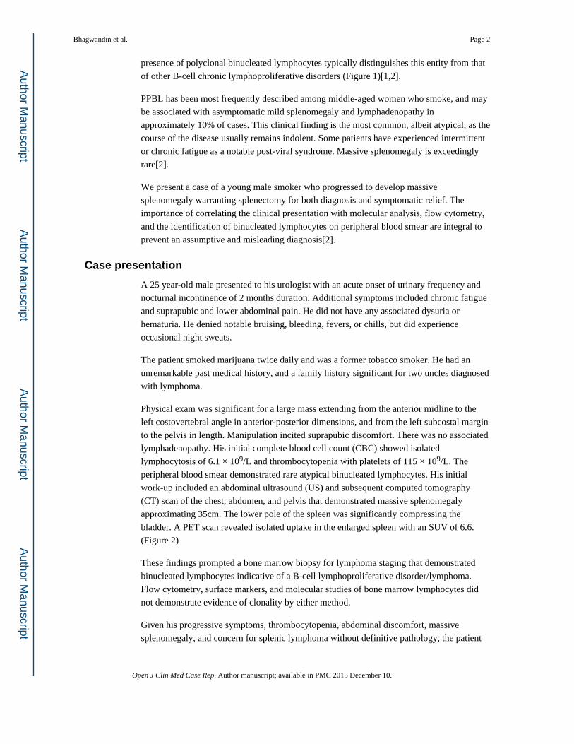
presence of polyclonal binucleated lymphocytes typically distinguishes this entity from that
of other B-cell chronic lymphoproliferative disorders (Figure 1)[1,2].
PPBL has been most frequently described among middle-aged women who smoke, and may
be associated with asymptomatic mild splenomegaly and lymphadenopathy in
approximately 10% of cases. This clinical finding is the most common, albeit atypical, as the
course of the disease usually remains indolent. Some patients have experienced intermittent
or chronic fatigue as a notable post-viral syndrome. Massive splenomegaly is exceedingly
rare[2].
We present a case of a young male smoker who progressed to develop massive
splenomegaly warranting splenectomy for both diagnosis and symptomatic relief. The
importance of correlating the clinical presentation with molecular analysis, flow cytometry,
and the identification of binucleated lymphocytes on peripheral blood smear are integral to
prevent an assumptive and misleading diagnosis[2].
Case presentation
A 25 year-old male presented to his urologist with an acute onset of urinary frequency and
nocturnal incontinence of 2 months duration. Additional symptoms included chronic fatigue
and suprapubic and lower abdominal pain. He did not have any associated dysuria or
hematuria. He denied notable bruising, bleeding, fevers, or chills, but did experience
occasional night sweats.
The patient smoked marijuana twice daily and was a former tobacco smoker. He had an
unremarkable past medical history, and a family history significant for two uncles diagnosed
with lymphoma.
Physical exam was significant for a large mass extending from the anterior midline to the
left costovertebral angle in anterior-posterior dimensions, and from the left subcostal margin
to the pelvis in length. Manipulation incited suprapubic discomfort. There was no associated
lymphadenopathy. His initial complete blood cell count (CBC) showed isolated
lymphocytosis of 6.1 × 10⁹/L and thrombocytopenia with platelets of 115 × 109/L. The
peripheral blood smear demonstrated rare atypical binucleated lymphocytes. His initial
work-up included an abdominal ultrasound (US) and subsequent computed tomography
(CT) scan of the chest, abdomen, and pelvis that demonstrated massive splenomegaly
approximating 35cm. The lower pole of the spleen was significantly compressing the
bladder. A PET scan revealed isolated uptake in the enlarged spleen with an SUV of 6.6.
(Figure 2)
These findings prompted a bone marrow biopsy for lymphoma staging that demonstrated
binucleated lymphocytes indicative of a B-cell lymphoproliferative disorder/lymphoma.
Flow cytometry, surface markers, and molecular studies of bone marrow lymphocytes did
not demonstrate evidence of clonality by either method.
Given his progressive symptoms, thrombocytopenia, abdominal discomfort, massive
splenomegaly, and concern for splenic lymphoma without definitive pathology, the patient
Bhagwandin et al. Page 2
Open J Clin Med Case Rep. Author manuscript; available in PMC 2015 December 10.
Author M
anuscriptA
uthor Manuscript
Author M
anuscriptA
uthor Manuscript

underwent a splenectomy for diagnostic and therapeutic purposes following scheduled
meningococcal, pneumococcal, and H. influenza vaccinations. He had an uncomplicated
post-operative course with resolution of his fatigue, pain, and urinary continence.
Results
Operative Details
Though most splenectomies are performed laparoscopically in our surgical unit, we
determined that a spleen of this size and weight would be better approached through an open
resection. The splenic artery was isolated and ligated to allow auto transfusion and drainage
of blood volume through the splenic vein to shrink the heavy and difficult to manipulate
spleen. After drainage of significant blood volume and resulting size decrease, the splenic
vein was divided along. The splenic ligaments were stretched and distorted due to the
increased size of the organ, and were sequentially divided. Blood loss was minimal. The
excised specimen weighed 4082 gm and measured 36.4×15.3×10.6 cm after auto transfusion
(Figures 3, 4). The patient was discharged on post-operative day 3 without any peri or post-
operative complications.
Histopathology analysis
There was prominent white pulp expansion of B lymphocytes in the germinal centers,
mantle zones, and marginal zones extending into the red pulp. These cells were not clonal by
flow cytometry or immunoglobulin gene rearrangement studies. A panel of immunostains
was negative for CD5, CD19, BCL 2, and HHV 8; as was an in situ hybridization study for
Epstein Barr virus. Regional lymph nodes revealed follicular hyperplasia, a sinus
histiocytosis pattern, and reactive germinal centers. Conventional cytogenetics revealed
normal karyotype and negative gene rearrangements. The i(3q) chromosomal abnormality
was negative and HLA-DR7 was not detected.
Discussion
Persistent and absolute lymphocytosis is attributed to chronic lymphoproliferative disorders
of B cells that include chronic lymphocytic leukemia (CLL), prolymphocytic leukemia
(PLL), splenic lymphoma, hairy cell leukemia (HCL), and mantle cell lymphoma (MCL).
These dyscrasias are predominantly the result of progressive expansion of an abnormal
clonal population of B lymphocytes in the blood, bone marrow, or tissues. All cells within
the clonal population display the same unique Ig gene rearrangement and demonstrate a
consistent chromosomal abnormality[3].
In 1982, a persistent polyclonal lymphocytosis of binucleated B lymphocytes was first
reported in three women by Gordon et al. The disorder is most commonly diagnosed by the
presence of binucleated lymphocytes as well as an increased polyclonal serum IgM level. It
was theorized that the process was reactive to the abnormal stimulation of B-cells that was
inherent among individuals with an underlying genetic predisposition. This study identified
the human leukocyte antigen HLA-DR7 in all 3 patients. Through molecular and
immunological analysis, this HLA haplotype has been identified among 26% of the
Caucasian population. Troussard et al. later described an additional isochromosome for the
Bhagwandin et al. Page 3
Open J Clin Med Case Rep. Author manuscript; available in PMC 2015 December 10.
Author M
anuscriptA
uthor Manuscript
Author M
anuscriptA
uthor Manuscript

long arm of chromosome 3, +i(3q), in 6 cases and premature chromosome condensation
(PCC) in all 7 cases of their series. Additionally, reports of familial PPBL and a case of
PPBL occurring among monozygotic twins have also suggested this strong genetic
predisposition[3,7].
In a series of 25 patients with PPBL, 77% were found to have a +i(3q) recurrent
chromosomal abnormality, whereas in the largest reported series of 111 patients, only 34%
of patients harbored this mutation. Furthermore, i(3q) is observed exclusively in non-
binucleated cells, which may account for its low accuracy as a tumor marker considering
that the population of lymphocytes may be primarily replaced by binucleated cells at the
time of diagnosis[2,10].
Although the etiology of PPBL has not been elucidated, an association with tobacco
smoking in the development of the polyclonal lymphocytosis has been considered. In the
largest series of 111 patients, 98% were smokers and two of the patients had a decrease of
lymphocytosis and the number of binucleated lymphocytes after smoking cessation.
Isochromosome +i(3q), however, did persist throughout the 2 years follow-up after tobacco
use[2,10].
Splenomegaly is rare in patients with PPBL though there are reports of progressive spleen
enlargement up to six years following diagnosis of PPBL. A series of 5 patients with
splenomegaly with PPBL presented by Del Giudice et al. found that 3 of those patients had
similar immunophenotypes of peripheral lymphocytes including expression of BCL-2 and
IgH rearrangements. They noted HLA-DR7 in all five cases. These patients had an
otherwise indolent clinical presentation and course of disease progression[2].
The long-term follow-up of 111 patients with typical PPBL revealed that 89% of patients
were symptom free after a median follow-up of 4.4 years. Two patients developed lung
cancer, one of whom died 9 years after diagnosis. Three additional cases of non-Hodgkins
Lymphoma (NHL) were observed as well as one case of cervical cancer, however, none of
the patients in the series developed significant splenomegaly warranting splenectomy[10].
Considering the long-term follow-up and event-free survival in these patients, any
aggressive medical or surgical intervention should be limited once a diagnosis has been
made. Awareness of PPBL, a high index of suspicion, and skepticism of a malignant
diagnosis of a B-cell lymphoproliferative disorder in the absence of immunophenotypic or
molecular evidence of clonality, splenectomy may be necessary to render the correct
diagnosis. The appropriate management of patients with PPBL relies on differentiating these
patients from those with other chronic lymphoproliferative B-cell disorders, secondary
malignancies, or lymphoma; and as a result close follow-up is recommended.
Conclusions
The PPBL patient reported here had an indolent course, but presented due to worsening
symptoms of massive splenomegaly causing a mass effect on surrounding organs. This case
supports past reports of PPBL associated with smoking and splenomegaly, though growth to
this size and weight appears exceedingly rare. Splenic resection was critical to establish the
Bhagwandin et al. Page 4
Open J Clin Med Case Rep. Author manuscript; available in PMC 2015 December 10.
Author M
anuscriptA
uthor Manuscript
Author M
anuscriptA
uthor Manuscript

correct diagnosis, and this case highlights that PPBL can present to the surgeon only with
binucleated lymphocytes and massive splenomegaly without other hematologic genetic
abnormities or being HLA-DR7+.
Acknowledgements
We would also like to acknowledge Dr. John Anastasi of the University of Chicago for his consultative diagnosis.
References
1. Tonelli S, Petronilla V, Sacchi S, et al. Persisistent polyclonal B lymphocytosis: morphological, immunological, cytogenetic and molecular analysis of an Italian case. Leuk Res. 2010; 24(10):877–879. [PubMed: 10996207]
2. Del Giudice I, Pileri SA, Rossi M, et al. Histopathological and molecular features of persistent polyclonal B-cell lymphocytosis (PPBL) with progressive splenomegaly. Br J Haematol. 2009; 144(5):726–731. [PubMed: 19133977]
3. Sun P, Juskevicius R. Histological and immunohistochemical features of the spleen in persistent polyclonal B-cell lymphocytosis closely mimic splenic B-cell lymphoma. Diagnostic Pathology. 2012; 7:107. [PubMed: 22901769]
4. Agrawal S, Matutes E, Voke J, et al. Persistent polyclonal B-cell lymphocytosis. Leuk Res. 1994; 18:791–795. [PubMed: 7934138]
5. Gordon DS, Jones BM, Browing SW, et al. Persistent polyclonal lymphocytosis of B-lymphocytes. NE Journal of Medicine. 1982; 307:232–236.
6. Troussard X, Cornet E, Lesesve JF, et al. Polyclonal B-cell lymphocytosis with binucleated lymphocytes (PPBL). OncoTargets and therapy. 2007; 1:59–66. [PubMed: 21127753]
7. Mossafa H, Malaure H, Maynadie M, et al. Persistent polyclonal B lymphocytosis with binucleated lymphocytes: a study of 25 cases. British Journal of Haematology. 1999; 104:486–493. [PubMed: 10086784]
8. Himmelmann A, Ruegg R, Fehr J. Familial persistent polyclonal B-cell lymphocytosis. Leuk Lymphoma. 2001; 41(1–2):157–160. [PubMed: 11342368]
9. Carr R, Fishlock K, Matutes E. Persistent polyclonal B-cell lymphocytosis in identical twins. Br J Haematol. 1997; 96(2):272–274. [PubMed: 9029012]
10. Cornet E, Lesesve JF, Mossafa H, et al. Long-term follow-up of 111 patients with persistent polyclonal B-cell lymphocytosis with binucleated lymphocytes. Leukemia. 2009; 23(2):419–422. [PubMed: 18668130]
11. Carstairs KC, Francombe WH, Scott JG, et al. Polyclonal lymphocytosis of B-lymphocytes induced by cigarette smoking? Lancet. 1985; ii:1094. [PubMed: 2860302]
Bhagwandin et al. Page 5
Open J Clin Med Case Rep. Author manuscript; available in PMC 2015 December 10.
Author M
anuscriptA
uthor Manuscript
Author M
anuscriptA
uthor Manuscript

Keypoints
Persistent polyclonal B-cell lymphocytosis (PPBL) is a rare lymphoproliferative
hematological disease characterized by binucleated lymphocytes.
Massive splenomegaly mimicked isolated splenic lymphoma in this patient requiring
splenectomy for persistent pain, symptoms, and diagnosis.
Bhagwandin et al. Page 6
Open J Clin Med Case Rep. Author manuscript; available in PMC 2015 December 10.
Author M
anuscriptA
uthor Manuscript
Author M
anuscriptA
uthor Manuscript
Figure 1. Blood smear comparing normal mononuclear lymphocytes (a) and binucleated lymphocytes
(b) found in our patient with splenomegaly.
Bhagwandin et al. Page 7
Open J Clin Med Case Rep. Author manuscript; available in PMC 2015 December 10.
Author M
anuscriptA
uthor Manuscript
Author M
anuscriptA
uthor Manuscript

Figure 2. CT and PET imaging reveal massive splenomegaly.
Bhagwandin et al. Page 8
Open J Clin Med Case Rep. Author manuscript; available in PMC 2015 December 10.
Author M
anuscriptA
uthor Manuscript
Author M
anuscriptA
uthor Manuscript

Figure 3. Intraoperative image demonstrating delivery of the enlarged spleen from the patient's
abdomen after division of the splenic hilum and vascular ligation.
Bhagwandin et al. Page 9
Open J Clin Med Case Rep. Author manuscript; available in PMC 2015 December 10.
Author M
anuscriptA
uthor Manuscript
Author M
anuscriptA
uthor Manuscript
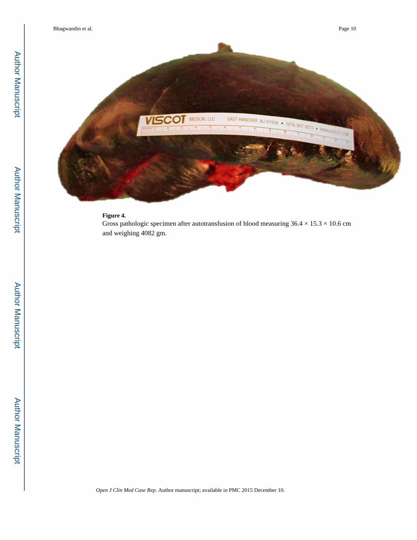
Figure 4. Gross pathologic specimen after autotransfusion of blood measuring 36.4 × 15.3 × 10.6 cm
and weighing 4082 gm.
Bhagwandin et al. Page 10
Open J Clin Med Case Rep. Author manuscript; available in PMC 2015 December 10.
Author M
anuscriptA
uthor Manuscript
Author M
anuscriptA
uthor Manuscript