hiatal hernia repair - department of surgery at · pdf filehiatal hernia repair irina kovatch,...
TRANSCRIPT

HIATAL HERNIA REPAIR
Irina Kovatch, PGY4Richmond University Medical Center
Morbidity and MortalityFebruary 17th, 2011
www.downstatesurgery.org

Case Presentation• 60 yo M with c/o vague abdominal
pain, left abdominal pulling x 1 year• Diagnosed with Type III hiatal hernia on
CT Abdomen• PMH: HTN• PSH: umbilical hernia repair• Meds: atenolol, norvasc, diovan, HCTZ• NKDA• Plan: elective laparoscopic repair
www.downstatesurgery.org

OR - Laparoscopy
• Large hiatal hernia containing stomach and omentum
• Contents reduced• Dissection of the sac with Harmonic scalpel• Primary repair of hiatal defect • Distal esophageal perforation with 50 Fr
bougie during Nissen fundoplication
www.downstatesurgery.org

OR - Open
• 3.5 cm intra-abdominal distal esophageal perforation
• Primary repair with interrupted 3-0 vicryl• Nissen fundoplication• Omentopexy• JP over repair• Pt left OR intubated to SICU
www.downstatesurgery.org

POD 1• Extubated in AM• TPN started, OOB to chair• In PM desaturation to 79% • CTA
• acute PE involving right main pulmonary artery
• fluid collection at the left lung base 14 x 8 cm
• Heparin drip
www.downstatesurgery.org

www.downstatesurgery.org

www.downstatesurgery.org

POD 2-7• Respiratory failure -> intubated• Daily fevers to 101.7, WBC 14• Cultures – NGTD• IV Abx• Wounds clean, dry, intact• Abdomen soft, ND• JP – white fibrinous output
www.downstatesurgery.org

POD 8
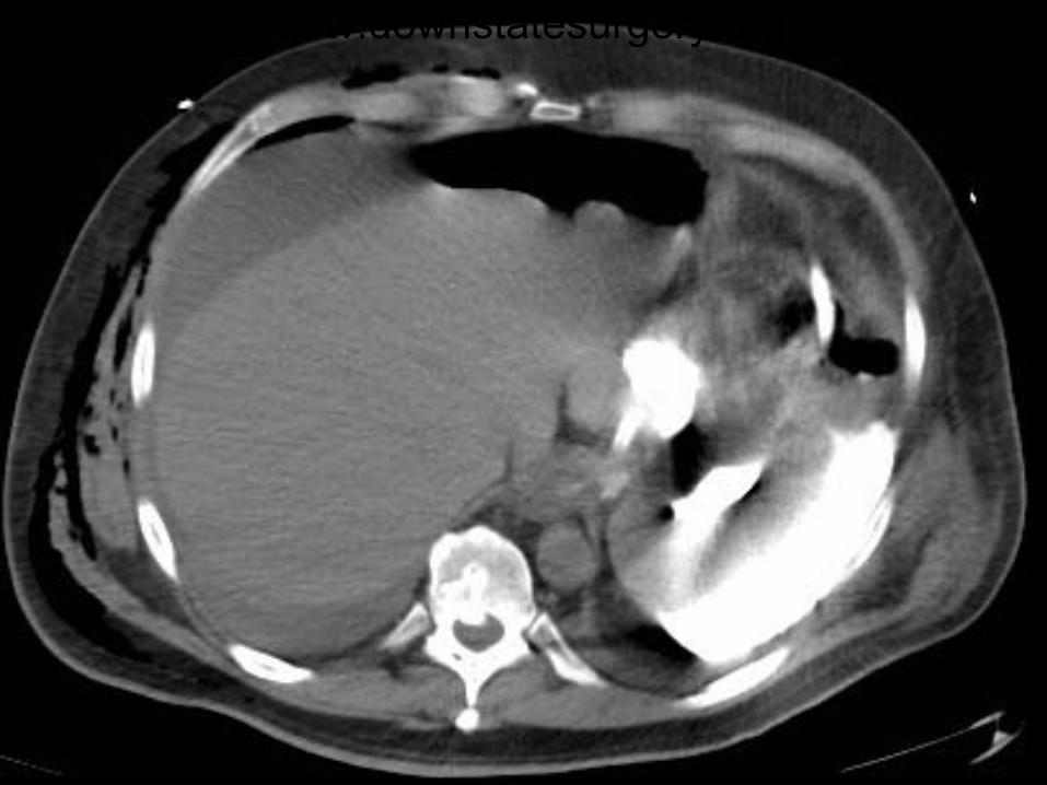
• Tm 103, WBC 35• Esophagram & CT w/contrast
– prominent extravasation at GE junction– Abdominal & pelvic ascites– 13 x 6 cm fluid collection in the site of
prior hernia– LLL atelectasis
www.downstatesurgery.org

www.downstatesurgery.org
www.downstatesurgery.org

OR• Ex-lap• Multiple intra-abdominal collections• Splenic capsule tear during mobilization
requiring transfusion• Multiple adhesions, unable to find tear• Drains: 2 sumps & 2 pediatric chest tubes• J-tube placement• Permacol mesh and 2 subq JPs
www.downstatesurgery.org

POD 10-16
• Unable to wean off the vent• L chest tube – serosanguinous fluid• Tube feeds • Continued fevers up to 102• WBC decreased to 15-20• Pt on broad spectrum IV Abx
www.downstatesurgery.org

POD 17
• CT Chest/Abd/Pelvis– Indeterminate findings for leak– Decrease in ascites and left
subphrenic collection– Residual hernia sac decrease in size
to 10 x 5 cm
www.downstatesurgery.org

www.downstatesurgery.org

www.downstatesurgery.org

POD 18-33
• POD 18 – bronchoscopy, BAL of LLL, percutaneous tracheostomy– edematous airway, large mucus
plug in LUL bronchus• POD 24 – CT Chest/Abd/Pelvis
– no leak, decrease in size of left subphrenic collection
• Continued fevers up to POD 33
www.downstatesurgery.org
POD 34-62
• Clears, trach collar, all drains out• POD 48 – UGIS: outpouching of distal
esophagus w/o extravasation• POD 56 - CT Chest/Abd/Pelvis – neg• Abx stopped, soft diet, decanulated• Transferred to rehab (prealbumin 20)
www.downstatesurgery.org

Questionswww.downstatesurgery.org

Hiatal Hernia• Etiology unknown• Pathophysiology - attenuation of the
phrenoesophageal membrane • Acquired weakness of tissue as a result of
aging or excess strain on the diaphragm• More common in
– older individuals– obese – patients with delayed gastric emptying
www.downstatesurgery.org
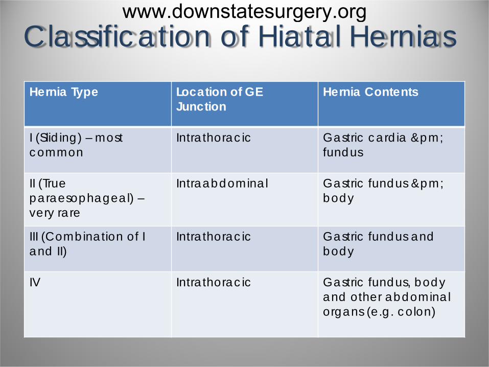
Classification of Hiatal HerniasHernia Type Location of GE
JunctionHernia Contents
I (Sliding) – most common
Intrathoracic Gastric cardia ±fundus
II (True paraesophageal) –very rare
Intraabdominal Gastric fundus ±body
III (Combination of I and II)
Intrathoracic Gastric fundus and body
IV Intrathoracic Gastric fundus, body and other abdominal organs (e.g. colon)
www.downstatesurgery.org

Hiatal Hernia Typeswww.downstatesurgery.org

Symptoms and Presentation• Most type I and III hiatal hernias are
diagnosed incidentally (UGIS or EGD) • Type II hernias often found on CXR (air-
fluid level in the chest) • Symptoms
– Type I: GERD – Type II/III: - epigastric pain,
postprandial fullness in the chest, dysphagia, respiratory problems
www.downstatesurgery.org

Acute Symptoms• Due to complete obstruction/strangulation
of the stomach within the chest• Type II hernias are at increased risk• Borchardt's triad is indicative of an
incarcerated hernia – chest pain– retching with inability to vomit– inability to pass NG tube
www.downstatesurgery.org

Pre-op Evaluation• EGD - to assess distal esophagus and
stomach for concomitant pathology• Studies difficult to acquire and interpret
for most type II and III hernias – motility studies– 24hr pH monitoring– gastric emptying studies
• CT Chest and Abdomen is not necessary for straightforward cases
www.downstatesurgery.org
Management
• Hiatal hernia is a mechanical abnormality - there is no non-operative treatment
• Presence of a sliding hiatal hernia alone does not mandate intervention
• Symptomatic patients with a type I hernia may be best served with an operative repair
www.downstatesurgery.org

Management• Traditional recommendation
– all patients with type II or III hernias should undergo surgical repair regardless of symptoms
– based on a report in 1967 documenting a mortality rate of 30% (6/21) in patients with paraesophageal hernia
www.downstatesurgery.org

Management• More recent recommendation
– elective repair in symptomatic and only select asymptomatic patients
– based on series of 23 patients with a paraesophageal hernia followed for a median of 78 months• no life-threatening complications • symptoms remained unchanged in
83% of these patients
www.downstatesurgery.org

Operative Objectives• Return the herniated content to its
anatomic position below the diaphragm• Resect hernia sac• Establish adequate esophageal length• Return GEJ to an intra-abdominal position• Repair hernia defect• Prevent recurrence while minimizing
associated morbidity
www.downstatesurgery.org

Operative Technique
• Laparoscopic– Transabdominal (currently most
common approach)– Transthoracic
• Open– Transabdominal– Transthoracic
www.downstatesurgery.org

Antireflux Procedure• Most sliding-type hernias are repaired
on the basis of symptoms• Most patients with type II hernias do
not have reflux symptoms • Many patients with type II hernias give
a history of GERD symptoms that spontaneously abated
• 30% of patients without GERD preoperatively will have GERD unmasked after hiatal hernia repair
www.downstatesurgery.org

www.downstatesurgery.org

Complications of HiatalHerniorrhaphy
• Intra-op complications– Perforation (esophagus, stomach)– Pneumothorax– Vagus nerve injury– Hemorrhage (splenic laceration, short
gastric vessels)
www.downstatesurgery.org

Complications of Hiatal Herniorrhaphy
• Post-op complications– Perforation (stricture, suture placement)– Dysphagia (mechanical vs. edema)– Early anatomic recurrence– Cardiac tamponade– Chylothorax– Pleural effusion
www.downstatesurgery.org
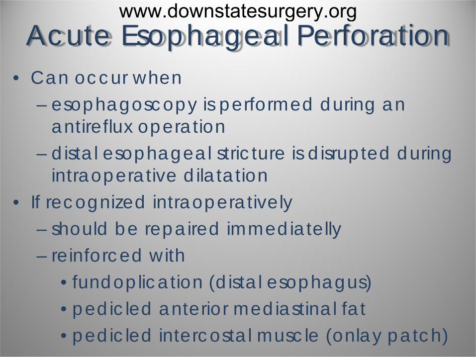
Acute Esophageal Perforation• Can occur when
– esophagoscopy is performed during an antireflux operation
– distal esophageal stricture is disrupted during intraoperative dilatation
• If recognized intraoperatively– should be repaired immediatelly– reinforced with
• fundoplication (distal esophagus)• pedicled anterior mediastinal fat • pedicled intercostal muscle (onlay patch)
www.downstatesurgery.org

Acute Esophageal Perforation• If involved tissues are not amenable to repair
(e.g. reflux stricture)– transthoracic esophagectomy with cervical
esophagogastric anastomosis– Thal fundic patch esophagoplasty
• uses gastric fundus as a “patch” • relies on healing of the opened, inflamed
distal esophagus • high incidence of suture line disruption
and mechanical complications
www.downstatesurgery.org

Delayed Esophageal Perforation
• May occur when esophageal sutures placed too deeply result in local mural necrosis
• 1st week post-op: fever, chest pain, or respiratory distress -> contrast study
• If perforation is diagnosed -> reoperation • Site of perforation is identified intraop (may
insufflate air through NGT)• Leak from fundoplication suture may be
closed and reinforced with omentum
www.downstatesurgery.org

Delayed Esophageal Perforation
• If leak is in the chest -> transthoracic approach– closure reinforced with a pedicled anterior
mediastinal fat, intercostal muscle or pleura• Jejunostomy feeding tube should be placed• Chest tube should be left near the thoracic
esophageal repair • Drain should be placed near transabdominally
repaired fundoplication
www.downstatesurgery.org

Esophageal Perforation Repair• Dissection of muscle to expose mucosal tear • Reapproximation of mucosa and submucosa• Reapproximation of the muscle • Optional limited esophagomyotomy 180
degrees opposite the site of injury • Reinforcement with a parietal pleura,
pedicled intercostal muscle flap, omentum, pericardium, visceral pleura or diaphragm
www.downstatesurgery.org

www.downstatesurgery.org

www.downstatesurgery.org

Ann Thorac Surg 2004;77:1475–83Evolving Options in the Management of Esophageal PerforationBrinster CJ, Singhal S, Lee L, et al
• Literature review of case series (559 patients) between 1990 and 2003 - 59% iatrogenic
• Surgical options– Primary or reinforced primary closure– Esophageal resection– Drainge alone– T-tube drainage– Exclusion and diversion
• Nonoperative management
www.downstatesurgery.org

Ann Thorac Surg 2004;77:1475–83Evolving Options in the Management of Esophageal PerforationBrinster CJ, Singhal S, Lee L, et al
• Mortality Rates– Operative Management (322 pts)
• Primary repair - 12%• Esophageal resection - 17%• Exclusion and diversion - 24%• Drainage alone - 37%
• Nonoperative Management (152 pts) – 18%
www.downstatesurgery.org

www.downstatesurgery.org

J Am Coll Surg Vol. 199, No. 6, December 2004: 991-3Complete Esophageal Diversion:A Simplified, Easily Reversible TechniqueKoniaris LG, Spector SA, Staveley-O’Carroll KF
• Propose simplified technique for complete esophageal diversion
• Report of 5 patients w/4 years follow-up• No leak, no stricture • Complete esophageal diversion may result in
leak or late stricture after reconstruction• Standard loop esophagostomies do not
provide complete diversion
www.downstatesurgery.org

www.downstatesurgery.org

www.downstatesurgery.org

www.downstatesurgery.org

www.downstatesurgery.org

J Thorac Cardiovasc Surg. 2007 Feb;133(2):333-8.Postoperative esophageal leak management with the Polyflex esophageal stent. Freeman RK, Ascioti AJ, Wozniak TC
• 21 patients with post-op esophageal leak treated with 27 stents– Esophagectomy (5)– Esophageal perforation repair (5)– Surgical antireflux procedure (4)– Endoscopic antireflux procedure (2)– Esophageal diverticulectomy (3)– Esophageal myotomy (2)
• Mean interval between surgical intervention and stent placement was 12 ± 8 days
www.downstatesurgery.org
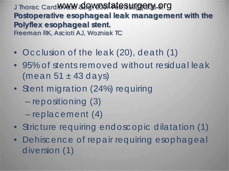
J Thorac Cardiovasc Surg. 2007 Feb;133(2):333-8.Postoperative esophageal leak management with the Polyflex esophageal stent. Freeman RK, Ascioti AJ, Wozniak TC
• Occlusion of the leak (20), death (1)• 95% of stents removed without residual leak
(mean 51 ± 43 days)• Stent migration (24%) requiring
– repositioning (3)– replacement (4)
• Stricture requiring endoscopic dilatation (1)• Dehiscence of repair requiring esophageal
diversion (1)
www.downstatesurgery.org