high-fidelity medical visualizationvis.lbl.gov/~jmeyer/papers/pp-9.pdf · high-fidelity medical...
TRANSCRIPT
HIGH-FIDELITY MEDICAL VISUALIZATION
Joerg Meyer
University of California, Irvine Biomedical Engineering Department
644E Engineering Tower Irvine, CA 92697-2625
[email protected] – (949) 824-9321
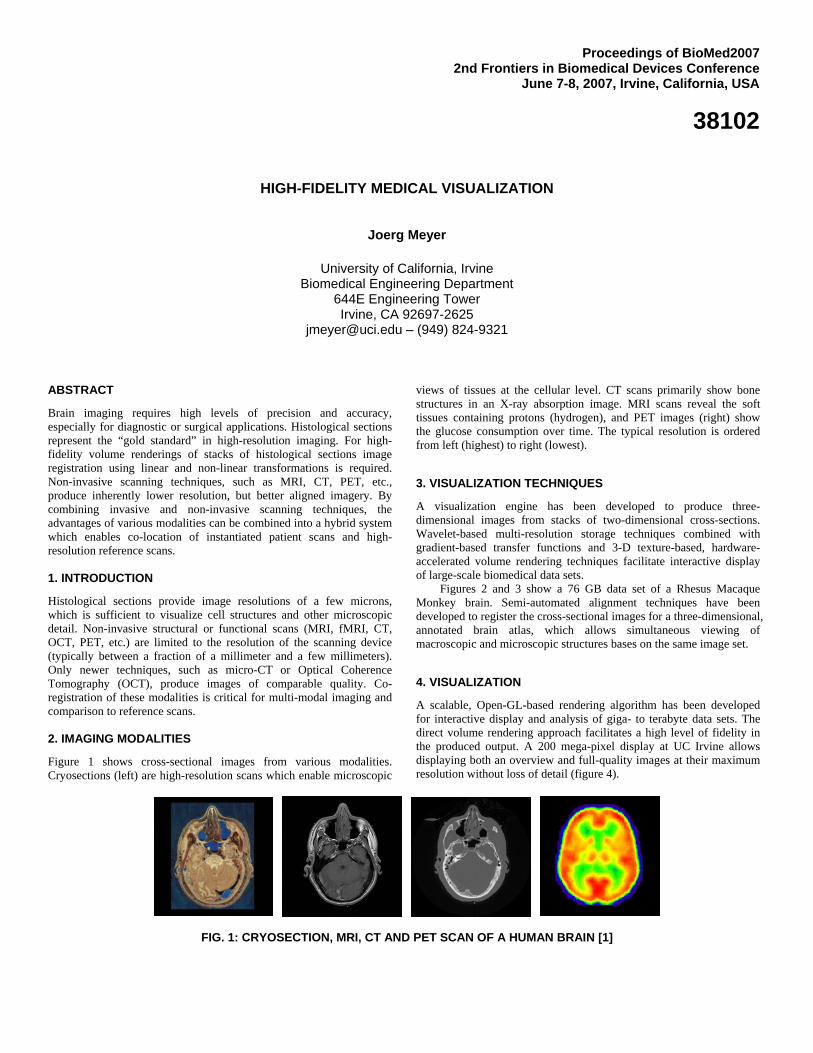
ABSTRACT Brain imaging requires high levels of precision and accuracy, especially for diagnostic or surgical applications. Histological sections represent the “gold standard” in high-resolution imaging. For high-fidelity volume renderings of stacks of histological sections image registration using linear and non-linear transformations is required. Non-invasive scanning techniques, such as MRI, CT, PET, etc., produce inherently lower resolution, but better aligned imagery. By combining invasive and non-invasive scanning techniques, the advantages of various modalities can be combined into a hybrid system which enables co-location of instantiated patient scans and high-resolution reference scans. 1. INTRODUCTION Histological sections provide image resolutions of a few microns, which is sufficient to visualize cell structures and other microscopic detail. Non-invasive structural or functional scans (MRI, fMRI, CT, OCT, PET, etc.) are limited to the resolution of the scanning device (typically between a fraction of a millimeter and a few millimeters). Only newer techniques, such as micro-CT or Optical Coherence Tomography (OCT), produce images of comparable quality. Co-registration of these modalities is critical for multi-modal imaging and comparison to reference scans. 2. IMAGING MODALITIES Figure 1 shows cross-sectional images from various modalities. Cryosections (left) are high-resolution scans which enable microscopic
views of tissues at the cellular level. CT scans primarily show bone structures in an X-ray absorption image. MRI scans reveal the soft tissues containing protons (hydrogen), and PET images (right) show the glucose consumption over time. The typical resolution is ordered from left (highest) to right (lowest).
3. VISUALIZATION TECHNIQUES A visualization engine has been developed to produce three-dimensional images from stacks of two-dimensional cross-sections. Wavelet-based multi-resolution storage techniques combined with gradient-based transfer functions and 3-D texture-based, hardware-accelerated volume rendering techniques facilitate interactive display of large-scale biomedical data sets.
Figures 2 and 3 show a 76 GB data set of a Rhesus Macaque Monkey brain. Semi-automated alignment techniques have been developed to register the cross-sectional images for a three-dimensional, annotated brain atlas, which allows simultaneous viewing of macroscopic and microscopic structures bases on the same image set.
4. VISUALIZATION A scalable, Open-GL-based rendering algorithm has been developed for interactive display and analysis of giga- to terabyte data sets. The direct volume rendering approach facilitates a high level of fidelity in the produced output. A 200 mega-pixel display at UC Irvine allows displaying both an overview and full-quality images at their maximum resolution without loss of detail (figure 4).
FIG. 1: CRYOSECTION, MRI, CT AND PET SCAN OF A HUMAN BRAIN [1]
Proceedings of BioMed20072nd Frontiers in Biomedical Devices Conference
June 7-8, 2007, Irvine, California, USA
38102
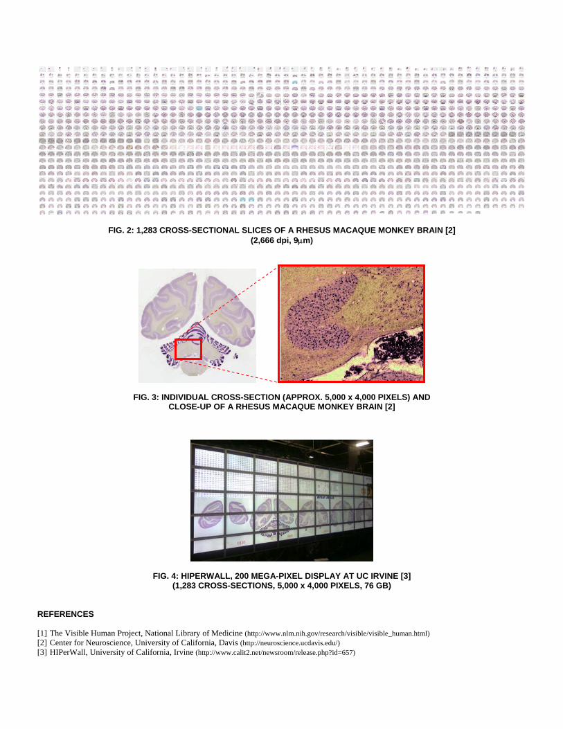
FIG. 2: 1,283 CROSS-SECTIONAL SLICES OF A RHESUS MACAQUE MONKEY BRAIN [2] (2,666 dpi, 9μm)
FIG. 3: INDIVIDUAL CROSS-SECTION (APPROX. 5,000 x 4,000 PIXELS) AND CLOSE-UP OF A RHESUS MACAQUE MONKEY BRAIN [2]
FIG. 4: HIPERWALL, 200 MEGA-PIXEL DISPLAY AT UC IRVINE [3] (1,283 CROSS-SECTIONS, 5,000 x 4,000 PIXELS, 76 GB)
REFERENCES [1] The Visible Human Project, National Library of Medicine (http://www.nlm.nih.gov/research/visible/visible_human.html) [2] Center for Neuroscience, University of California, Davis (http://neuroscience.ucdavis.edu/) [3] HIPerWall, University of California, Irvine (http://www.calit2.net/newsroom/release.php?id=657)