high-resolution solid-state carbon-13 nmr study of free and metal-complexed macrocyclic antibiotic...
TRANSCRIPT

7696 Biochemistry 1985, 24, 7696-7702
High-Resolution Solid-state 3C NMR Study of Free and Metal-Complexed Macrocyclic Antibiotic Ionophores Valinomycin, Nonactin, and Tetranactin:
Conformational Elucidation in Solid and Solution by Conformation-Dependent I3C Chemical Shifts
Ryoko Tabeta and Hazime Saita* Biophysics Division, National Cancer Center Research Institute, Tsukiji 5-chome, Chuo- ku, Tokyo 104, Japan
Received May 9. 1985
ABSTRACT: W e recorded high-resolution I3C N M R spectra of the macrocyclic antibiotic ionophores Val- inomycin, nonactin, and tetranactin in the solid state by the cross-polarization-magic angle spinning (CP-MAS) method, in order to gain insight into the use of conformation-dependent 13C chemical shifts as a convenient means to delineate a conformational change induced by metal ion complexation. The I3C peak splittings in the solid state are consistent with the symmetry properties of the ionophores as revealed by X-ray diffraction: C, symmetry in free tetranactin and S4 or S6 symmetry for a variety of metal complexes of nonactin and tetranactin or the K+ complex of valinomycin, respectively. Interestingly, many of the 13C N M R peaks of carbons in the skeletal backbones were significantly displaced (up to 8 ppm). The dis- placements of the peaks were explained by a conformational change as characterized by variations of torsion angles. Accordingly, we were able to obtain conformational features of Na' and Cs+ complexes of vali- nomycin, for which X-ray diffraction data are unavailable, on the basis of the displacements of the 13C N M R peaks. Further, we discuss conformational features of these complexes in chloroform solution, with reference to those observed in the solid state.
I t is well-known that the ionic permeabilities of alkali cations in biological membranes and lipid bilayers are strongly en- hanced by certain macrocyclic antibiotics such as valinomycin, enniatin, nonactin, etc. (Mueller & Rudin, 1967; Szabo et al., 1969; Ovchinnikov & Ivanov, 1974; Pressman, 1976). They possess high ionic selectivities depending upon a variety of molecular structures of antibiotics. Valinomycin and nonactin (Figure l ) , for instance, have a higher specific transport of K+ ion to a significant extent than Na+ ion (Mueller & Rudin, 1967; Ovchinnikov & Ivanov, 1974). A mode of action of these antibiotics can be interpreted in terms of mobile carriers (Pressman, 1976) in which the most important step is com- plexation of metal ions at the interface and the interior of membranes. Accordingly, a number of works have been di- rected to reveal conformational features of free and complexed antibiotics and the stability of complexes in solution, in order to obtain a clue in the understanding of such an ion selectivity. In particular, a nuclear magnetic resonance (NMR) study of valinomycin and its complexes with a variety of ions (Haynes et al., 1969; Ivanov et al., 1969; Ohnishi & Urry, 1969; Ohnishi et al., 1972; Grell et al., 1973; Pate1 & Tonelli, 1973; Davis & Tosteson, 1975; Bystrov et al., 1977) has shown that the conformation of ligand molecules plays an important role in accounting for the ionic selectivity. Interestingly, the con- formation of the K+ complex in nonpolar solvents turned out to be very similar to that of the solid state (Pinkerton et al., 1969; Neupert-Laves & Dobler, 1975), but the conformation of free valinomycin is solvent-dependent (Bystrov et al., 1977) and different from that of the solid state (Karle, 1975; Smith et al., 1975). By contrast, IH and I3C NMR studies (Prestegard & Chan, 1969, 1970; Pretsch et al., 1972) showed that the conformation and stability of the nonactin-K+ com- plex are almost identical with those of the Na+ complex, consistent with the data in the solid state by X-ray diffraction studies (Kilbourn et al., 1967; Dobler et al., 1969; Dobler, 1972; Dobler & Phizackerley, 1974).
To account for such an ion selectivity, it is essential to know
0006-2960/85/0424-7696$01.50/0
the conformations of ligand molecules in these metal complexes in the solid and, more preferably, in the membrane-bound state. No appropriate means, however, is available that is suitable for analyzing conformations of molecules bound to membranes. As a potential means for this purpose, we pre- viously showed that the conformation-dependent I3C chemical shifts, which are significantly varied with particular confor- mations, can be conveniently utilized as intrinsic probes of conformational characterization of a variety of molecules, (Taki et al., 1981; SaitB et al., 1981-1985). In fact, the I3C chemical shifts of individual amino acid residues in poly- peptides and proteins in the solid state are displaced by as large as 2-7 ppm, depending on the various types of conformations (Taki et al., 1981; SaitB et al., 1983-1985). In addition, the I3C chemical shifts of the C-I and C-X carbons at the gly- cosidic linkages of oligo- and polysaccharides in the solid state are displaced substantially among individual conformations or with respect to 13C chemical shifts in solution (Sait6 et al., 1981, 1982). Further, these displacements of peaks are found to vary with the torsion angles as defined for peptides or with rotation about the glycosidic linkages (SaitB et al., 1982, 1984, 1985).
We present here a novel approach to delineate the confor- mations of the free and complexed antibiotic ionophores valinomycin, nonactin, and tetranactin (Figure l ) , with K+, Na+, and Cs' cations, on the basis of the conformation-de- pendent 13C chemical shifts as determined by the cross-po- larization-magic angle spinning (CP-MAS) method (Pines et al., 1973; Schaefer & Stejskal, 1976). We found that sizable amounts of displacements of peaks (up to 8 ppm) are accom- panied by a conformational change of the ligand molecules as a result of formation of metal complexes.
EXPERIMENTAL PROCEDURES Valinomycin was purchased from Sigma
Chemical Co. (lot no. V-0627). Polycrystalline valinomycin was once recrystallized from ethanol-water solution. Vali-
Materials.
0 1985 American Chemical Society

S O L I D - S T A T E 1 3 C N M R O F I O N O P H O R E S VOL. 2 4 , N O . 2 6 , 1 9 8 5 7697
Table I: I3C Chemical Shifts of Free and Metal Ion Complexed Valinomycin in the Solid State ( f 0 . 4 ppm for the Solid; ppm from TMS) complexes
free Na+ K+ c s + solid soha solid soha solid soh" solid soha
172.6 171'4 173.5 i::::
174.6
78.4 79.9
172.5 172.9 174.3 176.1 175.8
175.1
78.6 78.8 78.9 79.7
;::: it;) 70.2 71.2 70.7 72.8 71.3 71.6 71.1 67.7 (c)
C,-N DL-Val' 61.5 (a) 60.2 61.7 61.5 62.1 61.9 61.3 61.5 55.4 (b) 58.7 61.1 61.7 61.3
Cb-H 31.1 30.1 30.3 30.4 30.1 30.3 30.5 30.5 28.3 28.5 28.5 28.1 28.5 28.1 28.9
28.3 28.8
19.3 19.1, 19.6 20.2 19.4 18.2 19.0 19.2 19.8 19.0
18.9 18.9 19.5 16.6 17.0 16.8 17.8 17.4 19.0 17.4
16.6 16.7 17.8 18.0
"In CDC1$ solution. bPeak position in Figure 2. CDistinction between the L and D enantiomers is not possible in the solid state (see text).
t 173.1 l7OA 172.5 :::::
173.2
172.2 171.9 (a)b 168.8 (b) 171.5
169.0
78.5
1 80.3 (a)
::::i(b. c)
t c=o L-Lac D-Hyi L-Val D-Val
c,-0 D-Hyi
L-Lac
CH3 20.2 19.5 19.0 20.0 20.4 20.3 19.4 20.0
16.9
0 L-Val D -Val
L -Lac D-Hyi Valinomycin 1
R=-eH3 : Nonactin ,2 R= -8kCH3 : Tetranactin ,3
I I
FIGURE 1: Chemical structure and numbering of valinomycin, no- nactin, and tetranactin.
n~mycin-~/,(KI, + KI,) complex was prepared by the method of Neupert-Laves & Dobler (1975). To valinomycin (230 mg) dissolved in 2 mL of ethyl acetate were added a few drops of aqueous solution of KI and I, while vigorously stirring. The dark brown crystalline complex thus obtained was isolated. Valinomycin-NaSCN and -CsSCN complexes were prepared in a similar manner.
Nonactin (needle crystals) was purchased from Boehring- er-Manheim GmbH, West Germany (lot no. 11221 lo), and used without further purification. Tetranactin was a generous gift from Chugai Pharmaceutical Co. Ltd., Tokyo (lot no. F850329, containing trinactin by 5%), and used without further purification. KSCN, NaSCN, and CsSCN complexes with nonactin were prepared by the procedure of Dobler (1972). To nonactin (1 50 mg) dissolved in 4 mL of ethyl acetate was added small amount of aqueous solution containing KSCN (21 mg), NaSCN, or CsSCN while vigorously stirring. Crystalline complexes were isolated after the reaction mixtures were allowed to stand at ambient temperature over 20 h and recrystallized from ethyl acetate-acetone solution. These metal
complexes with tetranactin were prepared by the procedure described above, except for use of acetone for solvent.
13C NMR Spectroscopy. Single-contact 13C CP-MAS NMR spectra were recorded on a Bruker CXP-300 spec- trometer operating at 75.46 MHz, equipped with a CP-MAS accessory. Samples were placed in an Andrew-Beams-type rotor machined from perdeuterated poly(methy1 methacrylate) and spun by compressed air. The contact time used was 1-5 ms, and the repetition time was 2 4 s. Spectral width and data points were 30 kHz and 8K, respectively. Spectra were ac- cumulated more than 1000 times. Chemical shifts were calibrated through external benzene and converted to the corrected value from tetramethylsilane (TMS).
High-resolution 13C NMR spectra in solution were recorded on a Bruker CXP-300 spectrometer. Chemical shifts were calibrated with respect to external TMS in a capillary.
RESULTS Valinomycin. Figure 2 illustrates the 75.46-MHz 13C CP-
MAS NMR spectra of crystalline free and complexed vali- nomycin with Na+, K+, and Cs+ ions. These 13C chemical shifts are summarized in Table I, together with the I3C chemical shift data of CDC13 solution. The 13C NMR spec- trum of the K+ complex is very similar to that of the Cs+ and Na' complexes, but significantly different from that of the free state. The I3C NMR peaks of these complexes in the solid state are easily assigned, as shown in the top trace, because these chemical shifts are very close to those of chloroform solution (<1.5 ppm). In the free state, however, the C,-0 signals of D-Hyi and L-Lac residues are split into three sets of peaks (a-c) with equal peak intensities, reflecting the presence of three residues in a molecule. Nevertheless, the C,-N peaks of D- and L-Val residues are split into two peaks (a and b), the peak intensity of the former being twice as intense as that of the latter. Several weak signals appear besides the three intense peaks of the Na' complex whose positions are very close to those of the K+ and Csf complexes. Obviously, these weak signals arose from uncomplexed vali- nomycin present together with the crystals of the Na' complex.
Figures 3 and 4 show the 75.46-MHz 13C CP-MAS NMR spectra of free and com-
Nonactin and Tetranactin.

7698 B I O C H E M I S T R Y I
T A B E T A A N D S A I T O
150 loo 50 O w m
FIGURE 2: The 75.46-MHz "C CP-MAS NMR spectra of free (A), NaSCN-complexed (B), ' /* (K13 + K15)-complexed (C), and CsSCN-complexed (D) valinomycin in the solid state. Numbers of accumulations are between 1500 and 2000. The peaks labeled ssb denote spinning sidebands from the carbonyl group.
V m y w NPI & l U J
W $71 - J
i
175 7 5 5 0 Ppm 0 FIGURE 3: The 75.46-MHz 13C CP-MAS NMR spectra of free (A), CsSCN-complexed (B), NaSCN-complexed (C), and KSCN-com- plexed (D) nonactin in the solid state. The peaks marked by the asterisk arose from the spinning sidebands. Number of transients are around 1500.
. . L I
, I
175 75 50 P P m O FIGURE 4: The 75.46-MHz "C CP-MAS NMR spectra of free (A), CsSCN-complexed (B), NaSCN-complexed (C), and KSCN-com- plexed (D) tetranactin in the solid state. Numbers of transients are about 1000.
plexed nonactin and tetranactin in the crystalline state, re- spectively. The I3C chemical shifts of K+-complexed nonactin and tetranactin and the Na+-tetranactin complex in the solid state are in good agreement with those of chloroform solution. Thus, the assignment of peaks by Pretsch et al. (1972) was extended to the 13C NMR signals of the solid state, except for the reversal of the C-2 and C-7 peaks of free nonactin. The spectral features of nonactin (Figure 3) and tetranactin (Figure 4) are very similar among the K+, Na+, and Cs+ complexes, although the C-3, C-6, and C-8 signals are split into the doublet peaks in both Cs+ complexes. The spectral profiles of the uncomplexed nonactin and tetranactin, however, are significantly different from those of the complexed ionophores. In particular, the relative peak intensity of the C-3, C-6, and C-8 signals is 1:4: 1 for uncomplexed tetranactin, although almost an equal peak intensity was noted for a various kinds of nonactin and tetranactin complexes. The manner of peak doubling of free tetranactin is well accounted for by the presence of C, symmetry as revealed by X-ray diffraction (Nawata et al., 1974). Namely, it is expected that the C-3 and C-8 signals are observed as doublet peaks: the remaining C-3 and C-8 peaks are accidentally superimposed upon the C-6 signal. This view is substantiated by the fact that the C-3 and C-8 peaks of CDC13 solution are resonated at the mid- points of the C-3 and C-6 and C-6 and C-8 peaks, respectively.
DISCUSSION Conformation-Dependent 13C Chemical Shifts. ( A ) Vali-
nomycin. Several crystalline modifications are known for free and K+-complexed valinomcyin, although moleular confor- mations are not strongly deviated among these polymorphs.
S O L I D - S T A T E 1 3 C N M R O F I O N O P H O R E S V O L . 2 4 , N O . 2 6 , 1 9 8 5 7699
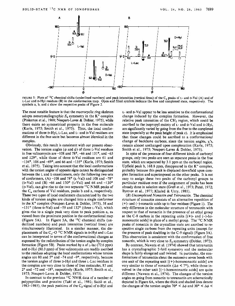
The most notable feature is that the macrocyclic ring skeleton adopts noncrystallographic S6 symmetry in the K+ complex (Pinkerton et al., 1969; Neupert-Laves & Dobler, 1975), while there exists no symmetrical property in the free molecule (Karle, 1975; Smith et al., 1975). Thus, the local confor- mations of three D-Hyi, L-Lac, and L- and D-Val residues are different in the free state but becomes almost identical in the complex.
Obviously, this result is consistent with our present obser- vation. The torsion angles (4 and I)) of three L-Val residues in free valinomycin are -108 and 78', -66 and 131', and -65 and 129', while those of three D-Val residues are 61 and -134', 106 and -69', and 66 and -135' (Karle, 1975; Smith et al., 1975). Taking into account that the local conformations with the torsion angles of opposite signs cannot be distinguished between the L and D enantiomers, only the following two sets of conformers, (A) -108 and 78' (L-Val) and 108 and -78' (D-Val) and (B) -66 and 130' (L-Val) and 66 and -130' (D-Val), can give rise to the two separate I3C NMR peaks of the C, carbons of Val residues, peaks b and a, respectively. These two types of local conformers characterized by the two kinds of torsion angles are changed into a single conformer in the Kf complex (Neupert-Laves & Dobler, 1975), 58 and -1 3 1 ' (three D-Val) and -59 and 132' (three L-Val), which gives rise to a single peak very close to peak position a, as viewed from the proximate position in the conformational map (Figure 5A). In Figure 5A, the 13C chemical shifts (un- derlined numbers) and peak intensities (vertical lines) are simultaneously illustrated. In a similar manner, the dis- placements of the C,-O I3C NMR signals in D-Hyi and L-Lac can be interpreted in terms of the conformational changes as expressed by the redistribution of the torsion angles by complex formation (Figure 5B). Peaks marked by a of L-lac (72.6 ppm) and D-Hyi (80.3 ppm) of free valinomycin are readily ascribed to the residues adopting the local conformations whose torsion angles are 80 and 5' and -74 and -9', respectively, because the torsion angles of three D-Hyi and three L-Lac residues in the complex are very close to those of free molecules, 83 and 2' and -72 and -18', respectively (Karle, 1975; Smith et al., 1975; Neupert-Laves 8i Dobler, 1975).
In contrast to the previous I3C NMR data of a number of polypeptides and proteins (Taki et al., 1981; Saitd et al., 1983-1985), the peak positions of the C, signal of D-Hyi and
L- and D-Val appear to be less sensitive to the conformational change induced by the complex formation. However, the relative peak intensities of the CH3 region, which could be ascribed to the isopropyl moiety of L- and D-Val and D-Hyi, are significantly varied by going from the free to the complexed state (especially at the peak height of peak c). It is emphasized that these changes could be ascribed to a conformational change of backbone carbons, since the torsion angles, x's, remain almost unchanged upon complexation (Karle, 1975; Smith et al., 1975; Neupert-Laves & Dobler, 1975).
In spite of the presence of four different kinds of carbonyl groups, only two peaks are seen as separate peaks in the free state, which are separated by 3.1 ppm at the carbonyl region. Upfield peak b, 168.8 ppm, disappeared in the K+ complex, probably because this peak is displaced downfield upon com- plex formation and superimposed on the other peaks. It is not easy to assign these two peaks of the carbonyl groups to particular residues even if the assignment of peaks has been already done in solution state (Grell et al., 1973; Patel, 1972; Bystrov et al., 1977; Khaled & Urry, 1981).
( E ) Uncomplexed Nonactin and Tetranactin. The chemical structure of nonactin consists of an alternative repetition of (+)- and (-)-nonactic acids up to four residues (Figure 1). The only difference in the molecular structure of tetranactin with respect to that of nonactin is the presence of an ethyl group at the C-8 carbon in the repeating units [(+)- and (-)-ho- mononactic acids] in place of a methyl group. The 13C NMR peaks of nonactin in the crystalline state are ascribed to re- spective single carbons from the repeating units (except for the presence of peak doubling in the C-9 signal) (Figure 3A). This observation is consistent with the conformation of free nonactin, which is very close to S, symmetry (Dobler, 1972).
By contrast, Nawata et al. (1974) showed that tetranactin has a crystallographic 2-fold symmetry and the molecular shape is fairly elongated and twisted. In particular, the con- formations of tetranactin about the successive seven bonds with one unit of the repeating unit [(+)-homononactic acids] are very similar to those of nonactin ( A 4 < 7'), while those in- volved in the other unit [(-)-homononactic acids] are quite different (Nawata et al., 1974). The changes of the torsion angles on going from nonactin to tetranactin are schematically depicted in Figure 6A, where the thick and shaded lines denote the changes of the torsion angles 70' < A 4 and 30' < A 4 <

7700 B I 0 C H E M I S T R Y T A B E T A A N D S A I T O
B
0
1 - 1 I 1 ' 1 \
\ 1 + 1 I I1 / - I
FIGURE 6: Schematic representation of the changes in the torsion angles between free nonactin and tetranactin (A) and of nonactin (and tetranactin) in going from the free state to the K', Na', and Cs' complexes (B). Data are based on X-ray diffraction studies by Dobler et al. (1968), Dobler & Phizackerley (1974), Dobler (1972), Iitaka et al. (1972), Nawata et al. (1974), and Sakamaki et al. (1976, 1977)). The thick and shaded lines represent variations of the torsion angles (A$), 70° < A$ and 30' < A$ < 70°, respectively.
70°, respectively (Dobler, 1972; Nawata et al., 1974). By analogy with the conformation-dependent I3C chemical
shifts of peptides and carbohydrates (Sait6 et al., 1982, 1984, 1985), it is natural to postulate that the I3C NMR signals of backbone carbons are related to pairs of nearby torsion angles. For instance, the C-1 signal is related to rotations about the 1'1 and 12 bonds (see Figure 1). Accordingly, it is expected from Figure 6A that the 13C NMR signals of tetranactin consist of two kinds of contributions, one from the peaks of the repeating units with nonactin-like local conformations (C-3 to C-8) and the other from the local conformers whose torsion angles are greatly different from those of nonactin. However, it is taken into account that the I3C chemical shifts of the C-7 and C-8 carbons (and also C-9, C-10, and C-11) should not be directly compared between tetranactin and nonactin (even if these two molecules adopt the same local conformations), because the ethyl group at the C-8 position of the former induces the displacements of peaks at the C-7 and C-8 chem- ical shifts through a simple substituent effect. Then, the 13C chemical shifts of the C-3, C-4, C-5, and C-6 carbons from the portion of the nonactin-like conformations of tetranactin should be resonated at positions similar to those of nonactin. Consistent with this expectation, the I3C chemical shifts of the C-5 and C-6 carbons are the same within the experimental errors between tetranactin and nonactin. The C-3 and C-4 chemical shifts of the former, however, are displaced downfield by 3.6 and 2.7 ppm as compared with those of the latter. It is probable that differences in the conformations of tetra- hydrofuran rings between tetranactin and nonactin are par- tially responsible for this discrepancy.
On the other hand, the intense peak at 76.0 ppm (relative peak intensity of 4 as compared with that of C-3 or C-8) is considered as an overlap of the C-3, C-6, and C-8 carbons from the other portion of the repeating unit whose torsion angles are greatly varied from those of nonactin. This assignment is confirmed by the following points. First, the resultant maximum displacement of the peak as a consequence of this assignment is 6.9 ppm and is within the range of the magnitude of conformation-dependent I3C chemical shifts so far observed (4 ppm). Second, this intense peak is not observed any more
for the metal complexes whose conformations are altered from the free molecule (see Figure 3). Third, the averaged 13C chemical shifts between the two types of conformers are ob- served in CDCl, solution as a result of a rapid chemical ex- change process (see Table 111). The presence of additional splittings of peaks due to the above-mentioned nonequivalence in (+) and (-) enantiomers of tetranactin is not clear for the region of the C-2, C-4, C-5, and C-9 carbons. However, the obvious splitting of the C-1 and C-10 signals is undoubtedly caused by the presence of the nonequivalence mentioned above. It is also pointed out that the C-9 methyl signal of nonactin could be ascribed to the presence of the similar magnetic nonequivalence between the two enantiomers in the crystals.
(C) Metal-Complexed Nonactin and Tetranactin. Obvi- ously, the spectral features of the I3C NMR spectra of met- al-complexed nonactin and tetranactin are significantly varied as compared to those of free molecules (Figures 3 and 4). This is unequivocally caused by a drastic conformational change of the ligand molecules by complex formations (Dobler, 1972; Dobler et al., 1969; Dobler & Phizackerley, 1974; Iitaka et al., 1972; Nawata et al., 1974; Sakamaki et al., 1976, 1977). In a similar manner to our argument about the comparison of the 13C NMR spectra between free nonactin and tetranactin as described above, the most affected torsion angles by the complex formations (thick lines, A@ > 70'; shaded lines, 70' > A@ > 30') are rotations about the 12,23, 3'6, and 88' bonds, irrespective of the variety of the metal ions (K+, Na', and Cs+) and ligand molecules (nonactin and tetranactin), as sche- matically illustrated in Figure 6B. Consistent with this ex- pectation, the displacements of the I3C chemical shifts of nonactin by formation of the K+ complex are seen for C-1 (6.6 ppm), C-2 (5.3 ppm), (2-10 (6.0 and 8.3 ppm), C-3 (2.4 ppm), C-6 (-2.0 ppm), and C-8 (-2.5 ppm), which can be unam- biguously ascribed to the changes of the torsion angles. Other significant displacements of peaks in the C-4 and C-5 carbons might be caused by a change of ring puckering in tetra- hydrofuran, as described in the previous section.
It is noteworthy, that the 13C chemical shifts of the C-1 to C-6 carbons of nonactin and tetranactin complexed with K+, Na', and Cs+ ions are the same within the experimental errors (f0.8 ppm) (see Tables I1 and 111). This observation shows that the local conformations of the ligand molecules in the six kinds of metal complexes (K+, Na', and Cs+ ions complexed with nonactin and tetranactin) are effectively identical, as viewed from the conformation-dependent I3C chemical shifts. In good agreement with this prediction by the 13C NMR data, the molecular conformations of these metal complexes by X-ray diffraction turned out to take approximate S4 symmetry, and differences of the torsion angles between the pairs of enantiomers are less than loo, as observed for K+-, Na+-, and Cs+-complexed nonactin and Cs+-tetranactin (Dobler, 1969; Dobler & Phizackerley, 1974; Sakamaki et al., 1977). Further, these torsion angles are in good agreement (<30°) among the six kinds of complexes (Dobler, 1969; Dobler & Phizackerley, 1974; Sakamaki et al., 1976, 1977) (Figure 6B). It is now clear that the conformation-dependent I3C chemical shifts can be conveniently utilized to clarify the conformations of un- complexed and metal-complexed nactins (nonactin, monactin, dinactin, and tetranactin) in the solid state. The present data also demonstrate that any conformational changes whose variations of the torsion angles are less than 10' cannot be recognized by displacements of the chemical shifts. On the contrary, variations of the torsion angles larger than 30' certainly induce substantial displacements of the conforma- tion-dependent 13C chemical shifts.

S O L I D - S T A T E 1 3 C N M R O F I O N O P H O R E S V O L . 2 4 , N O . 2 6 , 1 9 8 5 7701
Table 11: I3C Chemical Shifts of Free and Metal Ion Complexed Nonactin in the Solid and Solution (k0.4 ppm for the Solid; ppm from TMS) complexes
free c s + Na+ K+ solid soln" solid soh" solid soln" solid soln'
C- 1 c - 3 C-6 c - 8 c - 2 c - 7 c - 5 c - 4 c - 9
c -10
170.5 79.3 77.0 70.0 40.8 43.3 32.9 25.7 21.06 19.0b 8.5
174.1 80.0 76.3 69.0 45.1 42.2 31.3 28.0 20.4
12.7
177.8 83.0, 82.2 74.3 69.2, 68.2 46.2 44.8 32.2 28.6 20.9
15.6
174.6 80.3 75.9 68.7 45.3 42.5 31.3 28.2 20.5
13.0
178.3 81.5 74.4 68.3 45.1 43.3 31.3 28.5 20.8
16.0
172.2 80.9 74.5 67.9 45.8 44.2 31.2 28.9 20.8
15.0
177.1 81.7 75.0 67.5 46.1 44.5 30.9 28.9 20.2
16.8' 14.5b
1 177.2 81.8 74.2 66.9 45.9 44.2 31.3 28.6 20.9
15.0 ~~
"In CDCh solution (15 mg/mL). *Splittings of peaks arising from (+) and (-) enantiomers.
Table 111: "C Chemical Shifts of Free and Metal Ion Complexed Tetranactin in the Solid and Solution (f0.4 ppm for the Solid; ppm from TMS)
comolexes free c s + Na+ K+
solid soln" solid soln' solid soln' solid soln' c - 1 174.1, 172.5 174.3 177.7 177.0 177.9 177.7 177.5 177.7 c - 3 82.9, 76.0 79.8 82.9, 81.5 81.6 81.9, 80.9 80.9 81.6 81.8 C-6 76.0, 76.0 76.3 73.8 73.9 73.96 74.2 74.7 74.1 c - 8 72.2, 76.0 73.2 72.8 72.2 73.96 73.0 72.5 72.0 c - 2 43.9 45.0 45.1 45.9 45.4 46.0 45.2 46.0 c - 7 42.9, 40.8 39.9 42.4 41.6 42.1 42.6 42.2 42.6 c-5' 31.9 31.4 31.3 31.1 32.0 31.2 31.3 31.2
28.5 28.2
c - 4 28.4 28.0 29.0 c-9' 26.9 27.2 27.3 28.1 c-10 13.2, 7.5 12.8 16.0, 14.9 14.9 16.1, 14.9 15.2 15.0 15.3 c-11 9.2 9.2 11.4, 10.2 9.4 11.4, 10.4 10.1 10.9, 10.0 9.9
28'8 128.3 28.0 28.6 1 28.0
9.4 'In CDC13 solution (15 mg/mL). b T ~ o kinds of peaks are overlapped. 'Assignment of the C-5 and C-9 is reversed in Kyogoku et al. (1975).
It appears that the direct effect of the metal ions to the displacements of the 13C chemical shifts seems to be nominal, because no significant changes of the chemical shifts are seen among the complexes in spite of the larger differences in the ionic radii among Na, K, and Cs ions (Tables I1 and 111). Undoubtedly, this finding seems to justify the present approach to explain the displacements of peaks in terms of the con- formational changes. Nevertheless, the smaller splittings of peaks in the C-3 carbon of Cs+-complexed tetranactin and the C-3 and C-8 carbons of Cs+-complexed nonactin (< 1.4 ppm) could be ascribed to this direct effect of the Cs ion, because no difference of the torsion angles were found between the enantiomers of Cs+-complexed nonactiri and tetranactin. In this connection, it is pointed out that some of the bond dis- tances between the metal and ligand 0 atoms in the c s + complexes (3.03-3.07 A) are slightly smaller than the sum (3.09 A) of van der Waals and ionic radii of oxygen (1.40 A) and Cs (1.69 A) (Sakamaki et al., 1977). On the contrary, the distances of the metal to 0 atoms are usually larger than the sum of van der Waals and ionic radii for K and Na ions.
Conformations Revealed by the Conformation-Dependent 13C Chemical Shifts. On the basis of the foregoing discussion about the conformation-dependent 13C chemical shifts, we are now in a position to be able to apply this new methodology to clarify how conformations of these ionophores are altered by complex formation. In fact, it is now clear that confor- mations of the Na+ and Cs+ complexes of valinomycin, whose X-ray diffraction data are unavailable, are exactly the same as that of the K+ complex, as viewed from the conforma- tion-dependent 13C chemical shifts (Figure 2 and Table I). Obviously, this approach is straightforwardly applied to other
metal complexes of valinomycin, nonactin, and tetranactin to clarify their conformations in the solid state, with reference to the data described here.
Another important aspect of this approach is to reveal un- ambiguously whether or not the solid-state conformation is retained in a particular solvent system on the basis of direct comparison of the I3C chemical shifts between the solid and solution. The most notable feature is that the 13C chemical shifts of the K+ complexes of valinomycin, nonactin, and tetranactin and the Na' and Cs+ complexes with tetranactin are very similar to those of chloroform solution (Tables I-111), indicating that conformations achieved in the solid state are also retained in chloroform solution (Bystrov et al., 1977; Prestegard & Chan, 1969; Kyogoku et al., 1975). By contrast, the 13C NMR peaks of free ionophores and the rest of the metal complexes in chloroform are substantially different from those of the solid state because of the onset of rapid confor- mational fluctuation. In particular, no C, symmetrical ap- pearance of peaks is observed in free tetranactin dissolved in chloroform (Kyogoku et al., 1975).
ACKNOWLEDGMENTS
for a generous gift of tetranactin sample.
REFERENCES Bystrov, V. F., Gavrilov, Y . D., Ivanov, V. T., & Ovchinnikov,
Davis, D. G., & Tosteson, D. C. (1975) Biochemistry 14,
We are grateful to Chugai Pharmaceutical Co. Ltd., Tokyo,
Registry No. 1, 2001-95-8; 2, 6833-84-7; 3, 33956-61-5.
Yu, A. (1977) Eur. J . Biochem. 76, 63-82.
3962-3969.

7702 B I O C H E M IS T R Y A
T A B E T A A N D S A I T O
Dobler, M. (1972) Helv. Chim. Acta 55, 1371-1384. Dobler, M., & Phizackerley, R. P. (1974) Helv. Chim. Acta
Dobler, M., Dunitz, J. D., & Kilbourn, B. T. (1969) Helv.
Grell, E., Funck, T., & Sauter, H. (1973) Eur. J . Biochem.
Haynes, D. H. , Kowalsky, A., & Pressman, B. C. (1969) J .
Iitaka, Y., Sakamaki, T., & Nawata, Y. (1972) Chem. Lett.,
Ivanov, V. T., Laine, I. A., Abdulaev, N. D., Senyavina, L., Popov, E. M., Ovchinnikov, Yu. A., & Shemyakin, M. (1969) Biochem. Biophys. Res. Commun. 34, 803-8 1 1 .
Karle, I. L. (1975) J . Am. Chem. SOC. 97, 4379-4386. Khaled, M. A., & Urry, D. W. (1981) J . Chem. SOC., Chem.
Commun., 230-232. Kilbourn, B. T., Dunnitz, J. D., Pioda, L. A. R., & Simon,
W. (1967) J . Mol. Biol. 30, 559-563. Kyogoku, Y., Ueno, M., Akutsu, H., & Nawata, Y. (1975)
Biopolymers 14, 1049-1063. Nawata, Y., Sakamaki, T., & Iitaka, Y. (1974) Acta Crys-
tallogr., Sect. B: Struct. Crystallogr. Cryst. Chem. 830,
Neupert-Laves, K., & Dobler, M. (1975) Helv. Chim. Acta
Mueller, P., & Rudin, D. 0. (1967) Biochem. Biophys. Res. Commun. 26, 398-404.
Ohnishi, M., & Urry, D. W. (1969) Biochem. Biophys. Res. Commun. 36, 194-202.
Ohnishi, M., Fedarko, M. C., Baldeschwieler, J. D., & Johnson, L. F. (1972) Biochem. Biophys. Res. Commun.
Ovchinnikov, Yu. A., Ivanov, V. T., & Shkrob, A. M. (1974) Membrane Active Complexones, Elsevier Scientific, Am- sterdam.
57, 664-674.
Chim. Acta 52, 2573-2583.
34, 415-424.
Biol. Chem. 244, 502-505.
1225-1 230.
1047-1 053.
58, 432-442.
46, 3 12-320.
Patel, D. J. (1973) Biochemistry 12, 496-501. Patel, D. J., & Tonelli, A. E. (1973) Biochemistry 12,
486-496.
Pines, A., Gibby, M. G., & Waugh, J. S. (1973) J . Chem.
Pinkerton, M., Steinrauf, L. K., & Dawkins, P. (1969) Bio-
Pressman, B. C. (1976) Annu. Rev. Biochem. 45, 501-530. Prestegard, J . H., & Chan, S. I. (1969) Biochemistry 8,
Prestegard, J . H., & Chan, S. I. (1970) J . Am. Chem. SOC.
Pretsch, E., Vasak, M., & Simon, W. (1972) Helv. Chim. Acta
Saitb, H., Tabeta, R., & Harada, T. (1981) Chem. Lett.,
Saitb, H., Izumi, G., Mamizuka, T., Suzuki, S., & Tabeta, R. (1982) J . Chem. SOC., Chem. Commun., 1386-1388.
Sait6, H., Tabeta, R., Shoji, A., Ozaki, T., & Ando, I. (1983) Macromolecules 16, 1050-1057.
Sait6, H., Tabeta, R., Shoji, A., Ozaki, T., Ando, I., & Miyata, T. (1984) Biopolymers 23, 2279-2297.
Saitb, H., Tabeta, R., Shoji, A., Ozaki, T., Ando, I., & Asakura, T. (1985) in Magnetic Resonance in Biology and Medicine (Govil, G., Kheterapal, C. L., & Saran, A,, Eds.) pp 195-215, Tata McGraw-Hill, New Delhi.
Sakamaki, T., Iitaka, Y., & Nawata, Y. (1976) Acta Crys- tallogr., Sect. B: Struct. Crystallogr. Cryst. Chem. B32,
Sakamaki, T., Iitaka, Y., & Nawata, Y. (1977) Acta Crys- tallogr., Sect. B: Struct. Crystallogr. Cryst. Chem. B33,
Schaefer, J., & Stejskal, E. 0. (1976) J . Am. Chem. SOC. 98,
Smith, G. D., Duax, W. L., Langs, D. A,, DeTitta, G. T., Edmonds, J . W., Rohrer, D. C., & Weeks, C. M. (1975) J . Am. Chem. SOC. 97, 7242-7247.
Szabo, G., Eisenman, G., & Ciani, S. (1 969) J. Membr. Biol.
Taki, T., Yamashita, S., Satoh, M., Shibata, A,, Yamashita, T., Tabeta, R., & Saitb, H. (1 98 1) Chem. Lett., 1803-1 806.
Phys. 59, 569-590.
chem. Biophys. Res. Commun. 35, 512-518.
392 1-3927.
92, 4440-4446.
55, 1098-1 104.
571-574.
768-774.
52-59.
103 1-1032.
1 , 346-382.