high sensitivity of cap-feia rves v 5 and rves v 1 for diagnosis of vespula venom allergy
TRANSCRIPT
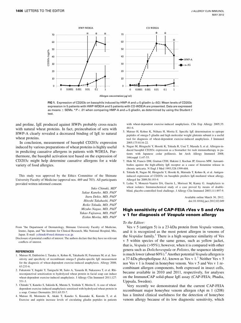
FIG 1. Expression of CD203c on basophils induced by HWP-A and v-5 gliadin (v-5G). Mean levels of CD203c
expression in 5 patients with HWP-WDEIA and 5 patients with CO-WDEIA are presented. Data are expressed
as means 6 SEMs. *P < .01 when comparing HMP-A and v-5 gliadin, as determined by using the Student ttest.
J ALLERGY CLIN IMMUNOL
MAY 2012
1406 LETTERS TO THE EDITOR
and proline, IgE produced against HWPs probably cross-reactswith natural wheat proteins. In fact, preincubation of sera withHWP-A clearly revealed a decreased binding of IgE to naturalwheat proteins.In conclusion, measurement of basophil CD203c expression
induced by various preparations of wheat proteins is highly usefulin predicting causative allergens in patients with WDEIA. Fur-thermore, the basophil activation test based on the expression ofCD203c might help determine causative allergens for a widevariety of food allergies.
This study was approved by the Ethics Committee of the Shimane
University Faculty of Medicine (approval nos. 469 and 703). All participants
provided written informed consent.
Yuko Chinuki, MDa
Sakae Kaneko, MD, PhDa
Itaru Dekio, MD, PhDa
Hitoshi Takahashi, PhDa
Reiko Tokuda, MD, PhDb
Mizuho Nagao, MD, PhDb
Takao Fujisawa, MD, PhDb
Eishin Morita, MD, PhDa
From athe Department of Dermatology, Shimane University Faculty of Medicine,
Izumo, Japan, and bthe Institute for Clinical Research, Mie National Hospital, Mie,
Japan. E-mail: [email protected].
Disclosure of potential conflict of interest: The authors declare that they have no relevant
conflicts of interest.
REFERENCES
1. Matsuo H, Dahlstr€om J, Tanaka A, Kohno K, Takahashi H, Furumura M, et al. Sen-
sitivity and specificity of recombinant omega-5 gliadin-specific IgE measurement
for the diagnosis of wheat-dependent exercise-induced anaphylaxis. Allergy 2008;
63:233-6.
2. Fukutomi Y, Itagaki Y, Taniguchi M, Saito A, Yasueda H, Nakazawa T, et al. Rhi-
noconjunctival sensitization to hydrolyzed wheat protein in facial soap can induce
wheat-dependent exercise-induced anaphylaxis. J Allergy Clin Immunol 2011;127:
531-3.
3. Chinuki Y, Kaneko S, Sakieda K, Murata S, Yoshida Y, Morita E. A case of wheat-
dependent exercise-induced anaphylaxis sensitized with hydrolysed wheat protein in
a soap. Contact Dermatitis 2011;65:55-7.
4. Matsuo H, Morimoto K, Akaki T, Kaneko S, Kusatake K, Kuroda T, et al.
Exercise and aspirin increase levels of circulating gliadin peptides in patients
with wheat-dependent exercise-induced anaphylaxis. Clin Exp Allergy 2005;35:
461-6.
5. Matsuo H, Kohno K, Niihara H, Morita E. Specific IgE determination to epitope
peptides of omega-5 gliadin and high molecular weight glutenin subunit is a useful
tool for diagnosis of wheat-dependent exercise-induced anaphylaxis. J Immunol
2005;175:8116-22.
6. Nagao M, Hiraguchi Y, Hosoki K, Tokuda R, Usui T, Masuda S, et al. Allergen-in-
duced basophil CD203c expression as a biomarker for rush immunotherapy in pa-
tients with Japanese cedar pollinosis. Int Arch Allergy Immunol 2008;
146(suppl 1):47-53.
7. Hide M, Francis DM, Grattan CEH, Hakimi J, Kochan JP, Greaves MW. Autoanti-
bodies against the high-affinity IgE receptor as a cause of histamine release in
chronic urticaria. N Engl J Med 1993;328:1599-604.
8. Tokuda R, Nagao M, Hiraguchi Y, Hosoki K, Matsuda T, Kohno K, et al. Antigen-
induced expression of CD203c on basophils predicts IgE-mediated wheat allergy.
Allergol Int 2009;58:193-9.
9. Leduc V, Moneret-Vautrin DA, Guerin L, Morisset M, Kanny G. Anaphylaxis to
wheat isolates: Immunochemical study of a case proved by means of double-
blind, placebo-controlled food challenge. J Allergy Clin Immunol 2003;111:897-9.
Available online March 30, 2012.doi:10.1016/j.jaci.2012.02.049
High sensitivity of CAP-FEIA rVes v 5 and rVesv 1 for diagnosis of Vespula venom allergy
To the Editor:Ves v 5 (antigen 5) is a 23-kDa protein from Vespula venom,
and it is recognized as the most potent allergen in venoms ofthe Vespidae family.1 There is a high sequence similarity of Vesv 5 within species of the same genus, such as yellow jacket,that is, Vespula (>95%); however, when it is compared with othergenera such as Dolichovespula or Polistes, the sequence identityis much lower (about 60%).2 Another potentialVespula allergen isa 37-kDa phospholipase A1, known as Ves v 1.1 Neither Ves v 5nor Ves v 1 is found in honeybee venom. Ves v 5 and Ves v 1 re-combinant allergen components, both expressed in insect cells,became available in 2010 and 2011, respectively, for analyseson the ImmunoCAP solid-phase IgE assay (CAP-FEIA; Phadia,Uppsala, Sweden).Very recently we demonstrated that the current CAP-FEIA
recombinant major honeybee venom allergen rApi m 1 (i208)has a limited clinical usefulness for the detection of honeybeevenom allergy because of its low diagnostic sensitivity, which
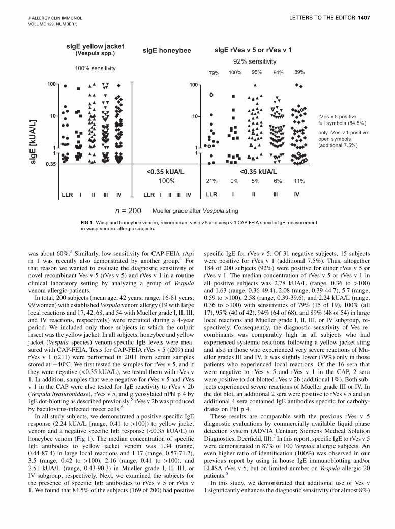
FIG 1. Wasp and honeybee venom, recombinant vesp v 5 and vesp v 1 CAP-FEIA specific IgE measurement
in wasp venom–allergic subjects.
J ALLERGY CLIN IMMUNOL
VOLUME 129, NUMBER 5
LETTERS TO THE EDITOR 1407
was about 60%.3 Similarly, low sensitivity for CAP-FEIA rApim 1 was recently also demonstrated by another group.4 Forthat reason we wanted to evaluate the diagnostic sensitivity ofnovel recombinant Ves v 5 (rVes v 5) and rVes v 1 in a routineclinical laboratory setting by analyzing a group of Vespulavenom allergic patients.In total, 200 subjects (mean age, 42 years; range, 16-81 years;
99 women) with established Vespula venom allergy (19 with largelocal reactions and 17, 42, 68, and 54 with Mueller grade I, II, III,and IV reactions, respectively) were recruited during a 4-yearperiod. We included only those subjects in which the culpritinsect was the yellow jacket. In all subjects, honeybee and yellowjacket (Vespula species) venom-specific IgE levels were mea-sured with CAP-FEIA. Tests for CAP-FEIA rVes v 5 (i209) andrVes v 1 (i211) were performed in 2011 from serum samplesstored at 2408C. We first tested the samples for rVes v 5, and ifthey were negative (<0.35 kUA/L), we tested them with rVes v1. In addition, samples that were negative for rVes v 5 and rVesv 1 in the CAP were also tested for IgE reactivity to rVes v 2b(Vespula hyaluronidase), rVes v 5, and glycosylated nPhl p 4 byIgE dot-blotting as described previously.5 rVes v 2b was producedby baculovirus-infected insect cells.6
In all study subjects, we demonstrated a positive specific IgEresponse (2.24 kUA/L [range, 0.41 to >100]) to yellow jacketvenom and a negative specific IgE response (<0.35 kUA/L) tohoneybee venom (Fig 1). The median concentration of specificIgE antibodies to yellow jacket venom was 1.34 (range,0.44-87.4) in large local reactions and 1.17 (range, 0.57-71.2),3.5 (range, 0.42 to >100), 2.16 (range, 0.41 to >100), and2.51 kUA/L (range, 0.43-90.3) in Mueller grade I, II, III, orIV subgroup, respectively. Next, we examined the subjects forthe presence of specific IgE antibodies to rVes v 5 or rVes v1. We found that 84.5% of the subjects (169 of 200) had positive
specific IgE for rVes v 5. Of 31 negative subjects, 15 subjectswere positive for rVes v 1 (additional 7.5%). Thus, altogether184 of 200 subjects (92%) were positive for either rVes v 5 orrVes v 1. The median concentration of rVes v 5 or rVes v 1 inall positive subjects was 2.78 kUA/L (range, 0.36 to >100)and 1.63 (range, 0.36-49.4), 2.08 (range, 0.39-44.7), 5.7 (range,0.59 to >100), 2.58 (range, 0.39-39.6), and 2.24 kUA/L (range,0.36 to >100) with sensitivities of 79% (15 of 19), 100% (all17), 95% (40 of 42), 94% (64 of 68), and 89% (48 of 54) in largelocal reactions and Mueller grade I, II, III, or IV subgroup, re-spectively. Consequently, the diagnostic sensitivity of Ves re-combinants was comparably high in all subjects who hadexperienced systemic reactions following a yellow jacket stingand also in those who experienced very severe reactions of Mu-eller grades III and IV. It was slightly lower (79%) only in thosepatients who experienced local reactions. Of the 16 sera thatwere negative to rVes v 5 and rVes v 1 in the CAP, 2 serawere positive to dot-blotted rVes v 2b (additional 1%). Both sub-jects experienced severe reactions of Mueller grade III or IV. Inthe dot blot, an additional 2 sera were positive to rVes v 5 and anadditional 4 sera contained IgE antibodies specific for carbohy-drates on Phl p 4.These results are comparable with the previous rVes v 5
diagnostic evaluations by commercially available liquid phasedetection system (ADVIA Centaur; Siemens Medical SolutionDiagnostics, Deerfield, Ill).7 In this report, specific IgE to rVes v 5were demonstrated in 87% of 100 Vespula allergic subjects. Aneven higher ratio of identification (100%) was observed in ourprevious report by using in-house IgE immunoblotting and/orELISA rVes v 5, but on limited number on Vespula allergic 20patients.5
In this study, we demonstrated that additional use of Ves v1 significantly enhances the diagnostic sensitivity (for almost 8%)

J ALLERGY CLIN IMMUNOL
MAY 2012
1408 LETTERS TO THE EDITOR
of diagnostic tests based on recombinant yellow jacket venomallergens. Nevertheless, rVes v 5 and rVes v 1 had missed 8% ofsubjects with established allergy. Testing for rVes v 2b added onlya minor contribution to diagnostic sensitivity. Primarily, 3 aller-gens were recognized as responsible for Vespula venom allergy,beyond Ves v 5 and Ves v 1, also Ves v 2, which occurs in iso-forms.1 Recently, a novel 100-kDa glycosylated protein with ho-mology to dipeptidyl peptidases with allergenic potential,namely, Ves v 3, was reported as a yellow jacket allergen.7 rVesv 3 showed IgE reactivity in approximately 50% of Vespula aller-gic subjects (in overall 54 tested)8 andmight be useful to diagnosethe few patients who are not identified with rVes v 5, 1, and 2.Clinically, we cannot afford to miss a patient who is sensitive to
insect venom; thus, the whole venom (that contains all the venomallergens as a single test) needs to be the first line of laboratoryevaluation. However, the identification of the disease-causinginsect venom in venom allergy is often difficult. In such cases,commercially available CAP-FEIA tests based on recombinantrVes v 5 and rVes v 1 allergens should be helpful for theserological dissection of Vespula venom allergy.
We thank Lucas Mach, Daniel Kolarich, and Friedrich Altmann, Depart-
ment of Applied Genetics and Cell Biology, University of Natural Resources
and Life Science, Vienna, Austria, for recombinant Ves v 2b.
Peter Koro�sec, PhDa
Rudolf Valenta, MDb
Irene Mittermann, PhDb
Nina �Celesnik, BSa
Mira �Silar, BSa
Mihaela Zidarn, MDa
Mitja Ko�snik, PhD, MDa
From athe University Clinic of Respiratory and Allergic Diseases, Golnik, Slovenia, andbthe Department of Pathophysiology and Allergy Research, Division of Immunopa-
thology, Center for Pathophysiology, Infectiology and Immunology, Medical Univer-
sity of Vienna, Vienna, Austria. E-mail: [email protected].
This study was supported in part by the Slovenian research agency (ARRS), Christian
Doppler Research Association, and the Austrian Science Fund (FWF).
Disclosure of potential conflict of interest: R.Valenta has received research support from
the Austrian Science Fund, CDG, Biomas, and Phadia. The rest of the authors declare
that they have no relevant conflicts of interest.
REFERENCES
1. Hoffman DR. Allergens in Hymenoptera venom, XXV: the amino acid sequences of
antigen 5 molecules and the structural basis of antigenic cross-reactivity. J Allergy
Clin Immunol 1993;92:707-16.
2. King TP, Lu G, Gonzalez M, Qian N, Soldatova L. Yellow jacket venom allergens,
hyaluronidase and phospholipase: sequence similarity and antigenic cross-reactivity
with their hornet and wasp homologs and possible implications for clinical allergy.
J Allergy Clin Immunol 1996;98:588-600.
3. Koro�sec P, Valenta R, Mittermann I, Celesnik N, Er�zen R, Zidarn M, et al. Low sen-
sitivity of commercially available rApi m 1 for diagnosis of honeybee venom al-
lergy. J Allergy Clin Immunol 2011;128:671-3.
4. Sturm GJ, Hemmer W, Hawranek T, Lang R, Ollert M, Spillner E, et al. Detection of
IgE to recombinant Api m 1 and rVes v 5 is valuable but not sufficient to distinguish
bee from wasp venom allergy. J Allergy Clin Immunol 2011;128:247-8.
5. Mittermann I, Zidarn M, Silar M, Markovic-Housley Z, Aberer W, Korosec P, et al.
Recombinant allergen-based IgE testing to distinguish honeybee and wasp allergy.
J Allergy Clin Immunol 2010;125:1300-7.
6. Kolarich D, Loos A, Leonard R, Mach L, Marzban G, Hemmer W, et al. A proteomic
study of the major allergens from yellow jacket venoms. Proteomics 2007;7:1615-23.
7. M€uller U, Johansen N, Petersen AB, Fromberg-Nielsen J, Haeberli G. Hymenoptera
venom allergy: analysis of double positivity to honey bee and Vespula venom by es-
timation of IgE antibodies to species-specific major allergens Api m 1 and Ves v 5.
Allergy 2009;64:543-8.
8. Blank S, Seismann H, Bockisch B, Braren I, Cifuentes L, McIntyre M, et al. Iden-
tification, recombinant expression, and characterization of the 100 kDa high molec-
ular weight Hymenoptera venom allergens Api m 5 and Ves v 3. J Immunol 2010;
184:5403-13.
Available online January 24, 2012.doi:10.1016/j.jaci.2011.12.975
Specific allergen concentration of WHO andFDA reference preparations measured usinga multiple allergen standard
To the Editor:Allergen measurements require well-defined allergen stan-
dards. Allergists rely on these measurements for dosing patientson immunotherapy with the aim of achieving maintenance dosesof 5 to 20 mg of specific allergen that have been associated withclinical efficacy.1 Allergists need to know that allergen measure-ments made by manufacturers are consistent and can reliably beused in clinical practice. Allergen measurements are widelyused in the indoor air quality industry to assess exposure inhomes, the workplace, schools, and commercial buildings. Theyare routinely used for assessing health risks associated with aller-gen exposure, for assessing the efficacy of allergen avoidance pro-cedures, and for developing new allergen control products.2
Measurements of allergens by ELISA rely on standards ofknown allergen concentration, but few national or internationalallergen standards exist. The World Health Organization/Inter-national Union of Immunological Societies (WHO/IUIS) Aller-gen Standardization Committee initiated a program to developpurified allergen standards for calibration of in vitro measure-ments. This initiative was funded by the European Union to de-velop certified reference materials for allergenic products(‘‘Development of Certified Reference Materials for AllergenicProducts and Validation of Methods for their Quantification,’’ ac-ronym CREATE). The aims of CREATE were to develop interna-tional reference materials with verifiable allergen content.3,4
Our goal was to apply the principles of allergen standardizationdeveloped in CREATE to other purified allergens. We developeda single ‘‘universal’’ allergen standard (UAS) for use in ELISAand in a fluorescent multiplex array for indoor allergens.5 Purifiedproteins are essential in multiplex systems to reduce nonspecificinteractions. Eight purified allergens (Der p 1, Der f 1, Der p 2,Fel d 1, Can f 1, Rat n 1, Mus m 1, and Bla g 2) were combinedin the UAS. The protein concentration of the purified allergenswas determined by amino-acid analysis, in keeping with CRE-ATE. A detailed validation of the UAS and comparison with pre-vious ELISA standards will be published elsewhere.6 Here, wereport the concentration of specific allergens in WHO/IUIS andUS Food and Drug Administration (FDA) reference preparationsusing the single multiallergen standard.Specific allergen concentrations of WHO/IUIS and FDA
reference preparations were determined by using ELISA(Table I). TheWHO/IUISDermatophagoides pteronyssinus stan-dard 82/518 has been widely used as a standard for measurementsof allergen exposure with a reported concentration of 12.5mg Derp 1 per ampoule.7 A value of 7.2 mg Der p 1 per ampoule wasobtained by using the UAS, which is similar to estimates of5 mg Der p 1 per ampoule reported previously.8,9 The WHO/IUIS dog hair standard has an assigned potency of 100,000 IUsper ampoule and contained 20.4 mg Can f 1 per ampoule as deter-mined by using the UAS.