high-throughput screening using multicellular tumor spheroids …1090154/fulltext01.pdf ·...
TRANSCRIPT

ACTAUNIVERSITATIS
UPSALIENSISUPPSALA
2017
Digital Comprehensive Summaries of Uppsala Dissertationsfrom the Faculty of Medicine 1334
High-throughput screening usingmulticellular tumor spheroids toreveal and exploit tumor-specificvulnerabilities
WOJCIECH SENKOWSKI
ISSN 1651-6206ISBN 978-91-554-9921-1urn:nbn:se:uu:diva-320598

Dissertation presented at Uppsala University to be publicly examined in Rosénsalen, Ing95/96, Akademiska Barnsjukhuset, Uppsala, Saturday, 10 June 2017 at 09:00 for the degree ofDoctor of Philosophy (Faculty of Medicine). The examination will be conducted in English.Faculty examiner: Doctor Krister Wennerberg (Institute for Molecular Medicine Finland(FIMM), University of Helsinki).
AbstractSenkowski, W. 2017. High-throughput screening using multicellular tumor spheroids toreveal and exploit tumor-specific vulnerabilities. Digital Comprehensive Summaries ofUppsala Dissertations from the Faculty of Medicine 1334. 50 pp. Uppsala: Acta UniversitatisUpsaliensis. ISBN 978-91-554-9921-1.
High-throughput drug screening (HTS) in live cells is often a vital part of the preclinicalanticancer drug discovery process. So far, two-dimensional (2D) monolayer cell cultures havebeen the most prevalent model in HTS endeavors. However, 2D cell cultures often fail torecapitulate the complex microenvironments of in vivo tumors. Monolayer cultures are highlyproliferative and generally do not contain quiescent cells, thought to be one of the main reasonsfor the anticancer therapy failure in clinic. Thus, there is a need for in vitro cellular models thatwould increase predictive value of preclinical research results. The utilization of more complexthree-dimensional (3D) cell cultures, such as multicellular tumor spheroids (MCTS), whichcontain both proliferating and quiescent cells, has therefore been proposed. However, difficulthandling and high costs still pose significant hurdles for application of MCTS for HTS.
In this work, we aimed to develop novel assays to apply MCTS for HTS and drug evaluation.We also set out to identify cellular processes that could be targeted to selectively eradicatequiescent cancer cells. In Paper I, we developed a novel MCTS-based HTS assay and found thatnutrient-deprived and hypoxic cancer cells are selectively vulnerable to treatment with inhibitorsof mitochondrial oxidative phosphorylation (OXPHOS). We also identified nitazoxanide, anFDA-approved anthelmintic agent, to act as an OXPHOS inhibitor and to potentiate the effectsof standard chemotherapy in vivo. Subsequently, in Paper II we applied the high-throughputgene-expression profiling method for MCTS-based drug screening. This led to discoverythat quiescent cells up-regulate the mevalonate pathway upon OXPHOS inhibition and thatthe combination of OXPHOS inhibitors and mevalonate pathway inhibitors (statins) resultsin synergistic toxicity in this cell population. In Paper III, we developed a novel spheroid-based drug combination-screening platform and identified a set of molecules that synergizewith nitazoxanide to eradicate quiescent cancer cells. Finally, in Paper IV, we applied ourMCTS-based methods to evaluate the effects of phosphodiesterase (PDE) inhibitors in PDE3A-expressing cell lines.
In summary, this work illustrates how MCTS-based HTS yields potential to reveal and exploitpreviously unrecognized tumor-specific vulnerabilities. It also underscores the importance ofcell culture conditions in preclinical drug discovery endeavors.
Keywords: spheroids, high-throughput screening, nitazoxanide, OXPHOS, gene expressionprofiling, statins, drug repositioning, 3D cell culture, quiescent cells
Wojciech Senkowski, Department of Medical Sciences, Cancer Pharmacology andComputational Medicine, Akademiska sjukhuset, Uppsala University, SE-75185 Uppsala,Sweden.
© Wojciech Senkowski 2017
ISSN 1651-6206ISBN 978-91-554-9921-1urn:nbn:se:uu:diva-320598 (http://urn.kb.se/resolve?urn=urn:nbn:se:uu:diva-320598)

“My apologies to great questions for small answers. Truth, please don't pay me much attention. Dignity, please be magnanimous. Bear with me, O mystery of existence, as I pluck the occasional thread
from your train.”
Wisława Szymborska, “Under One Small Star” (17 – 20) tr. Stanisław Barańczak and Clare Cavanagh
To my parents and my brother
Dla moich rodziców oraz brata


List of Papers
This thesis is based on the following papers, which are referred to in the text by their Roman numerals.
I Senkowski W, Zhang X, Olofsson MH, Isacson R, Höglund U, Gustafsson MG, Nygren P, Linder S, Larsson R and Fryknäs M, Three-dimensional cell culture-based screening identifies the anthelminthic drug nitazoxanide as a candidate for treatment of colorectal cancer. Molecular Cancer Therapeutics, 14(6); 1504-16 (2015)
II Senkowski W, Jarvius M, Rubin J, Lengqvist J, Gustafsson MG, Nygren P, Kultima K, Larsson R and Fryknäs M, Large-scale gene-expression screening in three-dimensional cell cul-tures identifies context-dependent drug responses. Cell Chemi-cal Biology, 23(11); 1428-1438 (2016).
III Senkowski W, Nazir M, Gustafsson MG, Nygren P, Larsson R and Fryknäs M, Drug combination screening in multicellular tumor spheroids identifies synthetic lethalities in quiescent can-cer cells. Manuscript (2017)
IV Nazir M, Senkowski W, Nyberg F, Blom K, Edqvist P, An-dersson C, Gustafsson MG, Nygren P, Larsson R and Fryknäs M, Targeting tumor cells based on PDE3A expression, Manu-script (2017)
Reprints were made with permission from the respective publishers.

Other papers, not included in the thesis
I Kashif M, Andersson C, Hassan S, Karlsson H, Senkowski W, Fryknäs M, Nygren P, Larsson R and Gustafsson MG, In vitro discovery of promising anti-cancer combinations using iterative maximisation of a therapeutic index. Scientific Reports 5: 14118 (2015)
II Fryknäs M, Zhang X, Bremberg U, Senkowski W, Olofsson MH, Brandt P, Persson I, D’Arcy P, Gullbo J, Nygren P, Kunz-Schughart L, Linder S and Larsson R, Iron chelators target both quiescent and proliferating cancer cells. Scientific Reports 6: 38343 (2016)
III Blom K, Senkowski W, Jarvius M, Berglund M, Rubin J, Len-hammar L, Parrow V, Andersson C, Loskog A, Fryknäs M, Nygern P and Larsson R, The anticancer effect of mebendazole may be due to M1 monocyte/macrophage activation via ERK1/2 and TLR8-dependent inflammasome activation. Im-munopharmacology and Immunotoxicology, in press (2017)

Contents
1. Introduction ............................................................................................... 111.1 Overview of current cancer treatment strategies ................................ 111.2 In vitro cancer models for drug discovery .......................................... 131.3 Drug discovery: high-throughput drug screening (HTS) ................... 131.4 Drug repositioning .............................................................................. 141.5 Multicellular tumor spheroids for HTS .............................................. 151.6 Targeting quiescent cancer cells ......................................................... 15
2. Aims .......................................................................................................... 17
3. Methods ..................................................................................................... 183.1 Drugs and libraries .............................................................................. 183.2 Cell lines ............................................................................................. 183.3 Cellular in vitro models ...................................................................... 19
3.3.1 Monolayer ................................................................................... 193.3.2 P-MCTS (‘proliferative spheroids’) ............................................ 193.3.3 Q-MCTS (‘quiescent spheroids’) ................................................ 19
3.4 Immunohistochemical staining ........................................................... 203.5 Drug tests and screening ..................................................................... 203.6 Cell proliferation and viability assays ................................................ 20
3.6.1 Fluorometric microculture cytotoxicity assay (FMCA) ............. 203.6.2 Spheroid GFP fluorescence intensity assay ................................ 203.6.3 TOX8 assay (resazurin-based) .................................................... 213.6.4 Spheroid-based clonogenic assay ............................................... 213.6.5 CellTiter-Glo 3D ......................................................................... 22
3.7 Mitochondrial activity measurements ................................................ 223.7.1 Oxygen consumption rate (OCR) measurements ....................... 223.7.2 JC-1 staining ............................................................................... 223.7.3 Staining for pimonidazole adducts in MCTS .............................. 23
3.8 In vivo experiments ............................................................................ 233.9 L1000 Gene Expression Profiling ...................................................... 233.10 Drug combinations and therapeutic synergy in MCTS .................... 24
4. Summary of the papers .............................................................................. 254.1 Paper I ................................................................................................. 25
4.1.1 Background and purpose ............................................................. 25

4.1.2 Compounds selectively toxic to Q-MCTS inhibit mitochondrial oxidative phosphorylation .................................................................... 254.1.3 Nitazoxanide is a suitable candidate for drug repositioning ....... 264.1.4 Nitazoxanide potentiates the therapeutic effect of a standard chemotherapeutic agent in vivo. .......................................................... 274.1.5 Conclusions ................................................................................. 29
4.2 Paper II ............................................................................................... 304.2.1 Background and purpose ............................................................. 304.2.2 Application of L1000 Gene Expression profiling to study 3D cell cultures at a large scale ........................................................................ 314.2.3 The dataset .................................................................................. 324.2.4 Up-regulation of mevalonate-pathway genes upon OXPHOS inhibition in Q-MCTS .......................................................................... 324.2.5 Synergistic activity of OXPHOS inhibitors and statins in Q-MCTS ................................................................................................... 324.2.6 Conclusions ................................................................................. 32
4.3 Paper III .............................................................................................. 344.3.1 Background and purpose ............................................................. 344.3.2 Drug combination screening in Q-MCTS ................................... 344.3.3 Identification of drug combinations targeting Q-MCTS ............ 344.3.4 Conclusions ................................................................................. 35
4.4 Paper IV .............................................................................................. 374.4.1 Background and purpose ............................................................. 374.4.2 PDE3A overexpression predicts sensitivity to PDE inhibition ... 374.4.3 Sensitivity to PDE inhibition in other cell culture models ......... 374.4.4 Conclusions ................................................................................. 38
5. Summary and final remarks ...................................................................... 405.1 New assays – development and application ....................................... 405.2 Application of MCTS in HTS yields novel findings .......................... 405.3 Culture conditions determine drug response ...................................... 415.4 A look ahead ....................................................................................... 42
6. Acknowledgements ................................................................................... 43
7. References ................................................................................................. 46

Abbreviations
2D 3D AML ATP CCCP CCD DNA DMSO FCCP FDA FMCA GFP HTS IC50 MCTS mRNA OCR OXPHOS P-MCTS PCPTCs PDE PDE3A RNA SD SEM Q-MCTS
Two-dimensional Three-dimensional Acute myeloid leukemia Adenosine triphosphate Carbonyl cyanide m-chlorophenyl hydrazone Charge-coupled device Deoxyribonucleic acid Dimethyl sulfoxide Carbonyl cyanide-4-(trifluoromethoxy)phenylhydrazone US Food and Drug Administration OR Fluorescein diacetate Fluorometric microculture cytotoxicity assay Green fluorescent protein High-throughput drug screening Half maximal inhibitory concentration Multicellular tumor spheroid(s) Messenger RNA Oxygen consumption rate Oxidative phosphorylation ‘Proliferative’ spheroids Primary cultures of patient tumor cells Phosphodiesterase Phosphodiesterase 3A Ribonucleic acid Standard deviation Standard error of the mean ‘Quiescent’ spheroids


11
1. Introduction
1.1 Overview of current cancer treatment strategies Cancer is a group of diseases that share a number of common traits. These traits are commonly called “hallmarks” – a term coming from the two most-cited publications in cancer research – “Hallmarks of Cancer” and “Hall-marks of Cancer: The Next Generation” by Hanahan and Weinberg [1, 2]. As listed by Hanahan and Weinberg, cancers can be characterized by perpet-ual and uncontrolled cellular proliferation (being a result of sustained pro-growth and insensitiveness to tumor suppressor signaling, evading senes-cence and cell death), deregulated cell metabolism, high genetic mutation rate, avoiding destruction by immune system while maintaining pro-inflammatory environment, generation of new blood vessels (angiogenesis) and invasion to distant tissues (metastasis). All these traits render cancers extremely variable and notoriously difficult to treat. Despite the substantial progress that has been made in the fight against can-cer, it still remains one of the leading causes of death worldwide, with 8.2 million of cancer-related deaths in 2012 [3]. The most common modality of cancer treatment is surgery (for solid tumors), often combined with chemo- and radiotherapy [4]. The latter both target processes involved in cellular growth and division. However, even though they have been successful to a large extent, they are not sufficient to eradicate cancer in many patients [4, 5]. Because they do not act selectively on cancer cells, but also affect healthy tissues, they yield serious side effects and dose-limiting toxicity. This is why in recent years a number of novel drugs that are commonly known as “tar-geted therapeutics” have emerged [5]. These molecules are designed to bind to and inhibit specific proteins, responsible for cancer growth. These thera-pies have also proven successful, but as of today, remain insufficient to fully cure many patients [4, 5]. Finally, another group of strategies to battle cancer have been designed to utilize patient’s immune system. These are called immunotherapies. Immunotherapeutic drugs can be antibodies or immune cells that specifically recognize cancer cells, thereby boosting immunologi-cal response, or abrogate immunosuppressive signals sent by cancer cells [6, 7]. Immunotherapies have in recent years largely improved clinical out-comes of cancer treatment[6-8]. However, even though the results have been

12
Figure 1.1 Schematic representation of cancer cell subtypes clonal evolution. Due to selective environmental pressures cancer cell clones undergo different changes on the genetic level. As a result, metastatic cancer is a heterogeneous popu-lation of various subclones, differently sensitive to therapy (Tx). From Greaves & Maley (2012) [9]; RightsLink license no.: 3871271396693.
promising, current immunotherapeutic agents are often not enough to suc-cessfully eradicate many types of cancer when used in mono- or combination therapy [4, 7, 10].
The main reason why many therapies fail is inherent or acquired drug re-sistance of cancer cells. This resistance stems from the fact that population of cancer cells within a single tumor or hematological malignancy is very heterogeneous [11, 12]. This heterogeneity is a result of a series of adapta-tions (caused by de novo genetic events and by selective pressures in local microenvironment) that different subpopulations of cancer cells undergo during early stages of cancer development [9, 12, 13]. In other words, one could say that the development of cancer is an evolutionary process. In the end, the quickly progressing disease consists not of a single subtype of cells that all carry the same set of genetic mutations, but rather multiple cell sub-sets that have undergone various molecular adaptations at genetic and, con-sequently, phenotypic level (Fig. 1.1) [9]. As a result, advanced tumors con-sist of rapidly dividing cancer cells, slow-dividing cells, but also non-proliferating quiescent cells (Fig. 1.2). These numerous adaptations render cancer cells differently sensitive to therapies [9, 12, 14]. Because most ther-

13
apies are targeted mainly at actively proliferating tumor cells (i.e. mecha-nisms targeted by standard chemo- and radiotherapies, such as replication of DNA) or at a single protein (aforementioned targeted therapies and immuno-therapies), only some cancer cell subsets will respond while others will re-main largely unaffected [9]. For this reason, in the recent years there has been an increased interest in using in vitro models that would capture the cancer heterogeneity already at the early stages of the anticancer drug devel-opment process [15-17].
1.2 In vitro cancer models for drug discovery For the past few decades, monolayer cell cultures – i.e. cells growing on flat surfaces of laboratory vessels – have been used as the predominant cellular in vitro cancer model at the early stages of drug discovery and development. However, this type of cell culture does not reflect a complex in vivo micro-environment of cancer, solid tumors in particular [16]. As demonstrated in numerous reports, cellular response to treatment in vitro is often highly de-pendent on the culture conditions used [18-21]. Thus, drugs identified as potent and promising in monolayer-based investigation often fail in further preclinical (i.e. in vivo testing in mouse models) or clinical stages of drug development [22, 23]. This in turn is one of the major reasons for the widely recognized problem of high failure rate of novel drug candidate molecules. Due to this and other issues, nowadays the development of a new anticancer agent lasts on average over 10 years and consumes resources exceeding 1, or – according to most recent reports – even 1.5 billion US dollars [22, 24].
1.3 Drug discovery: high-throughput drug screening (HTS) In the pharmaceutical industry setup, high-throughput drug screening (HTS) is often an early step of preclinical drug development. In HTS, thousands of chemicals are evaluated and compared simultaneously based on performance in predefined biological assay. HTS assays can be divided into two main subgroups: target- and phenotype-based. In target-based assays, one looks for molecules that specifically bind to and/or inhibit a known molecular tar-get. These assays are often performed using only the target molecule, with-out involvement of living cells [25]. In contrast, phenotype-based assays always involve living cells. In these assays, one looks for molecules that induce a phenotype of interest (i.e. cell apoptosis, cellular vesicle formation, nuclear translocation of tagged protein etc.) instead of being focused on the specified primary molecular target [26, 27].

14
Figure 1.2 Tumor heterogeneity. This colorectal cancer section was stained for expression of Ki67 protein (marker of cellular proliferation). Positively stained (brown, red arrows) cells are actively proliferating while negatively stained (blue, red circles) are quiescent. Source: Human Protein Atlas (www.proteinatlas.org).
1.4 Drug repositioning As mentioned above, the majority of candidate drugs identified in preclinical investigation fail at various stages of clinical trials, generating immense costs for the drug development industry and the society [23]. Thus, the con-cept of drug 'repositioning’ (or ‘repurposing’) has been gaining a lot of atten-tion in the recent years. Drug repositioning is utilizing known molecules with documented clinical use (e.g. approved drugs, but also those that were discontinued or withdrawn from the market) for new indications [28]. It is an attractive approach in drug development, because much of the safety and toxicity data of investigated compounds already exist. This can substantially reduce clinical trial-related costs and considerably shorten the time needed for the approval of the drug for a new indication [28]. Thus, multiple librar-ies containing thousands of compounds with clinical history have been gen-erated, in order to incorporate the drug repurposing approach already at the HTS step of drug discovery.

15
1.5 Multicellular tumor spheroids for HTS Monolayer cell cultures have been the predominant models in HTS. Howev-er, as described above, they do not mimic the complex conditions present in solid tumors in vivo. For instance, in monolayer cultures virtually all cells actively proliferate. In contrast, tumors in vivo comprise both proliferating and non-dividing (quiescent) cells (Fig. 1.2) – a result of chaotic vasculature within tumors and inequality in access to nutrients and oxygen [12]. The quiescent, non-proliferative cells persist in areas far from blood vessels, where glucose and oxygen are scarce [29]. Because they are non-dividing, they are often resistant to standard anti-proliferative therapy and are, at least partially, responsible for relapse of the disease [12, 29-31]. Moreover, due to their localization in deep tumor parenchyma, many drugs are not able to reach those cells [30]. For these reasons, 3D cell culture models, which comprise both proliferative and quiescent cells, have been suggested as more suitable for HTS [17, 32, 33]. The multicellular tumor spheroid (MCTS), a round-shaped microtissue consisting of tens of thousands cells, is one such model. In MCTS, one can observe gradients of glucose, lactate and oxygen, which result in genetic and phenotypic changes similar to those observed in in vivo tumors (Fig. 1.3) [15, 16, 32, 34-36]. Moreover, because of their tis-sue-like structure, MCTS facilitate the evaluation of penetrative properties of compounds under investigation [37]. MCTS have been used in cancer re-search since 1970’s and until today there have been multiple spheroid for-mation methods developed [15, 19, 21, 38-42]. However, for HTS one would need to obtain thousands of identical spheroids of well-characterized phenotype and, preferably, in at least 384-well microplate format. Because of these reasons and the model complexity, utilization of MCTS for HTS has been limited for decades.
1.6 Targeting quiescent cancer cells As demonstrated before by numerous research endeavors, compounds that are active against monolayer cell cultures often do not perform very well in spheroid-based assays [19, 21, 38]. Moreover, compounds identified in monolayer-based screens are different from those found in 3D-based investi-gation [19]. This suggests that 3D cell cultures are not only more resistant to treatment, but also that it is possible to find compounds preferentially toxic to cells in 3D environment. Thus, since 3D cell cultures comprise non-dividing, quiescent cells, their application for HTS could make it possible to identify drugs targeting these cell populations. This in turn could result in identification of novel drug combinations for the treatment of solid tumors.

16
Figure 1.3 Schematic representation of various gradients within MCTS. Demonstrated are glucose and ATP distribution, oxygen pressure, lactate gradient, cellular proliferation (proliferating S-phase cells in the outer layers of the spheroid), internal hypoxia and DNA damage accumulation. From Hirschhaeuser et al. (2010) [32]; RightsLink license no.: 3871290933152.

17
2. Aims
1. To develop tools and assays that utilize MCTS for HTS (Paper I and II);2. To apply these assays for MCTS-based evaluation of experimental mol-
ecules (Papers I – IV)3. To identify compounds with preferential activity towards quiescent,
nutrient- and oxygen-deprived cancer cells (Papers I – III);4. To identify novel drug targets in these cell populations (Papers I – IV);5. To find new candidate drug combinations for potential use in solid tu-
mors (Papers I-III).

18
3. Methods
3.1 Drugs and libraries For the spheroid-based screen in Paper I, we used the Pharmakon 1600 li-brary, a collection of 1600 clinically relevant compounds (i.e. FDA-approved drugs for various indications or candidate compounds that have reached clinical trials stage). In Paper II, we chose a panel of 22 drugs, 10 of which were inhibitors of mitochondrial oxidative phosphorylation (OXPHOS). The remaining 12 were standard cytotoxic compounds, kinase inhibitors and spheroid-relevant compounds (some of the most interesting hits from the screen in Paper I). For the screen in Paper III, we used the Pharmacologically Active Compound Library, which contains 1650 mole-cules with previously reported biological activity (both approved drugs and experimental compounds). In Paper IV, we evaluated previously known inhibitors of phosphodiesterases (PDE) alongside standard chemotherapeutic agents.
3.2 Cell lines In this work we have used a range of spheroid-forming cell lines, obtained from different vendors. Our initial spheroid-based screen of Pharmakon 1600 library (Paper I) was performed using HCT116 GFP, a colorectal can-cer cell line constitutively expressing green fluorescent protein (GFP), indis-pensible for our spheroid viability assay (see further). The 2D-based counter-screen was performed with the version of the cell line without GFP expres-sion. Follow-up experiments in Paper I involved HT-29 GFP cells, also a colorectal cancer cell line. In Paper II, besides HCT116 GFP and HT-29 GFP, we also used A549 GFP, a lung cancer cell line. In Paper III, we used HCT116 GFP and HT-29 GFP. In Paper IV, we used multiple cell lines, chosen for the experiments based on their expression levels of PDE3A. We also used primary cells from tumors of cancer patients. For more detailed information on the cell line origin and standard culture procedures, please see the individual papers.

19
Figure 3.1 HCT116 GFP spheroids stained (see 3.4 Immunological staining) for Ki67 (marker of proliferation) expression. Phase-contrast microphotographs of P- (left) and Q-MCTS (right) 7 days after cell seeding. In P-MCTS a large number of positively stained (proliferating) cells can be observed. In contrast, most cells within Q-MCTS are quiescent. Scale bar, 200 µm.
3.3 Cellular in vitro models
3.3.1 Monolayer Cells that are grown in a single layer, in flat-bottom, cell culture-treated 384-well plates.
3.3.2 P-MCTS (‘proliferative spheroids’) Most commonly used spheroid model. Cells are grown in 384-well Ultra-Low Attachment plates (U-bottom wells are covered with a layer of hydrogel that prevents cell attachment to the well bottom) as spheroids for 7 days before drug addition. The culturing medium is exchanged on days 4 and 7 of the culture, which results in maintaining high proliferation rates within sphe-roids (Fig. 3.1).
3.3.3 Q-MCTS (‘quiescent spheroids’) Cells are grown as spheroids in 384-well Ultra-Low Attachment plates for 7 days before drug addition and without medium change. This results in low glucose concentration and pH of the culturing medium on day 7 of the cul-

20
ture, at values similar to those observed within deep in vivo tumor paren-chyma [43, 44]. Cellular proliferation rate within Q-MCTS is low (Fig. 3.1).
3.4 Immunohistochemical staining In order to visualize various phenotypic characteristics (i.e. the extent of cellular proliferation, hypoxia etc.) spheroids were preserved in formalin, embedded in paraffin, sectioned and stained using suitable antibodies (for details, see following papers). They were counter-stained with hematoxylin and eosin and photographed using a phase-contrast microscope with a CCD camera.
3.5 Drug tests and screening All drugs were prepared as high-concentration stock solutions, using DMSO as solvent, unless specified otherwise. All drugs and libraries were added to experimental plates using Echo Liquid Handler 550 (Labcyte), an acoustic liquid dispenser, allowing precise and rapid liquid transfer (with precision to 2.5 nL). The final solvent concentration did not exceed 1% (mostly kept below 0.5%).
3.6 Cell proliferation and viability assays
3.6.1 Fluorometric microculture cytotoxicity assay (FMCA) Used for measuring cellular proliferation based on a number of cells with intact cell membrane. This was the primary assay used for monolayer cell culture viability tests in Papers I and IV. In FMCA, fluorescein diacetate (FDA) is added to experimental wells, where cells with intact cell membrane perform enzymatic hydrolysis of FDA. Then, the fluorescence intensity of fluorescein is measured in each well using an automatic plate reader and cell number is determined based on the fluorescent intensity, as compared with untreated control [45].
3.6.2 Spheroid GFP fluorescence intensity assay Primary spheroid viability assay used in Papers I – III. Spheroids formed from GFP-expressing cell lines are treated with experimental compounds and then the mean spheroid GFP fluorescence intensity is measured in each spheroid using an automated fluorescence microscope, ArrayScan VTI Reader

21
Figure 3.2 Overview of the GFP-based spheroid viability assay. (a) Fluorescence microphotographs of untreated (control; top) and drug-treated (bottom) spheroids. The treated spheroid has lost its GFP fluorescence. Both images were acquired using identical settings. (b) Composite image of the whole 384-well spheroid microplate containing both control and drug-treated spheroids. Image acquisition settings were identical in each well. (c) Mean spheroid GFP fluorescence intensity (quantified values) of each of the spheroids from the plate presented in (b). Z-factor of 0.79, calculated according to Zhang et al., indicates an excellent reproducibility of the assay and its full suitability for HTS applications [46].
(Cellomics Inc). As demonstrated before [21] and in Fig. 3.2, the assay is well-suited for the application in HTS.
3.6.3 TOX8 assay (resazurin-based) Used as a secondary viability assay for spheroid-based HTS in Paper I. TOX8 assay measures metabolic activity of cells. In TOX8, resazurin solu-tion is added to drug-treated spheroids. Metabolically active cells reduce resazurin to resorufin, which is strongly fluorescent (red). Then, fluores-cence is measured using the automatic plate reader and spheroid viability is determined (compared with fluorescence of untreated control wells). The TOX8 and GFP-based assays show high degree of concordance (see Paper I). Importantly, the TOX8 assay makes it possible to evaluate compounds that are fluorescent in the GFP spectrum and interfere with the GFP-based readout.
3.6.4 Spheroid-based clonogenic assay Used as a final viability assay in Papers I – III. Directly determines the po-tential of cells from spheroids to resume growth. Following drug treatment, spheroids are dispersed into single-cell suspensions. Then, cells are seeded in the fresh medium into 6-well culture-treated plates and left for re-growth for 10 days. Subsequently, colonies are fixed and stained using Giemsa dye. The treatment effects can be determined by comparing the number of colo-nies formed by treated cells with untreated controls. For schematic represen-tation of the protocol, see Figure 3.3.

22
Figure 3.3 Schematic representation of the spheroid-based clonogenic assay protocol
3.6.5 CellTiter-Glo 3D Used as a viability assay for spheroid experiments in Paper IV. CellTiter-Glo 3D determines number of viable cells based on the total amount of ATP present in the culture. In CellTiter-Glo 3D, which is a version of the assay designed to use in 3D cell cultures, drug-treated spheroids are first disrupted and then cells are lysed using CellTiter-Glo 3D solution. Subsequently, lu-minescence intensity, proportional to ATP concentration, is measured in each well using microplate reader. The percentage of metabolically active (‘viable’) cells is calculated based on the luminescence measurement, com-pared with untreated control.
3.7 Mitochondrial activity measurements The following methods were used in Paper I to assess the effects of the HTS hit compounds on mitochondria:
3.7.1 Oxygen consumption rate (OCR) measurements Oxygen consumption rate (OCR) is the speed at which cells utilize oxygen from the environment. It is used to indicate the respiratory activity of cells and the state of mitochondrial electron transport chain and inner mitochon-drial membrane potential.
3.7.2 JC-1 staining JC-1 is a fluorescent dye used for detection of the inner mitochondrial mem-brane polarization state [47]. JC-1 is present in the cytoplasm in its mono-meric form, in which it yields weak green fluorescence. On the contrary, cationic JC-1 aggregates accumulate in polarized mitochondria to yield strong red fluorescence of intensity proportional to the degree of the inner mitochondrial membrane polarization. In this way, one can distinguish cells with polarized, hyperpolarized or depolarized mitochondria (Fig. 3.4).

23
Figure 3.4 JC-1 staining. Fluorescent microphotographs of HCT-116 cells stained with Hoechst 33342 (nuclei) and JC-1 (polarized mitochondria). Left, untreated cells; middle, cells treated with an uncoupler of mitochondrial membrane potential, CCCP; right, cells treated with salinomycin. All images were acquired using identi-cal settings.
3.7.3 Staining for pimonidazole adducts in MCTS Immunological staining (see above) used for visualization of hypoxic areas within spheroids. Upon drug exposure, spheroids are treated with pimonida-zole, which irreversibly binds to thiol-containing (–SH groups) proteins un-der low oxygen pressure (below 10 mm Hg) [48]. After brief exposure to pimonidazole, spheroid sections are stained for pimonidazole adducts using suitable antibodies. The size of positively stained areas can be compared between sections of different spheroids to evaluate drug effects on mito-chondrial activity in three-dimensional culture conditions (see Fig. 4.3h)
3.8 In vivo experiments In Paper I, female NMRI nu/nu mice (‘nude’ mice) were used. These mice carry a double deletion in Foxn1 gene, which results in the absence of thy-mus, low number of T cells and dysfunctional immune system [49-51]. This renders nude mice unable to reject implanted cancer cells. In the experi-ments, mice were xenografted with HCT116 GFP cells. When tumors formed, mice were assigned into treatment groups (for details, see Paper I). The treatment effects were evaluated based on measurements of tumor vol-umes, weights (posthumous), GFP fluorescence intensity and animal body weight. The experiments were performed with approval from the ethical committee Stockholm North (N447/12).
3.9 L1000 Gene Expression Profiling In Paper II, we used the L1000 Gene Expression Profiling. In this method, each gene-expression profile is obtained from a single microplate well,

24
which allows performing gene expression-based HTS. After drug treatment, cells are lysed without prior transfer to a separate vessel. Then, they are fro-zen in -70°C and gene expression profiling is performed at Genometry facili-ty in Cambridge, MA, USA. Briefly, the expression of 978 predefined and carefully selected transcripts is measured directly. Subsequently, the expres-sion of the remaining ~21000 genes is inferred using an algorithm trained on thousands of historical datasets [52, 53].
3.10 Drug combinations and therapeutic synergy in MCTS In Papers II and III, we evaluated combinations of different drugs in MCTS (for details, see Paper II). Briefly, drugs were added into 384-well spheroid plates in quadruplicates of 6x9 concentration matrix (Paper II; six 2-fold dilutions of one drug vertically and nine 2-fold dilutions of the other hori-zontally) or in 3x3 and 3x4 matrix (Paper III). Then, the spheroid viability was assessed using GFP-based assay (see above). To characterize the type of interaction between two different drugs, we used the Bliss Independence model [54]. According to the model, one can predict the cell toxicity caused by a combination when drugs act independently using following formula:
𝑇! = 𝑇! + 𝑇! − 𝑇!𝑇!
Where: Tp – predicted toxicity; TA – toxicity of drug A used alone; TB – tox-icity of drug B used alone.
Based on predicted toxicity, we can characterize the interaction between drugs A and B using observed toxicity (To), as follows:
𝑇! > 𝑇! − 𝑠𝑦𝑛𝑒𝑟𝑔𝑖𝑠𝑡𝑖𝑐
𝑇! = 𝑇! − 𝑖𝑛𝑑𝑒𝑝𝑒𝑛𝑑𝑒𝑛𝑡
𝑇! < 𝑇! − 𝑎𝑛𝑡𝑎𝑔𝑜𝑛𝑖𝑠𝑡𝑖𝑐

25
4. Summary of the papers
4.1 Paper I Three-Dimensional Cell Culture-Based Screening Identifies the Anthelmin-thic Drug Nitazoxanide as a Candidate for Treatment of Colorectal Cancer Wojciech Senkowski, Xiaonan Zhang, Maria Hägg Olofsson, Ruben Isac-son, Urban Höglund, Mats Gustafsson, Peter Nygren, Stig Linder, Rolf Lars-son and Mårten Fryknäs Molecular Cancer Therapeutics; 14(6); 1504-16; 2015
4.1.1 Background and purpose In this paper, we aimed to perform screening of the Pharmakon 1600 library in order to identify compounds selectively toxic to quiescent cancer cells. We used HCT116 GFP Q-MCTS (quiescent spheroids formed without me-dium change over the culture period) as our in vitro model. We reasoned that screening an annotated compound library, which consists of compounds with reported clinical use, could result in identification of molecules that would be suitable for drug repositioning.
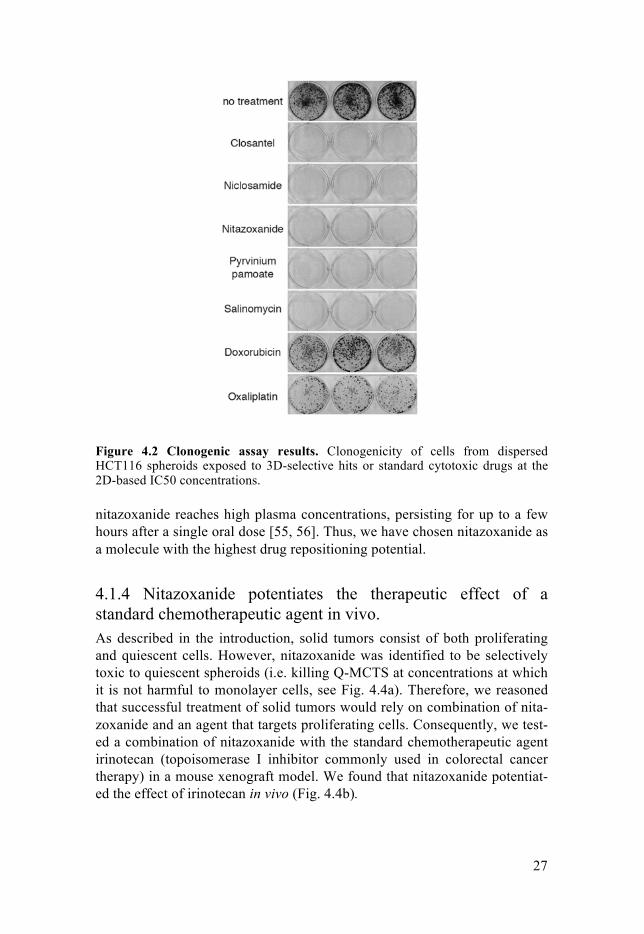
4.1.2 Compounds selectively toxic to Q-MCTS inhibit mitochondrial oxidative phosphorylation We performed screening of the Pharmakon 1600 library using GFP-based and TOX8 assays as readouts (For the screen overview, see Fig. 4.1). After hit validation, 41 Q-MCTS-active compounds were identified. Subsequently, we performed extensive dose-response experiments in monolayer cultures and Q-MCTS to identify compounds selectively toxic to spheroids. These experiments resulted in the identification of 12 “3D-selective” compounds. However, one needs to remember they were identified based on GFP-based viability assay, which does not directly indicate cellular death. Therefore, to identify molecules that are truly “3D-selective”, we tested the 12 compounds at their 2D-based IC50 concentrations (i.e. concentrations that caused 50% growth inhibition in monolayer cultures) in spheroid-based clonogenic assay. This experiment identified five molecules – nitazoxanide, niclosamide, closantel, pyrvinium pamoate and salinomycin – that fully abrogated re-growth of cells from dispersed spheroids (Fig. 4.2).

26
Figure 4.1 Overview of the screen
A literature search suggested that all five molecules target mitochondrial respiration, also known as oxidative phosphorylation (OXPHOS) – a pro-cess, in which most of cell’s ATP is produced. However, they inhibited OXPHOS through different mechanisms. Nitazoxanide, niclosamide and closantel were found to be uncouplers of mitochondrial membrane potential. Uncouplers are able to freely transfer protons across the mitochondrial membrane, equalizing the proton concentrations across the membrane, thereby causing an inability of the mitochondrion to utilize the proton gradi-ent to synthesize ATP through OXPHOS. The effects of the three uncouplers on OCR and sizes of hypoxic regions within spheroids closely resembled those of the standard uncoupler FCCP (Fig. 4.3a–d, h). Moreover, they in-duced depolarization of the mitochondria in JC-1 assay (Fig. 4.3g). In fact, all three compounds were found to share an identical structural pharmaco-phore. The other two compounds, pyrvinium pamoate and salinomycin, were found to inhibit OXPHOS through different mechanisms (Fig. 4.3e-h).
4.1.3 Nitazoxanide is a suitable candidate for drug repositioning Our experiments revealed that OXHPOS inhibitors are toxic to cells within spheroids exclusively upon prolonged and continuous exposure (see Paper I, Figure 4). Thus, we speculated that successful clinical application would rely on the high plasma concentration of the drug for a sustained period of time. Of the five final hit compounds, three – nitazoxanide, niclosamide and pyrvinium pamoate – are approved for use in humans. Of these, only
27
Figure 4.2 Clonogenic assay results. Clonogenicity of cells from dispersed HCT116 spheroids exposed to 3D-selective hits or standard cytotoxic drugs at the 2D-based IC50 concentrations.
nitazoxanide reaches high plasma concentrations, persisting for up to a few hours after a single oral dose [55, 56]. Thus, we have chosen nitazoxanide as a molecule with the highest drug repositioning potential.
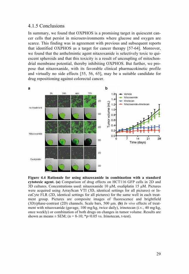
4.1.4 Nitazoxanide potentiates the therapeutic effect of a standard chemotherapeutic agent in vivo. As described in the introduction, solid tumors consist of both proliferating and quiescent cells. However, nitazoxanide was identified to be selectively toxic to quiescent spheroids (i.e. killing Q-MCTS at concentrations at which it is not harmful to monolayer cells, see Fig. 4.4a). Therefore, we reasoned that successful treatment of solid tumors would rely on combination of nita-zoxanide and an agent that targets proliferating cells. Consequently, we test-ed a combination of nitazoxanide with the standard chemotherapeutic agent irinotecan (topoisomerase I inhibitor commonly used in colorectal cancer therapy) in a mouse xenograft model. We found that nitazoxanide potentiat-ed the effect of irinotecan in vivo (Fig. 4.4b).

28
Figure 4.3 Characterization of effects of 3D-selective hit compounds on mito-chondrial respiration. (a-f), effects of FCCP (a), nitazoxanide (b), niclosamide (c), closantel (d), pyrvinium pamoate (e), and salinomycin (f) at various concentrations on OCR in 70,000 HCT116 cells, as measured by Seahorse XF analyzer. Loss of stimulation of OCR by addition of FCCP after uncoupler-induced mitochondrial respiration shutdown is highlighted with orange circles (b-d). Final hit compounds, oligomycin or FCCP, were added as indicated with dotted lines. Results are shown as mean ± SD; (n ≥ 3). (g) Left, effects of the final hit compounds at their 2D-based IC50 concentrations and CCCP (2.5 µmol/L) on mitochondrial membrane potential in HCT116 cells (2,500/well). Results in the graph are shown as means of JC-1 red fluorescence per cell + SD; (n ≥ 7). Right, fluorescence composite pictures of treated HCT116 cells. Cell nuclei were stained with Hoechst 33342 and polarized mito-chondria were stained with JC-1 probe. All pictures were acquired using identical settings. Magnification used was x20. (h) Effects of the final hit compounds or CCCP on hypoxia within HCT116 GFP Q-MCTS. Spheroids were treated with CCCP (2.5 µmol/L), nitazoxanide (3 µmol/L), niclosamide (1 µmol/L), closantel (15 µmol/L), pyrvinium pamoate (1 µmol/L), or salinomycin (2 µmol/L) for 4 or 24 hours. Subsequently, spheroids were treated with pimonidazole, sectioned, and hy-poxia was visualized by staining for pimonidazole adducts. Scale bar, 250 mm.
29
4.1.5 Conclusions In summary, we found that OXPHOS is a promising target in quiescent can-cer cells that persist in microenvironments where glucose and oxygen are scarce. This finding was in agreement with previous and subsequent reports that identified OXPHOS as a target for cancer therapy [57-64]. Moreover, we found that the anthelmintic agent nitazoxanide is selectively toxic to qui-escent spheroids and that this toxicity is a result of uncoupling of mitochon-drial membrane potential, thereby inhibiting OXPHOS. But further, we pro-pose that nitazoxanide, with its favorable clinical pharmacokinetic profile and virtually no side effects [55, 56, 65], may be a suitable candidate for drug repositioning against colorectal cancer.
Figure 4.4 Rationale for using nitazoxanide in combination with a standard cytotoxic agent. (a) Comparison of drug effects on HCT116 GFP cells in 2D and 3D cultures. Concentrations used: nitazoxanide 10 µM, oxaliplatin 15 µM. Pictures were acquired using ArrayScan VTI (3D, identical settings for all pictures) or In-cuCyte FLR (2D, identical settings for all pictures) for the same well in each treat-ment group. Pictures are composite images of fluorescence and brightfield (3D)/phase-contrast (2D) channels. Scale bars, 500 µm. (b) In vivo effects of treat-ment with nitazoxanide (gavage, 100 mg/kg, twice daily), irinotecan (i.v., 40 mg/kg, once weekly) or combination of both drugs on changes in tumor volume. Results are shown as means ± SEM; (n = 8-10; *p<0.05 vs. Irinotecan, t-test).

30
4.2 Paper II Large-Scale Gene Expression Profiling Platform for Identification of Con-text-Dependent Drug Responses in Multicellular Tumor Spheroids Wojciech Senkowski, Malin Jarvius, Jenny Rubin, Johan Lengqvist, Mats Gustafsson, Peter Nygren, Kim Kultima, Rolf Larsson and Mårten Fryknäs Cell Chemical Biology, 23(11); 1428-1438 (2016)
4.2.1 Background and purpose In the Paper II, we aimed to apply for the first time a high-throughput gene-expression profiling method to study effects of chemical perturbations in MCTS. As demonstrated in the Paper I, distinct cell culture models (i.e. monolayer cultures and Q-MCTS) from the same cell line are differentially sensitive to the same treatments. Thus, we speculated that it would likely be possible to observe early model-specific drug responses on the gene-expression level. In particular, we were interested in identifying processes that quiescent cells use to protect themselves from effects of OXPHOS in-hibitors. We hoped that we could identify targets, inhibition of which would result in synergistic anticancer activity with OXHPOS inhibitors in quiescent cells.
Figure 4.5 Schematic representation of experimental design. Three distinct mod-els (monolayer, P-MCTS and Q-MCTS) were treated with 22 drugs in dose-response for 6 hours. Subsequently, global gene-expression profiles were obtained using L1000 Gene Expression Profiling. Experiments were replicated 4 times (cells from 4 independent passages), resulting in 1065 independent model/compound profiles (Right, Gene-expression profiles (978 directly measured genes) of cells in 3 distinct models. Presented are all 1065 expression profiles. Data were hierarchically clus-tered vertically (one minus Pearson correlation distance metric).

31
Figure 4.6 Up-regulation of mevalonate pathway genes in Q-MCTS treated with OXPHOS inhibitors. (a) Results of the analysis of HumanCyc [66] pathway database. For each model, gene sets consisting of top 30 up-regulated genes (select-ed from landmarks (measured) gene space) by treatment with OXHPOS inhibitors were generated. Then, they were analyzed for enrichment in genes belonging to specific metabolic pathways. (b) Vehicle (DMSO)-normalized expression of 5 genes from mevalonate pathway in 3 distinct models treated with escalating doses of OXPHOS inhibitors or standard cytotoxic drugs. Results come from 4 independent experiments (each dose is replicated 4 times).
4.2.2 Application of L1000 Gene Expression profiling to study 3D cell cultures at a large scale For the experiments, we used 3 distinct cellular models: monolayer cell cul-tures, P-MCTS (‘proliferative spheroids’, formed with medium exchange during the culture period) and Q-MCTS (‘quiescent spheroids’, formed without medium change). All experiments were performed in biological quadruplicates, in 384-well microplates. We have exposed the three models to a panel of 22 drugs. Ten of the 22 were OXPHOS inhibitors (five final hit compounds from Paper I and five well-characterized reference compounds). The remaining 12 included standard cytotoxic drugs, kinase inhibitors and spheroid-relevant compounds (identified as potent hits in the screen from Paper I, albeit without reported activity on mitochondrial respiration). All drugs were used in dose-response setup with at least 3 different concentra-tions for each drug (for details, see Paper II). After 6-hour incubation with the compounds, cells were lysed (lysis protocol differed between models, since we have developed a new protocol suitable for 3D cell cultures – for details, see Paper II), supernatants frozen and shipped to Genometry. Gene expression profiling and all necessary quality checks were performed at Ge-nometry’s facility in Cambridge, MA, USA. For the overview of the experi-mental setup, see Fig. 4.5.
32
4.2.3 The dataset The experiments resulted in the generation of a dataset consisting of 1065 gene-expression profiles (Fig. 4.5). The analysis revealed profound trascrip-tional differences between the three models. Furthermore, to validate the dataset, we challenged various treatment responses against a reference com-pound signature database (LINCS; http://www.lincscloud.org/). All of the treatment-induced signatures we tested (both monolayer- and spheroid-based) linked correctly to their respective perturbagens in LINCS. This con-firms the reproducibility and quality of the dataset.
4.2.4 Up-regulation of mevalonate-pathway genes upon OXPHOS inhibition in Q-MCTS In order to identify model-specific responses to OXHPOS inhibitors, we performed a pathway database analysis. Briefly, we identified 30 genes that were most up-regulated upon exposure to OXPHOS inhibitors in each of the models (monolayer, P-MCTS and Q-MCTS). Then, we checked if any of these three gene sets was enriched in genes belonging to a specific metabolic pathway. The analysis revealed that OXPHOS inhibition resulted in strong up-regulation of the mevalonate-pathway genes in Q-MCTS, but not in P-MCTS or monolayer cultures (Fig. 4.6).
4.2.5 Synergistic activity of OXPHOS inhibitors and statins in Q-MCTS The above observations indicate that the mevalonate pathway up-regulation could serve as a protective response in quiescent, metabolically stressed cells upon OXPHOS inhibition. Therefore, we were interested to see if the simul-taneous inhibition of OXPHOS and the mevalonate pathway could result in synthetic lethality. Thus, we have performed combination treatment experi-ments using OXPHOS inhibitors and statins, inhibitors of mevalonate syn-thesis (and widely-used cholesterol-lowering drugs). We found that OXPHOS inhibitors and statins are synergistically toxic to Q-MCTS (exem-plified by nitazoxanide – Fig. 4.7a; and salinomycin – Fig 4.7d). These re-sults were confirmed using spheroids from other cell lines (A549 GFP, Fig. 4.7b; HT-29 GFP, Fig. 4.7c) and also in clonogenic assay (Fig. 4.7e).
4.2.6 Conclusions In summary, we applied for the first time a high-throughput gene-expression profiling method to study drug treatment effects in complex 3D cultures.

33
Figure 4.7 Therapeutic synergy of simvastatin and nitazoxanide. Combination treatment of HCT116 (a) A549 (b) or HT-29 (c) Q-MCTS with simvastatin and nitazoxanide. The values are the average percent inhibition of mean spheroid GFP intensity (i.e. percent toxicity). To visualize synergy, we applied a coloring scheme in which red squares indicate synergistic interaction and blue indicate antagonistic interaction. Q-MCTS were treated for 72 hrs, prior to viability measurements. Con-centrations used (2-fold dilutions): nitazoxanide 0.2 – 6.4 µM (HCT116) or 1 – 32 µM (A549 and HT-29) simvastatin 0.125 – 32 µM (all); n=4. Results are representa-tive of at least 3 independent experiments. (d) Bright-field/fluorescent (composite) microphotograph of a single combination experiment replicate in HCT116 Q-MCTS. Concentrations used (2-fold dilutions): salinomycin 0.1 – 3.2 µM; simvas-tatin 0.125 – 32 µM. Image acquisition settings were identical in each well. (e) Clonogenicity of cells from dispersed HCT116 Q-MCTS after 72-hr exposure to nitazoxanide and/or simvastatin at indicated concentrations. Results are representa-tive of nine replicates in three independent experiments.
With this approach, we identified unique model-specific drug responses. In particular, we found that cells within Q-MCTS up-regulate the mevalonate pathway genes upon inhibition of OXPHOS. Moreover, we found that OXPHOS inhibitors synergize with statins to eradicate cells in Q-MCTS. We believe that the described method could find a broad range of applications at different stages of drug development. Moreover, the identified drug combi-nation could have potential therapeutic implications for targeting quiescent cancer cells in in vivo solid tumors.

34
4.3 Paper III Drug combination screening in multicellular tumor spheroids identifies syn-thetic lethalities in quiescent cancer cells. Wojciech Senkowski, Madiha Nazir, Mats Gustafsson, Peter Nygren, Rolf Larsson and Mårten Fryknäs Manuscript (2017)
4.3.1 Background and purpose As demonstrated in Paper I, successful eradication of quiescent cancer cells from Q-MCTS is dependent on prolonged and continuous exposure to nita-zoxanide or other OXPHOS inhibitors (for details, see section 4.1.3 and Pa-per I, Figure 4). This could pose a substantial obstacle in future preclinical and clinical investigations concerning repurposing of nitazoxanide. Thus, it might be beneficial to identify molecules that increase anticancer activity of nitazoxanide in quiescent cancer cells. In order to do that, in Paper III we performed a drug combination screening in Q-MCTS of the library consist-ing of 1,650 pharmacologically active compounds.
4.3.2 Drug combination screening in Q-MCTS For the screen, we used Q-MCTS formed from HCT116 cells. We performed two separate screens in parallel (Fig. 4.8a). In the first one, library drugs were applied in combination with nitazoxanide. In the other (i.e. ‘counter-screen’), Q-MCTS were exposed to the library drugs without nitazoxanide addition. We used the GFP-based measurement of spheroid viability (see section 3.6.2 and Paper I for details). For each library compound we calcu-lated a ratio of the combination-treated spheroid viability to the viability of the spheroid treated with the compound alone. Subsequently, for further investigation, we selected 64 compounds that caused a pronounced potentia-tion of nitazoxanide’s efficacy (Fig. 4.8b). In order to focus on the most po-tent hits, we then tested these 64 compounds at a range of concentrations in combination with nitazoxanide (also used at different concentrations). This experiment resulted in the selection of 14 molecules that demonstrated the most pronounced synergistic activity (according to Bliss Independence mod-el) when combined with nitazoxanide (Fig. 4.8c).
4.3.3 Identification of drug combinations targeting Q-MCTS Next, we aimed to focus on compounds that demonstrate general activity against nutrient-deprived and hypoxic quiescent cells (Q-MCTS) rather than

35
Figure 4.8 Screen overview. (a) Schematic representation of the combinatorial screen design and selection criteria. (b) Ratios of combination-treated spheroid via-bility to the viability of spheroid exposed to compound alone (+NTZ/-NTZ ratio), calculated for each library compound and ordered from the lowest to the highest. Dashed lines indicate ‘primary potentiating hit’ (red) or ‘primary antagonistic hit’ (blue) thresholds. (c) A plot representing primary potentiating (red) or antagonistic (blue) hits ordered by synergy score from highest to lowest. Synergy scores for each compound are sums of excess over Bliss Independence values (see section 3.10) calculated for each concentration combination in 3x3 concentration matrix. Dashed lines indicate ‘synergistic hit’ (red) or ‘antagonistic hit’ (blue) thresholds.
a single cell line. Thus, we performed subsequent combination experiments using spheroids formed with an additional colorectal cancer cell line, HT-29. These experiments resulted in the selection of 10 molecules, toxic to Q-MCTS when combined with nitazoxanide in both cell lines (HCT116 and HT-29). Subsequently, in order to identify molecules with most pronounced synergistic activity in combination with nitazoxanide, we performed sphe-roid-based clonogenic assay. These experiments revealed seven molecules that fully abrogated the colony formation capacity of cells from Q-MCTS of both cell lines (Fig. 4.9). Notably, all of the seven molecules turned out to possess different primary molecular targets.
4.3.4 Conclusions In Paper III we applied MCTS for high-throughput screening of drug combi-nations. Using this approach, we were able to identify novel, previously un-

36
recognized drug interactions. The final seven hit compounds demonstrated pronounced synergistic activity when combined with nitazoxanide to eradi-cate nutrient-deprived and hypoxic quiescent cancer cells in spheroids from two different cell lines. Moreover, all final hit compounds target different cellular mechanisms. In conclusion, this work provides a rationale for per-forming combinatorial drug screening using 3D cell cultures.
Figure 4.9 Final hit compounds (a) Clonogenicity of cells from dispersed HCT116 Q-MCTS after 72-hr exposure to 1.5-µM nitazoxanide and/or 2-µM AS-252424,0.5-µM salinomycin, 2-µM LDE225, 4-µM ketoconazole, 0.5-µM nanchangmycin,4-µM PH-797804 or 4-µM cyclosporine. Results are representative of six replicatesin two independent experiments. (b) Clonogenicity of cells from dispersed HT-29Q-MCTS after 72-hr exposure to 8-µM nitazoxanide and/or 2-µM AS-252424, 1.5-µM salinomycin, 10-µM LDE225, 10-µM ketoconazole, 1.5-µM nanchangmycin, 5-µM PH-797804 or 5-µM cyclosporine. Results are representative of six replicates intwo independent experiments.

37
4.4 Paper IV Targeting tumor cells based on PDE3A expressionMadiha Nazir, Wojciech Senkowski, Frida Nyberg, Kristin Blom, Per-Henrik Edqvist, Claes Andersson, Mats Gustafsson, Peter Nygren, Rolf Larsson and Mårten FryknäsManuscript (2017)
4.4.1 Background and purpose Paper IV builds upon a previous report from our research group, in which phenotypic drug screening using a panel of cancer cell lines revealed that cervical cancer cell line HeLa is selectively vulnerable to inhibitors of phos-phodiesterase (PDE) enzymes [67]. This sensitivity was correlated with overexpression of phosphodiesterase 3A (PDE3A) mRNA in HeLa cells. Recently, it was also demonstrated that other cell lines overexpressing PDE3A mRNA are selectively sensitive to PDE inhibition [68]. Therefore, in Paper IV we aimed to evaluate, using more relevant cellular models, the applicability of PDE3A expression as a potential predictive biomarker of sensitivity to PDE inhibitors.
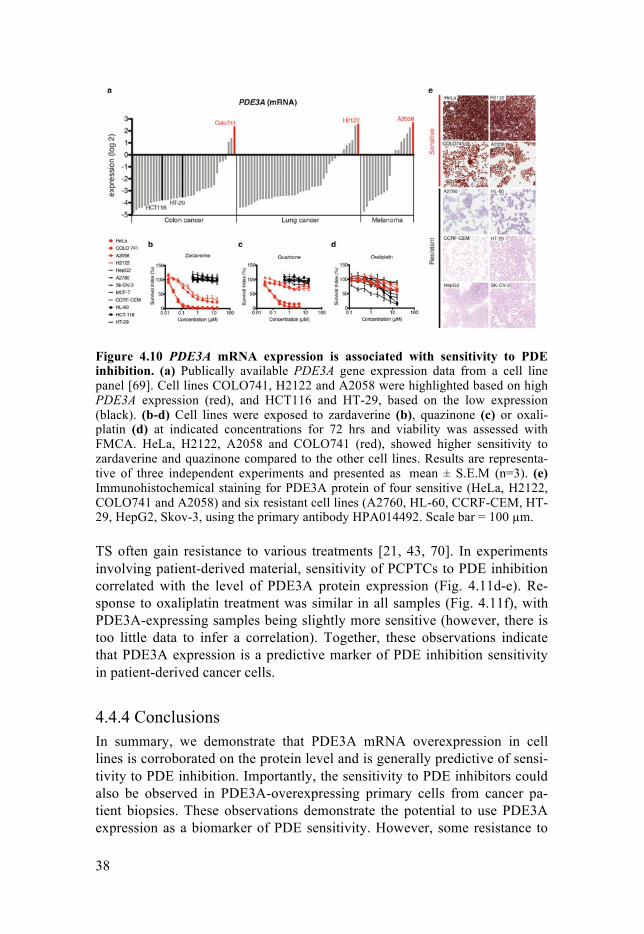
4.4.2 PDE3A overexpression predicts sensitivity to PDE inhibition First, we confirmed that the PDE3A mRNA overexpression is correlated with sensitivity to treatment with PDE inhibitors, zardaverine and quazinone, but not with a standard cytotoxic drug oxaliplatin in a panel of cell lines (Fig. 4.10a-d). Subsequently, we were interested to see whether the PDE3A mRNA overexpression in cell lines could also be observed at the protein level. We confirmed this hypothesis with immunohistochemical stainings, which revealed that cell lines reported to overexpress PDE3A mRNA also stain positively for PDE3A protein (Fig. 4.10e).
4.4.3 Sensitivity to PDE inhibition in other cell culture models Next, we set out to evaluate the effects of PDE inhibitors in other in vitro cancer models, i.e. in MCTS and primary cultures of patient tumor cells (PCPTCs). For MCTS formation, we used two PDE3A-overexpressing cell lines, HeLa and A2058 and three PDE3A-negative cell lines, HCT116, HT-29 and HepG2. While HeLa and A2058 cells, when cultured as MCTS, re-tained their PDE3A protein overexpression, only HeLa MCTS were sensi-tive to treatment with zardaverine and quazinone (Fig. 4.11a-b). All cell lines were resistant to oxaliplatin when grown as MCTS (Fig. 4.11c). These observations are in accordance with previous reports that cells grown as MC-
38
Figure 4.10 PDE3A mRNA expression is associated with sensitivity to PDE inhibition. (a) Publically available PDE3A gene expression data from a cell line panel [69]. Cell lines COLO741, H2122 and A2058 were highlighted based on high PDE3A expression (red), and HCT116 and HT-29, based on the low expression (black). (b-d) Cell lines were exposed to zardaverine (b), quazinone (c) or oxali-platin (d) at indicated concentrations for 72 hrs and viability was assessed with FMCA. HeLa, H2122, A2058 and COLO741 (red), showed higher sensitivity to zardaverine and quazinone compared to the other cell lines. Results are representa-tive of three independent experiments and presented as mean ± S.E.M (n=3). (e) Immunohistochemical staining for PDE3A protein of four sensitive (HeLa, H2122, COLO741 and A2058) and six resistant cell lines (A2760, HL-60, CCRF-CEM, HT-29, HepG2, Skov-3, using the primary antibody HPA014492. Scale bar = 100 µm.
TS often gain resistance to various treatments [21, 43, 70]. In experiments involving patient-derived material, sensitivity of PCPTCs to PDE inhibition correlated with the level of PDE3A protein expression (Fig. 4.11d-e). Re-sponse to oxaliplatin treatment was similar in all samples (Fig. 4.11f), with PDE3A-expressing samples being slightly more sensitive (however, there is too little data to infer a correlation). Together, these observations indicate that PDE3A expression is a predictive marker of PDE inhibition sensitivity in patient-derived cancer cells.
4.4.4 Conclusions In summary, we demonstrate that PDE3A mRNA overexpression in cell lines is corroborated on the protein level and is generally predictive of sensi-tivity to PDE inhibition. Importantly, the sensitivity to PDE inhibitors could also be observed in PDE3A-overexpressing primary cells from cancer pa-tient biopsies. These observations demonstrate the potential to use PDE3A expression as a biomarker of PDE sensitivity. However, some resistance to

39
PDE inhibitors in PDE3A-expressing cell lines was observed in the MCTS experiments. Thus, future research on application of PDE3A expression as a biomarker needs to involve the elucidation of potential resistance mecha-nisms.
Figure 4.11 Sensitivity to PDE inhibition in MCTS and PCPTCs. (a-c) MCTS viability, assessed with CellTiter-Glo 3D, after 72-hr exposure to zardaverine (a), quazinone (b) or oxaliplatin (c). Results are representative of three independent experiments and presented as mean ± S.E.M (n=3). (d-f) Viability of PCPTCs, assessed with FMCA after 72-hr exposure to zardaverine (d), quazinone (e) or oxal-iplatin (f). Results are presented as mean (n=2).

40
5. Summary and final remarks
5.1 New assays – development and application This work presents a research endeavor to develop and apply novel HTS assays using MCTS as a cellular model. These new methods include:
• Optimization of cell culture conditions to obtain three distinct cellular
models from the same cell line: cells growing as monolayer, prolifera-tive spheroids (P-MCTS) and quiescent spheroids (Q-MCTS) (Papers I and II);
• Spheroid-based HTS assay, utilizing cells constitutively expressing GFP in order to use GFP fluorescence as a surrogate marker of spheroid via-bility (Paper I);
• Application of L1000 Gene Expression Profiling to study drug-induced changes in global gene-expression in 3D cell cultures (Paper II);
• Spheroid-based screening platform to identify novel, previously unrec-ognized drug combinations (Paper III).
5.2 Application of MCTS in HTS yields novel findings Using these new assays, we focused on identifying processes that could be targeted to eradicate nutrient-deprived and hypoxic quiescent cancer cells. In order to achieve that, we used Q-MCTS as an in vitro cellular model. We found that nutrient-deprived quiescent cells are selectively vulnerable to inhibition of OXPHOS (Paper I). This finding is in accordance with a grow-ing body of evidence suggesting OXPHOS as a promising target in drug-resistant dormant cancer cells [58, 60, 61, 71]. We also found an FDA-approved anthelmintic drug nitazoxanide to be selectively toxic to quiescent cancer cells through uncoupling of mitochondrial membrane potential (Paper I). Anticancer activity in vivo, favorable pharmacokinetic profile and minor side effects render nitazoxanide a promising candidate for drug repurposing. Notably, recently there has been an increased interest in anti-diabetic drug metformin, which has been found to inhibit complex I of the mitochondrial electron transport chain, thereby inhibiting OXPHOS [72-74]. Metformin

41
has been reported to reduce the risk of developing certain types of cancer and demonstrated anticancer activity in vitro and in mouse models [59, 74-76]. However, as of now, it has failed to show clinical benefits in eradicating an already-existing cancer [77]. The reason for this might be low metformin plasma concentrations and unfavorable pharmacokinetic profile [76, 78]. Another notable example is IACS-10759, also an inhibitor of complex I, which is currently undergoing Phase I clinical trial in acute myeloid leuke-mia (AML) patients [79]. In Paper I, we observed that to successfully eradicate nutrient-deprived cells in Q-MCTS by OXPHOS inhibition, there was a requirement for prolonged and continuous exposure to the treatment. Thus, in Papers II and III we set out to identify processes that could be co-targeted for enhanced anticancer activity of OXPHOS inhibitors. In Paper II, using high-throughput gene expression profiling in 3D cell cultures, we were able to observe Q-MCTS-specific up-regulation of the mevalonate-pathway genes upon OXPHOS inhibition. Consequently, inhibitors of the mevalonate pathway – widely used cholesterol-lowering statins – demonstrated synergistic activity with OXPHOS inhibitors in Q-MCTS. In Paper III, we performed a combinatorial drug screen in Q-MCTS and identified several molecules that synergized with nitazoxanide. These two papers (II and III) demonstrate novel ap-proaches to characterize drug responses in various cell culture models and present novel, potentially therapeutically relevant findings. However, the in vivo effects of combining OXPHOS inhibitors with other agents, such as statins, are difficult to predict. Notably, rare but severe side effects of statins, including myopathy or rhabdomyolysis, were previously associated with inhibition of mitochondrial function [80]. Therefore, putative clinical tests of this and other combinations would have to be preceded with thorough toxici-ty experiments in animal models.
5.3 Culture conditions determine drug response Perhaps most importantly, this work demonstrates the relevance of cell cul-ture conditions in preclinical drug discovery endeavors. While much of cur-rent anticancer drug development seems to be focused on cancer genetics, the impact of cell culture conditions tends to be forgotten or neglected. As we demonstrate in all of the papers in this thesis, drug response of cancer cells can be profoundly changed by the physicochemical factors, such as local nutrient concentrations or pH. For instance, in Paper II, cells of the same cell line (i.e. with identical genetic background) are grown in three different culture conditions. As gene-expression profiling reveals, this pro-foundly impacts not only baseline gene expression of each model, but also response to treatment at both genetic and phenotypic levels. In Paper IV, a

42
cell line that overexpresses PDE3A is sensitive to PDE inhibition only when grown as monolayer and not when grown as spheroids, despite maintaining high PDE3A expression in both monolayer and spheroid culture. This sug-gests that changing culture conditions alone can induce unforeseen drug resistance mechanisms. These examples, in my mind, clearly demonstrate that culture conditions should be carefully considered in all cell-based exper-iments and that they deserve an emphasis at least equal to the one put upon genetics.
5.4 A look ahead 3D cell cultures have now become well-established tools in evaluating and developing cancer therapeutics. Recently, the emphasis has been put upon using patient-derived material (i.e. ‘organoids’) rather than cell lines for 3D cell culture experiments. With a few encouraging reports on the possibility of expansion and 3D culture of tumor cells from patients [81-83], one can easily see benefits of this approach over using standard cell lines. First, con-trary to cell lines, patient cells do not undergo a process of multiple passag-ing that reduces clonal diversity of the culture. Second, establishment of stable cancer cell lines involves usually a prolonged period of time, while patient-derived cultures are almost immediately ready-to-use. Third, with high take rates (successful culture and expansion in vitro), patient-derived cells provide an opportunity to more easily reproduce in vitro the genetic heterogeneity of cancer.
However, all current organoid-based drug-screening approaches fail to take many of the important culture conditions into account. In my opinion, the problem lies with the pre-assumption to simulate the whole, often large, heterogeneous tumor using a single type of an in vitro model. While empha-sis is put upon growing cells as multicellular aggregates and using extracel-lular matrix scaffolds, factors such as nutrient concentrations, pH, access to oxygen etc. remain non-physiological. As shown in Paper II, P-MCTS and Q-MCTS, both three-dimensional and both with the same genetic back-ground, still can respond to treatments in a completely different way, justbecause they persist in different nutrient concentrations. Thus, I believe thatin the future spheroid- and organoid-based research, one should try to notjust grow cells in 3D, but also to apply different culture conditions for thesame cellular material, in order to simulate as many different tumor-specificmicroenvironments as possible. As different cell populations within the samein vivo tumor can persist in completely different conditions, we need to ad-dress each of these microenvironments separately, if we aim to successfullytreat the whole tumor with the means of chemotherapy. I sincerely hope thatthese issues will be widely recognized and addressed in the near future.

43
6. Acknowledgements
This work could not be done without involvement of many great people, who contributed directly or indirectly. I feel that everyone, who made it pos-sible for me to get through this exciting, often frustrating, but also very re-warding period of life that was my PhD studies, deserves a mention here. So it’s gonna be long. That said, I would like to thank:
My supervisor, Mårten Fryknäs – you rock, man! I have learned from you much more than you probably suspect and I’m not gonna list all that here, cause it would take way too much space. So let me just say that working with you felt more like a scientific collaboration between equal partners, rather than a standard student-supervisor relationship. And, most important-ly, it also felt more like a friendship, and that meant a lot to me. Thank you for your great leadership, that you had time to discuss stuff with me basically every day, your scientific advice and life wisdom. I’m sure we’ve got more exciting challenges coming in the future!
My co-supervisor, Rolf Larsson, for being an amazing group leader and teaching me that sometimes one just needs to stop for a moment and say: “This is good enough” and submit the damn paper! I also deeply admire your constant excitement about science and how quickly you are able to move on after failures (which happen much more often than successes in science, but, thanks to you, it was easier for me to move on quickly as well).
My co-supervisor, Peter Nygren, for teaching me that in the end of the day we work for cancer patients and our findings need to be applicable. I think that in science, as much as we need excitement, we also need to always look at our results critically and question them all the time – so thanks for keeping us firmly on the ground! You and Rolf provided a perfect balance!
My co-supervisor, Mats Gustafsson, for your everyday kindness and for providing an invaluable insight of the computational scientist. As cancer research is relying on big data more and more, we need people like you, who can help us in making sense of all that.
Our awesome lab technicians and research assistants who taught me how to operate all the complicated machinery without causing millions of kronor

44
worth of damage; Lena Lenhammar, for your smile when I came to the department for the first time and your inexhaustible patience for my endless questions. Gunilla Frenne, for knowing everything and being always happy to share the knowledge. Nasrin Najafi, Malin Berglund, Anna-Karin Lannergård, Lena Fredriksson, Annika Jonasson, Jakob Rudfeldt and late Christina Leek, for making the place run smoothly, answering all my questions with a smile and being lots of fun both at work and outside of it.
Kristin Blom, for a delightful sense of humor and going an extra mile to help everyone. You rule!
All my collaborators and co-authors; Malin Jarvius for staying ahead of hardware problems with positive attitude (and funny jokes!); Xiaonan Zhang, Stig Linder and Maria Hägg-Olofsson from Karolinska Institute for great insights, discussions and the opportunity for me to learn a few techniques in your lab; Jenny Rubin for being lots of fun in the lab – it helps a lot!; Madiha Nazir for your kindness and sharing the burden of cell culture; Claes Andersson for making complicated issues approachable and a great sense of humor; Vendela Parrow, for a unique industry insight, inspir-ing discussions and advice about where to organize my PhD party; Justin Lamb and Willis Read-Button from Genometry, for incredible profession-alism that I could observe and learn from, friendliness and a great dinner in Boston; Jan Siljason, for coming up with miraculously good immunohisto-chemistry stainings that saved my whole project; Per-Henrik Edqvist and IngMarie Olsson for developing new IHC protocol and quality work; Kim Kultima, Julia Steinmetz and Johan Lengqvist for masterful proteomics; Urban Höglund and Ruben Isacson from Adlego AB for skillful perfor-mance of animal experiments.
Awesome roommates over the years; Henning Karlsson, for cool scientific discussions and your passion for science; Sara Strese, for great conversa-tions about books and life in general, so I could procrastinate even more; Caroline Haglund, Frida Nyberg and Sharmineh Mansoori, for being always friendly and for exchanging fresh gossip ;) ; Obaid Aftab and Mu-hammad Kashif, for your kindness and agreeing, without hesitation, to change your plans when I needed help with moving apartments on the day I broke a few ribs; Åsa Fransson, for being one of the most positive people I know.
Jessica and Gabor Schubert, Martin Dahlö, Björn Viklund, Sebastian DiLorenzo, Mao Mao, Christofer Bäcklin, Markus Mayrhofer and eve-ryone else from the Medsci Social group for warm welcome to the lab, awe-some nerdy discussions, keeping me updated with technological novelties I

45
didn’t even know existed, after-work dinners and keeping it in English when needed!
All other people that I met often in our corridor: Shibu Krishnan, Payam Emami, Stephanie Herman and Nils Anlind for joyful fikas and lunches; Effie Chantzi, Sadia Hassan, Anna-Karin Lidehäll, Kristin Bryon, Ilma Bertulyte, Hugo Kohnke, David Munro, Håkan Melhus, Gabriella Scor-do, Mia Wadelius, Pär Hällberg, Matilda Johnell, Eva Prado, Eva Frey-hult, Alexandra Ask, Anna Foyer, Nadja Lundström, Ulrica Ramqvist, Anna Lundberg, Joachim Gullbo, Anna Segerman, Sofie Collin, Lisa Rebello and Anna-Karin Hamberg, for contributing to the great atmos-phere at the department.
I would also like to thank all of my friends outside of work, especially:
The Polish bunch in Sweden – Kuba, Ula, little Leon, Tomek and Magda (and the cats: Kling, Klang, Docent and Tequila) – for brightening the Swedish darkness with humor, dinners, parties and hair everywhere.
Kim, Maciek and Delphine, for being awesome, positive people and for forcing me to participate in dinners and to socialize. Keep it up, guys!
Paweł and Eugene from the Master Program in Applied Biotechnology, for making the whole experience memorable and for the epic Balkan Madness trip in the midst of refugee crisis!
My lifelong friends from Andrychów (and Uraz) – Ad., Niewiera, Mateusz, Miłosz, Tomek and WojtKas – for keeping me grounded and sane, for sticking together for all these years, and for all the years to come! You know I love you guys!
Most importantly, I would like to thank my family (in Polish):
Chciałbym serdecznie podziękować:
Całej rodzinie z Andrychowa, Gdańska, Krakowa, Warszawy, Kobyłki, Miasteczka Krajeńskiego, Grabówna, Osieka nad Notecią i Uppsali za nieprzerwane wsparcie i zainteresowanie!
Piotrkowi, za to, że jesteś najlepszym bratem i przyjacielem jakiego można mieć.
Rodzicom, za to, że jesteście zawsze gotowi mnie wysłuchać, wspieracie mnie w moich decyzjach i że zawsze mam dokąd wracać.

46
7. References
1. Hanahan, D. and R.A. Weinberg, The Hallmarks of Cancer. Cell, 2000. 100(1):p. 57-70.
2. Hanahan, D. and Robert A. Weinberg, Hallmarks of Cancer: The NextGeneration. Cell, 2011. 144(5): p. 646-674.
3. Torre, L.A., et al., Global cancer statistics, 2012. CA: A Cancer Journal forClinicians, 2015. 65(2): p. 87-108.
4. DeSantis, C.E., et al., Cancer treatment and survivorship statistics, 2014. CA:A Cancer Journal for Clinicians, 2014. 64(4): p. 252-271.
5. Chabner, B.A. and T.G. Roberts, Chemotherapy and the war on cancer. NatRev Cancer, 2005. 5(1): p. 65-72.
6. Dougan, M. and G. Dranoff, Immune Therapy for Cancer. Annual Review ofImmunology, 2009. 27(1): p. 83-117.
7. Mellman, I., G. Coukos, and G. Dranoff, Cancer immunotherapy comes of age.Nature, 2011. 480(7378): p. 480-489.
8. Hoos, A. and C. Britten, The immuno-oncology framework: Enabling a new eraof cancer therapy. Oncoimmunology, 2012. 1(3): p. 334-339.
9. Greaves, M. and C.C. Maley, Clonal evolution in cancer. Nature, 2012.481(7381): p. 306-313.
10. Hoos, A., Development of immuno-oncology drugs - from CTLA4 to PD1 to thenext generations. Nat Rev Drug Discov, 2016. 15(4): p. 235-247.
11. Swanton, C., Intratumor Heterogeneity: Evolution through Space and Time.Cancer Research, 2012. 72(19): p. 4875-4882.
12. Trédan, O., et al., Drug Resistance and the Solid Tumor Microenvironment.Journal of the National Cancer Institute, 2007. 99(19): p. 1441-1454.
13. Nowell, P.C., The clonal evolution of tumor cell populations. Science, 1976.194(4260): p. 23-28.
14. McGranahan, N. and C. Swanton, Biological and Therapeutic Impact ofIntratumor Heterogeneity in Cancer Evolution. Cancer Cell, 2015. 27(1): p. 15-26.
15. Sutherland, R.M., Cell and environment interactions in tumor microregions: themulticell spheroid model. Science, 1988. 240(4849): p. 177-184.
16. Kunz-Schughart, L.A., M. Kreutz, and R. Knuechel, Multicellular spheroids: Athree-dimensional in vitro culture system to study tumour biology. InternationalJournal of Experimental Pathology, 1998. 79(1): p. 1-23.
17. Kunz-Schughart, L.A., et al., The use of 3-D cultures for high-throughputscreening: The multicellular spheroid model. Journal of BiomolecularScreening, 2004. 9(4): p. 273-285.
18. Herrmann, R., et al., Screening for compounds that induce apoptosis of cancercells grown as multicellular spheroids. Journal of Biomolecular Screening,2008. 13(1): p. 1-8.

47
19. Fayad, W., et al., Identification of Agents that Induce Apoptosis of Multicellular Tumour Spheroids: Enrichment for Mitotic Inhibitors with Hydrophobic Properties. Chemical Biology and Drug Design, 2011. 78(4): p. 547-557.
20. Strese, S., et al., Effects of hypoxia on human cancer cell line chemosensitivity. BMC Cancer, 2013. 13(1): p. 1-11.
21. Karlsson, H., et al., Loss of cancer drug activity in colon cancer HCT-116 cells during spheroid formation in a new 3-D spheroid cell culture system. Exp Cell Res, 2012. 318.
22. Hait, W.N., Anticancer drug development: the grand challenges. Nat Rev Drug Discov, 2010. 9(4): p. 253-4.
23. Hay, M., et al., Clinical development success rates for investigational drugs. Nat Biotech, 2014. 32(1): p. 40-51.
24. DiMasi, J.A., H.G. Grabowski, and R.W. Hansen, Innovation in the pharmaceutical industry: New estimates of R&D costs. Journal of Health Economics, 2016. 47: p. 20-33.
25. Fox, S., et al., High-Throughput Screening: Update on Practices and Success. Journal of Biomolecular Screening, 2006. 11(7): p. 864-869.
26. Hart, C.P., Finding the target after screening the phenotype. Drug Discovery Today, 2005. 10(7): p. 513-519.
27. Swinney, D.C. and J. Anthony, How were new medicines discovered? Nature Reviews Drug Discovery, 2011. 10(7): p. 507-519.
28. Ashburn, T.T. and K.B. Thor, Drug repositioning: identifying and developing new uses for existing drugs. Nat Rev Drug Discov, 2004. 3(8): p. 673-683.
29. Kyle, A.H., J.H.E. Baker, and A.I. Minchinton, Targeting Quiescent Tumor Cells via Oxygen and IGF-I Supplementation. Cancer Research, 2012. 72(3): p. 801-809.
30. Minchinton, A.I. and I.F. Tannock, Drug penetration in solid tumours. Nat Rev Cancer, 2006. 6(8): p. 583-592.
31. Fluegen, G., et al., Phenotypic heterogeneity of disseminated tumour cells is preset by primary tumour hypoxic microenvironments. Nat Cell Biol, 2017. 19(2): p. 120-132.
32. Hirschhaeuser, F., et al., Multicellular tumor spheroids: An underestimated tool is catching up again. Journal of Biotechnology, 2010. 148(1): p. 3-15.
33. LaBarbera, D.V., B.G. Reid, and B.H. Yoo, The multicellular tumor spheroid model for high-throughput cancer drug discovery. Expert Opinion on Drug Discovery, 2012. 7(9): p. 819-830.
34. Spencer, V.A., R. Xu, and M.J. Bissell, Gene expression in the third dimension: The ECM-nucleus connection. Journal of Mammary Gland Biology and Neoplasia, 2010. 15(1): p. 65-71.
35. Ghosh, S., et al., Three-dimensional culture of melanoma cells profoundly affects gene expression profile: A high density oligonucleotide array study. Journal of Cellular Physiology, 2005. 204(2): p. 522-531.
36. LaRue, K.E.A., M. Khalil, and J.P. Freyer, Microenvironmental Regulation of Proliferation in Multicellular Spheroids Is Mediated through Differential Expression of Cyclin-Dependent Kinase Inhibitors. Cancer Research, 2004. 64(5): p. 1621-1631.
37. Pampaloni, F., E.G. Reynaud, and E.H.K. Stelzer, The third dimension bridges the gap between cell culture and live tissue. Nat Rev Mol Cell Biol, 2007. 8(10): p. 839-845.
38. Vinci, M., et al., Advances in establishment and analysis of three-dimensional tumor spheroid-based functional assays for target validation and drug evaluation. BMC Biology, 2012. 10.

48
39. Tung, Y.C., et al., High-throughput 3D spheroid culture and drug testing usinga 384 hanging drop array. Analyst, 2011. 136(3): p. 473-478.
40. Ivascu, A. and M. Kubbies, Rapid generation of single-tumor spheroids forhigh-throughput cell function and toxicity analysis. Journal of BiomolecularScreening, 2006. 11(8): p. 922-932.
41. Li, Q., et al., 3D models of epithelial-mesenchymal transition in breast cancermetastasis: High-throughput screening assay development, validation, and pilotscreen. Journal of Biomolecular Screening, 2011. 16(2): p. 141-154.
42. Sodunke, T.R., et al., Micropatterns of Matrigel for three-dimensional epithelialcultures. Biomaterials, 2007. 28(27): p. 4006-4016.
43. Senkowski, W., et al., Three-Dimensional Cell Culture-Based ScreeningIdentifies the Anthelmintic Drug Nitazoxanide as a Candidate for Treatment ofColorectal Cancer. Molecular Cancer Therapeutics, 2015. 14(6): p. 1504-1516.
44. Harada, C.Y., et al., Microenvironments and Cellular Characteristics in theMicro Tumor Cords of Malignant Solid Tumors. International Journal ofMolecular Sciences, 2012. 13: p. 13949-13965.
45. Lindhagen, E., P. Nygren, and R. Larsson, The fluorometric microculturecytotoxicity assay. Nature Protocols, 2008. 3(8): p. 1364-1369.
46. Zhang, J.H., T.D.Y. Chung, and K.R. Oldenburg, A simple statistical parameterfor use in evaluation and validation of high throughput screening assays.Journal of Biomolecular Screening, 1999. 4(2): p. 67-73.
47. Smiley, S.T., et al., Intracellular heterogeneity in mitochondrial membranepotentials revealed by a J-aggregate-forming lipophilic cation JC-1.Proceedings of the National Academy of Sciences, 1991. 88(9): p. 3671-3675.
48. Varia, M.A., et al., Pimonidazole: A Novel Hypoxia Marker for ComplementaryStudy of Tumor Hypoxia and Cell Proliferation in Cervical Carcinoma.Gynecologic Oncology, 1998. 71(2): p. 270-277.
49. Flanagan, S.P., ‘Nude’, a new hairless gene with pleiotropic effects in themouse. Genetics Research, 1966. 8(03): p. 295-309.
50. Pantelouris, E.M., Absence of Thymus in a Mouse Mutant. Nature, 1968.217(5126): p. 370-371.
51. Nehls, M., et al., New member of the winged-helix protein family disrupted inmouse and rat nude mutations. Nature, 1994. 372(6501): p. 103-107.
52. Duan, Q., et al., LINCS Canvas Browser: interactive web app to query, browseand interrogate LINCS L1000 gene expression signatures. Nucleic AcidsResearch, 2014. 42(W1): p. W449-W460.
53. Peck, D., et al., A method for high-throughput gene expression signatureanalysis. Genome Biology, 2006. 7(7): p. R61-R61.
54. Bliss, C.I., The toxicity of poisons applied jointly. Annals of Applied Biology,1939. 26(3): p. 585-615.
55. Stockis, A., et al., Pharmacokinetics of nitazoxanide after single oral doseadministration in 6 healthy volunteers. Int J Clin Pharmacol Ther, 1996. 34: p.349-51.
56. Stockis, A., et al., Nitazoxanide pharmacokinetics and tolerability in man usingsingle ascending oral doses. Int J Clin Pharmacol Ther, 2002. 40: p. 213-20.
57. LeBleu, V.S., et al., PGC-1α mediates mitochondrial biogenesis and oxidativephosphorylation in cancer cells to promote metastasis. Nat Cell Biol, 2014.16(10): p. 992-1003.
58. Viale, A., et al., Oncogene ablation-resistant pancreatic cancer cells depend onmitochondrial function. Nature, 2014. 514(7524): p. 628-632.
59. Birsoy, K., et al., Metabolic determinants of cancer cell sensitivity to glucoselimitation and biguanides. Nature, 2014. 508(7494): p. 108-112.

49
60. Alvero, A.B., et al., Targeting the Mitochondria Activates Two Independent Cell Death Pathways in Ovarian Cancer Stem Cells. Molecular Cancer Therapeutics, 2011. 10(8): p. 1385-1393.
61. Zhang, X., et al., Induction of mitochondrial dysfunction as a strategy for targeting tumour cells in metabolically compromised microenvironments. Nat Commun, 2014. 5.
62. Wolf, Dieter A., Is Reliance on Mitochondrial Respiration a “Chink in the Armor” of Therapy-Resistant Cancer? Cancer Cell, 2014. 26(6): p. 788-795.
63. Viale, A., D. Corti, and G.F. Draetta, Tumors and Mitochondrial Respiration: A Neglected Connection. Cancer Research, 2015. 75(18): p. 3687-3691.
64. Weinberg, S.E. and N.S. Chandel, Targeting mitochondria metabolism for cancer therapy. Nat Chem Biol, 2015. 11(1): p. 9-15.
65. Stockis, A., et al., Nitazoxanide pharmacokinetics and tolerability in man during 7 days dosing with 0.5 g and 1 g b.i.d. Int J Clin Pharmacol Ther, 2002. 40: p. 221-7.
66. Romero, P., et al., Computational prediction of human metabolic pathways from the complete human genome. Genome Biology, 2005. 6(1): p. R2-R2.
67. Fryknäs, M., et al., Phenotype-Based Screening of Mechanistically Annotated Compounds in Combination with Gene Expression and Pathway Analysis Identifies Candidate Drug Targets in a Human Squamous Carcinoma Cell Model. Journal of Biomolecular Screening, 2006. 11(5): p. 457-468.
68. de Waal, L., et al., Identification of cancer-cytotoxic modulators of PDE3A by predictive chemogenomics. Nat Chem Biol, 2016. 12(2): p. 102-108.
69. Wagner, K.W., et al., Death-receptor O-glycosylation controls tumor-cell sensitivity to the proapoptotic ligand Apo2L/TRAIL. Nature Medicine, 2007. 13(9): p. 1070-1077.
70. Jacks, T. and R.A. Weinberg, Taking the Study of Cancer Cell Survival to a New Dimension. Cell, 2002. 111(7): p. 923-925.
71. Wenzel, C., et al., 3D high-content screening for the identification of compounds that target cells in dormant tumor spheroid regions. Experimental Cell Research, 2014. 323(1): p. 131-143.
72. El-Mir, M.-Y., et al., Dimethylbiguanide Inhibits Cell Respiration via an Indirect Effect Targeted on the Respiratory Chain Complex I. Journal of Biological Chemistry, 2000. 275(1): p. 223-228.
73. OWEN, M.R., E. DORAN, and A.P. HALESTRAP, Evidence that metformin exerts its anti-diabetic effects through inhibition of complex 1 of the mitochondrial respiratory chain. Biochemical Journal, 2000. 348(3): p. 607-614.
74. Wheaton, W.W., et al., Metformin inhibits mitochondrial complex I of cancer cells to reduce tumorigenesis. eLife, 2014. 3: p. e02242.
75. Evans, J.M.M., et al., Metformin and reduced risk of cancer in diabetic patients. BMJ, 2005. 330(7503): p. 1304-1305.
76. Pollak, M.N., Investigating Metformin for Cancer Prevention and Treatment: The End of the Beginning. Cancer Discovery, 2012. 2(9): p. 778-790.
77. Kordes, S., et al., Metformin in patients with advanced pancreatic cancer: a double-blind, randomised, placebo-controlled phase 2 trial. The Lancet Oncology, 2015. 16(7): p. 839-847.
78. He, L. and Fredric E. Wondisford, Metformin Action: Concentrations Matter. Cell Metabolism, 2015. 21(2): p. 159-162.
79. Protopopova, M., et al., Abstract 4380: IACS-10759: A novel OXPHOS inhibitor which selectively kill tumors with metabolic vulnerabilities. Cancer Research, 2015. 75(15 Supplement): p. 4380.

50
80. Dirks, A.J. and K.M. Jones, Statin-induced apoptosis and skeletal myopathy.American Journal of Physiology - Cell Physiology, 2006. 291(6): p. C1208-C1212.
81. Gao, D., et al., Organoid Cultures Derived from Patients with AdvancedProstate Cancer. Cell, 2014. 159(1): p. 176-187.
82. Boj, Sylvia F., et al., Organoid Models of Human and Mouse Ductal PancreaticCancer. Cell, 2015. 160(1–2): p. 324-338.
83. van de Wetering, M., et al., Prospective Derivation of a Living OrganoidBiobank of Colorectal Cancer Patients. Cell, 2015. 161(4): p. 933-945.


Acta Universitatis UpsaliensisDigital Comprehensive Summaries of Uppsala Dissertationsfrom the Faculty of Medicine 1334
Editor: The Dean of the Faculty of Medicine
A doctoral dissertation from the Faculty of Medicine, UppsalaUniversity, is usually a summary of a number of papers. A fewcopies of the complete dissertation are kept at major Swedishresearch libraries, while the summary alone is distributedinternationally through the series Digital ComprehensiveSummaries of Uppsala Dissertations from the Faculty ofMedicine. (Prior to January, 2005, the series was publishedunder the title “Comprehensive Summaries of UppsalaDissertations from the Faculty of Medicine”.)
Distribution: publications.uu.seurn:nbn:se:uu:diva-320598
ACTAUNIVERSITATIS
UPSALIENSISUPPSALA
2017