higher perceptual functions - pclcognitrn.psych.indiana.edu/busey/q551/pdfs/week7.pdfis...
TRANSCRIPT

104
Object Recognition
-Segregation of function
-Visual hierarchy
-What and where (ventral and dorsal streams)
-Single cell coding and ensemble coding
-Distributed representations of object categories
-Face recognition
-Object recognition as a computational problem
Higher Perceptual Functions

105
Segregation of function exists already in theearly visual system:
M channel (magnocellular): from M-type retinalganglion cells to magnocellular LGN layers to layer IVBof V1; wavelength-insensitive in LGN, orientationselectivity in V1 (“simple cells”), binocularity anddirection selectivity in layer IVB; processing visualmotion.
P channel (parvocellular): from P-type retinal ganglioncells to parvocellular LGN layers to interblob regions oflayer III in V1; many cells in LGN show coloropponency, cells in interblob regions of V1 have strongorientation selectivity and binocularity (“complexcells”), channel is also called P-IB; processing visualobject shape.
Functional Segregation

106
Segregation of function can also be found atthe cortical level:
- within each area: cells form distinct columns.
- multiple areas form the visual hierarchy …
Functional Segregation

107
The Visual Hierarchy
van Essen and Maunsell, 1983

108van Essen et al., 1990
The Visual Hierarchy
109
-functional segregation of visual features into separate(specialized) areas.-increased complexity and specificity of neuralresponses.- columnar groupings, horizontal integration withineach area.-larger receptive fields at higher levels.-visual topography is less clearly defined at higherlevels, or disappears altogether.-longer response latencies at higher levels.- large number of pathways linking each segregatedarea to other areas.- existence of feedforward, as well as lateral andfeedback connections between hierarchical levels.
The Visual Hierarchy

110
The Architecture of Visual Cortex
Mishkin and Ungerleider, 1983
Lesion studies in the macaque monkey suggest that there aretwo large-scale cortical streams of visual processing:
Dorsal stream (“where”)
Ventral stream (“what”)
111
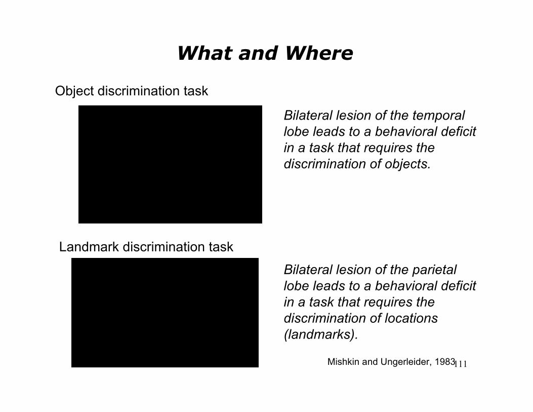
What and Where
Mishkin and Ungerleider, 1983
Object discrimination task
Landmark discrimination task
Bilateral lesion of the temporallobe leads to a behavioral deficitin a task that requires thediscrimination of objects.
Bilateral lesion of the parietallobe leads to a behavioral deficitin a task that requires thediscrimination of locations(landmarks).

112
The Architecture of Visual Cortex
motion
colorform
Lateral views of the macaque monkey brain

113
Single Cells and Recognition
What is the cellular basis for visual recognition (visuallong-term memory)?
1. Where are the cellular representationslocalized?
2. What processes generate theserepresentations?
3. What underlies their reactivation during recalland recognition?

114
Single Cells and Recognition
Visual recognition involves the inferior temporal cortex(multiple areas). These areas are part of a distributednetwork and are subject to both bottom-up (feature driven)and top-down (memory driven) influences.
Miyashita and Hayashi, 2000
115
Single Cells and Recognition
Characteristics of neural responses in IT:
1. Object-specific (tuned to object class), selectivefor general object features (e.g. shape)
2. Non-topographic (large RF)3. Long-lasting (100’s ms)
Columnar organization (“object feature columns”)Specificity has often rather broad range
(distributed response pattern)

116
Distributed Representations
Are there specific, dedicated modules (or cells) foreach and every object category?
No. – Why not?

117
Distributed Representations
Evidence → feature based and widely distributedrepresentation of objects across (ventral) temporalcortex.
What is a distributed representation?

118
Distributed Representations
Experiments conducted by Ishai et al.:
Experiment 1:1. fMRI during passive viewing2. fMRI during delayed match-to-sample
Experiment 2:1. fMRI during delayed match-to-sample with
photographs2. fMRI during delayed match-to-sample with line
drawings
Three categories: houses, faces, chairs.

119
Distributed Representations
Findings:
Experiment 1:Consistent topography in areas that most strongly
respond to each of the three categories.Modules?No - Responses are distributed (more so for non-face
stimuli)
Experiment 2:Are low-level features (spatial frequency, texture etc.)
responsible for the representation?No – line drawings elicit similar distributions of responses

120
Distributed Representations
From Ishai et al., 1999

121
Distributed Representations
From Ishai et al., 1999
houses
faces
chairs

122
Face recognition achieves a very high level ofspecificity – hundreds, if not thousands ofindividual faces can be recognized.
Face Recognition
Visual agnosia specific to faces: prosopagnosia.
High specificity of face cells → “gnostic units”,“grandmother cells”
Many face cells respond to faces only – andshow very little response to other object stimuli.

123
Face Recognition
Typical neural responses in the primate inferior temporalcortex:
Desimone et al., 1984

124
Face Recognition
Face cells (typically) do not respond to:1. “jumbled” faces2. “partial” faces3. “single components” of faces (although some
face-component cells have been found)4. other “significant” stimuli
Face cells (typically) do respond to:1. faces anywhere in a large bilateral visual field2. faces with “reduced” feature content (e.g. b/w,
low contrast)
Face cell responses can vary with: facialexpression, view-orientation
125
Face Recognition
Face cells are (to a significant extent) anatomicallysegregated from other cells selective forobjects. They are found in multiple subdivisionsacross the inferior temporal cortex (in particularin or near the superior temporal sulcus)

126
Face Recognition
Faces versus objects in a recent fMRI study (Halgren etal. 1999)

127
Object Recognition:Why is it a Hard Problem?
Objects can be recognized over huge variations inappearance and context!
Ability to recognize objects in a great number ofdifferent ways:object constancy (stimulus equivalence)
Sources of variability:- Object position/orientation- Viewer position/orientation- Illumination (wavelength/brightness)- Groupings and context- Occlusion/partial views
128
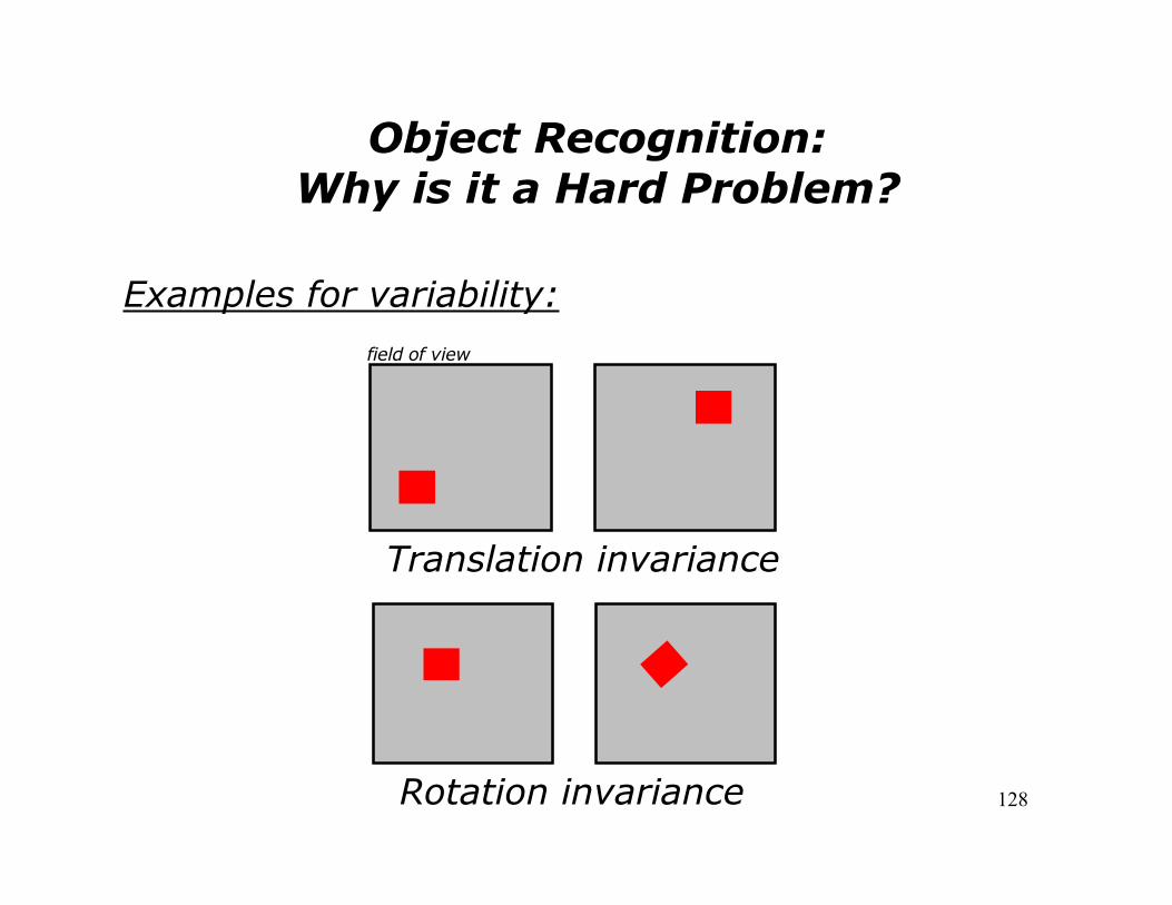
Object Recognition:Why is it a Hard Problem?
Examples for variability:field of view
Translation invariance
Rotation invariance
129
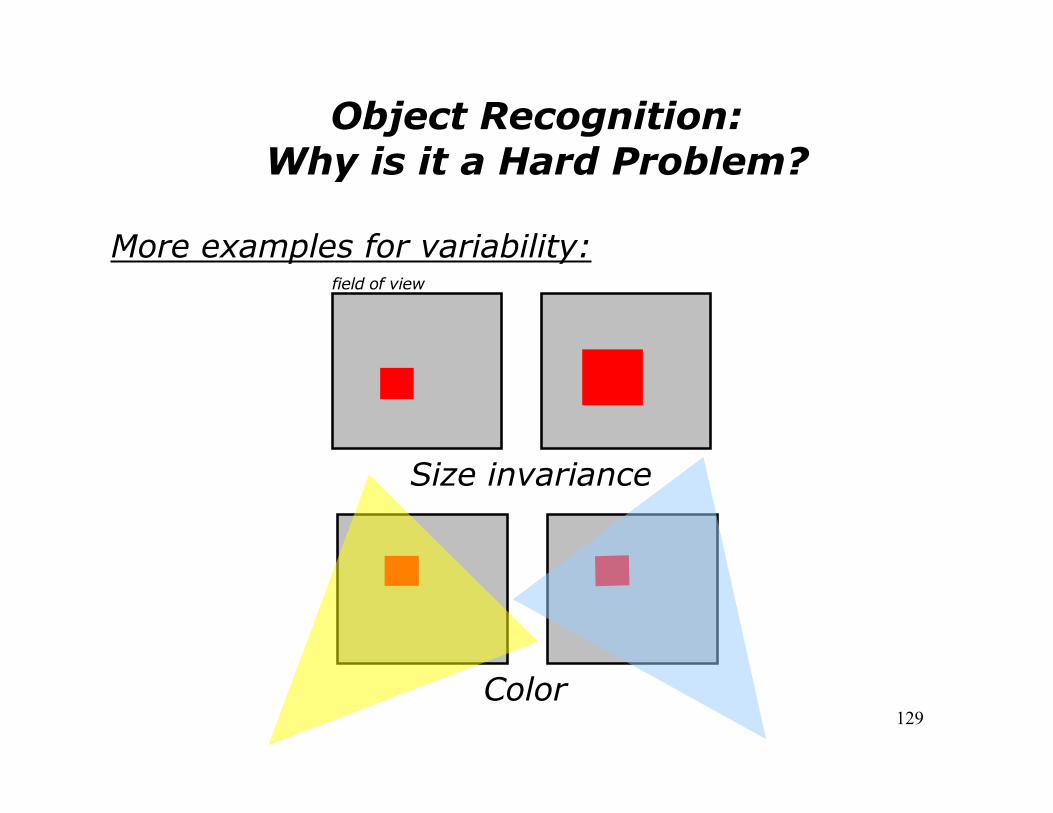
Object Recognition:Why is it a Hard Problem?
More examples for variability:field of view
Size invariance
Color

130
Object Recognition:Why is it a Hard Problem?
Variability in visual scenes:
field of view
Partial occlusionand presence of other objects
131
Object Recognition: Theories
Representation of visual shape (set of locations):
Viewer-centered coordinate systems:frame of reference: viewerexample: retinotopic coordinates, head-centeredcoordinateseasily accessed, but very unstable …
Environment-centered coordinate systems:locations specified relative to environment
Object-centered coordinate systems:intrinsic to or fixed to object itself (frame of reference:object)less accessible

132
Object Recognition: Theories
A taxonomy:
1. Template matching models (viewer-centered,normalization stage and matching)
2. Prototype models
3. Feature analysis model
4. Recognition by components (object-centered)

133
Object Recognition: Geons
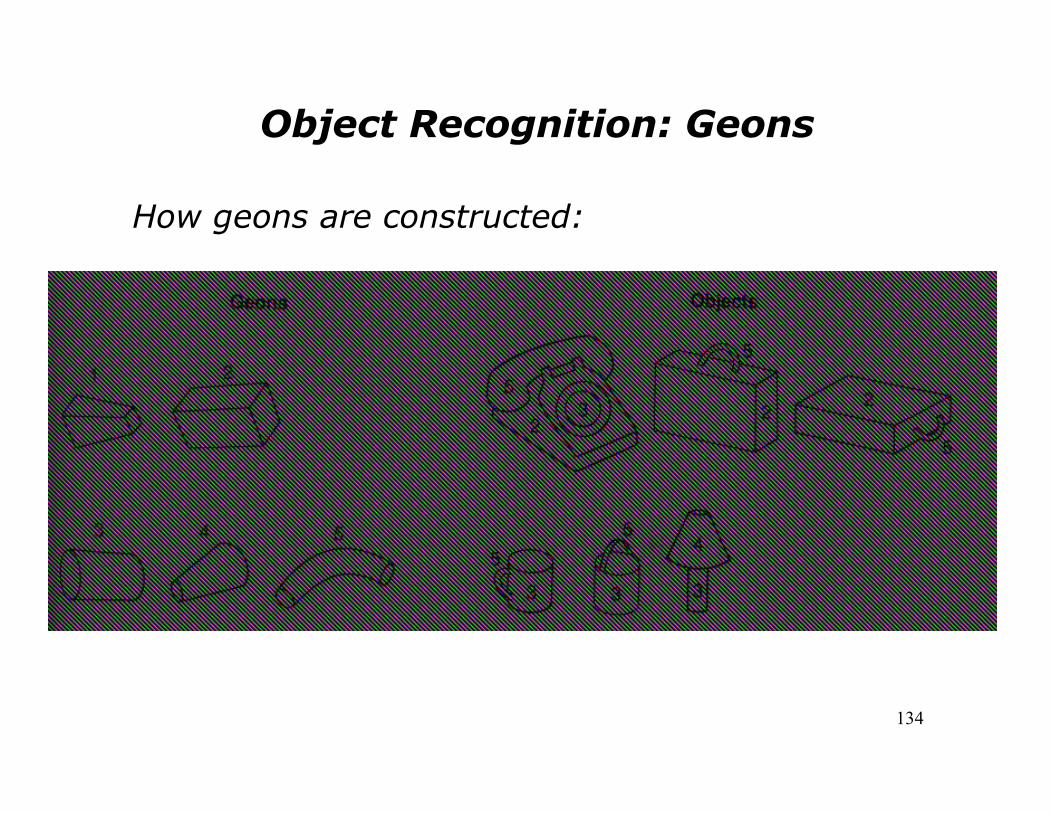
Theory proposed by Irv Biederman.
Objects have parts.
Objects can be described as configurations of a(relatively small) number of geometricallydefined parts.
These parts (geons) form a recognition alphabet.24 geons for four basic properties that areviewpoint-invariant.
134
Object Recognition: Geons
How geons are constructed:

135
Object Recognition: Geons
Irv Biederman, JCN, 2001
Geons in IT?
136
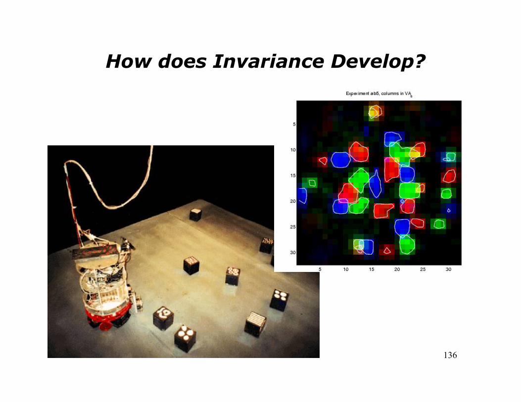
How does Invariance Develop?
137
Deficits of feature perception (such asachromatopsia) generally do not cause an inabilityto recognize objects.
Failure of knowledge or recognition = “agnosia”.(visual agnosia)
In visual agnosias, feature processing and memoryremain intact, and recognition deficits are limitedto the the visual modality. Alertness, attention,intelligence and language are unaffected.
Other sensory modalities (touch, smell) maysubstitute for vision in allowing objects to berecognized.
Higher Perceptual Functions: Agnosias
138
Apperceptive agnosia: perceptual deficit, affectsvisual representations directly, components ofvisual percept are picked up, but can’t beintegrated, effects may be graded, oftenaffected: unusual views of objects
Associative agnosia: visual representations areintact, but cannot be accessed or used inrecognition. Lack of information about thepercept. “Normal percepts stripped of theirmeaning” (Teuber)
This distinction introduced by Lissauer (1890)
Two Kinds of Agnosias
139
Apperceptive Agnosia
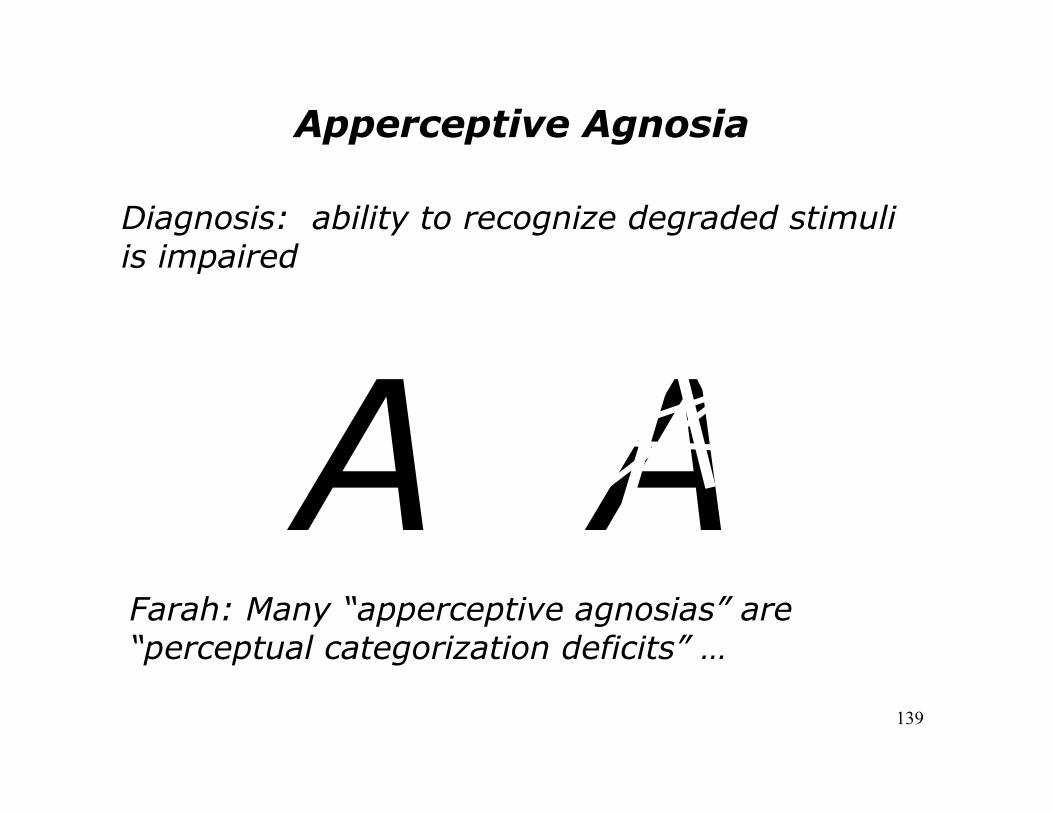
Diagnosis: ability to recognize degraded stimuliis impaired
A AFarah: Many “apperceptive agnosias” are“perceptual categorization deficits” …

140
Apperceptive Agnosia
Studies by E. Warrington:
Laterality in recognition deficits: patients withright-hemispheric lesions (parietal, temporal)showed lower performance on degraded imagesthan controls or left-hemispheric lesions.
Hypothesis: object constancy is disrupted (notcontour perception)
Experiment: Unusual views of objects – patientswith right-hemispheric lesions show acharacteristic deficit for these views.

141
Apperceptive Agnosia
Is “perceptual categorization deficit” a generalimpairment of viewpoint-invariant objectrecognition?
1. Patients are not impaired in everyday life(unlike associative agnosics).
2. They are not impaired in matching different“normal” views of objects, only “unusual views”.
3. Impairment follows unilateral lesions, notbilateral (as would be expected if visual shaperepresentations were generally affected).

142
Associative Agnosia
Patients do well on perceptual tests (degradedimages, image segmentation), but cannotaccess names (“naming”) or other information(“recognition”) about objects. Agnosics fail toexperience familiarity with the stimulus.
When given names of objects, they can(generally) give accurate verbal descriptions.
Warrington’s analysis places associativeagnosia in left hemisphere.

143
Associative Agnosia
Associative agnosics can copydrawings of objects butcannot name them (evidencefor intactness of perceptualrepresentations…) but…
144
Agnosia Restricted to SpecificCategories
Specific deficits in recognizing living versus non-livingthings.
Warrington and Shallice (1984): patients with bilateraltemporal lobe damage showed loss of knowledge aboutliving things (failures in visual identification and verbalknowledge).
Their interpretation: distinction between knowledgedomains – functional significance (vase-jug) versussensory properties (strawberry-raspberry).
Evolutionary explanation…

145
Agnosia Restricted to SpecificCategories
Another view: Damasio (1990)
Many inanimate objects are manipulated byhumans in characteristic ways.
Interpretation: inanimate objects will tend toevoke kinesthetic representations.
Agreeing with Warrington, difficulty is not due tovisual characteristics or visual discriminability.

146
Agnosia Restricted to SpecificCategories
Yet another view: Gaffan and Heywood (1993)
Presented images (line drawings) of animate andinanimate to normal humans and normalmonkeys, tachistoscopically (20 ms). Bothsubject groups made more errors in identifyinganimate vs. inanimate objects.
Interpretation: Living things are more similar toeach other than non-living things → “category-specific agnosia”
147
How is Semantic KnowledgeOrganized?
Category-based systemProperty-based system
Network model by Farah and McClelland (1991).

148
Prosopagnosia
Is face recognition “special”?Anatomical localizationFunctional independence
Associative visual agnosia (prosopagnosia): Lostability to recognize familiar faces.
Affects previous experience as well as(anterograde component) newly experiencedfaces.
Patients can recognize people by their voice,distinctive clothing, hairstyle etc.

149
Prosopagnosia
What is special about faces:
1. Higher specificity of categorization2. Higher level of expertise3. Higher degree of visual similarity4. Evolutionary significance
Can face and object recognition be dissociated?
Neuropsychological evidence suggests, yes (studyby McNeil and Warrington)
Also, remember Ishai et al. (object category map)