highly cross-linked polyethylene acetabular liners ... cross-linked polyethylene acetabular liners...
TRANSCRIPT
J O U R N A L O F T H E M E C H A N I C A L B E H AV I O R O F B I O M E D I C A L M A T E R I A L S 3 ( 2 0 1 0 ) 4 6 4 – 4 6 9
available at www.sciencedirect.com
journal homepage: www.elsevier.com/locate/jmbbm
Short communication
Highly cross-linked polyethylene acetabular liners retrievedfour to five years after revision surgery: A report of two cases
Matthew G. Teetera,b,∗, Douglas D.R. Naudiea,c, Kory D. Charronc,David W. Holdswortha,b,c
a Imaging Research Laboratories, Robarts Research Institute, P.O. Box 5015, London, ON, Canada, N6A 5K8bDepartment of Medical Biophysics, Schulich School of Medicine & Dentistry, The University of Western Ontario, London, ON, CanadacDivision of Orthopaedic Surgery, Schulich School of Medicine & Dentistry, The University of Western Ontario, London, ON, Canada
A R T I C L E I N F O
Article history:
Received 7 January 2010
Received in revised form
8 April 2010
Accepted 17 April 2010
Published online 27 April 2010
A B S T R A C T
There is currently considerable interest in the use of highly cross-linked polyethylene
(XLPE) acetabular liners for total hip arthroplasty (THA). In literature, only a single retrieval
analysis of one type of XLPE liner implanted for greater than four years exists. The purpose
of the present report is to quantify surface deviations in two XLPE liners implanted during
revision THA and retrieved between four to five years after implantation. The two XLPE
acetabular liners (Reflection, Smith and Nephew Inc., Memphis, TN) were retrieved from
patients undergoing their second revision surgery, at 4.90 and 4.07 years. The retrieved
liners and a new, non-implanted, unworn liner of the same size were scanned using
micro-computed tomography (micro-CT). Articular surface deviation maps were created
by comparing the retrievals to the unworn liner, based on the liner geometry obtained from
micro-CT. The linear penetration rates were found to be 0.018 and 0.008 mm/year. Localized
scratches and pits with deviations greater than 0.205 mm were also found on the articular
surfaces of both liners. The XLPE liners retrieved from the two cases demonstrated low
linear penetration rates. Regions with greater focal deviations were also apparent, likely
due to third-body wear. The results are consistent with previously published clinical follow-
ups of other XLPE liners.c© 2010 Elsevier Ltd. All rights reserved.
d
1. Introduction
There is currently considerable interest in the use of highlycross-linked polyethylene (XLPE) acetabular liners for totalhip arthroplasty (THA; Beksac et al., 2009; McCalden et al.,2009). The advantage of XLPE is an apparent reduction of wearrate versus conventional polyethylene (Beksac et al., 2009). A
∗ Corresponding author at: Imaging Research Laboratories, RobartsTel.: +1 519 663 3833; fax: +1 519 663 3900.
E-mail address: [email protected] (M.G. Teeter).
1751-6161/$ - see front matter c© 2010 Elsevier Ltd. All rights reservedoi:10.1016/j.jmbbm.2010.04.002
Research Institute, P.O. Box 5015, London, ON, Canada, N6A 5K8.
number of clinical follow-up studies on XLPE liners exceeding
five years have recently been published (Beksac et al., 2009;
McCalden et al., 2009; Bitsch et al., 2008; Engh et al., 2006;
Dorr et al., 2005). To our knowledge, only a single retrieval
analysis of one type of XLPE liner implanted for greater
than four years exists in literature (Knahr et al., 2007). That
study reports on two XLPE liners implanted during primary
.
J O U R N A L O F T H E M E C H A N I C A L B E H AV I O R O F B I O M E D I C A L M A T E R I A L S 3 ( 2 0 1 0 ) 4 6 4 – 4 6 9 465
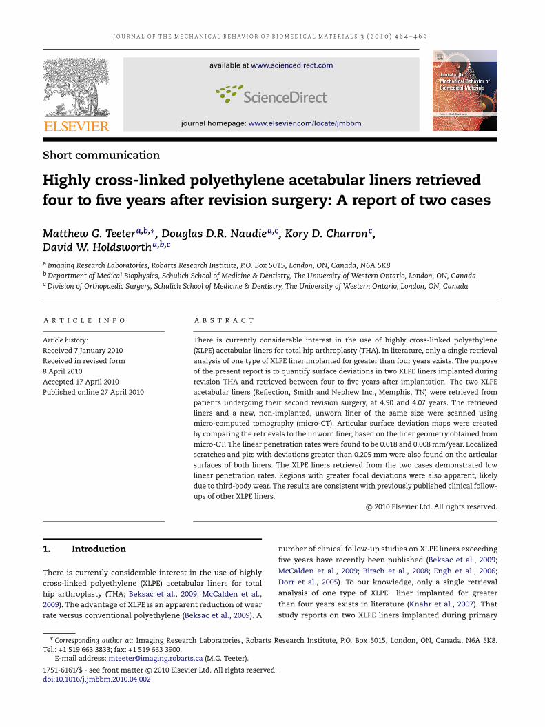
Fig. 1 – Radiographs for the two cases, prior to revision surgery and retrieval of the XLPE acetabular liners. A–B areanterior–posterior pelvic and lateral right hip views for case 1, respectively. C–D are anterior–posterior pelvic and lateralright hip views for case 2, respectively. Small tantalum beads, implanted at the time of the patient’s initial surgery, for usein a radiostereometric analysis (RSA) study are also visible in C and D.
THA. The purpose of the present report is to quantify surfacedeviations in two XLPE liners implanted during revision THAand retrieved between four to five years after implantation.
2. Patients and methods
2.1. Case 1
The patient was a 63 year-old man with ankylosing spondyli-tis who was 1.76 m tall and weighed 77 kg (body mass in-dex of 24.9). He underwent second-stage revision THA inOctober 2002, due to a previously infected THA. At thattime, the treating surgeon implanted an Echelon (Smith andNephew, Memphis, TN) porous bowed stem (size 18R, 260 mmlength, +15 mm), a 32 mm, +0 cobalt–chromium femoralhead, a Reflection porous acetabular cup (64 mm outer diam-eter, liner size H, standard size), and a Reflection acetabularliner (20 degree overhang, 32 mm inner diameter, 62–64 mmouter diameter, size H). The Reflection XLPE liner is man-ufactured from ram-extruded GUR 1050 polyethylene, andcold cross-linked using 10 Mrad of gamma irradiation withsubsequent melting (Willie et al., 2006). The polyethylene issterilized using ethylene oxide. The patient underwent apolyethylene liner and head exchange for recurrent infection
in April of 2003; at that time, he had replacement of his linerwith the same design (20 degree overhang, 32 mm inner di-ameter, 62–64 mm outer diameter, size H). Unfortunately, hedeveloped a recurrent deep periprosthetic infection from amethicillin sensitive Staphylococcus aureus (Fig. 1A–B). He un-derwent removal of his implants in March of 2008, at whichtime the liner was retrieved. There was no obvious biofilmon the surface of the retrieved liner. It had been in situ for4.90 years.
2.2. Case 2
The patient was a 74-year-old man who was 1.81 m talland weighed 98.5 kg (body mass index of 30.1). He hada primary cemented THA performed in October of 2002for osteoarthritis. He went on to aseptic loosening ofhis cemented femoral stem, and was revised to a size16 cemented Echelon (Smith and Nephew, Memphis, TN)175 mm length, standard collar in June of 2004 by a secondsurgeon. He unfortunately went on to aseptic subsidenceof his femoral stem and his cement mantle (Fig. 1C–D),and underwent re-revision THA to a cementless revisionarthroplasty in July of 2008. At the time of his mostrecent revision, the original acetabular cup was maintainedbut all other components were revised; these included a
466 J O U R N A L O F T H E M E C H A N I C A L B E H AV I O R O F B I O M E D I C A L M A T E R I A L S 3 ( 2 0 1 0 ) 4 6 4 – 4 6 9
cemented revision stem, a cobalt–chromium Taper FemoralHead (32mm diameter, 12/14 taper,+8 neck), and a Reflectionacetabular liner (20 degree overhang, 32 mm inner diameter,62–64 mm outer diameter, size H). The retrieved liner hadbeen in situ for 4.07 years. Currently, at an almost two-yearfollow-up, he has shown no signs of re-infection or furthercomplication, and appears to have achieved radiographic in-growth of his femoral stem.
2.3. Micro-CT scanning and 3D surface analysis
The liners were scanned with micro-computed tomography(micro-CT) and underwent 3D surface analysis using apreviously described approach (Teeter et al., 2010; Vicars et al.,2009; Bowden et al., 2005). A new, never-implanted liner ofthe same type as the retrieved liner (Reflection, Smith andNephew, Memphis, TN; 20 degree overhang, size H, 32 mminner diameter, 62–64 outer diameter) was also scannedto serve as an unworn reference for the surface analysis.Each liner was individually scanned and reconstructed usinga laboratory micro-CT scanner (eXplore Vision 120, GEHealthcare, London, Ontario). All scans were obtained usingan isotropic resolution of 50 µm over 1200 views, with 10frames averaged per view at an exposure time of 16 ms. TheX-ray tube voltage was 90 kV with a current of 40 mA. Thescans were reconstructed at the full 50 µm resolution. Thereconstructed scans of the liners were then analyzed with 3Dmicro-CT analysis software (MicroView v2.2, GE Healthcare,London, Ontario). Isosurface rendering was performed usinga threshold of −664 Hounsfield units with the highestpossible surface quality and no decimation. The resulting3D volume of each liner was saved in stereolithography fileformat. Gravimetric measurements were also obtained for thethree liners, and their masses were converted to volumesbased on the known polyethylene density (931 km/m3). Thegravimetric volumes were compared to the CT volumes toensure the accuracy of the micro-CT technique.
The surface file for each of the liners was then importedinto Geomagic Studio (Geomagic Inc., Research Triangle Park,N.C.) and converted to the wrap file format. The surface vol-ume was edited down to contain only the articular surface inorder to exclude artificial surface deviations (such as chiselmarks) in the non-articular surfaces, caused by damage dur-ing the retrieval process. The edited articular surfaces of thenew liner and retrieved liner were then imported into Geo-magic Qualify (Geomagic Inc., Research Triangle Park, N.C.),where the new liner was set as the reference object and theretrieved liner set as the test object. The two liners were co-aligned using an automated, iterative best-fit alignment algo-rithm.
Once co-aligned, a 3D comparison was performed to reportany deviation between the surfaces of the two liners. Thisanalysis was used to identify any focal deviations (such asscratching and pitting), in addition tomore diffuse patterns ofdeviation that correspond to femoral head penetration. Themaximum negative 3D deviation within the diffuse regionclosest to the center of the articular surface was used toestimate linear penetration. Deviations due to scratches andpits were not used for the calculation of linear penetration.The linear penetration rate for the first liner was calculated by
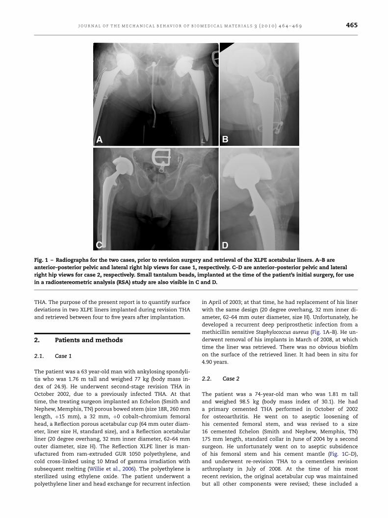
Fig. 2 – 3D rendered volume images of the retrieved XLPEacetabular liners scanned using micro-CT. Case 1 (A) wasimplanted for 4.90 years and case 2 (B) for 4.07 years. Theliners have been oriented as they were in vivo, with the lipin the superior direction.
dividing themaximum linear penetration by the implantationtime. The second liner was then imported into the software asthe test object and the process was repeated.
3. Results
Under visual inspection, the liner in the first case exhibitednumerous scratches on the articular surface. The liner inthe second case had only a few scratches on the articularsurface, but also had six small pits on the surface. Bothliners were yellowed on their backside surface (facing theacetabular shell), and the liner from the first case also hadyellowing of the articular surface. The scratching and pittingof the articular surfaces can be seen in the 3D renderedvolumes of the liners obtained using micro-CT (Fig. 2). Themean difference between the gravimetric and CT volumeswas 0.42%, with the CT volumes consistently lower than thegravimetric volumes.
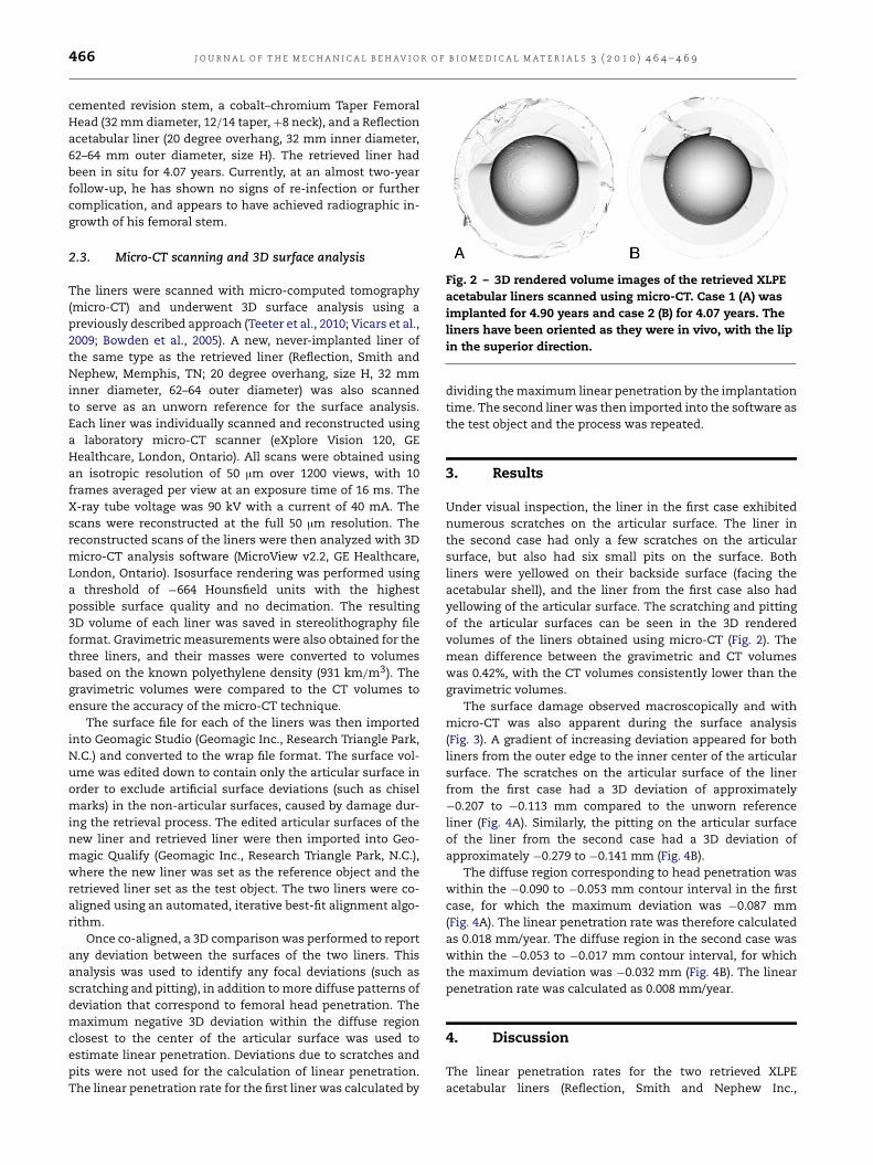
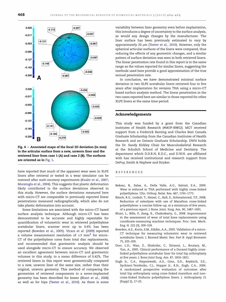
The surface damage observed macroscopically and withmicro-CT was also apparent during the surface analysis(Fig. 3). A gradient of increasing deviation appeared for bothliners from the outer edge to the inner center of the articularsurface. The scratches on the articular surface of the linerfrom the first case had a 3D deviation of approximately−0.207 to −0.113 mm compared to the unworn referenceliner (Fig. 4A). Similarly, the pitting on the articular surfaceof the liner from the second case had a 3D deviation ofapproximately −0.279 to −0.141 mm (Fig. 4B).
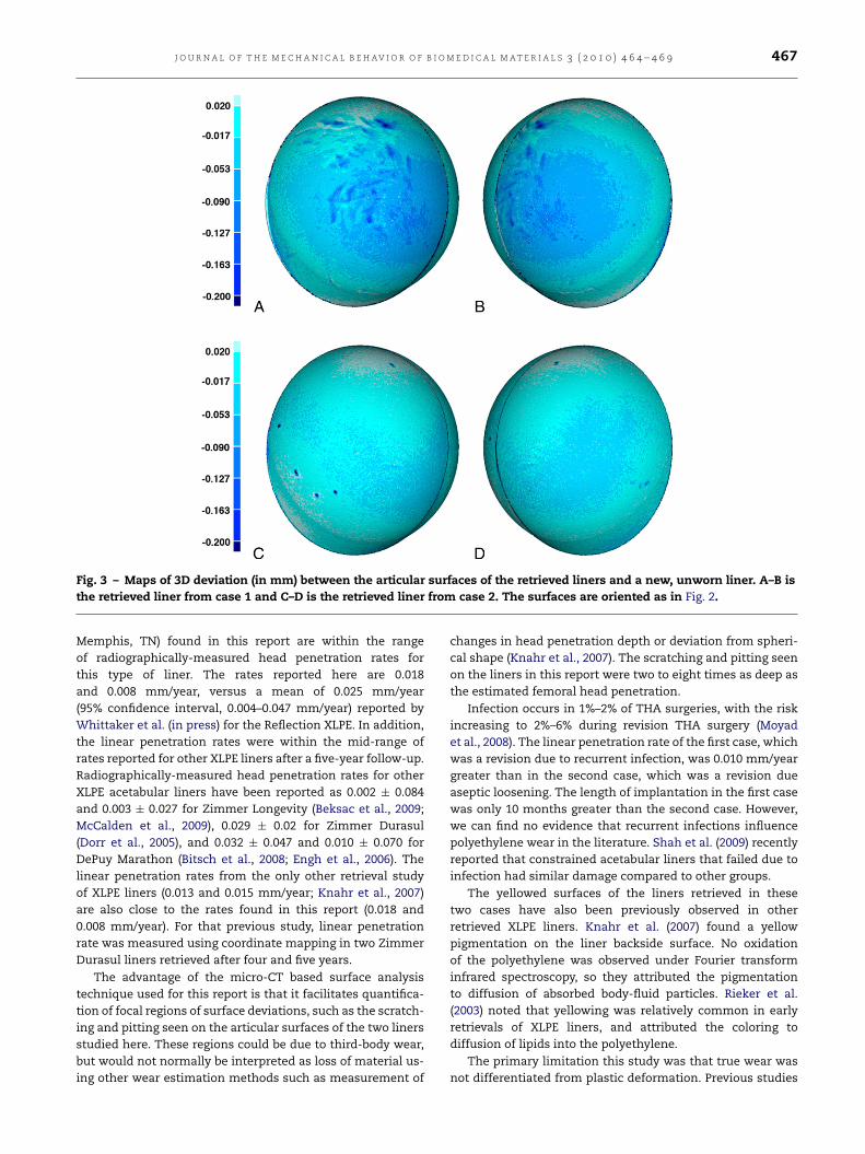
The diffuse region corresponding to head penetration waswithin the −0.090 to −0.053 mm contour interval in the firstcase, for which the maximum deviation was −0.087 mm(Fig. 4A). The linear penetration rate was therefore calculatedas 0.018 mm/year. The diffuse region in the second case waswithin the −0.053 to −0.017 mm contour interval, for whichthe maximum deviation was −0.032 mm (Fig. 4B). The linearpenetration rate was calculated as 0.008 mm/year.
4. Discussion
The linear penetration rates for the two retrieved XLPEacetabular liners (Reflection, Smith and Nephew Inc.,
J O U R N A L O F T H E M E C H A N I C A L B E H AV I O R O F B I O M E D I C A L M A T E R I A L S 3 ( 2 0 1 0 ) 4 6 4 – 4 6 9 467
Fig. 3 – Maps of 3D deviation (in mm) between the articular surfaces of the retrieved liners and a new, unworn liner. A–B isthe retrieved liner from case 1 and C–D is the retrieved liner from case 2. The surfaces are oriented as in Fig. 2.
Memphis, TN) found in this report are within the rangeof radiographically-measured head penetration rates forthis type of liner. The rates reported here are 0.018and 0.008 mm/year, versus a mean of 0.025 mm/year(95% confidence interval, 0.004–0.047 mm/year) reported byWhittaker et al. (in press) for the Reflection XLPE. In addition,the linear penetration rates were within the mid-range ofrates reported for other XLPE liners after a five-year follow-up.Radiographically-measured head penetration rates for otherXLPE acetabular liners have been reported as 0.002 ± 0.084and 0.003 ± 0.027 for Zimmer Longevity (Beksac et al., 2009;McCalden et al., 2009), 0.029 ± 0.02 for Zimmer Durasul(Dorr et al., 2005), and 0.032 ± 0.047 and 0.010 ± 0.070 forDePuy Marathon (Bitsch et al., 2008; Engh et al., 2006). Thelinear penetration rates from the only other retrieval studyof XLPE liners (0.013 and 0.015 mm/year; Knahr et al., 2007)are also close to the rates found in this report (0.018 and0.008 mm/year). For that previous study, linear penetrationrate was measured using coordinate mapping in two ZimmerDurasul liners retrieved after four and five years.
The advantage of the micro-CT based surface analysistechnique used for this report is that it facilitates quantifica-tion of focal regions of surface deviations, such as the scratch-ing and pitting seen on the articular surfaces of the two linersstudied here. These regions could be due to third-body wear,but would not normally be interpreted as loss of material us-ing other wear estimation methods such as measurement of
changes in head penetration depth or deviation from spheri-cal shape (Knahr et al., 2007). The scratching and pitting seenon the liners in this report were two to eight times as deep asthe estimated femoral head penetration.
Infection occurs in 1%–2% of THA surgeries, with the riskincreasing to 2%–6% during revision THA surgery (Moyadet al., 2008). The linear penetration rate of the first case, whichwas a revision due to recurrent infection, was 0.010 mm/yeargreater than in the second case, which was a revision dueaseptic loosening. The length of implantation in the first casewas only 10 months greater than the second case. However,we can find no evidence that recurrent infections influencepolyethylene wear in the literature. Shah et al. (2009) recentlyreported that constrained acetabular liners that failed due toinfection had similar damage compared to other groups.
The yellowed surfaces of the liners retrieved in thesetwo cases have also been previously observed in otherretrieved XLPE liners. Knahr et al. (2007) found a yellowpigmentation on the liner backside surface. No oxidationof the polyethylene was observed under Fourier transforminfrared spectroscopy, so they attributed the pigmentationto diffusion of absorbed body-fluid particles. Rieker et al.(2003) noted that yellowing was relatively common in earlyretrievals of XLPE liners, and attributed the coloring todiffusion of lipids into the polyethylene.
The primary limitation this study was that true wear wasnot differentiated from plastic deformation. Previous studies
468 J O U R N A L O F T H E M E C H A N I C A L B E H AV I O R O F B I O M E D I C A L M A T E R I A L S 3 ( 2 0 1 0 ) 4 6 4 – 4 6 9
Fig. 4 – Annotated maps of the focal 3D deviation (in mm)in the articular surface from a new, unworn liner and theretrieved liner from case 1 (A) and case 2 (B). The surfacesare oriented as in Fig. 1.
have reported that much of the apparent wear seen in XLPEliners after retrieval or tested in a wear simulator can berestored after melt-recovery experiments (Knahr et al., 2007;Muratoglu et al., 2004). This suggests that plastic deformationlikely contributed to the surface deviations observed inthis study. However, the surface deviations measured herewith micro-CT are comparable to previously reported linearpenetrations measured radiographically, which also do nottake plastic deformation into account.
Some limitations are associated with the micro-CT basedsurface analysis technique. Although micro-CT has beendemonstrated to be accurate and highly repeatable forquantification of volumetric wear in retrieved polyethyleneacetabular liners, scanner error up to 0.6% has beenreported (Bowden et al., 2005). Vicars et al. (2009) reporteda volume measurement resolution of ±3 mm3 for micro-CT of the polyethylene cores from total disc replacements,and recommended that gravimetric analysis should beused alongside micro-CT to ensure accuracy. We observedan excellent agreement between micro-CT and gravimetricvolumes in this study, to a mean difference of 0.42%. Theretrieved liners in this report were geometrically comparedto a new, unworn liner of the same size, rather than theiroriginal, unworn geometry. This method of comparing thegeometries of retrieved components to a never-implantedgeometry has been described for knees (Blunt et al., 2008)as well as for hips (Teeter et al., 2010). As there is some
variability between liner geometry even before implantation,this introduces a degree of uncertainty to the surface analysis,as would any design changes by the manufacturer. Theliner surface has been previously estimated to vary byapproximately 20 µm (Teeter et al., 2010). However, only thespherical articular surfaces of the liners were compared, thusreducing the effects of any geometric changes, and a similarpattern of surface deviation was seen in both retrieved liners.The linear penetration rate found in this report is in the samerange as the values reported for similar liners, suggesting themethods used here provide a good approximation of the trueannual penetration rate.
In conclusion, we have demonstrated minimal surfacedeviation in two XLPE acetabular liners retrieved four to fiveyears after implantation for revision THA using a micro-CTbased surface analysis method. The linear penetration in thetwo cases reported here are similar to those reported for otherXLPE liners at the same time period.
Acknowledgements
This study was funded by a grant from the CanadianInstitutes of Health Research (#MOP-89852). MGT receivedsupport from a Frederick Banting and Charles Best CanadaGraduate Scholarship from the Canadian Institutes of HealthResearch and an Ontario Graduate Scholarship. DWH holdsthe Dr. Sandy Kirkley Chair for Musculoskeletal Researchat the Schulich School of Medicine and Dentistry. Thedepartment which D.D.R.N, K.D.C., and D.W.H. are affiliatedwith has received institutional and research support fromDePuy, Smith & Nephew and Stryker.
R E F E R E N C E S
Beksac, B., Salas, A., Della Valle, A.G., Salvati, E.A., 2009.Wear is reduced in THA performed with highly cross-linkedpolyethylene. Clin. Orthop. Relat. Res. 467, 1765–1772.
Bitsch, R.G., Loidolt, T., Heisel, C., Ball, S., Schmalzrried, T.P., 2008.Reduction of osteolysis with use of Marathon cross-linkedpolyethylene: a concise follow-up, at a minimum of five years,of a previous report. J. Bone. Joint. Surg. Am. 90, 1487–1491.
Blunt, L., Bills, P., Jiang, X., Chakrabarty, G., 2008. Improvementin the assessment of wear of total knee replacements usingcoordinate-measuring machine techniques. Proc. Inst. Mech.Eng. H. 222 (3), 309–318.
Bowden, A.E., Kurtz, S.M., Edidin, A.A., 2005. Validation of a micro-CT technique for measuring volumetric wear in retrievedacetabular liners. J. Biomed Mater. Res. Part B: Appl Biomater75, 205–209.
Dorr, L.D., Wan, Z., Shahrdar, C., Sirianni, L., Boutary, M.,Yun, A., 2005. Clinical performance of a Durasol highly cross-linked polyethylene acetabular liner for total hip arthroplastyat five years. J. Bone Joint Surg. Am. 87, 1816–1821.
Engh Jr., C.A., Stepniewski, A.S., Ginn, S.D., Beykirch, S.E.,Sychterz-Terefenko, C.J., Hopper Jr., R.H., Engh, C.A., 2006.A randomized prospective evaluation of outcomes aftertotal hip arthroplasty using cross-linked marathon and non-cross-linked Enduron polyethylene liners. J. Arthroplasty 21(Suppl 2), 17–25.
J O U R N A L O F T H E M E C H A N I C A L B E H AV I O R O F B I O M E D I C A L M A T E R I A L S 3 ( 2 0 1 0 ) 4 6 4 – 4 6 9 469
Knahr, K., Pospischill, M., Kottig, P., Schneider, W., Plenk Jr., H.,2007. Retrieval analyses of highly cross-linked polyethyleneacetabular liners four and five years after implantation. J. BoneJoint Surg. Br. 89, 1036–1041.
McCalden, R.W., MacDonald, S.J., Rorabeck, C.H., Bourne, R.B.,Chess, D.G., Charron, K.D., 2009. Wear rate of highly cross-linked polyethylene in total hip arthroplasty. A randomizedcontrolled trial. J. Bone Joint Surg. Am. 91, 773–782.
Moyad, T.F., Thornhill, T., Estok, D., 2008. Evaluation andmanagement of the infected total hip and knee. Orthopedics31 (6), 581–588.
Muratoglu, O.K., Greenbaum, E.S., Bragdon, C.R., Jasty, M.,Freiberg, A.A., Harris, W.H., 2004. Surface analysis ofearly retrieved acetabular polyethylene liners: a compari-son of conventional and highly crosslinked polyethylenes.J. Arthroplasty 19 (1), 68–77.
Rieker, C.B., Konrad, R., Schön, R., Schneider, W., Abt, N.A., 2003.In vivo and in vitro surface changes in a highly cross-linkedpolyethylene. J. Arthroplasty 18 (7 Suppl 1), 48–54.
Shah, S.N., Kaye, R.J., Kelly, N.H., Su, E.P., Padgett, D.E., Wright,T.M., 2009. Retrieval analysis of failed constrained acetabularliners. J. Arthroplasty 24 (6 Suppl), 54–57.
Teeter, M.G., Naudie, D.D.R., Charron, K.D., Holdsworth, D.W.,2010. 3D surface deviation maps for analysis of retrievedpolyethylene acetabular liners using micro-computed tomog-raphy. J. Arthroplasty 25 (2), 330–332.
Vicars, R., Fisher, J., Hall, R.M., 2009. The accuracy and precisionof a micro computer tomography volumetric measurementtechnique for the analysis of in-vitro tested total discreplacements. Proc. Inst. Mech. Eng. H 223 (3), 383–388.
Whittaker, J.P., Charron, K.D., McCalden, R.W., MacDonald, S.J.,Bourne, R.B., 2009. Comparison of steady state femoral headpenetration rates between 2 highly cross-linked polyethylenesin total hip arthroplasty, J. Arthroplasty (in press).
Willie, B.M., Bloebaum, R.D., Ashrafi, S., Dearden, C., Steffensen,T., Hofmann, A.A., 2006. Oxidative degradation in highly cross-linked and conventional polyethylene after 2 years of real-timeshelf aging. Biomaterials 27 (10), 2275–2284.