highly divergent ssu rrna genes found in the marine
TRANSCRIPT

Introduction
The phototrophic ciliate Myrionecta rubra (= Meso-dinium rubrum) (Lohmann 1908, Jankowski 1976)(Mesodiniidae, Litostomatea) is nearly ubiquitous incoastal marine and estuarine habitats and has longbeen a curiosity to evolutionary biologists, perhaps
Highly Divergent SSU rRNA GenesFound in the Marine Ciliates Myrionecta rubraand Mesodinium pulex
Matthew D. Johnsona,1, Torstein Tengsb,2, David W. Oldachb, Charles F. Delwichec,and Diane K. Stoeckera
a Horn Point Laboratory, University of Maryland, Center for Environmental Science, PO Box 775, Cambridge, MD21613, USA
b Institute of Human Virology, University of Maryland, School of Medicine, Baltimore, MD, USAc Department of Cell Biology and Molecular Genetics/Plant Biology, University of Maryland, College Park, MD, USA
Submitted April 2, 2004; Accepted May 30, 2004Monitoring Editor: Mitchell L. Sogin
Myrionecta rubra and Mesodinium pulex are among the most commonly encountered planktonic cili-ates in coastal marine and estuarine regions throughout the world. Despite their widespread distribu-tion, both ciliates have received little attention by taxonomists. In order to better understand the phy-logenetic position of these ciliates, we determined the SSU rRNA gene sequence from cultures ofM. rubra and M. pulex. Partial sequence data were also generated from isolated cells of M. rubra fromChesapeake Bay. The M. rubra and M. pulex sequences were very divergent from all other ciliates,but shared a branch with 100% bootstrap support. Both species had numerous deletions and substi-tutions in their SSU rRNA gene, resulting in a long branch for the clade. This made the sequencesprone to spurious phylogenetic affiliations when using simple phylogenetic methods. Maximum like-lihood analysis placed M. rubra and M. pulex on the basal ciliate branch, following removal of am-biguously aligned regions. Fluorescent in situ hybridization probes were used with confocal laserscanning microscopy to confirm that these divergent sequences were both expressed in thecytoplasm and nucleolus of M. rubra and M. pulex. We found that our sequence data matched severalrecently discovered unidentified eukaryotes in Genbank from diverse marine habitats, all of whichhad apparently been misattributed to highly divergent amoeboid organisms.
beginning with Darwin (1839). The important eco-logical role of this ciliate is periodically made con-spicuous by massive non-toxic red tides in coastaland estuarine regions throughout the world, some ofwhich may exceed 100 square miles (Jiménez andIntriago 1987; Ryther 1967). M. rubra is well docu-mented to possess organelles of cryptophycean ori-gin, including plastids, mitochondria (Taylor et al.1969, 1971), and nuclei (Hibberd 1977; Oakley andTaylor 1978). While early studies debated whether
ORIGINAL PAPER
1Corresponding author;e-mail [email protected] address: Dana-Farber Cancer Institute, 44 Binney St,Boston, MA 02115, USA
1434-4610/04/155/03-347 $ 30.00/0
Protist, Vol. 155, 347–359, September 2004 http://www.elsevier.de/protistPublished online 14 September 2004 Protist

these organelles represented a true symbiosis orwere the result of sequestration from living prey (e.g.Taylor et al. 1969), more recent studies have pro-vided evidence for the latter, showing that growthand photosynthesis in M. rubra are dependent uponingesting free-living cryptomonads (Gustafson et al.2000).
Mesodinium (Stein 1863) (Mesodiniidae, Litosto-matea) is a commonly encountered genus of non-pigmented ciliates found in coastal marine, estuar-ine, and fresh water systems (Foissner et al. 1999).M. pulex is a heterotrophic ciliate that feeds uponbacteria, flagellates, algae, and ciliates (Dolan andCoats 1991; Foissner et al. 1999). Although there arefew described species in the genus besidesM. pulex, it is often confused with M. acarus andM. fimbriatum (Foissner et al. 1999).
Currently no sequence data are available for theMesodinidae, and only recently have efforts beenmade to determine small subunit (SSU) ribosomalRNA sequences for other familiar pelagic marine cil-iates (e.g. Snoeyenbos-West et al. 2002, Strueder-Kypke and Lynn 2003). Several recent PCR-basedstudies to assess microbial eukaryotic diversity inthe world’s oceans have led to the discovery of nu-merous unidentified SSU rRNA sequences, some ofwhich constitute new branches on familiar phyloge-netic lineages (e.g. López-Garcia et al. 2001). Manyof these new sequences can be attributed to welldescribed protistian groups such as alveolates (api-complexans, ciliates, colpodellids, dinoflagellates,perkinsids), while some are sequences with uncer-tain phylogenetic affiliation. However, many of therRNA genes of recognized marine protists have yetto be sequenced, suggesting that some of the newsequences may not be novel taxa. Recently Leanderet al. (2003) identified some of these novel se-quences as belonging to the colpodellids. In the pre-sent study we present evidence that several newlydescribed unidentified eukaryotic sequences withuncertain taxonomic affiliation also belong to a fa-miliar lineage of alveolates. Herein we present se-quence data that show Myrionecta and Mesodiniumshare similar and highly divergent SSU rDNA se-quences that suggest they are an early branchinglineage of ciliates, and use in situ hybridization toverify that these sequences are present and ex-pressed.
Results
SSU rRNA Gene Characteristics
The rDNA genes amplified for the Antarctic andChesapeake Bay M. rubra and the Chesapeake Bay
M. pulex were highly divergent compared to otherciliates and alveolates in general. The rDNA se-quences for both Myrionecta and Mesodinium wererelatively short, 1548 and 1543 bp respectively,compared to an average for alveolates of about1750. This was primarily due to coincident deletionsin the gene, for both taxa, in helices 10 (variable re-gion [V] 2) (~20bp), 11 (~9 bp), E23-1-7 (V4) (~35 bp),E23-14 (V4) (absent), and 43 (V7) (~23 bp) (based onWuyts et al. 2000 model). Furthermore both taxahave numerous substitutions in helices 8, 16, 18, 25,and 26 compared with all other alveolate taxa.
In situ Hybridization Analysis
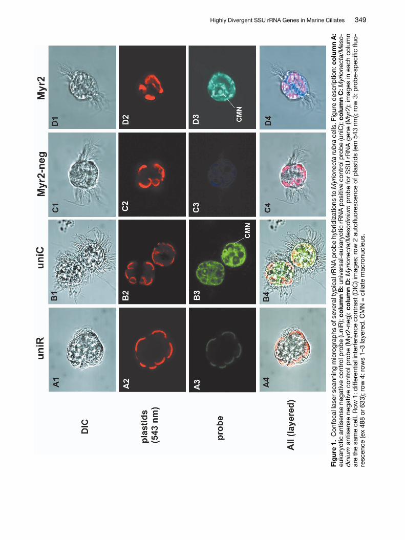
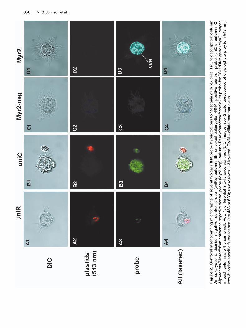
Due to the highly unusual nature of the Myrionectaand Mesodinium sequences we used fluorescent insitu hybridization (FISH) to determine whether thesesequences originated from the ciliates in question.In addition, confocal laser scanning microscopy(CLSM) was used to localize probe binding to thenucleolus, in order to verify that the probes were hy-bridizing to the targeted genome. An oligonucleotideprobe (Myr2) was designed from a variable region ofthe SSU rRNA molecule common to both Myri-onecta and Mesodinium, but with numerous substi-tutions in other alveolates and distantly related taxa(Table 2). We found that the Myr2 probe alwaysbound to rRNA within the ciliate cytoplasm and thenucleolus of the ciliate nuclei (Figs 1D3, 2D3), andnever to RNA in other taxa (data not shown). ManyM. rubra cells also possessed nuclei of their crypto-phyte prey, Teleaulax acuta, to which no binding ofthe Myr2 probe was observed (data not shown). Themacronuclei of both ciliates were found to have asingle large nucleolus or sphere filled with rRNA thatcomprised much of the volume of each macronu-cleus (Figs 1B3,D3). The universal eukaryotic posi-tive control probe uniC, labeled rRNA throughoutthe cell cytoplasm and within the nucleoi of all nu-clei, including those of cryptophyte prey when pre-sent (Figs 1B3, 2B3). No binding of any probes tonuclear DNA within the ciliates could be detected,nor of any negative control probes to any portion ofthe cell. Negative control probes consisted of anti-sense probes for both the universal eukaryote RNAprobe (uniR) (Figs 1A1–4, 2A1–4), and the Myri-onecta/Mesodinium probe (Myr2-neg) (Figs 1C1–4,2C1–4).
Phylogenetic Analysis
The Myrionecta/Mesodinium SSU rDNA sequencesshare high sequence similarity (7% sequence differ-ence, Table 1) in addition to the above-mentioned
348 M. D. Johnson et al.
Highly Divergent SSU rRNA Genes in Marine Ciliates 349
Fig
ure
1.C
onfo
cal l
aser
sca
nnin
g m
icro
grap
hs o
f sev
eral
typ
ical
rRN
A p
rob
e hy
brid
izat
ions
to M
yrio
nect
a ru
bra
cells
. Fig
ure
des
crip
tion:
co
lum
n A
:eu
kary
otic
ant
isen
se n
egat
ive
cont
rol p
rob
e (u
niR
); co
lum
n B
:uni
vers
al-e
ukar
yotic
rRN
A p
ositi
ve c
ontr
ol p
rob
e (u
niC
); co
lum
n C
: Myr
ione
cta/
Mes
o-d
iniu
man
tisen
se n
egat
ive
cont
rol p
rob
e (M
yr2-
neg)
; co
lum
n D
: Myr
ione
cta/
Mes
odin
ium
pro
be
for
SS
U r
RN
A g
ene
(Myr
2); i
mag
es in
eac
h co
lum
nar
e th
e sa
me
cell.
Row
1: d
iffer
entia
l int
erfe
renc
e co
ntra
st (D
IC) i
mag
es; r
ow 2
aut
oflu
ores
cenc
e of
pla
stid
s (e
m 5
43nm
); ro
w 3
: pro
be-
spec
ific
fluo-
resc
ence
(ex
488
or 6
33);
row
4: r
ows
1–3
laye
red
. CM
N =
cili
ate
mac
ronu
cleu
s.
350 M. D. Johnson et al.
Fig
ure
2.C
onfo
cal l
aser
sca
nnin
g m
icro
grap
hs o
f sev
eral
typ
ical
rR
NA
pro
be
hyb
ridiz
atio
ns t
o M
esod
iniu
m p
ulex
cells
. Fig
ure
des
crip
tion:
co
lum
nA
:eu
kary
otic
an
tisen
se
nega
tive
cont
rol
pro
be
(uni
R);
colu
mn
B:
univ
ersa
l-eu
kary
otic
rR
NA
p
ositi
ve
cont
rol
pro
be
(uni
C);
colu
mn
C:
Myr
ione
cta/
Mes
odin
ium
antis
ense
neg
ativ
e co
ntro
l pro
be
(Myr
2-ne
g); c
olu
mn
D: M
yrio
nect
a/M
esod
iniu
mp
rob
e fo
r S
SU
rR
NA
gen
e (M
yr2)
; im
ages
in e
ach
colu
mn
are
the
sam
e ce
ll. R
ow 1
: d
iffer
entia
l int
erfe
renc
e co
ntra
st (D
IC) i
mag
es;
row
2 a
utof
luor
esce
nce
of c
ryp
top
hyte
pre
y (e
m 5
43nm
);ro
w 3
: pro
be-
spec
ific
fluor
esce
nce
(em
488
or 6
33);
row
4: r
ows
1–3
laye
red
. CM
N =
cili
ate
mac
ronu
cleu
s.

Table 2. Probe and target sequence for Myrionecta rubra and Mesodinium pulex andcomparisons to other taxa; target region in bold and mismatches between target taxaand other taxa highlighted in gray.
Species Sequence
Outgroup taxaMastigamoeba invertens 5′..GAAATTCTTGGATTTATTAAAGATGAACTA..3′Diplonema papillatum 5′..GAAATTCTTAGATCGTAGGAAGACGAACTT..3′Echinamoeba therma 5′..GAAATTCTAGGATTAACTGAAAACAAACTA..3′
Other alveolatesDinophysis acuminata 5′..GAAATTCTTGGATTTGTTAAAGACGGACTA..3′Plasmodium vivax 5′..GAAATTCTTAGATTTTCTGGAGACAAACAA..3′
Other ciliatesDidinium nasutum 5′..GAAATTCTTGGATTTATTAAAGACTAACGT..3′Euplotes crassus 5′..GAAATTCTTTGAAATATTAAAGACTAACTT..3′Stentor roeseli 5′..GAAATTCTATGATTTATTAAAGACGAACTT..3′Loxodes magnus 5′..GAAATTCTTGGATTTACTGAAGACCAACTA..3′
Target taxaMyrionecta rubra 5′..GAAATTCTTGGACCGGACGAAGACGACCAG..3′Mesodinium pulex 5′..GAAATTCTTGGACCGGACGAAGACGATCAG..3′
ProbeMyr2 probe 3′..-------TTGGACCGGACGAAGAC------..5′
quences (e.g. Plasmodium vivax, Oxyrrhis marina), aresult that we attribute to branch-length effects(data not shown). In order to eliminate these effects,we removed ambiguously aligned regions of thedata set and proceeded with maximum likelihoodmethods.
common deletions, and formed a well-supportedclade in all analyses (100% bootstrap support,Figs 3,4). In preliminary phylogenetic analyses ofthese sequences using distance and maximum par-simony methods, the Myrionecta/Mesodinium cladeconsistently grouped with other highly divergent se-
Highly Divergent SSU rRNA Genes in Marine Ciliates 351
Table 1. Environmental clones in Genbank that are closely related to cultures of Myrionecta rubra and Meso-dinium pulex.
Clone or culture Accession Base pairs S′a Total gaps % identity––––––––––––––– ––––––––––––––– –––––––––––––––
number MRb MPb MR MP MR MP
Myrionecta rubra AY587129 1543 – – – 28 – 93Mesodinium pulex AY587130 1548 – – 28 – 93 –CB-MR-25c AY587131 662 – – 10 22 97 90DH145-EKD11 AF290065d 1474 1398 1052 7 14 98 91CCW100 AY180041e 1518 1320 1046 9 14 96 91CCW75 AY180032e 1519 1146 1357 10 11 92 98M43 AY331778f 1137 937 640 1 16 98 92M112 AY331777f 1173 1109 803 2 17 98 92M110 AY331783f 1141 1028 759 5 20 98 91
a bit score from BLAST search, b Myrionecta rubra and Mesodinium pulex, c Chesapeake Bay, Myrionecta rubraclone (partial sequences), d Lòpez-Garcia et al. 2001, e Stoeck and Epstein 2003, f Savin et al. 2004


Highly Divergent SSU rRNA Genes in Marine Ciliates 353
Figure 4. Gamma-corrected (Γ = 0.5444) maximum likelihood (-lnL = 15334.84); GTR model) tree with proportionof invariable sites (0.14153) using a small subunit rDNA alignment of 1479 sites. Base frequencies and substitu-tion rates were estimated with a distance-maximum likelihood search. Tree topology was found using stepwiseaddition and10× heuristic searches with TBR branch swapping and random-addition sequences. Numbers onbranches correspond to bootstrap values (100× with stepwise addition and a heuristic search with 2× random ad-dition and TBR branch swapping) for this ML tree.
� Figure 3. Gamma-corrected (Γ = 0.5576) distance-maximum likelihood (ML) (GTR model) tree (minimum evo-lution) with proportion of invariable sites (0.1079), using a small subunit rDNA alignment of 1474 sites. ML pa-rameters were estimated with Modeltest (Posada and Crandall 1998). Tree topology was found using stepwiseaddition and 25× heuristic searches with TBR branch swapping and random-addition sequences. Numbers onbranches correspond to bootstrap values (100× with stepwise addition and heuristic searches with 2× randomaddition and TBR branch swapping).

A matrix of 65 assorted protist taxa of 1474 char-acters and about 1300 nucleotides (923 were parsi-mony informative) was used for a Γ-corrected dis-tance-maximum likelihood (DML) search (generaltime reversible model) with proportion of invariablesites. In this analysis Myrionecta/Mesodinium cladebranched within the ciliates, albeit with low boot-strap support (64%) (Fig. 3). Included in this analysiswere several unidentified eukaryote clones with highsequence similarity to our Myrionecta/Mesodiniumsequences, and our Chesapeake Bay M. rubra se-quence (Table 1). These environmental taxa groupedstrongly with our Myrionecta and Mesodinium cul-tures (100% bootstrap support), revealing the likelysource of these clones (Fig. 3). All previous analysesof these environmental clones, in the absence of theMyrionecta/Mesodinium sequences, had placedthem outside of the alveolates with divergent amoe-boid taxa. Of the six environmental clones thatgrouped with Myrionecta and Mesodinium, five ap-peared to be closely related to M. rubra, with high %sequence identity (96–98%) and alignment bit score(S’), while one (CCW75) was most similar to M. pulex(98%) (Table 1).
A full ML analysis was then conducted on a nearlyidentical matrix (1479 bp) greatly reduced in taxa (32taxa) and composed mostly of alveolate taxa. ThisΓ-corrected ML (GTR model) analysis with pinvaralso revealed low bootstrap support to group theMyrionecta/ Mesodinium clade within the ciliates(55%) and alveolates (68%) (Fig. 4). In the ML-treethe ciliates, dinoflagellates, and apicomplexans allform monophyletic groups within the alveolates. TheMyrionecta/Mesodinium clade appears with theKaryorelictids and the Heterotrichs as the siblinggroup to all other ciliates. The other ciliate groupsformed a clade that received considerably higherbootstrap support (96%) then these basal taxa, andremoval of the Myrionecta /Mesodinium sequencesresulted in substantially higher maximum likelihoodbootstrap support for the ciliates as a whole (97%;data not shown). The basal placement of Myri-onecta/ Mesodinium was surprising given the tradi-tional placement of Myrionecta and Mesodiniumwithin the Litostomatea, raising the possibility thatthe model-based methods did not fully compensatefor branch-length effects.
Discussion
Myrionecta and Mesodinium (Mesodiniidae) belongto the Litostomatea, subclass Haptorida (Lynn andSmall 2000). Krainer and Foissner (1990) reclassifiedthe order Cyclotrichida Jankowski 1980, family
Mesodiniidae Jankowski 1980, as having the generaAskenasia, Rhabdoaskenasia, Mesodinium, andMyrionecta. However, Lynn (1991) remarked that thesomatic ciliature of Mesodinium are so dramaticallydifferent from any other litostomes that, if it is alitostome, it “has diverged significantly from the an-cestral stock”. Based on our SSU rDNA sequencesof Myrionecta and Mesodinium it is clear that eitherthese ciliates do not belong to the Litostomatea, ortheir SSU rDNA genes have diverged so greatly asto make them a poor phylogenetic marker.Litostomes generally form a well-supported mono-phyletic clade within the ciliates and share thesomewhat diagnostic deletions of helix E23-5 andportions of variable region 4 in the SSU rRNA gene(Wright et al. 1997). While M. rubra and M. pulexclearly have more extensive deletions and muchhigher substitution rates, an accurate placement ofthese taxa within the ciliates will require additionalsequence data from other genes.
Due to the highly divergent nature of these se-quences, we checked their validity using fluorescentin situ hybridization (FISH) and confocal laser mi-croscopy (CLSM). FISH probes have been usedsuccessfully to differentiate closely related speciesof Euplotes ciliates that appear morphologicallysimilar (Petroni et al. 2003). We found that the use ofCLSM with FISH adds an additional level of confi-dence in determining that a probe is binding to a tar-get genome, with the ability to localize binding of theprobe to RNA within the nucleolus. The FISH/CLSMresults clearly show that the probes designed forMyrionecta and Mesodinium, from highly variableand therefore taxon-specific regions hybridize torRNA both within the cytoplasm of the cells and theciliate macronucleus (Figs 1D3, 2D3).
The sequences determined in this study for M.rubra and M. pulex are highly divergent and of greatphylogenetic interest. Only after removing nearly allambiguously aligned regions of the rDNA alignmentwere we able to find support for these sequenceswithin the ciliate clade. The initial affiliation of theMyrionecta/Mesodinium sequences with other di-vergent taxa using certain distance and maximumparsimony methods in our analysis was alleviatedby more robust phylogenetic methods. This sug-gests that branch length may have been an impor-tant factor in previous analyses of similar environ-mental sequences that resulted in an affiliation toamoeboid taxa (i.e. Lòpez-Garcia et al. 2001; Savinet al. 2004; Stoeck and Epstein 2003). Long branchattraction (LBA) has been used to explain the phe-nomena of seemingly unrelated but fast-evolvingtaxa being drawn to one another in a tree (Philippeand Laurent 1998).
354 M. D. Johnson et al.
Recent studies of amplified and sequenced rDNAfrom ocean samples have revealed a great deal ofuncharacterized genetic diversity (e.g. Dawson andPace 2002; López-Garcia et al. 2001; van der Staayet al. 2001, ). Within this recently discovered diver-sity, alveolates have been among the most fre-quently recovered sequences (Moreira and López-Garcia 2002). These studies have been valuable inidentifying new branches of genetic diversity on fa-miliar lineages of organisms. However, not all ofthese novel sequences may represent novel organ-isms. We found several sequences within Genbankpurporting to represent novel eukaryotic diversity,which are closely affiliated or nearly identical to ourMyrionecta and Mesodinium sequences. These en-vironmental sequences have been determined fromdiverse habitats, including deep Antarctic water(López-Garcia et al. 2001), surface water from theBay of Fundy, CAN (Savin et al. 2004), and micro-aerobic water samples from Cape Cod, USA,(Stoeck and Epstein 2003). We have determinednearly identical partial sequences from cells of M.rubra isolated from Chesapeake Bay. Myrionectaand Mesodinium have a cosmopolitan oceanic, es-tuarine, and fresh water (for Mesodinium) distribu-tion (Foissner et al. 1999; Taylor et al. 1971). There-fore the wide geographical and ecological diversityof sites with matching environmental clones is notsurprising. The presence of M. rubra in microaerobicwater is also not unusual, as M. rubra has been ob-served to congregate near anoxic boundary layers instratified waters (Lindholm and Mörk 1990). In all ofthe above studies, clones matching our Myrionectasequence are described as having uncertain phylo-genetic ascription, and are weakly affiliated with var-ious amoeboid organisms such a Mastigamoeba(Lòpez-Garcia et al. 2001; Savin et al. 2004; Stoeckand Epstein 2003). We believe that these resultswere due primarily to branch length effects that werealleviated in the present study by removal of am-biguously aligned regions and using maximum likeli-hood methods.
In using sequence analysis alone, the artificialphylogenetic affiliation of Myrionecta and Meso-dinium with other divergent taxa is perhaps unavoid-able, due to the extremely divergent nature of theirSSU rDNA. Only by working with cultures and em-ploying FISH probes was it apparent that these se-quences belong to the respective ciliates and not toparasites or contaminants, as we initially suspected.While it is unclear if our analysis of the SSU rRNAgenes of these ciliates has succeeded in determin-ing their true phylogenetic position, it has revealed astriking example of divergent sequence evolutionwithin the ciliates.
These results are by no means a decisive charac-terization of the phylogenetic position of this group.It is possible that the rDNA gene may not be usefulfor interpreting the phylogeny of Myrionecta andMesodinium due to their highly accelerated substi-tution and deletion rates. Therefore the grouping ofM. rubra and M. pulex with Karyorelictids and Het-erotrichs has to be treated with some caution. WhilerDNA genes of ciliates generally have typical eu-karyotic substitution rates (e.g. Van de Peer andWachter 1997), other ciliate genes have been shownto have high rates of sequence divergence, such asthe elongation factor 1α (EF-1α) (Moreira et al.1999), actin (Villalobo et al. 2001), and histone (H4)(Berhard and Schlegel 1998; Katz et al 2004) genes.The resulting long branch lengths of the rDNA genefor Myrionecta and Mesodinium are one of the moredramatic found thus far in the alveolates, a group al-ready known for high genetic diversity and longbranches. In other phylogenetic clades, particularlythose of symbiotic or parasitic organisms, longbranches have been explained by asexuality andpopulation bottlenecks enhancing rDNA substitu-tion rates, relaxed selection on rDNA structure, orpositive selection for sequence change (Stiller andHall 1999). In the free-living heterotrophic dinoflagel-late O. marina, accelerated evolutionary rates arealso found in the rDNA gene, yet several protein-en-coding genes (actin, α-tubulin and β-tubulin) appearto be equally as divergent as other dinoflagellate ho-mologues (Saldarriaga et al. 2003). The protein-en-coding phylogenies of O. marina lend support toseveral plesiomorphic cellular characteristics andplace it at the base of the dinoflagellates near thePerkinsids, while the SSU rRNA gene groups it withGonyaulax as a more recent branch in the dinoflag-ellate tree (Saldarriaga et al. 2003). While our datasuggest that the Myrionecta/Mesodinium clade mayalso be an early and divergent branch of its phylum,like O. marina, the highly divergent rRNA gene phy-logeny of these ciliates could contradict future pro-tein phylogenies. However, additional support for analternative taxonomic classification for Myrionectaand Mesodinium may also stem from several un-usual phenotypic characteristics. These include thepresence of feeding tentacles with a unique 14-mi-crotubule structure (Lindholm et al. 1988), the com-plete absence of alveoli, an unusual somatic cilia-ture arrangement, and an unusual nuclear arrange-ment (two macronuclei and one micronuclei) (Tayloret al. 1971), all of which are synapomorphic withinthe Litostomatea. Currently we are working towardsdetermining the sequence of several protein-encod-ing genes for these taxa and accumulating pheno-typic data, in order to test these hypotheses.
Highly Divergent SSU rRNA Genes in Marine Ciliates 355

Methods
Culture conditions and cell isolations: Myrionectarubra was isolated from a nutrient enrichment ofwater collected in McMurdo Sound, Antarctica in1996 as described previously (Gustafson et al.2000). Cultures were maintained in 33 psu f/2 (-Si)culture media (Guillard 1976) and periodically fedthe cryptomonad, Teleaulax acuta. Mesodiniumpulex was isolated from an estuarine portion of theChoptank River, Cambridge, MD, USA, after enrich-ing river water with flagellate prey for several days.The culture was maintained at 15 ºC in 15psu sea-water, made from diluting full-strength seawater.Nutrients were not added directly to the M. pulexculture, except when carried over from adding itsprey, Rhodomonas sp. M. rubra cells were also iso-lated from the Choptank River, but all efforts to cul-ture them failed. Therefore multiple (10–50) M. rubracells were isolated from water samples, washedseveral times with clean media, added directly to 1×TE buffer (0.1 M tris-HCl, 0.01 M EDTA) and frozen(–20 ºC) for later PCR. Cultures of M. rubra andM. pulex are available upon request.
DNA extraction, PCR amplification and DNAsequencing: Cultures of M. rubra (~3×10⁴ cells ml–1)and M. pulex (~1000 cells ml–1) were centrifuged in50 ml centrifuge tubes at 4 ºC and 4000 g for 10 min.The Plant DNA Extraction Kit (Qiagen) was used andthe manufacturers protocol was followed. PCR wasconducted using 1× PCR buffer (TaqPro, Denville),0.2 µM nucleotides, 0.25 mg/ml bovine serum albu-min (BSA), 3 mM MgCl2, 0.4 µM primers, and 0.6 uTaq DNA polymerase, and were combined with10–20 ng of genomic DNA from cultures in a volumeof 25 µl. To amplify the SSU rDNA gene of isolatedM. rubra cells from Chesapeake Bay, the cells wereheated at 95 ºC for 2 min and 10 µl of the TE suspen-sion (described above) was then added to the PCRmix. The following general eukaryotic primers forsmall subunit (SSU) rRNA were used to amplify thegene from conserved regions: 4616, 4618 (Medlin etal. 1988; Oldach et al. 2000), 516 (CACATCTAAGGAAGGCAGCA), and 1416 (GAGTATGGTCGCAAGGCTGAA). PCR conditions were as follows: an initial 3min 95 ºC melting step, 40 cycles of 30 sec at 95 ºC(melting), 30 s at 55 ºC (hybridization), and 70 s at72 ºC (elongation), followed by a final 10 m 72 ºCelongation step. Products were then cloned using anInvitrogen TOPO TA cloning kit, following manufac-turers instructions. Colonies were then isolated andgene products were reamplified with PCR using thesame gene-specific primers. Cloned PCR productswere sequenced directly in both directions using theabove gene-specific primers and the BigDye termi-
nator kit (Perkin Elmer). All sequencing was con-ducted using an ABI 377. Species-specific SSUrDNA primers (Qiagen) were designed for all novelsequences identified from sequencing the SSUrDNA clone library, and all sequences were gener-ated at least 10 times. The species-specific primers,UNIDEUK(670R) (TATGAAGACTTGGTCTACCTTGA),UNIDEUK(880F) (ACTGAAACTATGCCAACTTGG),and UNIDEUK(1416R) GTTTCAGACTTGTGTCCATACTA), were used to verify the sequence from ourcultures and to amplify the SSU rRNA gene from en-vironmentally isolated cells.
Phylogenetic analysis: Contig sequences weregenerated using Sequencher (Gene Codes Corp.)and added to an alignment of sequences obtainedfrom Genbank. All alignments were created usingthe Clustal X algorithm (Thompson et al. 1997) andambiguous regions of the alignment found in highlyvariable regions were removed by eye in MacClade4.05 (Maddison and Maddison 1991). An alignmentmatrix was constructed of diverse alveolates andnumerous other lineages of protists, and is availableupon request.
The initial analysis of the data set was aimed atdetermining the relationship of our sequence data toa larger and more diverse group of eukaryotes, in-cluding most of the unidentified eukaryote environ-mental clones that shared high sequence similarityto our M. rubra and M. pulex cultures from Genbank.The analysis was performed using minimum evolu-tion (ME) gamma (Γ)-corrected (4 category: 0.5576)distance maximum likelihood (DML) analysis, withproportion of invariable sites (pinvar: 0.1079), esti-mated base frequencies (A: 0.2568, C: 0.2131, G:0.2856, T: 0.2445), and the general time-reversible(GTR) model for base substitutions (A-C: 1.2673, A-G: 2.4762, A-T: 1.4931, C-G: 0.9709, C-T: 34.2604,G-T: 1), selected using Modeltest version 3.04(Posada and Crandall 1998). For this analysis 57 in-group taxa and 8 outgroup taxa were used. Heuristicsearches (25×) were performed using step-wise ran-dom addition and tree bisection-reconstruction(TBR) branch swapping.
For the maximum likelihood analyses we first rana distance analysis to estimate base frequencies(A: 0.29258, C: 0.19152, G: 0.24914, T: 0.26676) andGTR substitution rates (A-C: 1.06649, A-G: 2.5439,A-T: 1.46564, C-G: 1.18343, C-T: 3.99694, G-T: 1)using stepwise addition and 25× random additionheuristic searches with TBR. These values werethen used for a Γ-corrected (0.5444) maximum likeli-hood (ML) analysis with pinvar (0.14153), usingstepwise addition and a 10× random additionheuristic search. This analysis included 26 ingrouptaxa and 6 outgroup taxa. The ML tree was found
356 M. D. Johnson et al.

10 of 10 times with a score of 15334.84. Bootstrapanalysis was performed on all trees, with the respec-tive initial model, on one hundred resampleddatasets using stepwise addition and a 2× randomaddition heuristic search. All phylogenetic analyseswere conducted in PAUP* version 4.0b (Swofford1999).
Fluorescence in situ hybridization and confo-cal microscopy: A fluorescence in situ hybridization(FISH) oligonucleotide probe, Myr2, labeled with 5-N-N′-diethyl-tetramethylindodicarbocyanine (Cy5)(see Table 1 for sequence) for M. rubra and M. pulexwas designed by eye from DNA alignments usingMacClade. All probes were ordered from Qiagen,and tested using Primer Express 1.0 (AppliedBiosystems) for possible complications due to sec-ondary structure. A positive control probe (uniC) la-beled with fluoroscein isothiocyante (FITC) wasused, capable of labeling all eukaryotic SSU rRNApresent in cells. Negative control probes included ananti-sense (reverse) probe of Myr2, called Myr2-neg(Cy5), as well as the anti-sense probe of the univer-sal probe uniC, called uniR (FITC). Both uniC anduniR were designed by Scholin et al. (1996) (see alsoMiller and Scholin 1998). To preserve M. rubra andM. pulex cells for hybridization they were added to4% paraformaldehyde with 5× SET (0.75 M NaCl,5 mM EDTA, 0.1 M Tris-HCl, pH 7.8). Cells werefixed for 12–24 hr prior to hybridization. The FISHprotocol was adopted from Miller and Scholin(1998). Preserved cells were gently filtered onto a2.0 µm nucleopore filter, using a 5 µm backing filter,and washed twice with hybridization buffer (finalconcentration: 5× SET, 1% IGEPAL-CA630, 31.25µg/ml polyadenylic acid). Cells were resuspended in0.5 ml of hybridization buffer and 5 ng/µl of probewas added. All hybridizations were conducted at45 ºC (determined empirically) in a water bath, for1–2 h. After hybridization, cells were filtered onto anew membrane and washed several times with45 ºC 5× SET. Cells were then resuspended in 45 ºC5× SET and incubated for 2–3 minutes, after whichthe cells were filtered and resuspended in 1 ml fresh5× SET buffer and stored at 4 ºC in the dark untilused for microscopy (<3 h).
A Zeiss LSM 510 confocal system attached to aZeiss inverted microscope, fitted with a C-Apochro-mat 63×/1.2 W lens, was used for viewing the FISHlabeled cells. Cells were added to a slide chamberand at least 50 cells for each treatment were ob-served by optically sectioning through the cell. Sin-gle scan or Z-stack imaging analysis was preformedthrough several representative cells for capturingimages. Images were captured using the multi-channel option at three wavelength settings: 1) blue
light (for FITC) excitation (ex) 488 nm, 2) far red(for Cy5) ex 633 nm, and 3) green (for phycoerythrinin chloroplasts) ex 543 nm. Emission filters foreach channel were as follows: blue: band pass505–560 nm, far-red: long pass (LP) 650nm, andgreen: LP 560 nm.
Genbank accession numbers: (U27500) Alexan-drium ostenfeldii, (AF239260) Amoebophrya sp.,(AF472555) Amoebophrya ex Scrippsiella sp.,(AF274256) Amphidinium semilunatum, (AF283305)Astasia longa, (AF548006) Babesia canis,(AB049999) Babesia rodhaini, (AF029763) Balantid-ium coli, (AF317831) Blepharisma americanum,(33317834) Bodo designis, (33330170) Cercomonassp., (AB062703) Chaetomorpha moniligera,(AB080308) Chlorella vulgaris, (CHU97109) Colepshirtus, (M97908) Colpoda inflata, (AB022819)Costaria costata, (AJ007275) Cyanoptyche gloeo-cystis, (AF111184) Cyclospora cercopitheci,(L19080) Cytauxzoon felis, (AF488386) Dasya sini-cola, (U57771) Didinium nasutum, (AB073117) Dino-physis acuminata, (4680238) Diplonema papillatum,(AF339490) Eimeria dipodomysis, (AF339492) Eime-ria peromysci, (M87327) Emiliania huxleyi,(29466123) Euglena pisciformis, (AJ305255) Eu-plotes crassus, (U97110) Frontonia vernalis,(AY187925) Geleia fossata, (AF274258) Gonyaulaxcochlea, (U17354) Ichthyophthirius multifiliis,(AF029762) Isotricha prostoma, (AF272046) Karlo-dinium micrum (= Gyrodinium galatheanum),(L31519) Loxodes magnus, (L26448) Loxophyllumutriculariae, (U73232) Mallomonas striata,(AF153206) Mastigamoeba invertens, (33309658)Massisteria marina, (AJ535164) Nitzschia frustulum,(AF022200) Noctiluca scintillans, (AF123294)Ochromonas sphaerocystis, (Y10570) Odontellasinensis, (AB033717) Oxyrrhis marina, (AF100314)Paramecium bursaria, (AJ310495) Pattersoniella vi-tiphila, (AB058362) Pavlova lutheri, (AY033488) Pfi-esteria piscicida, (AJ277877) Phacodinium metch-nikoffi, (U93235) Plasmodium vivax, (AF099183) Po-larella glacialis, (AF342746) Porphyra leucosticta,(AJ421145) Porphyridium aerugineum, (PVU97111)Prorodon viridis, (AJ246269) Prymnesium parvum,(U53127) Rhodomonas abbreviata (nucleomorph),(U53128) Rhodomonas abbreviata, (AF176940) Sar-cocystis hirsuta, (AJ428106) Staurastrum lunatum,(AF357913) Stentor roeseli, (AH009986) Sarcocystisneurona, (AF462060) Skeletonema pseudo-costatum, (33309650) Sponogomonas sp., (U97112)Strombidium purpureum, (AJ511862) Tetrahymenasp., (L02366) Theileria parva, (L31520) Trachelo-raphis sp., (38304358) Trypanosoma avium,(AF290065) uncultured marine eukaryote DH145-EKD11, (27802617) Uncultured eukaryote clone
Highly Divergent SSU rRNA Genes in Marine Ciliates 357
CCW100, (27802608) Uncultured eukaryote cloneCCW75, (AY331783) Uncultured marine eukaryoteclone m110, (AY331778) Uncultured marine eukary-ote clone m43, (AY331777) Uncultured marine eu-karyote clone m112.
Acknowledgements
We thank Tsetso Bachvaroff, Holly Bowers, RobertBrown, D. Wayne Coats, Dan Gustafson, Scott Hey-ward, and Wayne Litaker for their assistance andadvice during this project. We would especially liketo thank Chris Scholin for his generous guidancewith our in situ hybridization probe work. This re-search was funded by NSF award IBN-0131847.UMCES contribution 3783.
References
Altschul SF, Gish W, Miller W, Myers EW, Lipman DJ(1990) Basic local alignment search tool. J Mol Biol215: 403–410
Bernhard D, Schlegel M (1998) Evolution of histoneH4 and H3 genes I different ciliate lineages. J Mol Evol46: 344–354
Darwin C (1839) Journal of researches into the geol-ogy and natural history of the various countries visitedby the HMS Beagle under the command of CaptainFitzroy, R.N. from 1832–1836. 1st edition. Henry Col-burn, London
Dawson SC, Pace NR (2002) Novel kingdom-level eu-karyotic diversity in anoxic environments. Proc NatlAcad Sci USA 99: 8324–8329
Dolan JR, Coats DW (1991) A study of feeding inpredacious ciliates using prey ciliates labeled with fluo-rescent microspheres. J Plankton Res 13: 609–627
Foissner W, Berger H, Schaumburg J (1999) Identifi-cation of limnetic planktonic ciliates. Informations-berichte des Bayer. Landesamtes für Wasserwirt-schaft, Heft 3/99
Guillard RRL (1975) Culture of Phytoplankton forFeeding Marine Invertebrates. In Smith WL, ChanleyMH (eds) Culture of Marine Invertebrate Animals.Plenum Publishing Corp, New York, pp 29–60
Gustafson Jr DE, Stoecker DK, Johnson MD, VanHeukelem WF, Sneider K (2000) Cryptophyte algaeare robbed of their organelles by the marine ciliateMesodinium rubrum. Nature 405: 1049–1052
Hibberd, DJ (1977) Observations on the ultrastructureof the cryptomonad endosymbiont of the red water cil-iate Mesodinium rubrum. J Mar Biol Assoc UK 57:45–61
Jankowski AW (1976) Revision of the Classification ofthe Cyrtophorids. In Markevich AP, Yu I (eds) Materialsof the II All-Union Conference of Protozoology, Part I,General Protozoology, Naukova Dumka, pp 167–168
Jiménez R, Intriago P (1987) Observations on bloomsof Mesodinium rubrum in the upwelling area offEcuador. Oceanol Acta Special issue No 6: 145–154
Katz LA, Bornstein JG, Lasek-Nesselquist E, MuseSV (2004) Dramatic diversity of ciliate histone H4genes revealed by comparisons of patterns of substi-tutions and paralog divergences among eukaryotes.Mol Biol Evol 21: 555–562
Krainer KH, Foissner W (1990) Revision of the genusAskenasia Blochmann, 1895, with proposal of two newspecies, and description of Rhabdoaskenasia minimaN G, N Sp (Ciliophora, Cyclotrichida). J Eukaryot Mi-crobiol 37: 414–427
Leander BS, Kuvardina ON, Aleshin VV, MylnikovAP, Keeling PJ (2003) Molecular phylogeny and sur-phase morphology of Copodella edax (Alveolata): In-sights into the phagotrophic ancestral of apicomplex-ans. J Eukaryot Microbiol 50: 334–340
Lee SY (2001) Unalignable sequences and molecularevolution. Trends Ecol Evol 16: 681–687
Lindholm T, Lindroos P, Mörk AC (1988) Ultrastruc-ture of the photosynthetic ciliate Mesodinium rubrum.BioSystems 21: 141–149
Lindholm T, Lindroos P, Mörk AC (1990) Depth max-ima of Mesodinium rubrum (Lohmann) Hamburger andBuddenbrock- Examples from a stratified Baltic Seainlet. Sarsia 75: 53–64
López-Garcia P, Rodríguez-Valera F, Pedrós-AlióC, Moreira D (2001) Unexpected diversity of small eu-karyotes in deep-sea Antarctic plankton. Nature 409:371–656
Lynn DH (1991) The implications of recent descrip-tions of kinetid structure to the systematics of the cili-ated protists. Protoplasma 164: 123–142
Lynn DH, Small EB (2002) Phylum Ciliophora. In LeeJJ, Leedale GF, Bradbury P (eds) An Illustrated Guideto the Protozoa. Society of Protozoologists, Lawrence,pp 477–478
Maddison WP, Maddison DR (1992) MacClade-Anal-ysis of Phylogeny and Character Evolution. Sinauer,Sunderland, MA
Medlin L, Elwood HJ, Stickel S, Sogin ML (1988) Thecharacterization of enzymatically amplified eukaryotic16S-like rRNA-coding regions. Gene 71: 491–499
Miller PE, Scholin CA (1998) Identification and enu-meration of cultured and wild Pseudo-nitzschia (Bacil-lariophyceae) using species-specific LSU rRNA-tar-geted fluorescent probes and filter-based whole cellhybridization. J Phycol 34: 371–382
358 M. D. Johnson et al.

Moreira D, López-Garcia P (2002) The molecularecology of microbial eukaryotes unveils a hiddenworld. Trends Microbiol 10: 31–38
Moreira D, Guyader HL, Philippe H (1999) Unusuallyhigh evolutionary rate of the elongation factor 1 genesfrom the Ciliophora and its impact on the phylogeny ofeukaryotes. Mol Biol Evol 16: 234–245
Oakley BR, Taylor FJR (1978) Evidence for a new typeof endosymbiotic organization in a population of theciliate Mesodinium rubrum from British Columbia.BioSystems 10: 361–369
Oldach DW, Delwiche CF, Jakobsen KS, Tengs T,Brown, EG, Kempton JW, Schaefer EF, Bowers HA,Glasgow Jr HB, Burkholder JM, Steidinger KA,Rublee PA (2000) Heterodouplex mobility assay-guided sequence discovery: elucidation of the smallsubunit (18s) rDNA sequences of Pfiesteria piscicidaand related dinoflagellates from complex algal culturesand environmental sample DNA pools. Proc Natl AcadSci USA 97: 4303–4308
Petroni G, Rosati G, Vannini C, Modeo L, Dini F,Verni F (2003) In situ identification by fluorescentlylabeled oligonucleotide probes of morphologically sim-ilar, closely related ciliate species. Microbiol Ecol 45:156–162
Posada D, Crandall KA (1998) MODELTEST: testing themodel of DNA substitution. Bioinformatics 14: 817–818
Ryther JH (1967) Occurrence of red water off Peru.Nature 214: 1318–1319
Saldarriaga JF, McEwan ML, Fast NM, Taylor FJR,Keeling PJ (2003). Multiple protein phylogenies showthat Oxyrrhis marina and Perkinsus marinus are earlybranches of the dinoflagellate lineage. Int J Syst EvolMicrobiol 53: 355–365
Savin MC, Martin JL, LeGresley M, Giewat M,Rooney-Varga J (in press) Planktonic diversity in theBay of Fundy as measured by morphological andmolecular methods. Microbiol Ecol
Scholin CA, Buck KR, Britschgi T, Cangelosi,Chavez FP (1996) Identification of Pseudo-nitzschiaaustralis (Bacillariophyceae) using rRNA-targetedprobes in whole cell and sandwich hybridization for-mats. Phycologia 35: 190–197
Snoeyenbos-West OL, Salcedo T, McManus GB,Katz LA (2002) Insights into the diversity of choreotrichand oligotrich ciliates (Class: Spirotrichea) based ongenealogical analyses of multiple loci. Int J Syst EvolMicrobiol 52: 1901–1913
Stiller JW, Hall BD (1999) Long-branch attraction andthe rDNA model of early eukaryotic evolution. Mol BiolEvol 16: 1270–1279
Stoeck T, Epstein S (2003) Novel eukaryotic lineagesinferred from small-subunit rRNA analysis of oxygen-depleted marine environments. Appl Eviron Microbiol69: 2657–2663
Strüder-Kypke MC, Lynn DH (2003) Sequence anal-ysis of the small subunit rRNA gene conform the pa-raphyly of oligotrich ciliates sensu lato and supportthe monophyly of the subclass Oligotrichia andChoreotrichia (Ciliophora, Spirotrichea). J Zool 260:87–97
Swofford DL (1999) PAUP*: Phylogenetic AnalysisUsing Parsimony. Sinauer, Sunderland, MA
Taylor FJR, Blackbourn DJ, Blackbourn J (1969) Ul-trastructure of the chloroplasts and associated struc-tures within the marine ciliate Mesodinium rubrum(Lohmann). Nature 224: 819–821
Taylor FJR, Blackbourn DJ, Blackbourn J (1971) Thered-water ciliate Mesodinium rubrum and its “incom-plete symbionts”: a review including new ultrastruc-tural observations. J Fish Res Board Canada 28:391–407
Tengs T, Dahlberg OJ, Shalchian-Tabrizi K, Klave-ness D, Rudi K, Delwiche C, Jakobsen KS (2000)Phylogenetic analyses indicate that the 19’Hexanoy-loxy-fucoxanthin-containing dinoflagellates have ter-tiary plastids of haptophyte origin. Mol Biol Evol 17:718–729
Thompson JD, Gibson TJ, Plewniak F, Jeanmougin F,Higgins DG (1997) The CLUSTAL_X windows inter-face: flexible strategies for multiple sequence align-ment aided by quality analysis tools. Nucleic Acids Res25: 4876–4882
Van de Peer Y, De Wachter R (1997) Evolutionaryrelationships among the eukaryotic crown taxa takinginto account site-to-site rate variation in 18s rRNA.J Mol Evol 45: 619–630
Villalobo E, Perez-Romero P, Sanchez-Silva R, Tor-res A (2001) Unusual characteristics of ciliate actins.Int Microbiol 4: 167–174
Wright A-DG, Dehority BA, Lynn DH (1997) Phylo-geny of the rumen ciliates Entodinium, Epidinium andPolyplastron (Litostomatea: Entodiniomorphida) in-ferred from small subunit ribosomal RNA sequences.J Eukaryot Microbiol 44: 61–67
Wuyts J, De Rijk P, Van de Peer Y, Pison G,Rousseeuw P, De Wachter R (2000) Comparativeanalysis of more than 3000 sequences reveals the ex-istence of pseudoknots in area V4 of eukaryotic smallsubunit ribosomal RNA. Nucleic Acids Res 28:4698–4708
Highly Divergent SSU rRNA Genes in Marine Ciliates 359