histioteuthis reversa (verril, 1880) and h. bonnellii - eafp 6/figus.pdf · species, the reverse...
TRANSCRIPT
Bull. Eur. Ass. Fish Pathol., 30(6) 2010, 220
Larval anisakid nematodes of Histioteuthis reversa (Verril, 1880) and H. bonnellii (Férussac,
1835) (Cephalopoda: Teuthoidea) from Sardinian Channel (western Mediterranean)
J. Culurgioni, D. Cuccu, M. Mereu and V. Figus*
Dipartimento di Biologia animale ed Ecologia, Viale Poe�o 1, 09126 Cagliari, Italy
AbstractLarvae of two Anisakidae nematodes from the Histioteuthidae caught in Sardinian Channel (western Mediterranean) are described. Third-stage larvae of Lappetascaris sp. Type A (sensu Nagasawa and Moravec, 2002) measured about 16-28 mm long and occurred mainly encysted, not encapsuled, in the mantle musculature or free in the mantle cavity of the 4.26% of Histioteuthis bonnellii (Férussac, 1835) and of the 3.05% of H. reversa (Verril, 1880) examined. Larvae of Anisakis Type II (sensu Berland, 1961) were about 28-29 mm long and were found only in H. bonnellii (P%=1.83), free in the mantle cavity or adhered to the stomach wall. These findings represent new host reports for both Lappetascaris sp. Type A and Anisakis Type II larvae in Histioteuthidae, and new geographical record for the genus Lappetascaris Rasheed, 1965 in Mediterranean waters.
IntroductionThe Histioteuthidae (Cephalopoda: Teuthoidea) is a mesopelagic family inhabiting all the world oceans composed of 1 genus with 13 species. (Voss et al., 1998).
In the Mediterranean Sea, as reported by Voss et al. (1998) this family is represented by 2 species, the reverse jewel squid Histioteuthis reversa (Verril, 1880) and the umbrella squid H. bonnellii (Férussac, 1835) inedible for humans. Although this, their preference for high productive waters ascribes them a key role in the marine food chain both as predators (Oshima et al., 1969; Hochberg, 1983) and preys (Bello, 1991, 1996; Voss et al., 1998; Santos et al., 1999; Xavier et al., 2007),
allowing the transmission of parasites in the marine ecosystem. In fact, as cephalopods, they work as intermediate, paratenic or reservoir hosts in the nematode life cycle (Oshima, 1972; Hochberg, 1983, 1990; Pascual et al., 1995; Pascual et al., 1996).
To date, reports on nematode parasites of Mediterranean Histioteuthidae are restricted to an unidentified larva found in H. bonnellii from France by Dollfus (1958).
During a study concerning the biology and distribution of cephalopod stocks in Sardinian waters (Cuccu et al., 2007), parasitological examinations were carried out.
* Corresponding author’s e-mail: [email protected]

Bull. Eur. Ass. Fish Pathol., 30(6) 2010, 221
This paper provides an important integration in the knowledge of parasites of Mediterranean cephalopods indicating a new host for the nematode Anisakis sp. Type II (sensu Berland, 1961) and new hosts and a new geographical record for the genus Lappetascaris Rasheed, 1965.
Materials and methodsSamples of 141 H. reversa and of 164 H. bonnelli were collected between April 2005 and April 2009 in the Sardinian Channel (western Mediterranean). Trawlings were conducted during commercial bottom fishing, in 7 different hauls comprised between 38°36’ and 39°07’ N and 8°13’ and 9°31’ E (Figure 1), at a depth range of 550-700 m.
Immediately a�er landing, in the laboratory, dorsal mantle length (DML) and total body weight (TBW) for each fresh specimen were taken according to Roper and Voss (1983),
and sex and gonad maturity stage were assessed (Voss et al., 1998). Then, the squids were examined for parasites using standard diagnostic techniques.
The larval nematodes detected were collected and counted, then washed in physiological saline and observed as fresh mounts, fixed in 70° ethanol and cleared in lactophenol. Figures were made with a drawing tube and a Nikon digital camera connected to an Olympus microscope. All measurements are presented in micrometers (μm).
The parasitological indices as Prevalence (P%), Mean Intensity (MI), and Intensity range were calculated according to Bush et al. (1997).
ResultsSix out 141 H. reversa examined (P%=4.26) were found infected by third-stage anisakid larvae of the genus Lappetascaris Rasheed,
Figure 1. Map of the Sardinian Channel (western Mediterranean). a-f, hauls in the fishing area for H. reversa and H. bonnellii.
Bull. Eur. Ass. Fish Pathol., 30(6) 2010, 222
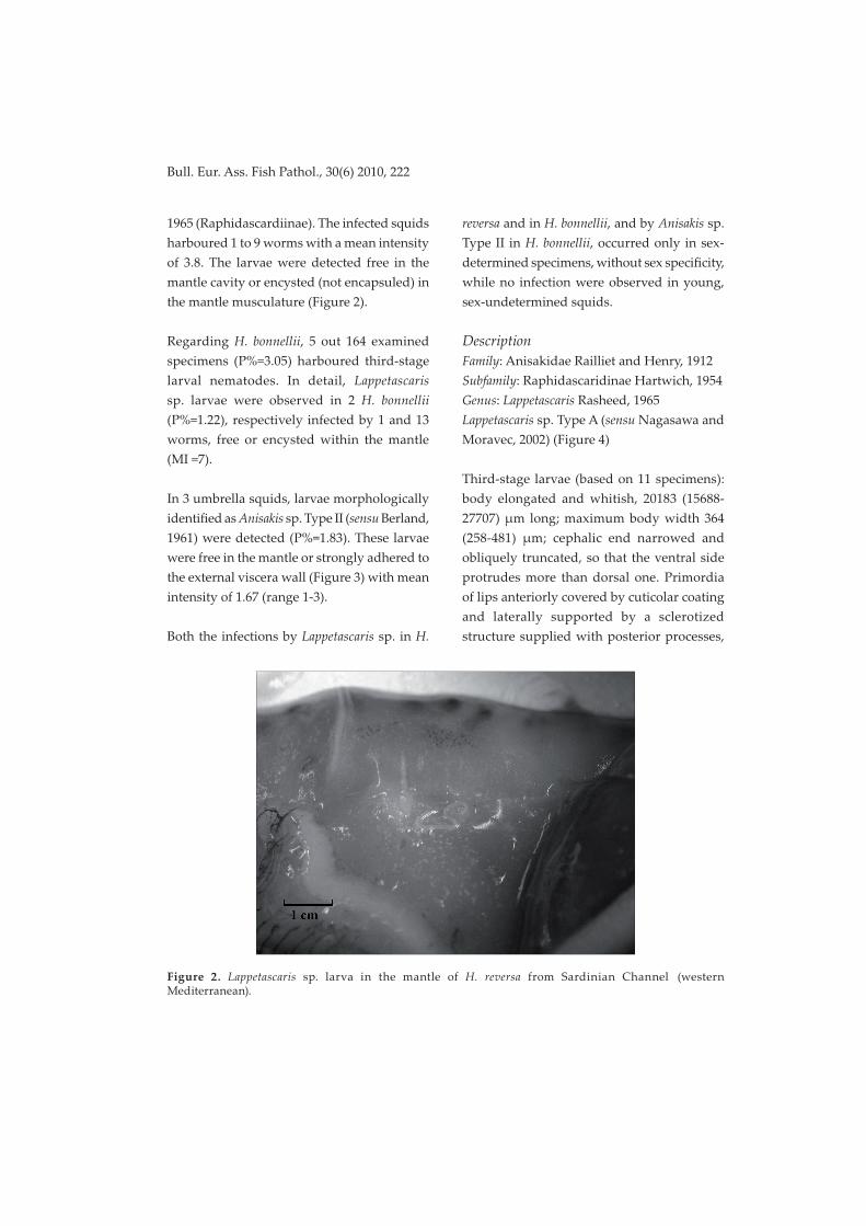
1965 (Raphidascardiinae). The infected squids harboured 1 to 9 worms with a mean intensity of 3.8. The larvae were detected free in the mantle cavity or encysted (not encapsuled) in the mantle musculature (Figure 2).
Regarding H. bonnellii, 5 out 164 examined specimens (P%=3.05) harboured third-stage larval nematodes. In detail, Lappetascaris sp. larvae were observed in 2 H. bonnellii (P%=1.22), respectively infected by 1 and 13 worms, free or encysted within the mantle (MI =7).
In 3 umbrella squids, larvae morphologically identified as Anisakis sp. Type II (sensu Berland, 1961) were detected (P%=1.83). These larvae were free in the mantle or strongly adhered to the external viscera wall (Figure 3) with mean intensity of 1.67 (range 1-3).
Both the infections by Lappetascaris sp. in H.
reversa and in H. bonnellii, and by Anisakis sp. Type II in H. bonnellii, occurred only in sex-determined specimens, without sex specificity, while no infection were observed in young, sex-undetermined squids.
DescriptionFamily: Anisakidae Railliet and Henry, 1912Subfamily: Raphidascaridinae Hartwich, 1954Genus: Lappetascaris Rasheed, 1965Lappetascaris sp. Type A (sensu Nagasawa and Moravec, 2002) (Figure 4)
Third-stage larvae (based on 11 specimens): body elongated and whitish, 20183 (15688-27707) μm long; maximum body width 364 (258-481) μm; cephalic end narrowed and obliquely truncated, so that the ventral side protrudes more than dorsal one. Primordia of lips anteriorly covered by cuticolar coating and laterally supported by a sclerotized structure supplied with posterior processes,
Figure 2. Lappetascaris sp. larva in the mantle of H. reversa from Sardinian Channel (western Mediterranean).

Bull. Eur. Ass. Fish Pathol., 30(6) 2010, 223
ventral side of the whole structure 73 (65-88) μm long. Oesophagus cylindrical, 1883 (1427-2920) μm long; oesophageal gland distinct; ventriculus oval, 243 (172-373) μm long and 120 (74-156) μm wide. Intestinal caecum extending always behind the level of excretory pore, 1633 (1060-2533) μm long; ventricular appendix very long, measuring 13129 (8970-19951) μm; intestine straight, rectum hyaline with three drop-shaped rectal glands. Nerve ring situated 369 (278-421) μm from anterior extremity; distance between excretory pore and anterior extremity 461 (411-495) μm; tail conical with slightly inflated extremity, 255 (179-377) μm long, bearing a caudal spike apparently formed by two segments, 7.1 (6.5-8.1) μm long.
Representative specimens are deposited in the Museo di Zoologia del Dipartimento di Biologia Animale ed Ecologia (University of Cagliari).
Report detailsHost: H. bonnellii, umbrella squid and H.
reversa, reverse jewel squid (Cephalopoda: Teuthoidea: Histioteuthidae).Site of infection: mantle musculature, mantle cavity.Locality: Mediterranean Sea: Sardinian Channel (haul d: 38°55’N and 9°21’E / 39°02’N and 9°16’E, 9 Dec 2005, 22 Jun and 12 Oct 2007, 11 Jan 2008, 4 Feb 2008 ― H. reversa, 18 Jan 2008 ― H. bonnellii; haul b: 38°36’N and 8°35’E / 38°42’N and 8°51’E, 7 Apr 2009 ― H. bonnellii).
CommentsThe nematodes collected correspond in their morphology to the third-stage larvae of Lappetascaris sp. described first by Nagasawa and Moravec (1995) in the common squid Todarodes pacificus (Steenstrup, 1880) from the Sea of Japan, and to Lappetascaris sp. Type A found by the same authors (2002) in Thysanoteuthis rhombus Troschel, 1857, Ommastrephes bartramii (Lesueur, 1821), Onychoteuthis borealijaponica Okada, 1927, and Gonatopus borealis Sasaki, 1923 from the central
Figure 3. Anisakis sp. Type II larvae encysted in the visceral wall of H. bonnellii.

Bull. Eur. Ass. Fish Pathol., 30(6) 2010, 224
Figure 4. Third-stage larvae of Lappetascaris sp. from Histioteuthis reversa from Sardinian Channel (western Mediterranean). A, anterior part of body; B, general view; C, cephalic end; D, caudal end. cc, cuticolar coating of the lips primordia; cs, caudal spike; ep, excretory pore; i, intestine; ic, intestinal caecum; nr, nerve ring; oe, oesophagus; pl, primordium of lips; ps, process of sclerotized support of lips primordia; r, rectum; rg, rectal glands; ss, sclerotized support of lips primordia; v, ventriculus; va, ventricular appendix.

Bull. Eur. Ass. Fish Pathol., 30(6) 2010, 225
and northern Pacific Ocean.
As reported by Nagasawa and Moravec (1995), another description of third-stage larvae of the same genus is given by Moravec and Scholz in 1991 from the cyprinid fish Paralaubuca cf. typus, but the same authors assert that the two forms belong to different congeneric species.
To date, the genus Lappetascaris comprises only one species, L. lutjanii, first described by Rasheed (1965) from Lutjanus sp. (Bloch, 1790) (Lutjanidae) and Hilsa ilisha (Hamilton, 1822) (Clupeidae) from Karachi waters (Pakistan), then detected by other authors in marine, brackish and fresh water teleosts from India, Pakistan (Soota and Dey-Sarkar, 1980; De, 1990) and from Atlantic coasts of Brazil (Vicente et al., 2002).
This is the first report of the presence of the genus Lappetascaris both in Mediterranean Sea and in the Histioteuthidae.
DescriptionFamily: Anisakidae Railliet & Henry, 1912 Subfamily: Anisakinae Chabaud, 1965Genus Anisakis Dujardin, 1845Anisakis sp. Type II (sensu Berland, 1961) (Figure 5)
Third-stage larvae (based on 2 specimens): body length 28552 (28311-28792) μm; maximum body width 831 (828-833) μm; cuticle transversely finely striated; anterior extremity rounded, ventral lip with one larval tooth 21.6 (21-22.2) μm long. Oesophagus straight, 1904 (1875-1932) μm long; ventriculus white, relatively short, 592 (516-667) μm long and 335 (291-379) μm wide; intestine straight,
rectum short with three rounded rectal glands. Distance of nerve ring from anterior extremity 323 (322-324) μm; excretory pore ventral, situated at 48 (48-48) μm from anterior extremity; tail conical, without mucron, 270 (225-314) μm long.
Representative specimens are deposited in the Museo di Zoologia del Dipartimento di Biologia Animale ed Ecologia (University of Cagliari).
Report detailsHost: H. bonnelliiSite of infection: stomach wall; mantle cavity.Locality: western Mediterranean: Sardinian Channel (haul d: 38°55’N and 9°21’E / 39°02’N and 9°16’E, 15 Apr 2005, 5 May 2006; haul b: 38°36’N and 8°35’E / 38°42’N and 8°51’E, 3 Apr 2009).
CommentsThese larvae differ from those of Anisakis sp. Type I (sensu Berland, 1961) and are characterized by a short ventriculus with a horizontal ventriculo-intestinal junction and a long conical tail without a mucron, corresponding to Anisakis sp. Type II (sensu Berland, 1961). This larval morphotype was reported in Mediterranean Sea from several species of fish and cephalopods (Ma�iucci et al., 1986, 2001; Orecchia et al., 1989; Culurgioni et al., 2006). Nowadays, Anisakis sp. Type II morphotype comprises three genetically distinct species: A. physeteris Baylis, 1923, A. brevispiculata Dollfus, 1966 (Ma�iucci et al., 2001), and A. paggiae (Ma�iucci et al., 2005).
This is the first report of the presence of third-stage larvae of Anisakis sp. Type II in H. bonnelli from Mediterranean Sea.

Bull. Eur. Ass. Fish Pathol., 30(6) 2010, 226
Figure 5. Third-stage larvae of Anisakis sp. Type II from Histioteuthis bonnellii from Sardinian Channel (western Mediterranean). A, anterior part of body; B, caudal end. i, intestine; nr, nerve ring; oe, oesophagus; r, rectum; rg, rectal glands; vt, ventral tooth; v, ventriculus.
DiscussionConcerning Lappetascaris sp., it is noticeable the first observation of this genus in Mediterranean and, considering the role of cephalopods as intermediate or paratenic hosts in the transmission of Anisakidae, it would be important to deepen the study on the life-cycle of this parasite, identifying its first hosts, probably euphasiids and other shrimps and its definitive hosts. Regarding the la�er, since adults of the single species of this genus, L. lutianji, as reported above, has been detected in teleosts both from Indian Ocean and from Atlantic coasts of Brasil (Rasheed, 1965; Soota and Dey-Sarkar 1980; De, 1990; Vicente et al., 2002), it would be interesting to pursue further investigations on histioteuthids-feeding Mediterranean teleosts as Xiphias gladius L. (Bello, 1991, 1996).
It’s known that Anisakis sp. Type II larvae lack morphologically useful characters for an identification to the specific level by light microscopy. On the other hand, geographical data concerning the distribution of A. physeteris, A. brevispiculata and A. paggiae, ascribed to the Anisakis sp. Type II morphotype (Ma�iucci et al., 2005) suggest that the specimens described in the present work may be A. physeteris, also reported recently by Farjallah et al. (2008) from the Northern Tunisian coasts, i.e. Sardinian Channel. This hypothesis is also supported by the fact that H. bonnellii is one of the most important preys of the sperm whale Physeter catodon L. in Atlantic and in Mediterranean waters (Santos et al., 1999; Roberts, 2003) well known as the main definitive host of A. physeteris (Ma�iucci et al., 2001).

Bull. Eur. Ass. Fish Pathol., 30(6) 2010, 227
ReferencesBello G (1991). Role of Cephalopods in the diet of the swordfish, Xiphias gladius, from the Eastern Mediterranean Sea. Bulletin of Marine Science 49 (1-2), 312-324.
Bello G (1996). Teuthophagous predators as collectors of oceanic cephalopods: the case of the Adriatic Sea. Bolle�ino Malacologico 32 (14), 71-78.
Berland B (1961). Nematodes from some Norwegian marine fishes. Sarsia 2, 1-50.
Bush AO, Lafferty KD, Lotz JM and Shostak AW (1997). Parasitology meets ecology on its terms: Margolis et al. revisited. Journal of Parasitology 83 (4), 575-583.
Cuccu D, Mereu M, Loi B, Sanna I and Cau A (2007). The squid family Histioteuthidae in the Sardinian waters. Biologia Marina Mediterranea 14 (2), 262-263.
Culurgioni J, D’Amico V, Coluccia E, Mulas A and Figus V (2006). Metazoan parasite fauna of conger eel Conger conger L. from Sardinian waters (Italy). I�iopatologia 3, 253-261.
De NC (1990). Some new data on the morphology of the nematode Lappetascaris lutjani Rasheed, 1965 (Nematoda: Anisakidae). Folia Parasitologica 37 (4), 295-299.
Dollfus RP (1958). Copépodes, Isopodes et Helminhes parasites de Céphalopodes de la Méditerranée et de l’Atlantique Européen. Faune marine des Pyrénées Orientales 1, 61-72.
Farjallah S, Ben Slimane B, Busi M, Paggi L, Amor N, Blel H, Said K and D’Amelio S (2008). Occurrence and molecular identification of Anisakis spp. from the North African coasts of Mediterranean Sea. Parasitology Research 102, 371-379.
Hochberg FG (1983). The parasites of cephalopods: a review. Memoirs of the National Museum Victoria 44, 109-145.
Hochberg FG (1990). Diseases of Mollusca: Cephalopoda. Diseases caused by protistans and metazoans. In “Diseases of Marine Animals. Vol. III. (Cephalopoda to Urochordata)” (O. Kinne, Ed.), pp 21-227. Biologische Anstalt Helgoland, Hamburg.
Ma�iucci S, Nasce�i G, Bullini L, Orecchia P and Paggi L (1986). Genetic structure of Anisakis physeteris, and its differentiation from the Anisakis simplex complex (Ascaridida: Anisakidae). Parasitology 93, 383-387.
Ma�iucci S, Paggi L, Nasce�i G, Abollo E, Webb SC, Pascual S, Cianchi R and Bullini L (2001). Genetic divergence and reproductive isolation between Anisakis brevispiculata and Anisakis physeteris (Nematoda: Anisakidae). International Journal of Parasitology 31, 9-14.
Ma�iucci S, Nasce�i G, Dailey M, Webb SC, Barros NB, Cianchi R and Bullini L (2005). Evidence for a new species of Anisakis Dujardin, 1845: morphological description and genetic relationships between congeners (Nematoda: Anisakidae). Systematic Parasitology 61, 157-171.
Nagasawa K and Moravec F (1995). Larval anisakid nematodes of Japanese common squid (Todarodes pacificus) from the Sea of Japan. Journal of Parasitology 81 (1), 69-75.
Nagasawa K and Moravec F (2002). Larval anisakid nematodes from four species of squid (Cephalopoda: Teuthoidea) from the central and western North Pacific Ocean. Journal of Natural History 36, 883-891.
Orecchia P, Paggi L, Ma�iucci S, Di Cave D and Catalini N (1989). Infestazione da larve di Anisakis simplex A e Anisakis physeteris in specie i�iche dei mari italiani. Parassitologia 31, 37-43.
Oshima T, Shimazu T, Koyama H and Akahane H (1969). On the larvae of the genus Anisakis (Nematoda: Anisakinae) from the euphasiids. Japanese Journal of Parasitology 18, 241-248.

Bull. Eur. Ass. Fish Pathol., 30(6) 2010, 228
Oshima T (1972). Anisakis and anisakiasis in Japan and adjacent areas. In “Progress of Medical Parasitology in Japan. Vol. 4” (K. Morishita, Y. Komiya, H. Matsubayashi, Eds.), pp 301-393. Meguro Parasitological Museum, Tokio.
Pascual S, Gonzàlez C, Arias C and Guerra A (1995). Helminth infection in the short-finned squid Illex coindetii (Cephalopoda, Ommastrephidae) off NW Spain. Diseases of Aquatic Organisms 23, 71-75.
Pascual S, Gestal C, Estevez JM, Rodriguez H, Soto M, Abollo E and Arias C (1996). Parasites in commercially exploited cephalopods (Mollusca: Cephalopoda) in Spain: an update perspective. Aquaculture 142, 1-10.
Rasheed S (1965). On a Remarkable New Nematode, Lappetascaris lutjani gen. et sp. nov. (Anisakidae: Ascaridoidea) from Marine Fishes of Karachi and an Account of Thynnascaris inquies (Linton, 1901) n. comb. and Goezia intermedia n. sp.. Journal of Helminthology XXXIX (4), 313-342.
Roberts SM (2003). Examination of the stomach contents from a Mediterranean sperm whale found south of Crete, Greece. Journal of Marine Biological Association of the United Kingdom 83 (3), 667-670.
Roper CFE and Voss GL (1983). Guidelines for taxonomic descriptions of Cephalopod species. Memoirs of the National Museum Victoria 44, 49-63.
Santos MB, Pierce GJ, Boyle PR, Reid RJ, Ross HM, Pa�erson IAP, Kinze CC, Tougaard S, Lick R, Piatkowski U and Hernandez-Garcia V (1999). Stomach contents of sperm whale Physeter macrocephalus stranded in the North Sea 1990-1996. Marine Ecology Progress Series 183, 281-294.
Soota TD and Dey-Sarkar SR (1980). On three species of the nematode genus Cucullanus Mueller 1777, and a note on Lappetascaris
lutjani Rasheed, 1965 from Indian marine fishes. Records of the Zoological Survey of India 76, 1-6.
Vicente JJ, Mincarone MM and Pinto RM (2002). First Report of Lappetascaris lutjani Rasheed, 1965 (Nematoda, Ascaridoidea, Anisakidae) Parasitizing Trachipterus arawatae (Pisces, Lampridiformes) on the Atlantic Coast of Brazil. Memórias do Instituto Oswaldo Cruz 97 (1), 93-94
Xavier J, Clarke MR, Magalhães MC, Stowasser, G, Blanco C and Cherel Y (2007). Current status of using beaks to identify cephalopods: III International Workshop and training course on Cephalopod beaks, Faial island, Azores, April 2007. Arquipélago - Life and Marine Sciences 24, 41-48.
Voss NA, Nesis KN and Rodhhouse PG (1998). The cephalopod family Histioteuthidae (Oegopsida): Systematics, biology and biogeography. In “Systematics and Biogeography of Cephalopods (Vol. II)” (N.A. Voss, M. Vecchione, R.B. Toll, M.J. Sweeney, Eds.), pp 293-372. Smithsonian Contributions to Zoology.