histocompatibility and hematopoietic transplantation in
TRANSCRIPT

Histocompatibility and HematopoieticTransplantation in the Zebrafish
The Harvard community has made thisarticle openly available. Please share howthis access benefits you. Your story matters
Citation de Jong, Jill L. O., and Leonard I. Zon. 2012. Histocompatibilityand hematopoietic transplantation in the zebrafish. Advances inHematology 2012:282318.
Published Version doi:10.1155/2012/282318
Citable link http://nrs.harvard.edu/urn-3:HUL.InstRepos:10448842
Terms of Use This article was downloaded from Harvard University’s DASHrepository, and is made available under the terms and conditionsapplicable to Other Posted Material, as set forth at http://nrs.harvard.edu/urn-3:HUL.InstRepos:dash.current.terms-of-use#LAA
Hindawi Publishing CorporationAdvances in HematologyVolume 2012, Article ID 282318, 8 pagesdoi:10.1155/2012/282318
Review Article
Histocompatibility and HematopoieticTransplantation in the Zebrafish
Jill L. O. de Jong1 and Leonard I. Zon2
1 Section of Hematology-Oncology and Stem Cell Transplant, Department of Pediatrics, The University of Chicago,KCBD 5120, Chicago, IL 60637, USA
2 Stem Cell Program and Division of Hematology/Oncology, Children’s Hospital Boston-Dana, Farber Cancer Institute,Howard Hughes Medical Institute, Harvard Stem Cell Institute, and Harvard Medical School, Boston, MA 02115, USA
Correspondence should be addressed to Jill L. O. de Jong, [email protected]
Received 7 March 2012; Accepted 1 May 2012
Academic Editor: Jason Berman
Copyright © 2012 J. L. O. de Jong and L. I. Zon. This is an open access article distributed under the Creative Commons AttributionLicense, which permits unrestricted use, distribution, and reproduction in any medium, provided the original work is properlycited.
The zebrafish has proven to be an excellent model for human disease, particularly hematopoietic diseases, since these fishmake similar types of blood cells as humans and other mammals. The genetic program that regulates the development anddifferentiation of hematopoietic cells is highly conserved. Hematopoietic stem cells (HSCs) are the source of all the blood cellsneeded by an organism during its lifetime. Identifying an HSC requires a functional assay, namely, a transplantation assayconsisting of multilineage engraftment of a recipient and subsequent serial transplant recipients. In the past decade, severaltypes of hematopoietic transplant assays have been developed in the zebrafish. An understanding of the major histocompatibilitycomplex (MHC) genes in the zebrafish has lagged behind transplantation experiments, limiting the ability to perform unbiasedcompetitive transplantation assays. This paper summarizes the different hematopoietic transplantation experiments performed inthe zebrafish, both with and without immunologic matching, and discusses future directions for this powerful experimental modelof human blood diseases.
1. Introduction
In the past few decades, the zebrafish has emerged as anoutstanding vertebrate animal model for studying develop-mental hematopoiesis (reviewed in [1, 2]). In this same timeframe, the understanding of the biology of adult hematopoi-etic stem cells has also blossomed, predominantly due tohematopoietic transplantation experiments performed inmice (reviewed by Orkin and Zon in [3]). To capitalize on theadvantages of the zebrafish model (small size, high fecundity,rapid maturation, external fertilization, and the ability toperform large-scale genetic and chemical screens), a zebra-fish hematopoietic transplantation assay was needed.
Developing a transplantation assay in the zebrafishrequired a different approach than that used in mice. Whiledifferential expression of CD45 isoforms is generally usedto distinguish between donor and recipient cells in murinetransplant assays, these reagents are not available for
zebrafish. Instead, scientists have utilized transgenic technol-ogy to make zebrafish expressing green fluorescent protein(GFP) or other fluorochromes under the influence of anubiquitous or a tissue-specific promoter. These fluorescentlylabeled donor cells are transplanted into fluorochrome-negative recipients, and engraftment is monitored at varioustime points after transplant.
2. A History of HematopoieticTransplantation in Zebrafish
2.1. Adult Marrow Cells into Embryos. The first hematopoi-etic transplant experiments in zebrafish were performed byTraver et al., whose work was published in 2003 [4]. Thislandmark paper was the first to report the evaluation ofzebrafish kidney marrow cells including separation of themajor blood cell lineages by flow cytometry, a method which
2 Advances in Hematology
Erythroid
LymphoidPrecursors
Myeloid
Forward scatter (FSC)
104
103
102
101
100
200 400 600 800 1000
Side
sca
tter
(SS
C)
(a)
GFP
Lymphoid Myeloid Precursors
44% 91% 70%
100
80
60
40
20
0100 101 102 103
Max
(%
)
100
80
60
40
20
0100 101 102 103
100
80
60
40
20
0100 101 102 103
FL1-H:: gfp FL1-H:: gfp FL1-H:: gfp
(b)
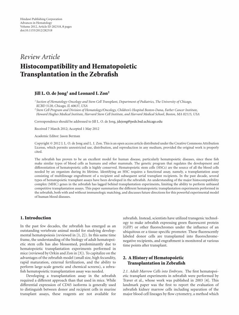
Figure 1: Flow cytometry analysis of zebrafish whole kidney marrow from a marrow transplant recipient. Zebrafish transplant recipientswere irradiated and injected with 5 × 10⁵ marrow cells from a transgenic β-actin:GFP donor. Whole kidney marrow from a representativerecipient was dissected 3 months later and analyzed by flow cytometry. (a) The forward scatter (FSC) versus side scatter (SSC) profile ofzebrafish whole kidney marrow shows four cell populations: erythroid, lymphoid, myeloid, and precursor cells. (b) Histograms for GFPexpression of cells within the lymphoid, myeloid and precursor gates show multilineage engraftment with GFP+ donor cells (blue lines). Thered lines show GFP expression in a wild-type-negative control fish.
is currently the standard procedure for identifying multi-lineage engraftment after hematopoietic transplantation inzebrafish (Figure 1(a)). In addition, hematopoietic trans-plantation was used to rescue two different mutant embryos.The Vlad tepes (gata1−/−) mutation is homozygous lethalby 14 days after fertilization, and these embryos have acomplete absence of erythroid cells [5]. Approximately 100–1000 whole kidney marrow (WKM) cells from a gata1-GFP transgenic donor were injected into the circulation ofgata1−/− zebrafish embryos 48 hours after fertilization (hpf).While untransplanted control embryos did not survive past14 dpf, 20–60% of the transplant recipients survived longterm, up to 8 months after transplant. All surviving recipientshad circulating GFP+ red blood cells, indistinguishable fromthe gata1-GFP donors [4].
Taking these embryonic transplant experiments one stepfurther, donor marrow was isolated from double transgenicβ-actin-GFP/gata1-dsRED fish, in order to monitor donor-derived cells from multiple lineages. The β-actin-GFP trans-gene is expressed by almost all zebrafish cell types, including
all leukocytes. Erythrocytes do not express βactin, so theyare marked by the gata1-dsRED transgene instead. For theseexperiments, the transplant recipients were bloodless (bls)mutants, a dominant, partially penetrant mutation resultingin absent primitive hematopoiesis, but preserved adulthematopoiesis [6]. Injection of double-positive WKM cellsinto 48 hpf bls mutants allowed independent tracking ofGFP+ leukocytes and dsRED+ erythrocytes in the recipientembryos [4]. Sustained multilineage donor-derived cellswere visible in the circulation of transplant recipients at8 weeks after transplantation, indicating successful engraft-ment of long-term hematopoietic repopulating cells.
2.2. Adult Marrow Cells into Adult Recipients. Following upon their transplantation experiments into embryos, Traveret al. subsequently performed transplantation of WKMcells into adult recipients [7]. After using ionizing radia-tion as pretransplant conditioning to ablate the recipient’shematopoietic cells, including the immune system, approx-imately 1 × 10⁶ β-actin-GFP/gata1-dsRED donor marrow
Advances in Hematology 3
(a) (b)
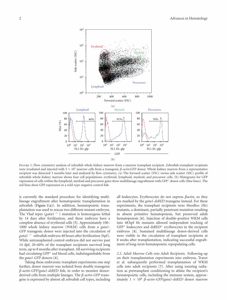
Figure 2: Direct visualization of engrafted GFP+ and mCherry+ marrow donor cells in casper recipients. 40 × 103 WKM cells from atransgenic ubiquitin:GFP donor were mixed with 80 × 103 WKM cells from a transgenic ubiquitin:mCherry donor and injected into thecirculation of a casper recipient fish. The photos are taken 4 weeks after transplantation and show engraftment of (a) GFP+ and (b) mCherry+
cells in the kidney (white arrows).
cells were delivered into the recipient’s circulation by directintracardiac injection. When irradiated with 40 Gy, a lethaldose, all the untransplanted animals died by 14 days afterirradiation. However, >70% of the animals receiving WKMcells after irradiation were rescued, and survived at least 30days after irradiation. As in the experiments with embry-onic transplant recipients, GFP+ leukocytes and dsRED+
erythrocytes were visible in the circulation of the engraftedadult recipients using fluorescence light microscopy [7].FACS analysis of recipient WKM showed robust multilineageengraftment with >86% GFP+ cells up to 8 weeks aftertransplant (Figure 1(b)).
2.3. Embryonic HSCs into Embryos. Similar to murine embr-yonic HSCs, the first HSCs in the developing zebrafish arelocated in the aorta-gonad-mesonephros (AGM) [8]. Initialexperiments to identify these HSCs in zebrafish relied uponanatomic similarities with murine embryonic HSCs. Cellsexpressing cmyb, runx1, and CD41 are observed in the ventralwall of the dorsal aorta in zebrafish embryos 24–36 hpf[9–12], similar to the expression noted in the ventral wallof the aorta in murine embryos [13]. These cmyb+ andrunx1+ cells were presumed to be embryonic definitiveHSCs, although functional evaluation of these cells waslacking. Using CD41 as another marker of embryonic HSCs,Bertrand et al. sorted CD41+/gata1− donor cells by flowcytometry from CD41-eGFP/gata1-dsRED double transgenicembryos at 72 hpf [14]. These cells were then injected intothe sinus venosus of age-matched wild-type embryos. Withinone day after transplant, donor-derived cells were observedin the caudal hematopoietic tissue (CHT) and thymi ofrecipients. Although the transplanted donor cells had beendsRED negative, subsequent erythroid differentiation ofengrafted cells revealed dsRED+ cells in the circulation ofrecipients [14]. These experiments helped to prove thatCD41+ cells in the AGM are capable of colonizing definitivehematopoietic organs, namely, the thymus and CHT, indeveloping zebrafish, and therefore, this population includesthe first developing HSCs in the embryo.
2.4. A Competitive Transplantation Assay for Chemical Screen-ing. Capitalizing on the relative ease of in vivo chemicalscreening using the zebrafish model, Li et al. have utilizeda competitive hematopoietic transplantation assay to search
for chemicals that enhance hematopoietic engraftment(manuscript submitted). Marrow cells from βactin-GFP fishwere incubated ex vivo in chemicals from a panel of morethan 2000 known bioactive compounds. After pretreatment,the βactin-GFP WKM was mixed at a standard ratio withWKM from commercially available red Glofish, and trans-planted into casper recipient fish [15]. Normally kidneymarrow fluorescence is not visible in an adult animal due tothe presence of pigmentation in the skin. However, casper fishare homozygous for two pigment mutations, roy and nacre,and therefore have transparent skin, allowing visualizationof engrafted fluorescent marrow cells in vivo. Unlike priorstudies examining engraftment at a single time point byFACS analysis of multilineage WKM populations, this screenalso followed the level of GFP+ and RFP+ cells in thekidney of anesthetized recipients at several time points aftertransplant (Figure 2). The ratio of green : red marrow cellsby fluorescence microscopy in vivo was highly correlatedwith the green : red ratio measured by flow cytometry ofthe dissected WKM cell preparation. All chemicals identifiedin the screen that stimulated enhanced engraftment werealso tested in murine transplants to validate the effects inan immune-matched mammalian transplant assay. In total,ten compounds were identified in the screen that resulted inenhanced green : red ratio, and these are currently undergo-ing further evaluation.
3. Importance of Immune Matchingin Hematopoietic Transplantation
None of the transplantation experiments described to thispoint took into account any aspect of immunologic match-ing, as isogenic and congenic fish lines were not available.This fact highlights another significant difference betweenmurine and zebrafish marrow transplants, namely that mu-rine donors and recipients are congenic and hence immuno-logically identical. In contrast, although many commonlyused zebrafish lines (e.g., AB, Tubingen, and wik) havebeen repeatedly incrossed through decades of laboratory use,attempts to generate truly isogenic or congenic zebrafishlines have largely failed due to inbreeding depression suchthat these fish lines could no longer be maintained [16]. Inaddition, sex skewing of clutches, whereby a generation ofsiblings was all the same sex, has also hindered the ability

4 Advances in Hematology
to maintain highly inbred fish lines. Despite this disadvan-tage, significant progress has still been made developinghematopoietic transplantation methods in the zebrafish overthe past decade, as described above.
As more sophisticated transplantation experiments aredesigned to ask more complex questions about stem cell biol-ogy, the need for immune matching becomes more critical.When transplanting any allogeneic tissue into an adult recip-ient with a competent immune system, one would expect alack of immune matching to result in rejection of the trans-planted tissue (reviewed in [17]). In the zebrafish, immunematching is not required in embryonic recipients youngerthan 5 days after fertilization, as thymic development is notapparent until then [18]. By 4–6 weeks after fertilization, thecellular and humoral immune system is fully functional andwould be capable of rejecting any transplanted tissue thatwas not histocompatible [19, 20]. Pretransplant conditioningwith radiation is commonly used to suppress the immunesystem of adult murine and zebrafish recipients, and in thecase of hematopoietic transplants to give the added advan-tage of clearing the marrow niche. For zebrafish recipientsreceiving a sublethal dose of radiation, the transplantedtissue is still rejected once the recipient’s immune cellsrecover, approximately 4 weeks after irradiation [21].
Another consequence of immune mismatch betweentransplant donors and recipients occurs uniquely in thesetting of hematopoietic transplantation. When engraftedimmune cells recognize the recipient as “nonself,” an im-mune response is mounted against the recipient’s tissuesresulting in graft-versus-host disease (GVHD), a phenome-non that is also observed clinically in human allogeneicbone marrow transplant [22]. Therefore, the importance ofimmune matching in hematopoietic transplantation impactsnot only initial engraftment, but also the health and survivalof the recipient if the engrafted hematopoietic cells attack thehost.
4. Methods to QuantitateHematopoietic Engraftment
Comparing the function of two HSC populations involves acompetitive hematopoietic transplantation assay where bothpopulations engraft in the same transplant recipient(reviewed by Purton and Scadden in [23]). This experimentaldesign is required when mutant marrow cells from one donorare hypothesized to have defective hematopoietic engraft-ment. The mutant cells are transplanted into the recipienttogether with a radio-protective dose of wild-type marrowcells. If the mutant HSCs are defective, the wild-type HSCswill out-compete them, and the donor chimerism of therecipient will highly favor the wild-type donor cells. Withoutthese wild-type HSCs to rescue the recipient, lack of engraft-ment of the mutant cells would likely result in the recipient’sdeath, and there would be no blood or marrow cells toevaluate at the end of the experiment. Using a competitiveexperimental design ensures that all the recipients surviveuntil the end of the experiment and their data are includedin the final analyses. In the event that the mutant marrow has
normal HSC function, the donor chimerism would revealan equal mix of engrafted hematopoietic cells from bothdonors. Immune matching of both donors and the recipientis an essential component of any competitive hematopoietictransplantation assay. Otherwise, one cannot rule out biasedimmune rejection of one donor’s cells compared to theother, and the engraftment “winner” may merely reflectimmunologic differences and not a difference in stem cellbiology.
A variation of the competitive hematopoietic transplan-tation assay is the limit dilution assay. This method is thegold standard for quantitating HSC content and also requiresall donors and recipients to be immunologically matched.This assay involves transplantation of serially diluted marrowcells such that fewer and fewer marrow cells are given tosubsequent transplant recipients, while a constant numberof wild-type marrow cells are given simultaneously to radio-protect the recipients. Engraftment and donor chimerismare evaluated for each recipient, and then Poisson statisticsare used to calculate the number of long-term repopulatingcells contained in the original marrow population [24]. Theability to perform these competitive and quantitative exper-iments using zebrafish HSCs will be essential to characterizestem cell mutants and asking questions about HSC biology.Therefore, a better understanding of the histocompatibilitygenes in the zebrafish is needed so that these assays can beperformed with proper immune matching.
5. Histocompatibility Antigens in ZebrafishCompared with Other Vertebrates
One of the first multimegabase regions of the human genometo be sequenced, the human major histocompatibility com-plex (MHC) locus, is located on chromosome 6p21.31 andcontains over 200 identified genes within a 3.6× 10⁶ basepairspan [25]. The classical class I and class II genes within theMHC region are the central cell surface proteins responsiblefor determining tissue histocompatibility of an allograft. Thisgene-dense region also contains a number of other genesimportant for the immune response, including antigen-processing genes such as proteasome subunit β type (PSMB),complement genes, and the peptide transporters TAP1 andTAP2 [26, 27].
Class I MHC molecules are polymorphic transmembraneproteins with three immunoglobulin-like domains that areexpressed on virtually all cell types. They bind noncovalentlyto β2-microglobulin and present endogenously derived pep-tides to CD8+ T lymphocytes (reviewed in [28]). Althoughclass I and II proteins share a similar three-dimensionalstructure, class II MHC molecules are heterodimeric com-plexes consisting of an alpha chain and a beta chain, witheach chain containing two immunoglobulin-like domains.They present lysosomally derived peptide antigens to CD4+
T lymphocytes, and their expression is limited to B-lympho-cytes, macrophages, and other antigen-presenting cells.
While most jawed vertebrate species possess linked classI and II genes located within a single chromosomal locussimilar to the human MHC, the bony fishes are unique in

Advances in Hematology 5
Table 1: Mean percentage of GFP+ cells in engrafted recipient zebrafish receiving MHC-matched or -unmatched donor marrow.
Only Chr 19 matched [35] Chr 1, 8, 19 all matched
Myeloid matched 47.86± 30.9P = 0.0002
52.36± 25.43P = 0.0036
Myeloid unmatched 6.45± 1.77 11.58± 7.03
Lymphoid matched 10.51± 19.88P = 0.05
9.51± 12.32P = 0.047
Lymphoid unmatched 1.28± 0.38 3.47± 4.601
Data are mean ± S.D.
that they have class I and II genes located on distinct chro-mosomes [29]. In the zebrafish, at least three relevant locihave been identified. Chromosome 19 contains class I genesas well as some antigen-processing genes, making the locussyntenic to the human MHC locus [30, 31]. However, thereare no class II genes on chromosome 19. Instead the zebrafishclass II alpha and beta genes are located on chromosome 8[26, 32]. Chromosome 1 contains additional class I genes,termed “ze” genes, which appear most similar to mammaliannonclassical Class I genes [33]. Finally, the “L” genes, classI genes unique to teleost fish, are located on chromosomes3 and 8, although they are less polymorphic than otherclass I genes, and their precise function is not clear [34].While DNA sequence analyses of the zebrafish MHC genesshow similarities with MHC genes of many species, virtuallyno data are available to evaluate the function or even thecell-surface expression of the class I and II genes in zebrafish.Prior to the transplantation experiments described below, nofunctional evaluation of any zebrafish MHC genes had beenperformed.
6. Immune-Matched HematopoieticTransplants in Zebrafish
Following up on the adult marrow transplant experimentspublished in 2004 [7], subsequent adult transplantationexperiments sought to evaluate long-term hematopoieticengraftment greater than 12 weeks after transplant. Hav-ing observed poor survival in random donor long-termhematopoietic transplantation experiments (J. L. O. de Jongand L. I. Zon, unpublished data), immune typing of thezebrafish MHC genes was a logical step to ensure thatgraft rejection and/or GVHD were not contributing to therecipient mortality. In these first hematopoietic transplantexperiments with immune matching, the class I MHCgenes at the chromosome 19 locus were typed for all thesibling progeny of a single mating pair [35]. Genotypingwas achieved by preparing DNA from fin clips of individ-ual fish, then using a panel of PCR primers to amplifyMHC gene sequences. The amplified fragments were thensequenced to identify the specific MHC genes present ineach individual animal. As expected, there were four MHChaplotypes represented within this family, and approximately25% of the progeny fell into each of the four genotypes.WKM cells from β-actin-GFP+ donor fish of each MHCgenotype were transplanted into GFP-negative siblings of
the same MHC genotype and also into unrelated wild-type recipients, presumed to be mismatched. Survival anddonor chimerism were significantly improved in the matchedrecipients compared with the unmatched recipients (Table1), indicating the importance of immune matching at thechromosome 19 MHC locus for hematopoietic engraftment[35]. These experiments were the first functional evaluationof any zebrafish MHC genes in a transplantation assay.
These first experiments did not specifically type for classII genes located on chromosome 8, or other class I genes onother chromosomes. It may be that coincidental matching atthe class II locus occurred for a significant number of therelated “matched” recipients in these experiments, therebycontributing to improved donor chimerism.
We expected that immune matching at the class II locuswould also be important for hematopoietic engraftment.Therefore, we performed additional transplantation exper-iments matching the donors and recipients at three separateloci: the two class I loci on chromosomes 1 and 19 and theclass II locus on chromosome 8. 2.5 × 10⁵ WKM cells fromβ-actin-GFP+ donor fish were transplanted into both com-pletely matched recipients and unmatched, unrelated recip-ients. Long-term engraftment at 3 months after transplantshowed similar donor chimerism results as the transplantexperiments with matching at only the chromosome 19 locus(Table 1). These data suggest that matching of the class Igenes at the chromosome 19 locus is the most importantfor tissue histocompatibility in a transplantation assay, andthat the additional MHC loci on chromosomes 1 and 8play a minimal role. Further experiments are underway toindividually test the class I genes on chromosome 1 and theclass II genes on chromosome 8 to determine the contrib-ution, if any, of these loci to histocompatibility in tissuetransplantation.
7. Optimizing Survival of HematopoieticTransplant Recipients
Survival of zebrafish hematopoietic transplant recipients isoften difficult to predict from one experiment to the next.We have implemented a number of changes to the initiallypublished transplantation protocol to address the problemof poor survival after transplant. While lack of histocom-patibility may play a role for some animals, a number ofother factors also appear to be important. In our experience,younger fish have better survival than older fish, and optimal

6 Advances in Hematology
recipients are approximately 3-4 months of age (J. L. O. deJong and L. I. Zon, unpublished data). This may be due tocolonization of older fish with bacterial or fungal pathogensthat overwhelm and kill the immune-compromised hostafter transplantation. Maintaining excellent water quality isalso critically important to recipient survival. We hypothe-sized that treatment with prophylactic antibiotics for a fewdays immediately after transplant might improve survival.However, placing transplant recipients “off system” in fishwater containing antibiotics paradoxically caused decreasedsurvival, as fish being treated in this way suffered fromquickly deteriorating water quality and high ammonialevels (C. Lawrence, personal communication). While it isimpractical to keep a therapeutic level of antibiotics in thelarge volume of water circulating through an entire aquaticsystem, the ability to maintain water quality at a consistentlyhigh standard resulted in improved survival of our transplantrecipients, even without antibiotics.
Determining the appropriate radiation dose for pre-transplant conditioning of recipient fish has also provenmore challenging than initially anticipated. Water can greatlyattenuate the radiation dose over a short distance. For exam-ple, at a depth of 1 cm of water, we have observed that theradiation dose at the bottom of the dish is decreased by about10–15% compared with the radiation dose at the surface ofthe water (J. L. O. de Jong, unpublished data). Therefore,it is critically important that fish be placed in a minimalvolume and depth of water to ensure that all recipientsreceive an equivalent radiation dose. The minimum lethaldose of radiation for zebrafish was first reported to be 40 Gy[7]. However, subsequent work showed that this dose was notoptimal for pretransplant conditioning, as the mortality offish was 100%, even after receiving a radio-protective dose ofWKM cells. A sublethal dose of 25 Gy provided for maximalsurvival with engraftment, so this was the dose selected formost experiments [35]. This result suggests that while thehematopoietic compartment is the most radiation-sensitivetissue in the zebrafish, as in mammals, there is a narrowtherapeutic index for lethal radiation damage to other tis-sues. To minimize the radiation injury to nonhematopoietictissues, many protocols for murine and human bone marrowtransplants utilize fractionated radiation dosing. We havenow initiated a standard conditioning protocol of 30 Gysplit into two equal fractions of 15 Gy, where the twofractions are given 24 hours apart. The survival of theserecipients is comparable to animals receiving 25 Gy as asingle dose (J. L. O. de Jong, unpublished data). Finally,we have observed that different fish lines have varyingsensitivities to radiation. For example, when comparing fishfrom the AB strain that have been bred to homozygosityat the MHC loci, some were significantly more sensitive toa given radiation dose than others (Figure 3). This resultsuggests that a radiation dose-response titration should beperformed for each strain of recipients to be transplanted inorder to determine the optimal radiation dose. Alternatively,conditioning with chemotherapeutic medications such ascyclophosphamide [36] could be used, although these havenot been tested for pretransplant conditioning of zebrafishdonors.
0
0.2
0.4
0.6
0.8
1
0 10 20 30 40
UDA UBA
Days after irradiation
CG1 UXA2
Surv
ival
(%
)
Figure 3: Survival of different zebrafish lines in response to radia-tion. Kaplan-Meier survival curves are shown for four differentzebrafish strains after irradiation with a total dose of 25 Gy, deliv-ered in two equal fractions of 12.5 Gy separated by 24 hours.Twenty one fish were irradiated in each group. CG1 is a clonalhomozygous diploid fish line generated by parthenogenesis [21, 38].UDA, UXA2, and UBA are inbred zebrafish lines derived from asingle mating pair of AB parents [35]. Each line was named for thehomozygous class I MHC gene at its chromosome 19 locus. Theresults demonstrate 100% mortality for the CG1 fish by day 22,and by day 37 for the UDA fish. In contrast, the UBA and UXA2fish lines both had approximately 80% survival at 40 days afterirradiation.
8. Future Directions for HematopoieticTransplantation in the Zebrafish
Although HSC transplantation is a commonly used treat-ment modality for human diseases, including many malig-nancies, blood disorders, and immune deficiencies, thisprocedure continues to have high morbidity and mortality.Difficulties include selecting an optimally matched allogeneicdonor, prolonged immune suppression with susceptibilityto deadly infections, delayed and/or incomplete immunereconstitution, and maximizing the graft-versus-tumor effectwhile minimizing graft-versus-host disease. A zebrafishmodel for hematopoietic transplantation permitting in vivoinvestigation of these challenges would provide a basis tounderstand the biological mechanisms involved and identifypossible solutions to address them.
8.1. Parthenogenesis to Develop Homozygous Diploid FishLines. The lack of isogenic and congenic fish lines is aserious handicap for future transplantation experiments withzebrafish. To overcome this barrier, gynogenetic fish lineshave been utilized in recent years to successfully transplantliver tumors, acute lymphoblastic leukemia cells, and rhab-domyosarcoma tumor cells into unirradiated immunolog-ically identical adult recipients [21, 37]. Developing thesehomozygous diploid clonal fish lines is labor intensive, timeconsuming and inefficient [38, 39]. However, once a robust

Advances in Hematology 7
line is generated, it can be used to make transgenic donorswith fluorochrome-labeled marrow cells. These donors couldthen be used to perform competitive HSC transplants usingimmunologically identical donors and recipients. Develop-ing a homozygous diploid fish line from casper fish would beeven more useful, as the advantages of analyzing engraftmentat many time points could also be realized in the settingof an immune-matched competitive transplant. Efforts arecurrently underway to generate these fish.
8.2. Minor Histocompatibility Antigens. Further work willalso be valuable to identify all the specific class I and IIgenes important for histocompatibility in the zebrafish,both for a basic understanding of zebrafish immunology,as well as the implications for optimizing future transplantexperiments. When a zebrafish mutant has a postulated HSCdefect, scientists need to have immune-matched recipientsto test whether marrow cells from the mutant zebrafishhave flawed engraftment in a competitive transplantationassay. Without immune matching, such an assay will bedifficult to interpret. The ability to immunotype any randomzebrafish, and thereby select appropriately matched donorsand recipients would allow for a much quicker time frameto perform these experiments, compared with generationsof inbreeding, which may be unsuccessful given the historyof prior attempts to generate such inbred zebrafish lines.However, even having a donor with “perfect” matching atthe MHC locus, human bone marrow transplant recipientsare still at risk for GVHD, likely due to mismatched minorhistocompatibility antigens on other chromosomes. There-fore, identifying both major and minor histocompatibilityantigens throughout the genomes that are relevant fortransplant rejection and GVHD in the zebrafish will becritical to prospectively determine optimally matched donorsand recipients. This information will clearly be useful forzebrafish experiments, as described above. In addition, iden-tifying significant minor histocompatibility antigens in thezebrafish would suggest minor histocompatibility antigensthat may also be relevant for human bone marrow trans-plantation and GVHD. Such work may impact the selectionof human bone marrow transplant donors to minimize thispotentially devastating outcome after human BMT.
8.3. Developing a Zebrafish Model for GVHD. Finally, in theprocess of fully characterizing the zebrafish histocompatibil-ity genes, we expect to identify recipients with GVHD. Todate, we have observed transplant recipients that developsevere edema and ascites resulting in flaring of their scales.This condition in the zebrafish is generically termed “dropsy”and likely can result from a myriad of causes. We postulatethat in the setting of hematopoietic transplantation, someof these recipient fish may have GVHD, although furtherwork is needed to fully characterize the “dropsy” pheno-type after transplant and confirm the pathophysiology ofthis diagnosis. By characterizing the GVHD phenotype inzebrafish and developing a zebrafish model of GVHD, onecould exploit the advantages of genetic and small molecule-based screening to further characterize the pathways that
regulate GVHD. Such experiments may discern mechanismsto minimize GVHD while maximizing the graft-versus-leukemia effect in bone marrow transplant patients.
9. Conclusion
As a model for human disease, the zebrafish holds numerousadvantages. Gaining knowledge of the functional Class I andII genes in the zebrafish will enhance our understandingof basic zebrafish biology, as well as the ability to use thisversatile animal model to ask questions about tissue trans-plantation, including hematopoietic stem cells, other normaltissues and cancers cells. This work will likely inform mam-malian biology, improving our understanding of humanHSCs, and has the potential to impact the treatment ofpatients undergoing bone marrow transplantation.
Acknowledgment
The authors would like to thank Dr. V. Binder for helpfuldiscussions and for providing the photos in Figure 2. J. L.O. de Jong is supported by grants from the NIH NIDDK(5K08DK074595, 1R03DK091497). L. I. Zon is supported byHHMI, and grants from the NIH NHLBI (5R01HL48801-19)and NIDDK (5RO1DK53298-14).
References
[1] L. Jing and L. I. Zon, “Zebrafish as a model for normal andmalignant hematopoiesis,” Disease Models & Mechanisms, vol.4, no. 4, pp. 433–438, 2011.
[2] D. Carradice and G. J. Lieschke, “Zebrafish in hematology:sushi or science?” Blood, vol. 111, no. 7, pp. 3331–3342, 2008.
[3] S. H. Orkin and L. I. Zon, “Hematopoiesis: an evolving para-digm for stem cell biology,” Cell, vol. 132, no. 4, pp. 631–644,2008.
[4] D. Traver, B. H. Paw, K. D. Poss, W. T. Penberthy, S. Lin, and L.I. Zon, “Transplantation and in vivo imaging of multilineageengraftment in zebrafish bloodless mutants,” Nature Immuno-logy, vol. 4, no. 12, pp. 1238–1246, 2003.
[5] S. E. Lyons, N. D. Lawson, L. Lei, P. E. Bennett, B. M. Wein-stein, and P. P. Liu, “A nonsense mutation in zebrafish gata1causes the bloodless phenotype in vlad tepes,” Proceedings ofthe National Academy of Sciences of the United States of Amer-ica, vol. 99, no. 8, pp. 5454–5459, 2002.
[6] E. C. Liao, N. S. Trede, D. Ransom, A. Zapata, M. Kieran, andL. I. Zon, “Non-cell autonomous requirement for the blood-less gene in primitive hematopoiesis of zebrafish,” Develop-ment, vol. 129, no. 3, pp. 649–659, 2002.
[7] D. Traver, A. Winzeler, H. M. Stern et al., “Effects of lethalirradiation in zebrafish and rescue by hematopoietic cell trans-plantation,” Blood, vol. 104, no. 5, pp. 1298–1305, 2004.
[8] A. Medvinsky and E. Dzierzak, “Definitive hematopoiesis isautonomously initiated by the AGM region,” Cell, vol. 86, no.6, pp. 897–906, 1996.
[9] C. E. Burns, T. DeBlasio, Y. Zhou, J. Zhang, L. Zon, and S. D.Nimer, “Isolation and characterization of runxa and runxb,zebrafish members of the runt family of transcriptional regula-tors,” Experimental Hematology, vol. 30, no. 12, pp. 1381–1389,2002.
8 Advances in Hematology
[10] M. L. Kalev-Zylinska, J. A. Horsfield, M. V. Flores et al.,“Runx1 is required for zebrafish blood and vessel develop-ment and expression of a human RUNX-1-CBF2T1 transgeneadvances a model for studies of leukemogenesis,” Develop-ment, vol. 129, no. 8, pp. 2015–2030, 2002.
[11] D. Ma, J. Zhang, H. F. Lin, J. Italiano, and R. I. Handin, “Theidentification and characterization of zebrafish hematopoieticstem cells,” Blood, vol. 118, no. 2, pp. 289–297, 2011.
[12] H. F. Lin, D. Traver, H. Zhu et al., “Analysis of thrombocytedevelopment in CD41-GFP transgenic zebrafish,” Blood, vol.106, no. 12, pp. 3803–3810, 2005.
[13] T. E. North, M. F. de Bruijn, T. Stacy et al., “Runx1 expressionmarks long-term repopulating hematopoietic stem cells in themidgestation mouse embryo,” Immunity, vol. 16, no. 5, pp.661–672, 2002.
[14] J. Y. Bertrand, A. D. Kim, S. Teng, and D. Traver, “CD41+cmyb+ precursors colonize the zebrafish pronephros by anovel migration route to initiate adult hematopoiesis,” Devel-opment, vol. 135, no. 10, pp. 1853–1862, 2008.
[15] R. M. White, A. Sessa, C. Burke et al., “Transparent adultzebrafish as a tool for in vivo transplantation analysis,” CellStem Cell, vol. 2, no. 2, pp. 183–189, 2008.
[16] M. Shinya and N. Sakai, “Generation of highly homogeneousstrains of zebrafish through full sib-pair mating,” Genes Gen-omes Genetics, vol. 1, no. 5, pp. 377–386, 2011.
[17] J. Chinen and R. H. Buckley, “Transplantation immunology:solid organ and bone marrow,” Journal of Allergy and ClinicalImmunology, vol. 125, no. 2, supplement 2, pp. S324–S335,2010.
[18] C. E. Willett, A. Cortes, A. Zuasti, and A. G. Zapata, “Earlyhematopoiesis and developing lymphoid organs in the zebra-fish,” Developmental Dynamics, vol. 214, no. 4, pp. 323–336,1999.
[19] S. H. Lam, H. L. Chua, Z. Gong, T. J. Lam, and Y. M.Sin, “Development and maturation of the immune systemin zebrafish, Danio rerio: a gene expression profiling, in situhybridization and immunological study,” Developmental &Comparative Immunology, vol. 28, no. 1, pp. 9–28, 2004.
[20] D. M. Langenau and L. I. Zon, “The zebrafish: a new model ofT-cell and thymic development,” Nature Reviews Immunology,vol. 5, no. 4, pp. 307–317, 2005.
[21] A. C. Smith, A. R. Raimondi, C. D. Salthouse et al., “High-throughput cell transplantation establishes that tumor-initiat-ing cells are abundant in zebrafish T-cell acute lymphoblasticleukemia,” Blood, vol. 115, no. 16, pp. 3296–3303, 2010.
[22] M. E. Flowers, Y. Inamoto, P. A. Carpenter et al., “Comparativeanalysis of risk factors for acute graft-versus-host disease andfor chronic graft-versus-host disease according to NationalInstitutes of Health consensus criteria,” Blood, vol. 117, no. 11,pp. 3214–3219, 2011.
[23] L. E. Purton and D. T. Scadden, “Limiting factors in murinehematopoietic stem cell assays,” Cell Stem Cell, vol. 1, no. 3,pp. 263–270, 2007.
[24] C. Taswell, “Limiting dilution assays for the determination ofimmunocompetent cell frequencies. I. Data analysis,” Journalof Immunology, vol. 126, no. 4, pp. 1614–1619, 1981.
[25] S. Beck, D. Geraghty, H. Inoko, and L. Rowen, “Completesequence and gene map of a human major histocompatibilitycomplex. The MHC sequencing consortium,” Nature, vol. 401,no. 6756, pp. 921–923, 1999.
[26] N. Kuroda, F. Figueroa, C. O’hUigin, and J. Klein, “Evidencethat the separation of Mhc class II from class I loci in the zebra-fish, Danio rerio, occurred by translocation,” Immunogenetics,vol. 54, no. 6, pp. 418–430, 2002.
[27] B. W. Murray, H. Sultmann, and J. Klein, “Analysis of a 26-kbregion linked to the Mhc in zebrafish: genomic organizationof the proteasome component β/transporter associated withantigen processing-2 gene cluster and identification of five newproteasome β subunit genes,” Journal of Immunology, vol. 163,no. 5, pp. 2657–2666, 1999.
[28] J. Neefjes, M. L. Jongsma, P. Paul, and O. Bakke, “Towardsa systems understanding of MHC class I and MHC class IIantigen presentation,” Nature Reviews Immunology, vol. 11,no. 12, pp. 823–836, 2011.
[29] J. Bingulac-Popovic, F. Figueroa, A. Sato et al., “Mapping ofmhc class I and class II regions to different linkage groups inthe zebrafish, Danio rerio,” Immunogenetics, vol. 46, no. 2, pp.129–134, 1997.
[30] V. Michalova, B. W. Murray, H. Sultmann, and J. Klein, “Acontig map of the mhc class I genomic region in the zebrafishreveals ancient synteny,” Journal of Immunology, vol. 164, no.10, pp. 5296–5305, 2000.
[31] J. G. Sambrook, F. Figueroa, and S. Beck, “A genome-widesurvey of major histocompatibility complex (MHC) genes andtheir paralogues in zebrafish,” BMC Genomics, vol. 6, article152, 2005.
[32] H. Sultmann, W. E. Mayer, F. Figueroa, C. O’hUigin, and J.Klein, “Organization of mhc class II B genes in the zebrafish(Brachydanio rerio),” Genomics, vol. 23, no. 1, pp. 1–14, 1994.
[33] C. P. Kruiswijk, T. T. Hermsen, A. H. Westphal, H. F. J.Savelkoul, and R. J. M. Stet, “A novel functional class I lineagein zebrafish (Danio rerio), carp (Cyprinus carpio), and largebarbus (Barbus intermedius) showing an unusual conservationof the peptide binding domains,” Journal of Immunology, vol.169, no. 4, pp. 1936–1947, 2002.
[34] J. M. Dijkstra, T. Katagiri, K. Hosomichi et al., “A third broadlineage of major histocompatibility complex (MHC) class Iin teleost fish; MHC class II linkage and processed genes,”Immunogenetics, vol. 59, no. 4, pp. 305–321, 2007.
[35] J. L. de Jong, C. E. Burns, A. T. Chen et al., “Characteri-zation of immune-matched hematopoietic transplantation inzebrafish,” Blood, vol. 117, no. 16, pp. 4234–4242, 2011.
[36] I. V. Mizgirev and S. Revskoy, “A new zebrafish model forexperimental leukemia therapy,” Cancer Biology and Therapy,vol. 9, no. 11, pp. 895–902, 2010.
[37] I. V. Mizgireuv and S. Y. Revskoy, “Transplantable tumor linesgenerated in clonal zebrafish,” Cancer Research, vol. 66, no. 6,pp. 3120–3125, 2006.
[38] I. Mizgirev and S. Revskoy, “Generation of clonal zebrafishlines and transplantable hepatic tumors,” Nature Protocols, vol.5, no. 3, pp. 383–394, 2010.
[39] G. Streisinger, C. Walker, N. Dower, D. Knauber, and F. Singer,“Production of clones of homozygous diploid zebra fish (Bra-chydanio rerio),” Nature, vol. 291, no. 5813, pp. 293–296, 1981.