histoire des ultrasons - · pdf filehistory of ultrasound in obstetrics and gynecology, part 1...
TRANSCRIPT

24/08/08 15:58History of Ultrasound in Obstetrics and Gynecology, Part 1
Page 1 sur 19http://www.ob-ultrasound.net/history1.html
A short History of the development ofUltrasound in Obstetrics and Gynecology
Dr. Joseph Woo
[ Part 1 ] [ Part 2 ] [ Part 3 ] [ Site Index ]
read this first
he story of the development of ultrasound applications in medicine should probably start with the history of measuringdistance under water using sound waves. The term SONAR refers to Sound Navigation and Ranging. Ultrasoundscanners can be regarded as a form of 'medical' Sonar.
As early as 1826, Jean-Daniel Colladon , a Swiss physicist, had successfully used an underwater bell to determine thespeed of sound in the waters of Lake Geneva. In the later part of the 1800s, physicists were working towards defining thefundamental physics of sound vibrations (waves), transmission, propagation and refraction.One of them was Lord Rayleigh in England whose famous treatise "the Theory of Sound"published in 1877 first described sound wave as a mathematical equation, forming the basis offuture practical work in acoustics. As for high frequency 'ultrasound', Lazzaro Spallanzani, anItalian biologist, could be credited for it's discovery when he demonstrated in 1794 the abilityof bats navigating accurately in the dark was through echo reflection from high frequencyinaudible sound. Very high frequency sound waves above the limit of human hearing weregenerated by English scientist Francis Galton in 1876, through his invention, the Galtonwhistle.
The real breakthrough in the evolution of high frequency echo-sounding techniques camewhen the piezo-electric effect in certain crystals was discovered by Pierre Curie and hisbrother Jacques Curie in Paris, France in 1880. They observed that an electric potential wouldbe produced when mechanical pressure was exerted on a quartz crystal such as theRochelle salt (sodium potassium tartrate tetrahydrate). The reciprocal behavior of achieving amechanical stress in response to a voltage difference was mathematically deduced fromthermodynamic principles by physicist Gabriel Lippman in 1881, and which was quicklyverified by the Curie brothers. It was then possible for the generation and reception of 'ultrasound' that are in thefrequency range of millions of cycles per second (megahertz) which could be employed in echo sounding devices. Furtherresearch and development in piezo-electricity soon followed.
Underwater sonar detection systems were developed for the purposeof underwater navigation by submarines in World war I and inparticular after the Titanic sank in 1912. Alexander Belm in Vienna,described an underwater echo-sounding device in the same year. Thefirst patent for an underwater echo ranging sonar was filed at theBritish Patent Office by English metereologist Lewis Richardson,one month after the sinking of the Titanic. The first working sonarsystem was designed and built in the United States by CanadianReginald Fessenden in 1914. The Fessenden sonar was an
electromagnetic moving-coil oscillator that emitted a low-frequency noise and then switched to a receiver to listen forechoes. It was able to detect an iceberg underwater from 2 miles away, although with the low frequency, it could notprecisely resolve its direction.
The turn of the century also saw the invention of the Diode and the Triode, allowing powerfulelectronic amplifications necessary for developments in ultrasonic instruments. Powerful highfrequency ultrasonic echo-sounding device was developed by emminent French physicist PaulLangévin and Russian scientist Constantin Chilowsky, then residing in France. Patents werefiled in France and the United States. They called their device the 'hydrophone'. Thetransducer of the hydrophone consisted of a mosaic of thin quartz crystals glued between twosteel plates with a resonant frequency of 150 KHz. Between 1915 and 1918 the hydrophonewas further improved in classified research activities and was deployed extensively in thesurveillance of German U-boats and submarines. The first known sinking of a submarinedetected by hydrophone occurred in the Atlantic during World War I in April,1916.
Langevin's hydrophones had formed the basis of the development of naval pulse-echo sonar inthe following years. By the mid 1930s, many ocean liners were equipped with some form ofunderwater echo-sounding range display systems.
In another development, the first successful radio range-finding experiment occurred in 1924,when British physicist Edward Appleton used radio echoes to determine the height of theionosphere. The first practical RADAR system (Radio Detection and Ranging, and usingelectromagnetic waves rather than ultrasonics) was produced in 1935 by another Britishphysicist Robert Watson-Watt, and by 1939 England had established a chain of radar stationsalong its south and east coasts to detect aggressors in the air or on the sea. World war II sawrapid developments and refinements in the naval and military radar by researchers in theUnited States.
24/08/08 15:58History of Ultrasound in Obstetrics and Gynecology, Part 1
Page 2 sur 19http://www.ob-ultrasound.net/history1.html
Such radar display systems had been the direct precursors of subsequent 2-dimensionalsonars and medical ultrasonic systems that appeared in the late 1940s. Books such as the"Principles of Radar" published by the Massachusetts Institute of Technology (M I T)Radar school staff in 1944 detailed the techniques of oscilloscopic data presentation whichwere employed in medical ultrasonic research later on (see below). Two other engineeringadvances probably had also influenced significantly the development of the sonar, in terms of
the much needed data aqusition capabilities: the first digital computer (the Electronic Numerical Integrator andComputer -- the ENIAC) constructed at the University of Pennsylvania in 1945, and the invention of the point-contacttransister in 1947 at AT & T's Bell Laboratories.
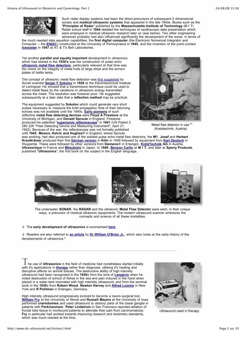
Yet another parallel and equally important development in ultrasonicswhich had started in the 1930's was the construction of pulse-echoultrasonic metal flaw detectors, particularly relevant at that time wasthe check on the integrity of metal hulls of large ships and the armourplates of battle tanks.
The concept of ultrasonic metal flaw detection was first suggested bySoviet scientist Sergei Y Sokolov in 1928 at the Electrotechnical Instituteof Leningrad. He showed that a transmission technique could be used todetect metal flaws by the variations in ultrasionic energy transmittedacross the metal. The resolution was however poor. He suggestedsubsequently at a later date that a reflection method may be practical.
The equipment suggested by Sokolov which could generate very shortpulses necessary to measure the brief propagation time of their returningechoes was not available until the 1940s. Early pioneers of suchreflective metal flaw detecting devices were Floyd A Firestone at theUniversity of Michigan, and Donald Sproule in England. Firestoneproduced his patented "supersonic reflectoscope" in 1941 (US-Patent 2280 226 "Flaw Detecting Device and Measuring Instrument", April 21,1942). Because of the war, the reflectoscope was not formally publisheduntil 1945. Messrs. Kelvin and Hughes® in England, where Sproulewas working, had also produced one of the earliest pulse-echo metal flaw detectors, the M1. Josef and HerbertKrautkrämer produced their first German version in Köln in 1949 followed by equipment from Karl Deutsch inWuppertal. These were followed by other versions from Siemens® in Erlangen, KretzTechnik AG in Austria,Ultrasonique in France and Mitsubishi in Japan. In 1949, Benson Carlin at M I T, and later at Sperry Products,published "Ultrasonics", the first book on the subject in the English language.
The underwater SONAR, the RADAR and the ultrasonic Metal Flaw Detector were each, in their uniqueways, a precursor of medical ultrasonic equipments. The modern ultrasound scanner embraces the
concepts and science of all these modalities.
The early development of ultrasonics is summarised here.
Readers are also referred to an article by Dr William O'Brien Jr., which also looks at the early history of the
developments of ultrasonics.^
he use of Ultrasonics in the field of medicine had nonetheless started initiallywith it's applications in therapy rather than diagnosis, utilising it's heating anddisruptive effects on animal tissues. The destructive ability of high intensityultrasound had been recognised in the 1920s from the time of Langévin when henoted destruction of school of fishes in the sea and pain induced in the hand whenplaced in a water tank insonated with high intensity ultrasound; and from the seminalwork in the 1930s from Robert Wood, Newton Harvey and Alfred Loomis in NewYork and R Pohlman in Erlangen, Germany.
High intensity ultrasound progressively evolved to become a neuro-surgical tool.William Fry at the University of Illinois and Russell Meyers at the University of Iowaperformed craniotomies and used ultrasound to destroy parts of the basal ganglia inpatients with Parkinsonism. Peter Lindstrom in San Francisco reported ablation offrontal lobe tissue in moribound patients to alleviate their pain from carcinomatosis.Fry in particular had worked towards improving research and dosimetry standards,which was much needed at the time.

24/08/08 15:58History of Ultrasound in Obstetrics and Gynecology, Part 1
Page 3 sur 19http://www.ob-ultrasound.net/history1.html
Ultrasonic energy was also extensively used in physical and rehabilitation medicine. Jerome Gersten at the University ofColorado reported in 1953 the use of ultrasound in the treatment of patients with rheumatic arthritis. Other reseacherssuch as Peter Wells in Bristol, England, Douglas Gordon in London and Mischele Arslan in Padua, Italy employedultrasonic energy in the treatment of Meniere's disease.
Uses of ultrasonic energy in the 1940s. Left, in gastric ulcers. Right, in arthritis
The 1940s saw exuberant claims made in some sectors on the effectiveness of ultrasound as an almost "cure-all" remedy,abeit the lack of much scientific evidence. This included conditons such as arthritic pains, gastric ulcers, eczema, asthma,thyrotoxicosis, haemorrhoids, urinary incontinence, elephanthiasis and even angina pectoris! Cynicism and concern overharmful tissue damaging effects of ultrasound were also mounting, which had curtailing consequences on thedevelopment of diagnostic ultrasound in the years that followed.
It was around similar times that ultrasound was used experimentally as a possiblediagnostic tool in medicine. H Gohr and Th. Wedekind at the Medical University ofKoln in Germany in 1940 presented in their paper "Der Ultraschall in der Medizin" thepossibility of ultrasonic diagnosis basing on echo-reflection methods similar to that usedin metal flaw detection. They suggested that the method would be able to detecttumours, exudates or abscesses. However they were unable to publish convincingresults from their experiments. Karl Theo Dussik, a neurologist/ psychiatrist at theUniversity of Vienna, Austria, who had begun experiments in the late 1930s, wasgenerallly regarded as the first physician to have employed ultrasound in medicaldiagnosis.
Dussik, together with his brother Friederich, a physicist, attempted to locate brain tumorsand the cerebral ventricles by measuring the transmission of ultrasound beam throughthe skull. Dussik presented his initial experiments in a paper in 1942 and further resultsafter the end of the second world war in 1947. They called their procedure"hyperphonography".
They used a through-transmission technique with two transducers placed on eitherside of the head, and producing what they called"ventriculograms", or echo images of the ventricles of thebrain. Pulses of 1/10th scond were produced at 1.2 MHz.Coupling was obtained by immersing the upper part of thepatient's head and both transducers in a water bath and thevariations in the amount of ultrasonic power passing betweenthe transducers was recorded photographically on heat-sensitivepaper as light spots (not on a cathode-ray screen). It was anearliest attempt at the concept of 'scanning ' a human organ.Although their apparatus appeared elaborate with thetransducers mounted on poles and railings, the images producedwere very rudimentary 2-dimensional rows of mosaic lightintensity points. They had also reasoned that if imaging theventricles was possible, then the technique was also feasible fordetecting brain tumors and low-intensity ultrasonic waves couldbe used to visualize the interior of the human body.
Nevertheless, the images that Dussik produced were later thought to be artifactual by W Güttner and others at theSiemens Laboratory, Erlangen, Germany in 1952 and researchers at the M.I.T. (see below), as it had become apparentfrom further experiments that the reflections within the skull and attenuation patterns produced by the skull werecontributing to the attenuation pattern which Dussik had originally thought represented changes in acoustic transmissionsthrough the cerebral ventricles in the brain. Research basing on a similar transmission technique was not further pursued,both by Dussik, or at the M. I. T.. For more information read Dussik .
In nearby Germany, Heinrich Netheler , a physician at the Luebeck-South Hospital in Hamburg, was operating in 1945 asmall repair facility for medical equipments at the Hamburg university hospital at Eppendorf and had a mission ofdeveloping inventive medical products. Professor Hansen, his superior, suggested to him in that year to develop anultrasonic tomographic equipment for medical use basing on the concept of the RADAR. Important pioneering reseachwork started at the Eppendorf University Hospital. Nevertheless, due to a lack of funds right after the war, the equipmentdesigns had not reached the stage of actual fabrication. In the mid 1940s, German physician Wolf-Dieter Keidel at thePhysikalisch-Medizinischen Laboratorium at the University of Erlangen, Germany, also studied the possibility of usingultrasound as a medical diagnostic tool, mainly on cardiac and thoracic measurements. Having discussed with researchersat Siemens, he conducted his experiments using the transmission technique with ultrasound at 60 KHz, and rejected thepulse-reflection method. He was only able to make satisfactory recordings of intensity variations in relation to cardiacpulsations. He envisaged much more difficulties would be encounterd with the reflection method. In the First Congress of

24/08/08 15:58History of Ultrasound in Obstetrics and Gynecology, Part 1
Page 4 sur 19http://www.ob-ultrasound.net/history1.html
Ultrasound in Medicine held in Erlangen, Germany in May, 1948, Dussik and Keidel presented their papers onultrasound employed in medical diagnosis. These were the only two papers that discussed ultrasound as a diagnostictool. The other papers were all on its therapeutic use.
In France , French scientists who were in the study of ultrasonics,namely André Dognon and André Dénier and several others atthe research center in Salpêtrière in Paris also embarked onultrasound insonation experiments before the 1950s. Dénierpublished his theoretically work on ultrasound transmission in1946, among many other works on ultrasound used in therapy,and suggested the possibiity of "Ultrasonoscopie". This was atransmission technique and recordings made on a micro-amperemeter and oscilloscope. Equipments were fabricated from 'therapy'counterparts and various electrical current values were determinedon different body tissues. Attempts to display voltages as Lissajousfigures on the oscilloscope were made. However the work wasunsuccessful in producing useful structural images and relatedinstruments were not constructed. André Dénier published in 1951his book, "Les Ultras-sons -- Appliques a la Medecin". Nearly theentire book was devoted to ultrasonics used in the treatment ofvarious diseases and only a small portion of the text was on ultrasound diagnostics.
Systematic investigations into using ultrasound as a diagnostic tool finally took off inthe United States in the late 1940s. The time was apparently ripe for this to happen. Theconcept of applying ultrasonics to medicine had progressively matured, so were theavailable equipments and electronics after the war. George Ludwig, a graduate from theUniversity of Pennsylvania in 1946 was on active duty as junior Lieutenant at the NavalMedical Research Institute in Bethseda, Maryland. There, he began experiments onanimal tissues using A-mode industrial flaw-detector equipment. Ludwig designedexperiments to detect the presence and position of foreign bodies in animal tissues and inparticular to localise gallstones, using reflective pulse-echo ultrasound methodologysimilar to that of the radar and sonar in the detection of foreign boats and flying objects.A substantial portion of Ludwig's work was considered classified information by the Navyand was not published in medical journals. Although Ludwig's work had started at aconsiderably earlier date, notice of his work was not released to the public domain untilOctober 1949 by the United States Department of Defence. The June '49 report isconsidered the first report of its kind on the diagnostic use of ultrasound from the UnitedStates.
Ludwig systematically explored physical characteristics of ultrasound in various tissues, including beef and organs fromdogs and hogs. To address the issue of detecting gallstones in the human body, he studied the acoustic impedance ofvarious types of gallstones and of other tissues such as muscle and fat in the human body, employing different ultrasonicmethodologies and frequencies. His collaborators included Francis Struthers and Horace Trent, physicists at the NavalResearch Laboratory, and Ivan Greenwood, engineer from the General Precision Laboratories, New York, and theDepartment of Research Surgery, University of Pennsylvania. Ludwig also investigated the detection of gallstones(outside of the human body) using ultrasound, the stones being first embedded in pieces of animal muscle. Very shortpulses of ultrasound at a repetition rate of 60 times per second were employed using a combined transmitter/ receivertransducer. Echo signals from the reflected soundwaves were recorded on the oscilloscope screen. Ludwig was able todetect distinct ultrasonic signals corresponding to the gallstones. He reported that echo patterns could sometimes beconfusing, and multiple reflections from soft tissues could make test results difficult to interpret. Ludwig also studiedtransmission through living human extremities, to measure acoustic impedance in muscle. These investigations alsoexplored issues of attenuation of ultrasound energy in tissues, impedance mismatch between various tissues and relatedreflection coefficients, and the optimal sound wave frequency for a diagnostic instrument to achieve adequate penetrationof tissues and resolution, without incurring tissue damage. These studies had helped to build the scientific foundation forthe clinical use of ultrasound.
In the following year, Greenwood and General Precision Laboratories madeavailable commercially the "Ultrasonic Locator" which Ludwig used for "use inMedicine and Biology". Suggested usage indicated in the sales informationleaflet already included detection of heart motion, blood vessels, kidney stonesand glass particles in the body. Ludwig's pulse-reflection methodology andequipment in his later experiments on sound transmission in animal tissueswere after earlier designs from the work of John Pellam and John Galt in1946 at the Electronics and Acoustics research laboratories of theMassachusetts Institute of Technology (M. I. T.), which was on themeasurement of ultrasonic transmission through liquids. The M. I. T. was thenvery much at the forefront of electronics and ultrasonics research. A significantamount of physical data and instrumentation electronics were already in placein the second half of the 1940s, on the characteristics of ultrasoundpropagation in solids and liquids.
Among other important original findings, Ludwig reported the velocity of soundtransmission in animal soft tissues was determined to be between 1490 and 1610 meters per second, with a meanvalue of 1540 m/sec. This is a value that is still in use today. He also determined that the optimal scanning frequency ofthe ultrasound transducer was between 1 and 2.5 MHz. His team also showed that the speed of ultrasound and acousticimpedance values of high water-content tissues do not differ greatly from those of water, and that measurements fromdifferent directions did not contribute greatly to these parameters.
Ludwig went on to collaborate with the Bioacoustics laboratory at the M. I. T.. His work with physicist Richard Bolt (who,at the age of 34 was appointed Director of a newly conceived Acoustics Laboratory at M. I. T.), neurosurgeon H Thomas
24/08/08 15:58History of Ultrasound in Obstetrics and Gynecology, Part 1
Page 5 sur 19http://www.ob-ultrasound.net/history1.html
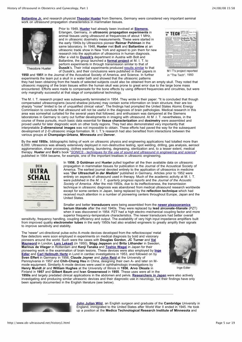
Ballantine Jr. and research physicist Theodor Hueter from Siemens, Germany were considered very important seminalwork on ultrasound propagation characteristics in mammalian tissues.
Prior to 1949, Hueter had already been involved at Siemens,Erlangen, Germany, in ultrasonic propagation experiments inanimal tissues using ultrasound at frequencies of about 1 MHz,and in ultrasonic dosimetry measurements. These were started inthe early 1940s by Ultrasonics pioneer Reimar Pohlman in thesame laboratory. In 1948, Hueter met Bolt and Ballantine at anultrasonic trade show in New York and agreed to join them for newresearch into the application of ultrasonics in human diagnosis.After a visit to Dussik's department in Austria with Bolt andBallantine, the group launched a formal project at M. I. T. toperform experiments in through transmission similar to that ofDussik 's. Their initial experiments produced results similar to thatof Dussik's, and their conclusions were published in their papers in
1950 and 1951 in the Journal of the Acoustical Society of America, and Science. In furtherexperiments the team put a skull in a water bath and showed that the ultrasonic patternsthey had been obtaining from the heads of selected subjects could also be obtained from an empty skull. They noted thatultrasonic mapping of the brain tissues within the human skull was prone to great error due to the large bone massencountered. Efforts were made to compensate for the bone effects by using different frequencies and circuitries, but wereonly marginally successful at that stage of computational technology.
The M. I. T. research project was subsequently terminated in 1954. They wrote in their paper: "It is concluded that thoughcompensated ultrasonograms (sound shadow pictures) may contain some information on brain structure, their are toosharply "noise" limited to be of unqualified clinical value". The findings had prompted the United States Atomic EnergyCommission to conclude that ultrasound will not be useful in the diagnosis of brain pathologies. Medical research in thisarea was somewhat curtailed for the several years that followed, and enthusiasm was dampened at the Siemenslaboratories in Germany to carry out further developments in imaging with ultrasound. At M .I. T. nevertheless, in thecourse of these pursuits, much basic data essential for tissue characterization and dosimetry were assembled andproved useful for later diagnostic work on other body regions. They had also demonstrated very importantly thatinterpretable 2-dimensional images was not impossible to obtain. These efforts had paved the way for the subsequentdevelopment of 2-D ultrasonic image formation. M. I. T.'s research had also benefited from interactions between thevarious groups at Champaign-Urbana, Minnesota and Denver.
By the mid 1950s, bibliographic listing of work on ultrasonic physics and engineering applications had totalled more than6,000. Ultrasonics was already extensively deployed in non-destructive testing, spot welding, drilling, gas analysis, aerosolagglomeration, shear processing, clothes washing, laundering, degreasing, sterilization and, to a lesser extent, medicaltherapy. Hueter and Bolt's book "SONICS - techniques for the use of sound and ultrasound in engineering and science"published in 1954 became, for example, one of the important treatises in ultrasonic engineering.
In 1956, D Goldman and Hueter pulled together all the then available data on ultrasonicpropagation in mammalian tissues for publication in the Journal of the Acoustical Society ofAmerica. The earliest journal devoted entirely to the application of ultrasonics in medicinewas "Der Ultraschall in der Medizin" published in Germany. Articles prior to 1952 wereentirely on aspects of ultrasound used in therapy. Much of the academic activity at M. I. T.were published in the M. I. T. quarterly progress reports and the Journal of the AcousticSociety of America. After the mid-1950s, due to its ineffectiveness, the transmissiontechnique in ultrasonic diagnosis was abandoned from medical ultrasound research worldwideexcept for some centers in Japan, being replaced by the reflection technique which hadreceived much attention in a number of pioneering centers throughout Europe, Japan and theUnited States.
Smaller and better transducers were being assembled from the newer piezoceramicsbarium titanate after the mid 1940s. They were replaced by lead zirconate-titanate (PZT)when it was discovered in 1954. PZT had a high electro-mechanical coupling factor and moresuperior frequency-temperature characteristics. The newer transducers had better overall
sensitivity, frequency handling, coupling efficiency and output. The availability of very high input impedance amplifiers builtfrom improved quality electrometer tubes in the early 1950s had also enabled engineers to greatly amplify their signalsto improve sensitivity and stability.
The 'newer' uni-directional pulse-echo A-mode devices developed from the reflectoscope/ metalflaw detectors were soon employed in experiments on medical diagnosis by bold and visionarypioneers around the world. Such were the cases with Douglas Gordon, JC Turner and ValMayneord in London, Lars Leksell (in 1950), Stigg Jeppson and Brita Lithander in Sweden,Marinus de Vlieger in Rotterdam and Kenji Tanaka and Toshio Wagai in Japan for theirpioneering work in the examination of brain lesions. These devices were also employed by IngeEdler and Carl Hellmuth Hertz in Lund in cardiac investigations in 1953, and followed on bySven Effert in Germany in 1956, Claude Joyner and John Reid at the University ofPennsylvania in 1957 and Chih-Chang Hsu in China, designing their own A- and later on M-mode equipment. Similarily A-mode devices were used in ophthalmologic investigations byHenry Mundt Jr and William Hughes at the University of Illinois in 1956, Arvo Oksala inFinland in 1957 and Gilbert Baum and Ivan Greeenwood in 1955. These uses were all in the1950s and largely predated clinical applications in the abdomen and pelvis. Researchers in Japan were also activelyinvestigating and producing similar ultrasonic devices and their diagnostic use in neurology, but their findings have onlybeen sparsely documented in the English literature (see below).
John Julian Wild, an English surgeon and graduate of the Cambridge University inEngland, immigrated to the United States after World War II ended in 1945. He tookup a position at the Medico Technological Research Institute of Minnesota and

24/08/08 15:58History of Ultrasound in Obstetrics and Gynecology, Part 1
Page 6 sur 19http://www.ob-ultrasound.net/history1.html
started his investigations with ultrasound waves on the thickness of the bowelwall in various surgical conditions, such as paralytic ileus and obstruction. Workingwith Donald Neal, an engineer, Wild published their work in 1950 on uni-directionalA-mode ultrasound investigations into the thickness of surgical intestinal materialand later on the properties of gastric malignancies. They noted that malignanttissue was more echogenic than benign tissue and the former could be diagnosedfrom their density and failure to contract and relax. Wild's original vision of theapplication of ultrasound in medical diagnosis was more of a method of tissuediagnosis from the intensity and characteristics of different returning echos ratherthan as an imaging technique. Between 1950 and 51, he also collaborated with LyleFrench at the department of Neurosugery in making diagnosis of brain tumors usingultrasound, although they had not found the method to be very helpful.
Donald Neal was soon deployed to regular navalservices at the naval air base after the Koreanwar. John Reid, a newly graduating electricalengineer, was engaged through a grant from the
National Cancer Institute as the sole engineer to build and operate Wild's ultrasonicapparatus. The device which they first used was an ultrasonic instrument which hadbeen designed by the U.S. Navy for training pilots in the use of the radar, with which itwas possible to practise 'flying' over a tank of water covering a small scale map ofenemy territory. " We have a tissue radar machine scaled to inches instead of miles bythe use of ultrasound". Wild and Reid soon built a linear hand-held B-mode instrument,a formidable technical task In those days, and were able to visualise tumours bysweeping from side to side through breast lumps. The instrument operated at afrequency of 15 megahertz. In 1952 they published the Landmark paper: "Application ofEcho-Ranging Techniques to the Determination of Structure of Biological Tissues". Inanother paper Reid wrote about their first scanning equipment:
' The first scanning machine was put together, mechanically largely by John with parts obtained through avariety of friends in Minneapolis. I was able to modify a standard test oscilloscope plug-in board. We wereable to make our system work, make the first scanning records in the clinic, and mail a paper off toScience Magazine within the lapsed time of perhaps ten days. This contribution was accepted in early1952 and became the first publication ( to my knowledge ) on intensity-modulated cross-sectionultrasound imaging. It appeared even before Douglass Howry's paper from his considerably moreelaborate system at the end of the same year.'
In May 1953 they produced real-time images at 15 megahertz of cancerousgrowths of the breast. They had also coined their method 'echography' and'echometry', suggesting the quantitative nature of the investigation. By 1956, Wildand Reid had examined 117 cases of breast pathology with their linear real-time B-mode instrument and had started work on colon tumour diagnosis and detection.Analysis of the breast series showed promising results for pre-operative diagnosis.Malignant infiltration of tissues surrounding breast tumours could also be resolved.
Wild and Reid had also invented and described the use of A-mode trans-vaginaland trans-rectal scanning transducers in 1955. Despite these, Wild was notcommended for his unconventional research methods at the time. His results wereconsidered difficult to interpret and lacked overall stability. Intellectual and financialsupport for Wild's research dwindled, and legal disputes and politics also hamperedfurther governmental grants. His work was eventually supported only by privatefunds which ran scarce and his data apparently received much less recognitionthan they deserved.
John Reid completed his MS thesis in 1957 on focusing radiators. In addition hehad importantly verified that dynamic focusing was practical. After leaving Wild'slaboratory he pursued his doctoral degree at the University of Pennsylvania. From1957-1965 he worked on echocardiography, producing and using the first suchsystem in the United States, with cardiologist Claude Joyner.
Visit John Wild's own site on his discoveries and current activities.
Read also: "The scientific discovery of sonic reflection of soft tissue and application of ultrasound to
diagnostic medicine and tumor screening" by John J Wild (Press Release at the Third Meeting of the WorldFederation for Ultrasound In Medicine and Biology, 1982).
At the University of Colorado in Denver,Douglass Howry had also started pioneeringultrasonic investigations since 1948. Howry, aradiologist working at the Veteran'sAdministration Hospital, had concentrated moreof his work on the development of B-modeequipment, displaying body structures in a 2-dimensional and sectional manner "comparable tothe actual gross sectioning of structures in thepathology laboratory". Published works from the M IT Radar school staff served as initial referencematerial on techniques in data presentation.
He was able to demonstrate an ultrasonic echointerface between structures or tissues, such asthat between fat and muscle, so that the individual structures could be outlined.Supported by his nephrologist friend and colleague Joseph Homles, who was

24/08/08 15:58History of Ultrasound in Obstetrics and Gynecology, Part 1
Page 7 sur 19http://www.ob-ultrasound.net/history1.html
then the acting director of the hospital's Medical Research Laboratories, Howryproduced in 1951 with William Roderic Bliss and Gerald J Posakony, bothengineers, the 'Immersion tank ultrasound system' *, the first 2-dimensional B-mode (or PPI, plan position indication mode) linear compound scanner. Twodimensional cross-sectional images were published in 1952 and 1953, whichconvincingly demonstrated that interpretable 2-D images of internal organ
structures and pathologies could be obtained with ultrasound. The team produed the formal motorized ' Somascope', acompound circumferential scanner, in 1954. The transducer of the somascope was mounted around the rim of a largemetal immersion tank filled with water . The machine was able to make compound scans of an intra-abdominal organ fromdifferent angles to produce a more readable picture. The sonographic images were referred to as 'somagrams'. Thediscovery and apparatus were reported in the Medicine section of the LIFE Magazine® in 1954.
The 'Pan-scanner' *, where the transducer rotatedin a semicircular arc around the patient, wasdeveloped in 1957. The patient sat on a modifieddental chair strapped against a plastic window of asemicircular pan filled with saline solution, while thetransducer rotated through the solution in asemicircular arc. The achievement was commendedby the American Medical Association in 1958 atits scientific meeting in San Francisco, and theteam's exhibit was awarded a Certificate of Meritby the association.
The work of Douglass Howry, Joseph Holmesand his team is necessarily the most importantpioneering work in B-mode ultrasound imaging andcontact scanning in the United States that had beenthe direct precursor of the kind of ultrasound
imaging we have today. Pioneering designs in electronic circuitries were alsomade in conjunction with the development of the B-scan, these included thepulse-echo generator circuitry, the limiter and log amplification circuitry and the demodulator and time gain compensationcircuitries.
The Howry/ Holmes systems, although capable of producing 2-D, accurate, reproducible images of the body organs,required the patient to be totally or partially immersed in water, and remained motionless for a length of time. Migration tolighter and more mobile versions of these systems, particularly with smaller water-bag devices or transducers directly incontact and movable on the body surface of patients were imminently necessary.
Read notes and see more pictures from Gerald Posakony on the early Howry scanners here.
Homles, together with consultantengineers William Wright andRalph (Edward) Meyerdirk, andsupport from the U. S. Public HealthServices and the University ofColorado, continued to fabricate anew prototype compound contactscanner, which had the transducerin direct contact with the patient'sbody and suspended on movingrailings above the patient. Theapparatus and the usuage ofultrasound scanning were reportedin the May 22 issue of the TIMEMagazine in 1964.
After working on the project for about 2 years, the team finallycame up with an innovative multi-joint articulated-arm compound
contact scanner with wire mechanisms and electronic position transducing potentiometers. The transducer could bepositioned by hand and moved over the scanning area in various directions by the operator. In 1962, with blessing fromHolmes, Wright and Meyerdirk left the University to form the Physionics Engineering® Inc. at Longmont, Colorado, toproduce and market their scanner.
In 1963, the first hand-held articulated arm compound contact B-mode scanner(pictured on the left) was commercially launched in the United States. The launch wasreported in the Longmont "Daily Times-Call" in 1963. This was the start of the mostpopular design in the history of static ultrasound scanners, that of the articulated-armscanning mechanism.
Physionics® was acquired by the Picker Corporation in 1967. Picker continued toproduced improved versions of the design right into the 1980s.
Much of the later work in clinical ultrasound was followed up by Homles and hiscolleagues, Stewart Taylor , Horace Thompson and Kenneth Gottesfeld in Denver.The group published some of the earliest papers in obstetrical and gynecologicalultrasound from North America. Douglass Howry had moved to Boston in 1962 where heworked at the Massachussetts General Hospital until he passed away in 1969.
24/08/08 15:58History of Ultrasound in Obstetrics and Gynecology, Part 1
Page 8 sur 19http://www.ob-ultrasound.net/history1.html
Earliest Wright-Meyerdirk scanner console with one of the first images from a practical commercial articulated-arm scanner. Portability was also emphasized.
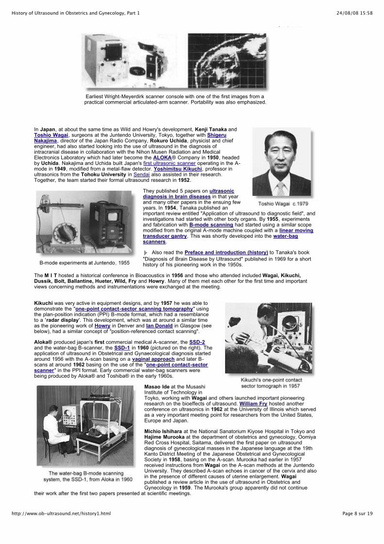
In Japan, at about the same time as Wild and Howry's development, Kenji Tanaka andToshio Wagai, surgeons at the Juntendo University, Tokyo, together with ShigeruNakajima, director of the Japan Radio Company, Rokuro Uchida, physicist and chiefengineer, had also started looking into the use of ultrasound in the diagnosis ofintracranial disease in collaboration with the Nihon Musen Radiation and MedicalElectronics Laboratory which had later become the ALOKA® Company in 1950, headedby Uchida. Nakajima and Uchida built Japan's first ultrasonic scanner operating in the A-mode in 1949, modified from a metal-flaw detector. Yoshimitsu Kikuchi, professor inultrasonics from the Tohoku University in Sendai also assisted in their research.Together, the team started their formal ultrasound research in 1952.
They published 5 papers on ultrasonicdiagnosis in brain diseases in that yearand many other papers in the ensuing fewyears. In 1954, Tanaka published animportant review entitled "Application of ultrasound to diagnostic field", andinvestigations had started with other body organs. By 1955, experimentsand fabrication with B-mode scanning had started using a similar scopemodified from the original A-mode machine coupled with a linear movingtransducer gantry. This was shortly developed into the water-bagscanners.
Also read the Preface and introduction (history) to Tanaka's book
"Diagnosis of Brain Disease by Ultrasound" published in 1969 for a shorthistory of his pioneering work in the 1950s.
The M I T hosted a historical conference in Bioacoustics in 1956 and those who attended included Wagai, Kikuchi,Dussik, Bolt, Ballantine, Hueter, Wild, Fry and Howry. Many of them met each other for the first time and importantviews concerning methods and instrumentations were exchanged at the meeting.
Kikuchi was very active in equipment designs, and by 1957 he was able todemonstrate the "one-point contact-sector scanning tomography" usingthe plan-position indication (PPI) B-mode format, which had a resemblanceto a 'radar display'. This development, which was at around a similar timeas the pioneering work of Howry in Denver and Ian Donald in Glasgow (seebelow), had a similar concept of "position-referenced contact scanning".
Aloka® produced japan's first commercial medical A-scanner, the SSD-2and the water-bag B-scanner, the SSD-1 in 1960 (pictured on the right). Theapplication of ultrasound in Obstetrical and Gynaecological diagnosis startedaround 1956 with the A-scan basing on a vaginal approach and later B-scans at around 1962 basing on the use of the "one-point contact-sectorscanner" in the PPI format. Early commercial water-bag scanners werebeing produced by Aloka® and Toshiba® in the early 1960s.
Masao Ide at the MusashiInstitute of Technology inToyko, working with Wagai and others launched important pioneeringresearch on the bioeffects of ultrasound. William Fry hosted anotherconference on ultrasonics in 1962 at the University of Illinois which servedas a very important meeting point for researchers from the United States,Europe and Japan.
Michio Ishihara at the National Sanatorium Kiyose Hospital in Tokyo andHajime Murooka at the department of obstetrics and gynecology, OomiyaRed Cross Hospital, Saitama, delivered the first paper on ultrasounddiagnosis of gynecological masses in the Japanese language at the 19thKanto District Meeting of the Japanese Obstetrical and GynecologicalSociety in 1958, basing on the A-scan. Murooka had earlier in 1957received instructions from Wagai on the A-scan methods at the JuntendoUniversity. They described A-scan echoes in cancer of the cervix and alsoin the presence of different causes of uterine enlargement. Wagaipublished a review article in the use of ultrasound in Obstetrics andGynecology in 1959. The Murooka's group apparently did not continue
their work after the first two papers presented at scientific meetings.

24/08/08 15:58History of Ultrasound in Obstetrics and Gynecology, Part 1
Page 9 sur 19http://www.ob-ultrasound.net/history1.html
Also read a short History of the development of Medical Ultrasonics in Japan.
ohn Wild was back in England in 1954 to give a lecture on his new discovery and this was attended by ValMayneord, Professor of medical physics at the Royal Cancer Hospital (now the Royal Marsden) who had also beenexperimenting with the Kelvin & Hughes® MK llB metal flaw-detector in neurological diagnosis. Among the audience wasIan Donald who was then Reader in Obstetrics and Gynaecology at the St. Thomas Hospital Medical School in Londonand was about to take up the appointment of Regius Chair of Midwifery at Glasgow University. Donald was quick to
realize what ultrasound had to offer.# Wild, while returning to Minnesota, had mainly concentrated his investigations onthe diagnosis of tumors of the breast and colon using 15 MHz probes which had tissue penetrations of only up to 2 cm. In1956, Wild published his landmark paper on the study of 117 breast nodules, reporting an accuracy of diagnosis of over90 percent. Despite that, the ultrasonic method of tissue diagnosis which he so popularised did not reach the point of wideacceptance. Pioneering work in ultrasonic diagnosis in the field of Obstetrics and Gynaecology however, soon took offin Glasgow, Scotland.
The following is an excerpt from an article in the University of Glasgow publication 'Avenue' No. 19: January 1996entitled ' Medical Ultrasound ---- A Glasgow Development which Swept the World ', by Dr. James Willocks MD,who had best described the circumstances of Donald 's early work :
' Ultrasound scanning is a householdword. Every mother knows it and many havepictures to prove it. It is painless, safe andreliable. Its success since its beginnings 40years ago is truly astonishing. It started inGlasgow in the University Department ofMidwifery under Professor Ian Donald andseemed a rather crazy experiment at thetime. But Ian Donald was no backroomboffin, but a full-blown flamboyant consultantat the sharp edge of one of medicine's mostacute specialities - a colourful character ofJohnsonian richness for whom I am a veryinadequate Boswell.
He was born in Cornwall in December 1910,the son and grandson of Scottish doctors.His school education began in Scotland andfinished in South Africa. He returned toEngland in 1931 and graduated in medicineat St Thomas's Hospital Medical School in1937. In 1939 he joined the RAF where hisservice was distinguished. He was decorated for gallantry for entering a burningbomber with the bombs still in it, to rescue injured airmen. Service in the RAFstimulated his interest in gadgetry of all kinds and he became familiar with radar andsonar, a technique which had been devised by the French physicist, Paul Langevin inthe First World War as a possible method of submarine detection.
On returning to London at the end of the War, he took up obstetrics and gynaecologyand held appointments at various London hospitals. His first research work wasdirected towards respiratory problems in the newborn, and he devised apparatus tohelp babies breathe when respiration did not get off to a flying start. Because of hisinterest in machines, Ian was known as 'Mad Donald' by some of his Londoncolleagues, who caricatured him as a crazy inventor, but his talent was spotted by thatgreat university statesman, Sir Hector Hetherington, and he was appointed to theRegius Chair of Midwifery at the University of Glasgow in 1954 .......... .
His interest soon turned to theidea that sonar could be used formedical diagnosis and the ideawas first put into practice on 21July 1955, when he visited theResearch Department of theboilermakers Babcock & Wilcox atRenfrew on the invitation of oneof the directors, who was thehusband of a grateful patient. Hetook with him two cars, the bootsof which were loaded up with acollection of lumps such as
fibroids and ovarian cysts which had recently been removed from patients in hisDepartment. He carried out some experiments with an industrial ultrasonic metalflaw detector on these tumours, and on a large lump of steak which the company

24/08/08 15:58History of Ultrasound in Obstetrics and Gynecology, Part 1
Page 10 sur 19http://www.ob-ultrasound.net/history1.html
had kindly provided as control material. (No one had the appetite for the steakafterwards!) Later he formed a link with the Kelvin & Hughes Scientific InstrumentCompany, and particularly with a young technician called Tom Brown. Quite byaccident, Tom Brown had heard the strange tale of a professor who was attempting touse a metal flaw detector to detect flaws in women. He telephoned Professor Donaldand suggested a meeting, and it was not long before Donald and Brown together withDr John MacVicar, later Professor of Obstetrics & Gynaecology in the University ofLeicester, plunged into an intensive investigation into the value of ultrasound indifferentiating between cysts, fibroids and any other intra abdominal tumours thatcame their way.
Early results were disappointing and theenterprise was greeted with a mixture ofscepticism and ridicule. However, adramatic case where ultrasound saved apatient's life by diagnosing a huge, easilyremovable, ovarian cyst in a woman whohad been diagnosed as having inoperablecancer of the stomach, made people takethe technique seriously. 'From this point',Ian Donald wrote, 'there could be noturning back'. Results eventuallyappeared in print in The Lancet of 7June 1958 under the arid title'Investigation of Abdominal Masses byPulsed Ultrasound'. This was probablythe most important paper on medical diagnostic ultrasound ever published. Ten yearslater all doubt had been cast away and Ian Donald was able to review the early historyof ultrasound in a characteristic , forthright manner. 'As soon as we got rid of thebackroom attitude and brought our apparatus fully into the Department with aninexhaustible supply of living patients with fascinating clinical problems, we were ableto get ahead really fast. Any new technique becomes more attractive if its clinicalusefulness can be demonstrated without harm, indignity or discomfort to the patient........ . Anyone who is satisfied with his diagnostic ability and with his surgical resultsis unlikely to contribute much to the launching of a new medical science. He shouldfirst be consumed with a divine discontent with things as they are. It greatly helps, ofcourse, to have the right idea at the right time, and quite good ideas may come,Archimedes fashion, in one's bath.'
In 1959 Ian Donald noted that clear echoes could be obtainedfrom the fetal head and began to apply this information. Ibecame involved shortly afterwards, and indeed was given theproject to play with on my own. At the Royal Maternity Hospital,Rottenrow, there was no separate room to examine the patientsand not even a cupboard in which to keep the apparatus, so mycolleague, the physicist Tom Duggan, and I pushed it about ona trolley and approached patients in the wards for permission toexamine them at the bedside. Glasgow women are wonderfuland they accepted all this without demur ........ . We applied themethod of fetal head measurement to assess the size and
growth of the foetus. When the Queen Mother's Hospital opened in 1964 it becamepossible to refine the technique greatly. My colleague Dr. Stuart Campbell (nowProfessor at King's College Hospital, London) did this and fetal cephalometrybecame the standard method for the study of fetal growth for many years.
Within the next few years it became possible to study pregnancy from beginning toend and diagnosis of complications like multiple pregnancy, fetal abnormality andplacenta praevia (which causes life threatening haemorrhage) became possible.Professor Donald had gathered around him a team of talented young doctors andtechnologists, including the research engineers John Fleming and Angus Hall, whowere engaged by the University when the Kelvin Hughes company was closed in1966.
John Fleming has continued at the Queen Mother's Hospital as the technical geniusbehind all developments, and is also in charge of the valuable historical collectionabout diagnostic ultrasound. Practically all apparatus is now Japanese in origin, butthe contribution of Scottish engineering to the development of medical ultrasoundshould never be forgotten. '
Ian Donald was also aware of the work of Howry in the United States and Kikuchi in Japanin the early 1950s, and had referenced these pioneers alongside with the work of Wild andReid in his Lancet paper in 1958. Donald had felt that it was his fortune to have started withthese historical A-mode and B-mode instruments instead of the apparatus that Wild andHowry had used, as these involved high frequency transducers (and hence associated withpoor penetration into tissues) or a water-bath arrangement which could both become
deterrants to further development in a medical setting ##. Aside from this, Donald had onmany occasions remarked that a lot of his developments in ultrasound was from a stroke of

24/08/08 15:58History of Ultrasound in Obstetrics and Gynecology, Part 1
Page 11 sur 19http://www.ob-ultrasound.net/history1.html
accident, coincidence and luck. The 'full bladder' was one, which he only discovered in 1963.That the fetal head, being a symmetrical skull bone could be easily demonstrable andmeasured accurately by a beam of ultrasound in an A-scan was another, as was theopportunity of meeting up with a number of important administrators on the way and workingwith the very bright engineer Tom Brown from Kelvin & Hughes®.
Brown, at the age of 24, invented and constructed with Ian Donald theprototype of the world's first Compound B-mode (plan-position indication,PPI) contact scanner in 1957. The transducer operated at 2.5 MHz. Theprototype was progressively improved to become the Diasonograph®manufactured commercially by Smith Industrials of England which hadtaken control of the Kevin and Hughes Scientific Instrument Company in1961.
For a detailed account of the pioneering development of the prototypes, readan important unpublished paper by Tom Brown entitled Development ofultrasonic scanning techniques in Scotland 1956-1979.
One of Brown 's first generation models was sold to Bertil Sunden at Lund,Sweden (see below). The console design of the Diasonograph® came fromDugald Cameron who was then an industrial design student at the Glasgowschool of Art. Brown also invented and patented an elaborate and expensiveautomated compound contact scanner in 1958 and it was at themachine's exhibition in London in 1960 that Ian Donald met for the first time Douglass Howry from the United States whohad been using the much larger size water-tank circumferential scanner for several years (see above). Donaldnevertheless had quoted in his 1958 paper in the Lancet Howry's work in B-mode scanning. The meeting had alsoinfluenced Howry and his team into producing a similar compound contact scanner like the Donald's although this hadrapidly evolved into the multi-joint articulated arm version.
A brief description of the working of the prototype compound contact scanner (which eventually developed into theDiasonograph®) was given by Donald and Brown in 1958, the same concept and design were extended into the latercommercial models:
" ..... A probe containing both transmitting, and receiving
transducers is mounted on a measuring jig, which is placed
above the patient's bed. The probe is free to move vertically
and horizontally and, as it does so, operates two linear
potentiometers, which give voltage outputs proportional to its
horizontal and vertical displacements from some reference
point. The probe is also free to rotate in the plane of its
horizontal and vertical freedom, and transmits its rotation via
a linkage to a sine-cosine potentiometer. The voltage outputs
from this system of potentiometers control an electrostatic
cathode-ray tube, so that the direction of the linear time-
base sweep corresponds to the inclination of the probe, and
the point of origin of the sweep represents the instantaneous
position of the probe. The apparatus is so calibrated that the
same reflecting point will repeat itself in exactly the same
position on the cathode-ray tube screen from whatsoever
angle it is scanned, and likewise a planar interface comes to
be represented as a consistent line.
The echoes picked up by the probe are displayed on three oscilloscope screens: an A-scope display, a combined
B-scope and PPI display on a long-persistence screen for monitoring: and a similar screen and display of short
persistence with a camera mounted in front of it. The probe is moved slowly from one flank, across the abdomen
to the other flank being rocked to and fro on its spindle the whole time to scan the deeper tissues from as many
angles as possible. ....."
The automated scanner which Brown originally designed to overcome the effects of motion variables did not catch onwell, while the Diasonograph® was sold to many other parts of Britain and Europe including Sweden, London andBristol, the place where another ultrasound pioneer, Peter NT Wells, a medical physicist, had been developing a differentversion of the multi-joint articulated arm scanner (basing on the Diasonograph electronics), independently from hisAmerican counterpart.
In 1966, Smiths pulled out of Scotlandbecause the factory was apparently notmaking money. At the same time the USSupreme Court ruled against Smiths in favourof Automation Industries (formerly theSperry Company) of Denver on the questionof the so-called "Firestone patents" (FloydFirestone's patent on flaw-detection devicesin 1942, see above). As part of thesettlement, Smiths undertook to withdrawboth from the industrial and medicalapplications of ultrasound, and Automationacquired title to the collection of Smiths'patents on these subjects. This included theBrown patents on 2-D contact scanning.Smiths sold the medical business to Nuclear Enterprises (G.B.) Ltd. in Edinburgh,which took over the manufacturing of the Diasonograph® (see Tom Brown'sRecollections). Ian Donald had to set up his own Department of Ultrasonic Technology
24/08/08 15:58History of Ultrasound in Obstetrics and Gynecology, Part 1
Page 12 sur 19http://www.ob-ultrasound.net/history1.html
at the Queen Mother's Hospital. He had John Fleming and Angus Hall back to helphim. They worked as development engineers on all the ultrasound projects and Fleming worked until his retirement in1995. He is co-ordinator of the BMUS historical collection and oversees the ultrasonic equipments at the HunterianMuseum, University of Glasgow. By 1968, Brian Fraser and Alan Cole at Nuclear Enterprises revamped the mechanicaland valve design and redeveloped a new electronic system using semiconductor technology. The resulting "NE 4102"became a very popular instrument, and was used in most British hospitals and many European ones.
For a detailed account of the pioneering
development of medical ultrasonics inGlasgow, Scotland, read the biography ofTom Brown and an important unpublishedpaper by Brown on the Development ofultrasonic scanning techniques inScotland 1956-1979.
Visit the pictorial presentation: Scenes
from the History of Ultrasound from theBritish Medical Ultrasound Society(BMUS) Historical Collection. Co-ordinator:Mr. John Fleming.
Read also Peter Well's article on the
History of the development of ultrasonography.
Joseph Holmes and Ian Donald had subsequently become good friends across theAtlantic and Ian Donald and John Fleming were invited to speak on their experiencesat the International Conference at Pittsburg hosted by Homles and others in 1965.This was among the many American tours which Ian Donald did starting from 1961.He spoke about Homles in a speech he gave in 1967 to the World Federation for Ultrasound in Medicine and Biology(WFUMB), 'I think Joe Holmes has done more than anyone to pull us all together from our several pathways'. Holmesbecame the founding editor of the Journal of Clinical Ultrasound in 1973.
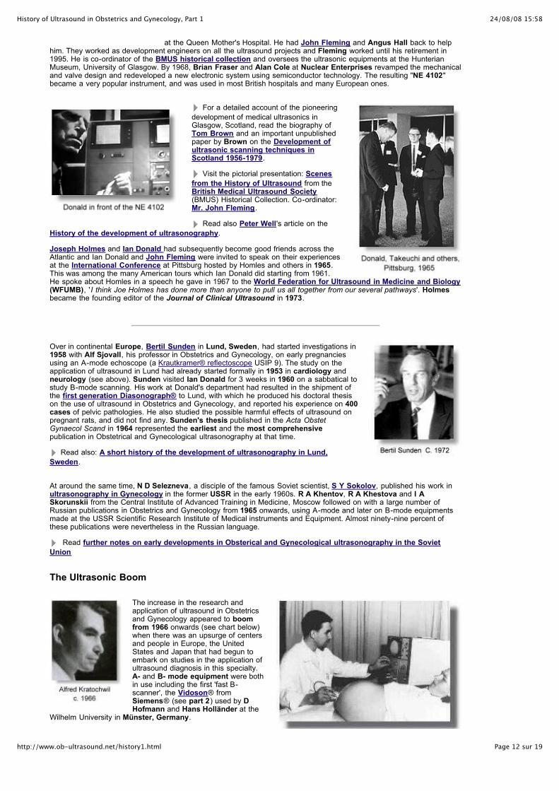
Over in continental Europe, Bertil Sunden in Lund, Sweden, had started investigations in1958 with Alf Sjovall, his professor in Obstetrics and Gynecology, on early pregnanciesusing an A-mode echoscope (a Krautkramer® reflectoscope USIP 9). The study on theapplication of ultrasound in Lund had already started formally in 1953 in cardiology andneurology (see above). Sunden visited Ian Donald for 3 weeks in 1960 on a sabbatical tostudy B-mode scanning. His work at Donald's department had resulted in the shipment ofthe first generation Diasonograph® to Lund, with which he produced his doctoral thesison the use of ultrasound in Obstetrics and Gynecology, and reported his experience on 400cases of pelvic pathologies. He also studied the possible harmful effects of ultrasound onpregnant rats, and did not find any. Sunden's thesis published in the Acta ObstetGynaecol Scand in 1964 represented the earliest and the most comprehensivepublication in Obstetrical and Gynecological ultrasonography at that time.
Read also: A short history of the development of ultrasonography in Lund,
Sweden.
At around the same time, N D Selezneva, a disciple of the famous Soviet scientist, S Y Sokolov, published his work inultrasonography in Gynecology in the former USSR in the early 1960s. R A Khentov, R A Khestova and I ASkorunskii from the Central Institute of Advanced Training in Medicine, Moscow followed on with a large number ofRussian publications in Obstetrics and Gynecology from 1965 onwards, using A-mode and later on B-mode equipmentsmade at the USSR Scientific Research Institute of Medical instruments and Equipment. Almost ninety-nine percent ofthese publications were nevertheless in the Russian language.
Read further notes on early developments in Obsterical and Gynecological ultrasonography in the Soviet
Union
The Ultrasonic Boom
The increase in the research andapplication of ultrasound in Obstetricsand Gynecology appeared to boomfrom 1966 onwards (see chart below)when there was an upsurge of centersand people in Europe, the UnitedStates and Japan that had begun toembark on studies in the application ofultrasound diagnosis in this specialty.A- and B- mode equipment were bothin use including the first 'fast B-scanner', the Vidoson® fromSiemens® (see part 2) used by DHofmann and Hans Holländer at the
Wilhelm University in Münster, Germany.
24/08/08 15:58History of Ultrasound in Obstetrics and Gynecology, Part 1
Page 13 sur 19http://www.ob-ultrasound.net/history1.html
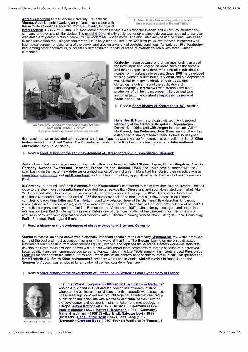
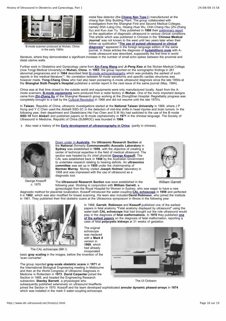
Alfred Kratochwil at the Second University Frauenklinik,Vienna, Austria started working on placental localisation withthe A-mode scanner he acquired from Paul Kretz, founder ofKretzTechnik AG in Zipf, Austria. He soon learned of Ian Donald 's work with the B-scan and quickly collaborated thecompany to develop a similar device. The model 4100 originally designed for ophthalmologic use was adapted to carry anarticulated-arm gantry (pictured below) for the abdominal B-scan mode. The articulated-arm design he found, was easierto manipulate than the Glasgow counterpart. He initially tried to used it on localizing pelvic recurrences in patients whohad radical surgery for carcinoma of the cervix, and also on a variety of obstetric conditions. As early as 1972, Kratochwilhad, among other endeavours, successfully demonstrated the visualisation of ovarian follicles with static B-modeultrasound.
Kratochwil soon became one of the most prolific users ofthe instrument and worked on areas such as the breastsand other surgical conditions, where he also published anumber of important early papers. Since 1968 he developedtraining courses in ultrasound in Vienna and his departmentwas visited by many hundreds of radiologists andobstetricians to learn about the applications ofultrasonography. Kratochwil was probably the mostproductive of all the investigators in Europe and wasinstrumental to the constantly improving designs atKretzTechnik AG.
Read a Short history of Kretztechnik AG, Austria.
Hans Henrik Holm, a urologist, started the ultrasoundlaboratory at the Gentofte Hospital in Copenhagen,Denmark in 1964, and with Jorgen Kristensen, AllenNortheved, Jan Pedersen, Jens Bang among others hadestablished a strong research team. Holm also designed
their version of an articulated-arm scanner which subsequently was taken up for commercial production at Smith KlineInstrument® in the United States. The Copenhagen center had in time become a leading center in Interventionalultrasound, even up to this day.
Read a short history of the early development of ultrasonography in Copenhagen, Denmark.
And so it was that the early pioneers in diagnostic ultrasound from the United States , Japan, United Kingdom, Austria,Germany, Sweden, Switzerland, Denmark , France , Poland, Holland, USSR and China have all started with the A-scan basing on the metal flaw detector or a modification of the instrument. Many had first started their investigations inneurology, cardiology and ophthalmology, and only later on did they apply ultrasonic techniques to the abdomen andpelvis.
In Germany, at around 1950 both Siemens® and Krautkrämer® had started to make flaw-detecting equipment. Locatedclose to the steel industry Krautkrämer® provided better service than Siemens® and soon dominated the market. AfterW Güttner and others had shown the impracticality of the transmission technique in 1952, Siemens had lost interest indiagnostic ultrasound. Around the end of 1956 the company decided to stop producing flaw-detection equipmentcompletely. It was Inge Edler and Carl Hertz in Lund who adapted three of the Siemens® flaw detectors for cardiacinvestigations in 1957 (see above), and these were introduced back into hospitals in Germany. After a lapse of almost 10years, the company developed the first fast B-scanner, the Vidoson in 1967, suitable for gynecological and abdominalexamination (see Part 2). Germany was nevertheless one of the more 'prolific' of the European countries in terms ofcenters in early ultrasonic applications and research, with publications coming from Muchen, Erlangen, Bonn, Heidelberg,Berlin, Frankfurt, Freiburg and Bochum.
Read a history of the development of ultrasonography at Siemens, Germany.
Vienna in Austria, as noted above was 'historically' important because of the company Kretztechnik AG which producedsome of the best and most advanced machines in the world at that time. The B-scan , basing on more sophisticatedinstrumentation emanating from radar sciences quickly evolved and replaced the A-scans. Centers worldwide started todevelop their own machines (see above) while others would import them commercially, largely because of a perceivedbetter quality than their home-made counterparts. For example, in the late 1960s some Finnish centers used Physionics /Picker® machines from the United States and French and Italian centers used scanners from Nuclear Enterprise® andKretzTechnik AG. Smith Kline Instruments® scanners were used in Spain, Aloka® models in Brussels and theSiemens® Vidoson was employed by a number of centers outside of Germany.
Read a short history of the development of ultrasound in Obstetrics and Gynecology in France.
The "First World Congress on Ultrasonic Diagnostics in Medicine"was held in Vienna in 1969 and the second in Rotterdam in 1972where an increasing number of papers in this specialty was presented.These meetings identified and brought together an international groupof clinicians and scientists who started to contribute heavily towardsthe developments of ultrasonic instrumentation and methodology. InEurope, Alfred Kratochwil (1966), (Austria), D Hofmann (1966),Hans Hollander (1966), Manfred Hansmann (1966), (Germany),Malte Hinselmann (1968) (Switzerland), Salvator Levi (1967)(Brussels), Hans Henrik Holm (1967), Jens Bang (1967)(Denmark), Georges Boog (1969), Francis Weill (1969) (France), I

24/08/08 15:58History of Ultrasound in Obstetrics and Gynecology, Part 1
Page 14 sur 19http://www.ob-ultrasound.net/history1.html
Roszkowski I (1968), Jerzy Groniowski (1968), (Poland), PaavoPystynen (1966), Pekka Ylöstalo (1971), Pentti Jarvinen (1968), Pentti Jouppila (1970)(Finland), J Hernandez (1970), R Montero (1970), Fernando Bonilla-Musoles (1971) (Spain), Bruno Damascelli(1967), L Roncoroni (1967), Alberto Zacutti (1968), C Brugnoli (1968), Achille Ianniruberto (1970) (Italy), E Kalamara(1972), M Bulic (1972), Asim Kurjak (1973) (Yugoslavia, now Croatia), Juriy Wladimiroff (1974) (Netherlands), MFalus (1969), M Sobel (1969), L Kun (1973), P Bosze (1973) (Hungary), among many others, soon followed up with theirmany publications in obstetrical and gynecological sonography, although much of what was published was not in theEnglish language. [The year in parenthesis denoted the year in which publications in Obstetrics and Gynecology from theparticular author first appeared in the literature]. The delegates of 13 European ultrasound societies met in Basel,Switzerland in 1972 to form the European Federation of Societies for Ultrasound in Medicine and Biology(EFSUMB).
Read a brief history of the development of medical ultrasonics in Poland.
In the United Kingdom, Ellis Barnett, Patricia Morley, HughRobinson, Usama Abdulla in Glasgow, Peter Wells in Bristol, A CChristie in Aberdeen, E I Kohorn, Stuart Campbell in London (see Part2), Hyton Meire and Pat Farrant in Middlesex, and Christopher Hill atthe Royal Marsden continued to make very important contributions inmany areas.
Barnett and Morley's book in 1974: "Clinical Diagnostic Ultrasound" wasthe first book (including publications from the United States) devoted toabdominal B-mode ultrasonography. Peter Wells in particular, was thesingle most important contributor to the advancement of ultrasoundtechnology in Britain. Stuart Campbell eventually became one of theworld's most well-known researcher and teacher in the field of Obstetrical
and Gynecological ultrasound. The British Medical Ultrasound group was formed in 1969 by members of the HospitalPhysicists Association and the British Institute of Radiology. The group later changed its name and became officially theBritish Medical Ultrasound Society (BMUS) in 1977.
Read the early history of Obstetrical and Gynecological ultrasound in Finland.
Back in the United States , J Stauffer Lehman, in Hahnemann,Philadelphia was instrumental in the early 1960's to the continuingdevelopment of ultrasound technology in the United States . Hisassociation with Smith Kline Instruments® had been catalytic to thecompany's production of water-bag and contact B-mode scanners ontop of their existing line of A- and M- mode equipments forechocardiography. The LIFE® magazine made an introduction toUltrasound scanning at Lehman's laboratory in their January andSeptember issues in 1965. The Family Circle® magazine alsoreported on the medical use of ultrasound in their October 1966 issue.
Lehman's equipment was nevertheless cumbersome and expensive tofabricate and later on a smaller company, Hoffrel took up the
production of his machine. After the expiration of SKI's contract, Lehmann turned to use the articulated arm scanneroriginally invented and produced by the Physionics Inc in Longmont, Colorado (later on acquired by the PickerCorporation and further expanding its development).
Barry Goldberg joined Lehman in 1968 and expanded the research. He published extensively on a variety of subjectsincluding echocardiography and interventional ultrasonography and was on record the first to describe fetal cephalometryin 1965 outside of Britain and Europe. George Evans, then a young Radiologist, was responsible for organizing theservice and several important research projects. With his team was Marvin Ziskin. Together they have introducedultrasound to the Radiological community in the United States and convincing them of the technique's clinical value. Lajosvon Micsky, working at the St. Luke's Medical Center in New York, was also one of the important pioneers of abdominalas well as endoscopic sonographic equipment. He established a bioacoustical laboratory at the center in 1963 anddevised many innovative abdominal, trans-vesical, rectal and trans-vaginal scanners.
Also read an article "Obstetric US imaging: the First 40 Years" by Dr. Barry Golberg.
Articulated arm scanners such as the PortaScan from Physionics Inc® produced in the mid-1960s had become themost popular format in compound contact B-scanners in the United States and throughout the world. Other earliestmanufacturers of similar devices included the UniRad Corporation®. Newer machines soon followed from manufacturersin the United States and worldwide. These included the Picker® Laminograph 102, the KretzTechnik AG Combison 1and 2, the Nuclear Enterprise® Diasonograph 4102 (pictured above), the Aloka® SSD-10 compound contact scanner(pictured below) and the Toshiba® TSL systems. Jan C Somer and Nicolaas Bom in the Netherlands introduced thephased-array and linear-array transducers respectively in 1968 and 1971 (see Part 2).
Louis M Hellman, Mitsunao Kobayashi, Ross Brown,George Leopold, Roy Filly , Roger Sanders, ArthurFleischer, Kenneth Taylor , Fred Winsberg, JohnHobbins and William Cochrane were among those whoproduced a substantial amount of work from the early 1970son the application of ultrasound relating to Obstetrics andGynecology and had contributed much to moving themodality forward. Winsberg had a particular interest in real-time scanners and he was the first to use the German

24/08/08 15:58History of Ultrasound in Obstetrics and Gynecology, Part 1
Page 15 sur 19http://www.ob-ultrasound.net/history1.html
Vidoson® real-time scanner (see part 2) in North America(at the McGill University in Montreal, Canada) in 1970.One of the very earliest textbooks in sonography in theEnglish language aside from Bertil Sunden 's thesis wasfrom Kobayashi, Hellmen and Cromb: "Atlas ofUltrasonography in Obstetrics and Gynaecology" published in 1972.
The American Institute of Ultrasound in Medicine (AIUM) which was founded in1952 by a group of physicians engaged primarily in the use of ultrasound in physicalmedicine only started to accept members in the diagnostic arena in 1964. Diagnosticultrasound has since then become the mainstream application in the organization. The"First International Conference on Diagnostic Ultrasound" was held in Pittsburgh,Pensylvannia in 1965 and was well attended by most of the ultrasound pioneers.
The Journal of Ultrasound in Medicine, the official journal of the AIUM, was inaugurated in 1982 replacing the Journalof Clinical Ultrasound as the association's main vehicle of communication with it's members. George Leopold was itsfounding editor. By the mid-1970s important producers of articulated compound B-scanners in the United States includedthe Picker Corp®, Smith Kline Instruments®, the UniRad Corporation®, Searle Ultrasound®, Rohe Scientific®,Litton Medical Systems® and Metrix Inc®. A list of manufacturers of static compound contact scanners as at 1975can be found here.
The number of publications in the world literature each year on the application of ultrasound in Obstetricsand Gynecology rose from 1(Ian Donald's paper) in 1958 to 296 in 1978. In the first 10 years, mostpublications were of a general descriptive nature and had similar titles to the effect of "The use of
ultrasonography in Obstetrics and Gynecology".ref
In Japan, Shigemitsu Mizuno, HisayaTakeuchi, Koh Nakano and Masao Arimafollowed up the ultrasound work at the JuntendoUniversity in Tokyo, and experimented with newversions of the A-mode transvaginal scanner.The first ultrasound scan of a 6-week gestationalsac by vaginal A-scan was reported in theJapanese language in 1963. From 1962, thegroup worked extensively with the water-bag B-scanner, the Aloka SSD-1 and was very active inmany areas and producing a huge number ofresearch publications, ranging from earlypregnancy diagnosis to cephalometry to
placentography. They also reported on a large series of pelvic tumors in1965, and in the following 2 years switched from the water-bag contactscanner to the articulated-arm compound contact scanner, the SSD-10.Another group consisting of T Tanaka, I Suda and S Miyahara startedresearches into the different stages of pregnancy in 1964.
Shigemitsu Mizuno, Hisaya Takeuchi and their team also demonstrated in 1965 an endovaginal scanner for pelvicexamination using the plan-position indication (PPI) B-mode format. The device was mannually rotated and the resultingdisplay was very similar to a circular military 'radar" display. Used either transrectally or transvaginally, It was capable ofproducing some meaningful pictures of the pelvic organs. See Hisaya Takeuchi for a list of early work from the group.
The Japan Society of Ultrasonics in Medicine was officially formed in 1962. In the 1970s important work started at theTottori Uinversity, Toyko under Kazuo Maeda, particularly on doppler fetal cardiotocography and at the University ofToyko under Shoichi Sakamoto. Toshiba® produced their first A-mode scanner, the SSA-01A and the compoundcontact B-scanner, the TSL system in 1967. Hitachi® produced their first A-mode (the EUA-1) and B-mode scanner(EUB-1) in 1971 and 72 respectively.
Also read a short history of the development of Medical Ultrasonics in Japan.
In the Republic of China, Shih An founded in 1958 the ShanghaiUltrasonic Medical Research group at the Sixth People's Hospital ofShanghai and his team included Tao-Hsin Wang and Shih-Yuan An. Inthe same year they started ultrasonic investigations using a modifiedmetal flaw detector (the Chiang Nan Type I) manufactured at the
24/08/08 15:58History of Ultrasound in Obstetrics and Gynecology, Part 1
Page 16 sur 19http://www.ob-ultrasound.net/history1.html
metal flaw detector (the Chiang Nan Type I) manufactured at thechiang Nan Ship Building Plant. The group collaborated withinvestigators from the Shanghai First and Second Medical Colleges,namely Shih-Liang Chu, Hsiang-Huei Wu, Chih-Chang Hsu (Zhi-ZhangXu) and Kuo-Juei Yu. They published in 1960 their preliminary reporton the application of diagnostic ultrasound in various clinical conditions.This article which was published in Chinese in the 'Chinese MedicalJournal' was not known to the west until two years later when theirfollow-up publication "The use of pulsed ultrasound in clinicaldiagnosis" appeared in the foreign language edition of the samejournal. In these articles the diagnosis of hydatidiform mole with A-mode ultrasound was described, supposedly the first time in world
literature, where they demonstrated a significant increase in the number of small echo spikes between the proximal anddistal uterine walls.
Further work in Obstetrics and Gynecology came from Xin-Fang Wang and Ji-Peng Xiao at the Wuhan Medical College(now Tongji Medical University) in Wuhan, China. In 1963, the group reported on the sonographic findings in 261abnormal pregnancies and in 1964 described fetal M-mode echocardiography which was probably the earliest of suchreports in the medical literature°°. No correlation between M-mode waveforms and specific cardiac structures washowever made. Yong-Chang Chou who had also been pioneering A-mode ultrasound diagnosis since the late 1950s atthe Shanghai Sixth People's Hospital published a similar report in the next issue of the same journal (May, 1964).
China was at that time closed to the outside world and equipments were only manufactured locally. Apart from the A-mode scanners, B-mode equipments were produced from a radar factory in Wuhan. One of the more important designscame from Zhi-Zhang Xu of the Shanghai Research group working at the ZhongShan Hospital. Regrettably progress wascompletely brought to a halt by the Cultural Revolution in 1966 and did not resume until the late 1970's.
In Taiwan, Republic of China, ultrasonic investigations started at the National Taiwan University in 1966, where J PHung and Y C Chen used the Aloka® SSD-2C in the detection of mid-line shifts in head injuries and brain tumors. In thefollowing year, their department and Obsteticians Hsi-Yao Chen and S M Wu had switched to the use of the B-modeSSD-10 from Aloka® and published papers on B-mode cephalometry in 1971 in the chinese language. The Society ofUltrasound in Medicine, Republic of China (SUMROC) was founded in 1984.
Also read a history of the Early development of ultrasonography in China. (partly in chinese).
Down under in Australia , the Ultrasonic Research Section atthe National (formerly Commonwealth) Acoustic Laboratory inSydney was established in 1959, with the objective of creating acenter of technical expertise in the field of medical ultrasound. Thesection was headed by it's chief physicist George Kossoff. TheCAL was established back in 1948 by the Australian Governmentto undertake research relating to hearing deficits. An ultrasonicscommittee was set up in 1955 under the chairmanship ofNorman Murray. Murray visited Joseph Holmes' laboratory in1958 and was impressed with the use of ultrasound as adiagnostic tool.
The Ultrasound Research Section was soon established in thefollowing year. Working in conjunction with William Garrett, agynecologist from the Royal Hospital for Women in Sydney, who was eager to have a new
diagnostic method for placental localization, Kossoff introduced the water-coupling CAL echoscope in 1959 and perfectedit in 1962, which was also modified for breast scanning. His team also included David Robinson, who joined the Institutein 1961. They published their first obstetric scans at the Ultrasonics symposium in Illinois in the following year.
In 1968, Garrett, Robinson and Kossoff published one of the earliestpapers in fetal anatomy "Fetal anatomy displayed by ultrasound" using thewater-bath CAL echoscope that had brought out the role ultrasound wouldplay in the diagnosis of fetal malformations. In 1970 they published oneof the ealiest papers on the diagnosis of fetal malformation, reporting acase of fetal polycystic kidneys at 31 weeks of gestation.
The originalechoscopewas replacedwith a Mark IIversion in1969, whichhad alreadyincoporated
basic gray scaling in the images, before the invention of the'scan-converter'.
The group reported gray-scale obstetric scans in 1971 atthe International Biological Engineering meeting in Melbourneand then at the World Congress of Ultrasonic Diagnosis inMedicine in Rotterdam in 1973. David Carpenter joined theSection in 1968, and headed the Engineering Researchsubsection. Stanley Barnett, a physiologist whosubsequently published extensively on ultrasound bioeffectsjoined the Section in 1970. Kossoff and his team developed sophisticated annular dynamic phased-arrays in 1974which was installed in the mark II water-coupling echoscope.
In 1975, they constructed the UI Octoson, a rapid multi-transducer water-bath scanner which had then incoporated the

24/08/08 15:58History of Ultrasound in Obstetrics and Gynecology, Part 1
Page 17 sur 19http://www.ob-ultrasound.net/history1.html
In 1975, they constructed the UI Octoson, a rapid multi-transducer water-bath scanner which had then incoporated thenew scan-converter, improved annular array transducers and more powerful computing electronics that had allowed forsuperior compound scans to be completed in less than 1 second. The scanning mechanism of the Octoson is completelyimmersed in the coupling tank and the patient, lying prone, is examined from below. (see also Part 2)
Also read An historical look at ultrasound as an Australian innovation by Kaye A Griffiths.
Interestingly about terminologies in the early days: At the "First InternationalConference on Diagnostic Ultrasound" in Pittsburgh, Pensylvannia in 1965, CharlesGrossman, editor of the proceedings made the following comments:
" ..... The terminology of diagnostic ultrasonics, like that of any newscience, is still in the formative stage ...... The first term to describe thediagnostic medical procedure involving the application of ultrasoundappears to be ultrasonoscopie and was suggested by Denier in 1946.Dussik in 1947 used hyperphonography for the transmission technicand Ballantine, Bolt, Hueter and Ludwig in 1950 ultrasonicventriculography........ Wild and Reid in 1952 tried to simplify theterminology and changed the term ultrasonoscope to echoscope, intheir words 'to correspond with the word stethoscope.' They furthersuggested the terms 'echograph ' for the equipment and 'echograms'for the records as well as unidimensional echography for A-scope andtwo- dimensional echography for B-scan presentations. In the sameyear (1952) Howry and Bliss referred to their instrument as the'somascope' and to the B-Scan recordings as 'somagrams'. Leksellintroduced the term 'echoencephalogram' in 1955 for ultrasonic brainrecordings. In my own publications the medical procedure of ultrasonicdiagnostic examination of the brain is termed 'sonoencephalography ',whereas the graph is called 'sonoencephalogram'...... . In Japan, theycall it 'ultrasono-tomogram' and in Australia 'echograms' ......... ".
Most have quickly settled with calling this "diagnostic medical procedure involving theapplication of ultrasound" 'ultrasonography' and the images recorded'ultrasonograms '. In North America, the terms 'sonography' and 'sonogram ' soonbecame more fashionable, and technical staff performing the procedure became knownas 'sonographers '. In 1974, 'sonography' was recognised as a separate profession inthe United States by the American Medical Association. On the other hand,"Ultrasonography" is now used as the sole MeSH keyword in Medline indexes todelineate the subject.
Go to [ Part 2 ] and [ Part 3 ]
Some of the early references:
Curie. J.P., Curie. (1880) Développement par pression de l'é'lectricite polaire dans les cristaux hémièdres à faces inclinées. C.R. Acad. Sci.(Paris) 91:294.Chilowsky C.M. Langévin. M.P. (1916) Procídés et appareil pour production de signaux sous-marins dirigés et pour la localsation à distancesd'obstacles sons-marins. French patent no. 502913.Langévin, M.P. (1928) Lés ondes ultrasonores. Rev Gen Elect 23:626.Firestone, F.A. (1945) The supersonic reflectoscope for interior inspection. Met. Progr, 48:505-512.Firestone, F.A. (1945) The supersonic reflectoscope, an instrument of inspecting the interior of solid parts by means of sound waves. J. Acoust.Soc. Am. 17:287-299.Desch, C.H., Sproule, D.O. and Dawson, W.J. (1946) The detection of cracks in steel by means of supersonic waves. J. Iron and Steel Inst.(1964):319.Tanaka, K., Miyajima, G., Wagai, T., Yasuura, M. Kikuchi, Y and Uchida, R. Detection of intracranial anatomical abnormalities by ultrasound.Tokyo Med. J. 69:525. (1950).Tanaka, K. (1952) Application of ultrasound to diagnostic field. Electr. Ind. 3.Miyajima, G., Wagai, T., Fukushima, Y., Uchida, R. and Hagiwara, I. (1952) Detection of intracranial disease by pulsed ultrasound. Tokyo Med. J.72:37
Dussik, K.T. (1942) Uber die moglichkeit hochfrequente mechanische schwingungen als diagnostisches hilfsmittel zu verwerten. Z Neurol Psychiat174:153.Dussik, K.T. (1942) On the possibility of using ultrasound waves as a diagnostic aid. Neurol. Psychiat. 174:153-168.Dussik, K.T., Dussik, F. and Wyt, L. (1947) Auf dem Wege Zur Hyperphonographie des Gehirnes. Wien. Med. Wochenschr. 97:425-429Dussik, K.T. (1948) Ultraschall Diagnostik, in besondere bei Gehirnerkrankungen, mittels Hyperphongraphie Z. Phys. Med. 1:140-145.Dussik, K.T. (1949) Zum heutigen stand der medizinischen ultraschallforschung. Wien. Klin. Wochenschr. 61:246-248.
Ludwig, G.D., Bolt, R.H., Hueter, T.F. and Ballantine, H.T. (1950) Factors influencing the use of ultrasound as a diagnostic aid. Trans. Am.Neurol. Assoc. 225-228

24/08/08 15:58History of Ultrasound in Obstetrics and Gynecology, Part 1
Page 18 sur 19http://www.ob-ultrasound.net/history1.html
Ludwig, G.D. and Ballantine, H.T. (1950) Ultrasonic irradiation of nervous tissue. Surgical Forum, Clinical Congress of the American College ofSurgeons P. 400.Ludwig, G.D. (1950) The velocity of sound through tissues and the acoustic impedance of tissues. J. Acoust. Soc. Am. 22:862-866Ludwig, G.D. and Struthers, F.W. Detecting gallstones with ultrasonic echoes. Electronics 23:172-178. (1950).Wild, J.J., French, L.A. and Neal, D. Detection of cerebral tumours by ultrasonic pulses. Cancer 4:705. (1950).Wild, J.J. The use of ultrasonic pulses for the measurement of biological tissues and the detection of tissue density changes. Surgery 27:183-188.(1950).Wild, J.J., Neal, D. (1951) Use of high frequency ultrasonic waves for detecting changes in texture in living tissue. Lancet 1:655.Wild, J.J. and Reid, J.M. (1952) Application of echo-ranging techniques to the determination of structure of biological tissues. Science 115:226-230. Wild, J.J. and Reid, J.M. (1957) Current developments in ultrasonic equipments of medical diagnosis. IRE Trans. Ultrason. Engng. 5:44-56.Howry, D.H. (1952) The ultrasonic visualization of soft tissue structures and disease processes. J. Lab. Clin. Med. 40:812-813.Howry, D.H. and Bliss, W.R. (1952) Ultrasonic visualization of soft tissue structures of the body. J. Lab. Clin. Med. 40:579-592.Howry, D.H. (1958) Development of an ultrasonic diagnostic instrument. Am. J. Phys. Med. 37:234. Holmes, J.H., Howry, D.H., Posakony, G.J. and Cushman, C.R. (1954) The ultrasonic visualization of soft tissue structures in the human body.Trans. Am. Clin. Climatol. Assoc. 66:208-223
Donald, I., MacVicar, J. and Brown, T.G. (1958) Investigation of abdominal masses by pulsed ultrasound. Lancet 1:1188-1195. Donald, I. (1961) Ultrasonic radiations: Diagnostic applications. Tools of Biological Research 3rd Series. Blackwell Scientific Publications, Oxford.pp. 148-155.Donald, I. And Brown, T.G. (1961) Diagnostic applications of ultrasound. Proc. 3rd. Int. Conf. Med. Electron. London. P. 458.Donald, I. And Brown, T.G. (1961) Demonstration of tissue interfaces within the body by ultrasonic echo sounding. Br. J. Radiol. 34:539-546. Donald, I. (1962) Clinical applications of ultrasonic techniques in obstetrical and gynaecological diagnosis. Br. J. Obstet. Gynaecol. 69:1036.Donald, I. (1962) SONAR: A new diagnostic scho-sounding technique in obstetrics and gynaecology. Proc. Roy. Soc. Med. 55:637-638.Donald, I. (1974) SONAR. The Story of an experiment.Ultrasound Med Biol 1:109-117.
Acknowledgements:
Images of George Ludwig and his early ultrasound equipment courtesy of the Ludwig Family. Reproduced with permission.Image of the Denver somascope reproduced with permission from Mr. GJ Posakony.Images of the Denver pan-scanner and Dr. Toshio Wagai were reproduced with permission from Dr. Barry Goldberg, Chairman of the ArchivesCommittee of the AIUM. Pictures of this, as well as other early scanners can be found in the Eastman Kodak Health Sciences publication,"Medical Diagnostic Ultrasound: A retrospective on its 40th anniversary" by Drs. Goldberg and Kimmelman published in 1988.Image of Dr. John Wild courtesy of Dr. Wild. ^ Dr. William O'Brien Jr., Professor, Bioaccoustic Research Laboratory, Department of Electrical and Computer Engineering, University of Illinois.** Courtesy of KretzTechnik AG, Zipf, Austria.Images of the NE 4102 reproduced with permission from Dr. RG Law, from his book 'Ultrasound in Clinical Obstetrics', John Wright and Sons Ltd,Bristol, 1980.# Press release, Third meeting of the Federation of Ultrasound in Medicine and Biology, Brighton, England, July 1982.## from "Sonar -- the Story of an Experiment" by Professor Ian Donald which appeared in Ultrasound in Medicine and Biology, vol 1 pp109-117,1974. Pictures of Professors Bertil Sunden and Salvator Levi courtesy of Professor Levi.^^ Courtesy of the Department of Ultrasonics, Polish Academy of Science. ref raw data from "Ultrasound in Biomedicine - Cumulative Bibliography of the World Literature to 1978" by Drs. Denis White, Geraldine Clark,Joan Carson and Elizabeth White. Pergamon Press 1982.¥ The story of the early development of sonar in Glasgow was vividly narrated in the article "Sonar -- the Story of an Experiment" by ProfessorIan Donald which appeared in Ultrasound in Medicine and Biology, vol 1 pp109-117, 1974. °° Personal communications from Professor Xin-Fang Wang and Dr. Jing Deng, University College, London.Other important references for this Internet article included:"The constitution of diagnostic ultrasound in Insight and industry, On the dynamics of technological change in medicine", by S Blume. Cambridge,Massachusetts The MIT Press, 1992: 74-118."In the image of science. Negotiating the development of diagnostic ultrasound in the cultures of surgery and radiology in Technology and culture"by Koch E. Society for the History of Technology, 1993; 34:858-893."Seeing with sound: A study of the development of medical images" by Edward Yoxen, in "The social construction of technological systems: Newdirections in the sociology and history of technology", Bijker W., Hughes T., Pinch T., (ed). The MIT Press, Cambridge, Massachusetts. 1987:281-303."Ultrasound in Medicine - A review". Kenneth Erikson, Francis Fry and Joie Jones, IEEE Transactions on Sonics and Ultrasonics, vol. SU-21 no.3, July 1974."Historical Review - The history of Echocardiography" by Inge Edler and Kjell Lindstrom, Ultrasound in Med. & Biol., Vol. 30, No. 12, pp. 1565 -1644, 2004. "Diagnostic Ultrasound: Historical Perspective" by Dr. Joseph Holmes. Diagnostic Ultrasound, D.L. King (ed). Mosby 1974."The History of Ultrasound in Gynecology 1950 - 1980" by Professor Salvator Levi : Ultrasound in Medicine and Biology, vol 23 pp481-552, 1997"Assessing the Risks for Modern Diagnostic Ultrasound Imaging". William D. O¡!Brien, Jr."Early history of Diagnostic ultrasound:The role of the American Radiologists" by Drs.Goldberg, Gramiak and Freimanis : American Journal ofRoentgenology, vol 160, pp 189-194, 1993"Diagnostic Ultrasound during the early years of A.I.U.M." by Dr. Joseph Homles : Journal of Clinical Ultrasound, vol 8, pp 299-308, 1980."A History of AIUM" by Dr. Joseph Holmes, 1980."An historical review of Ultrasonic Investigations at the National Acoustic Laboratories" by Dr. George Kossoff : Journal of Clinical Ultrasound, vol3, pp 39-44, 1975."Ultrasound in Biomedicine - Cumulative Bibliography of the World Literature to 1978" by Drs. Denis White, Geraldine Clark, Joan Carson andElizabeth White. Pergamon Press 1982."Radiology - An illustrated History" by Professor Ronald L. Eisenberg. Mosby Year Book 1992."Ultrasonic diagnosis in Gynecology and Obstetrics" by S Mizuno. Vol 19, no.2, Nippon Sanka Fujinka Gakkai Zasshi pp.171-175, 1967. (inJapanese)."The Dawn of Diagnostic Ultrasound" by Toshio Wagai, 1987. (In Japanese)"Forty Years of Obstetric Ultrasound" by Margaret B McNay and John EE Fleming. Ultrasound in Medicine and Biology 25:3-56, 1999."Looking at the Unborn: Historical Aspects of Obstetric Ultrasound" - Witness seminar transcript. Wellcome Witness to Twentieth CenturtyMedicine. E M Tansey, D A Christie, eds. January 2000."Diagnostic Ultrasound -- Proceedings of the first International Conference, University of Pittsburg, 1965. Edited by CC Grossman, JH Holmes, CJoyner and EW Purnell. Plenum Press, New York. 1066.
- Every effort has been made to ensure accuracy in dates, persons and events.- It is not possible to include all the names who have contributed significantly to the advancement of Obstetrical and Gynecological sonography,some who may have been less well-known than the others and some who may not have published so extensively in the English language.Apologies are extended to those whose contribution has not been fully credited in this article.
All original contents Copyright 1998-2002 Joseph SK Woo MBBS, FRCOG. All Rights Reserved.
This website is listed in the Humbul Humanities Hub, Resource Discovery Network, University of Oxford, the Wellcome Library for the History ofMedicine and Science Links website of the United States National Science Teachers Association.

24/08/08 15:58History of Ultrasound in Obstetrics and Gynecology, Part 1
Page 19 sur 19http://www.ob-ultrasound.net/history1.html
Search site
Go to [ Part 2 ] and [ Part 3 ]

24/08/08 15:58History of Ultrasound in Obstetrics and Gynecology, Part 2
Page 1 sur 23http://www.ob-ultrasound.net/history2.html
A short History of the development ofUltrasound in Obstetrics and Gynecology
Dr. Joseph Woo
[ Part 1 ] [ Part 2 ] [ Part 3 ] [ Site Index ]
Pushing ahead, new technology and new techniques
The A-mode scan had been used for early pregnancy assessment (detection of fetal heart beat), cephalometry andplacental localization in Europe, Britain, United States, Japan, China, USSR, Poland and Australia in the early 1960s,the measurement of the biparietal diameter (BPD) having been invented by Ian Donald in 1961 and further expanded inhis department by James Willocks, basing on improvements in the 'bright-up' markers and the electronic caliper system.The measurements were done 'blindly' without actually seeing the structures under study. Visualising the gestational sacby B-mode ultrasound was first described by the Donald and MacVicar team in 1963. In 1965, they were able todemonstrate a 5-weeks gestational sac. The Gestational sac diameters in the assessment of fetal maturity wasdescribed by Lou M Hellman and M Kobayashi in 1969 and by Pentti Joupilla (Finland), Salvator Levi (Brussels) andE Reinold (Vienna) in 1971 in relation to early pregnancy complications. Kobayashi also described the ultrasonicappearance of extra-uterine pregnancy using bi-stable B-mode ultrasound in 1969. Kenneth Gottesfeld in Denverreported in 1970 a large series of patients where fetal death in utero was diagnosed solely on bistable ultrasound scan.
The ability to recognise and confirm the presence of fetal cardiac action in earlypregnancy was considered to be one of the most indispensible use of ultrasonography(and still is). Although detection of fetal hreartbeat by the A-scan and audio dopplerultrasound (the first "Doptone" was invented in 1965, see section below on doppler) hadbeen variously reported by early groups such as Wang (1964, M-mode from 10 weeks),Kratochwil (1967, vaginal A-scan from 7 weeks), Bang and Holm (1968, A- and M-modefrom 10 weeks), it was not until 1972 that Hugh Robinson in Glasgow, basing onimproved instrumentation reported a practically useful 100% detection of fetal cardiacaction from 7 weeks onwards. The fetus was first located with B- scan ultrasound andthe heartbeat obseved with a directed beam in A- and M-mode (also see below). Thisbreakthrough has profound implication in the management of early pregnacy bleedingand threatened miscarriages.
B-mode placentography was successfully reportedin 1966 by the Denver group in the United Statesand the Donald group in 1967 (Usama Abdulla).
Ultrasonic diagnosis of molar pregnancies was described as early as 1963 by thesame group.
Stuart Campbell's landmark publication in 1968 "An improved method of fetalcephalometry by ultrasound" described the use of both the A- and B-mode scan tomeasure the fetal biparietal diameter. This elegant and practical 'maneuver' hadquickly become standard practice in an ultrasound examination of the fetus for thenext 10 years. Operating the static scanner skillfully and effectively has also becomea crafted art. In 1971, with improvements in the caliper system, Campbell andNewman published normograms for the biparietal diameter from the 13th weeks ofgestation and has made cephalometry a standard tool for the assessment of fetalgrowth and maturity. Many early paper in cephalometry followed in the late 1960ssuch as those from Boog in France, Khentov in the USSR, Zacutti and Brugnoli in Italy, Kratochwil in Austria andPystynen and Ylostalo in Finland.

24/08/08 15:58History of Ultrasound in Obstetrics and Gynecology, Part 2
Page 2 sur 23http://www.ob-ultrasound.net/history2.html
The early bistable oscilloscopic B-scan image at the level of the BPD and the A-scan tracing showingcephalic (P and D) and midline echoes (M). The distance between the 2 cephalic echoes is the BPD.
Without scan converters on-screen (oscilloscope) measurements on the B-mode image are not possible.Very accurate measurements can however be made using the A-scan calipers.
Two years later in 1973, measurement of the fetal crown-rump length was describedby Hugh Robinson in Glasgow who was then a research registrar. Life sizemagnification of the images had become possible with the newer machines whichenabled accurate measurements to be made on early embryos. In 1972, the Scottishgroup, basing on the ultrasonic findings, expounded the concept of 'blighted ovum'in Obstetrics, first described by Ian Donald in 1967, which had changed considerablythe concept and management of pregnancies with vaginal bleeding in the firsttrimester.
Horace Thompson in Denver has described in 1965measurement of the thoracic circumference (TC) as amethod for studying fetal growth. The measurement had anaccuracy of within 3cm in 90% of the patients. Thompson
also introduced the idea of fetal weight estimation basing on the TC and estimates werefound to be accurate to within 300 grams in 52% of patients. The resolution of images at thattime however, did not in general allow for very accurate measurement of the fetal trunk.Manfred Hansmann in Bonn, Germany re-introduced the thoracic circumference in 1972and similarily correlated it with the birth weight of the fetus. In the same year he also describedintrauterine transfusion under ultrasonic guidance. William Garrett and David Robinson inSydney had also reported on measurement of the fetal trunk area as a means to assess fetalsize. The group used a water-bath Echoscope which by 1970 had already incorporated somedegree of gray scale capabilitiy allowing for better visualization of the fetal trunk.
With the static B-scan, Campbell reported the diagnosis of an anencephalic fetus at 17 weeks in 1972 and the diagnosisof spina bifida in 1975. Both had appeared as landmark papers in the Lancet. They were the first cases of theseconditions in which a correct diagnosis by ultrasound had effectively led to a termination of pregnancy. In 1975, theCampbell group introduced the measurement of the abdominal circumference which has since then remained the mostimportant single parameter to assess fetal weight and nutrition. Circumference measurements of the fetal trunk isconsidered superior to diameter measuements as the former is less affected by the change in shape of the fetal body. Itwas not until 1979 that Margi Mantoni and Jan Fog Pederson in Copenhagen first described the visualization of theyolk sac, using the static B-scan.
Also read a brief discussion on the historical aspects of the fetal trunk circumference measurements.
It was round about this time in the History of Ultrasonography (the mid-70s) that several important developments inultrasound instrumentation took place.
Gray scale and the Scan Converter
Pioneering designs in electronic circuitries were made in conjunction with the developmentof the B-scan, these included the pulse-echo generator circuitry, the limiter and logamplification circuitry and the demodulator and time gain compensation circuitries. Early B-scanners employed threshold detection which registered echoes on a phorsphorous coatedoscilloscope screen as dots of light. A 'storage' or 'bi-stable' cathode-ray tube was used.Echoes above a certain amplitude are displayed as dots of constant intensity and echoes ofa lesser amplitude below the threshold were not depicted. The images were often recordedunsatisfactorily on ordinary 35mm photographic film. The situation was improved with theadvent of the black and white "peel-apart" polaroid® instant film which had become

24/08/08 15:58History of Ultrasound in Obstetrics and Gynecology, Part 2
Page 3 sur 23http://www.ob-ultrasound.net/history2.html
available from 1959 onwards in both England, Europe and the United States. Although therewas good representation of size, shape and position, the images did not depict differences inecho amplitude. It was apparent that some sort of gray-scaling was imminently necessary toexpand the diagnostic capability and accuracy of a B-scan.
The most important innovation inultrasound imaging subsequent to the
invention of the compound contact scanner was the advent of the scanconverter. The cathode ray tube had a low dynamic range of about 16decibels. Early attempts at creating a 'gray scale' was through the use ofa short-persistence oscilloscope and varying the time of shutter exposurein the photography. This could manage roughly 4 shades of gray in thefinal picture but the process was difficult to control and resultsunpredictable. In later developments prior to the appearance of the scanconverter, echoes were compressed using sophisticated logarithmiccompression amplifiers to accomodate the maximum amount ofinformation into the range and a useful degree of grey-scaling could bemanaged with this principle.
Machines such as that developed in Glasgow in the mid-1950's wereactually grey-scale ready from the outset. The function of the signalprocessor was to provide a degree of time-domain pulse shaping, in anattempt to separate echoes arriving closely-spaced in time and secondlyto enable the display to record the very large dynamic range of signals (atleast 60 dB, a range of signal amplitudes of more than 1000 to 1) which
were received, without going into 'hardlimiting' at the top end, or suppression ofsmall echoes at the bottom end. Thefocus at that time was rather more on"spatial noise" reduction by signal integration as it was on accommodating the largedynamic range of the received signals.
The analog scan converter which was hailed as a profound "invention" in the field ofimage processing and reproduction, used a silicon oxide/silicon target that acted as acapacitance matrix and was then raster-scanned by an electron beam that 'read' it anddisplayed the information on a standard television monitor unit. By doing so, computer-processor technology, which was just up and coming at about the same time, could beapplied to process the signal. Images could then be scaled, calipers moved andapplied on-screen (something that had changed entirely the way measurements aremade), gray-scaling applied to the images and the resultant image recorded on a variety
of media including videotape, emulsion films and thermal printer devices.
The application of true gray scaling had evloved from the work of the Kossoff group at the Ultrasonic Institute inSydney ( formerly the National Acoustic Laboratories ), Australia.
George Kossoff, chief physicist and director of the Ultrasonic Research Section,had been inventing and refining ultrasound apparatus for a variety of purposesincluding ophthalmic applications since 1959. Together with William Garrett, agynecologist, George Radovanovitch and David Carpenter, two brilliantengineers, they published their new scan converter with gray scale capabilities in1973, basing on work which they had already started in 1969. They demonstratedthat the clinically relevant echoes that affected the magnitude of reflected echoeswere those that came from the internal texture of soft tissues, and in gray-scaleimaging they optimised the signal processing to display this textural information.By about ' 73-' 74 other centers in Britain and Europe have also published ontheir version of gray-scale equipments. In 1975 the Kossoff group constructedand demonstrated the UI Octoson®, a rapid water-bath scanner employing 8annular dynamic phased-array transducers which achieved it's scans by acombination of mechanical rocking and sequential pulsed-echo operations. The machine
produced very impressive images at that time compared with the European counterparts.
A similar mechanism was evaluated at the Royal Marsden Hospital in England at the same time under Kenneth Taylorand David Carpenter, who was visiting engineer from the Kossoff group. Together with Christopher Hill and VRMcCready the group published their experience with gray scale imaging in 1973 and demonstrated their version of thecompound gray-scale contact scanner. (Taylor moved to Yale University in 1975). Gray scale equipments had soonbecome widely available commercially by 1976. Gray scale sonography, as Kossoff had put it "had the shortest transitionphase between development and acceptance" because improvements in the quality and 'interpretability' of the imageswere truely dramatic. The addition of gray scale had been instrumental at that point in time to the evolvement of themeasurement of the fetal abdominal circumference, first discribed by the Campbell group at King's College Hospital;and to the assessment of fetal malformations and gynecological pathologies.

24/08/08 15:58History of Ultrasound in Obstetrics and Gynecology, Part 2
Page 4 sur 23http://www.ob-ultrasound.net/history2.html
A bistable b-
scan image ofthe maternalabdomenshowingabdominalcircumferenceand placentausing acompoundcontact scanner( Diasonograph®) without gray-scale in the late1960s.
B-scan
image with grayscale of asimilar section ofthe maternalabdomenshowingabdominalcircumferenceand placentausing theNuclearEnterprise® NE4102 in the late1970s
A gray scale
Octoson® imageof the abdominalcircumferenceand placenta inthe late 1970s.The Octoson®producedsuperior imagesas compared toarticulated armscanners butloosed out onmobility andflexibility.

24/08/08 15:58History of Ultrasound in Obstetrics and Gynecology, Part 2
Page 5 sur 23http://www.ob-ultrasound.net/history2.html
A gray scale
longitudinal scanof a section ofthe fetal trunkand placentamade with thevery popularPicker® 80Lstatic scanner inthe early 1980s.Despite the verygood imagesthat could beobtained withthese machines,they were soonreplaced by thenew real-timescanners.
With advancement in computer electronics, the analog scan converter wassoon being replaced by digital scan converters (DSC). Albert Waxman andothers at Searle Ultrasound produced one of the earliest DSCs in 1976 with a256 by 240 pixels memory. Position and velocity data were fed through a PDP-11 mini-computer (Digital Equipment Corporation) and logic circuits sampledthe data from the analog front end and wrote it to the correct place in DRAM,and at the same time reading out the data continuously for display on agrayscale video monitor.
The digital scan converter provided for enhancement in peak detectionoperation for larger pulse echo amplitudes, and the integration-normalizationanalysis for smaller amplitudes, resulting in great improvements in the greyscale quality and resolution of the ultimate diagnostic image. Non-uniformitiesand distortions, both of linearity and of gray scale that were associate withanalog converters could be avoided. Small echo amplitudes emanating frominterfaces between similar tissues bearing useful image information could be preserved. Multiple image storage capabilitywas improved, and write and erase speed were also much enhanced to permit real time imaging (see below).
In the late 70s' DRAM memory boards (mainly from Intel®) and analog-digital converter electronics were expensivenecessitating new and more targeted designs. New general-purpose microprocessors (see below) were starting to appearwhich gradually replaced the large PDP-11 (picture). The early Searle design also incorporated digital differential analyzer,rate multiplier and peak value detector circuits. Image clarity was very substantially improved from the analogcounterparts. The bulky size (kitchen cabinet) of the static machines with more advanced digital conversions seen at thattime was partly contributed to by the computer electronics housed inside. A number of designs even up to the first fewyears of the 1980s still incorporated the entire PDP-11 inside the console. By this time new and advancedmicroprocessors such as the 8086 from Intel® and the 6800 from Motorola® were coming into the market (see below).
Hitachi in Japan produced their scanner, the EUB-20Z in 1978 incorporating what they claimed to be the world's first DSCin an array ultrasound scanner. David Robinson and George Kossoff in Australia also described one of the earlier DSCsin 1978, employing a 512 by 512 pixels digital memory. The images were stable and the simultaneous read and writecapabilities allowed for versatility in processing the image. 4 bit (16 shades of gray) and 5 bit (32 shades) machines hadbecome available. Aloka® in Japan produced a similar digital device in the same year and was incoporated into theirproduction models.
Subsequent developments in pulse-echo imaging and scan conversion was based on the recognition that ultrasonicechoes originate not only from major interfaces but also from the smallest mechanical structures of the human body. Theadvancements were therefore directed towards the detection of small echoes in the presence of noise and to display thesubsequent information in the fullest dynamic range of spatial details and echo amplitudes and calling for smaller spotsize and wider range of brightness levels in the display.
With early scanners made of vacumn tubes there was often problem of drifts andnumeric instability which required periodic re-calibration. Another importantconsideration in the early days was the assumed velocity of ultrasounds in human tissues.Different centers adopted different values, ranging from 1540 m/s, 1580 m/s and 1600 m/s,and this had to be stated in their reports and papers. By about the early 1980s and afterseveral meeting of the experts, the ultrasound community throughout the world settled for1540 m/s. This universal acceptance is important because with different velocitycalibrations measurements like the biparietal diameter will end up with different normals.
Attenuation charcteristics, axial and lateralresolution, fluid enhancement characteristics
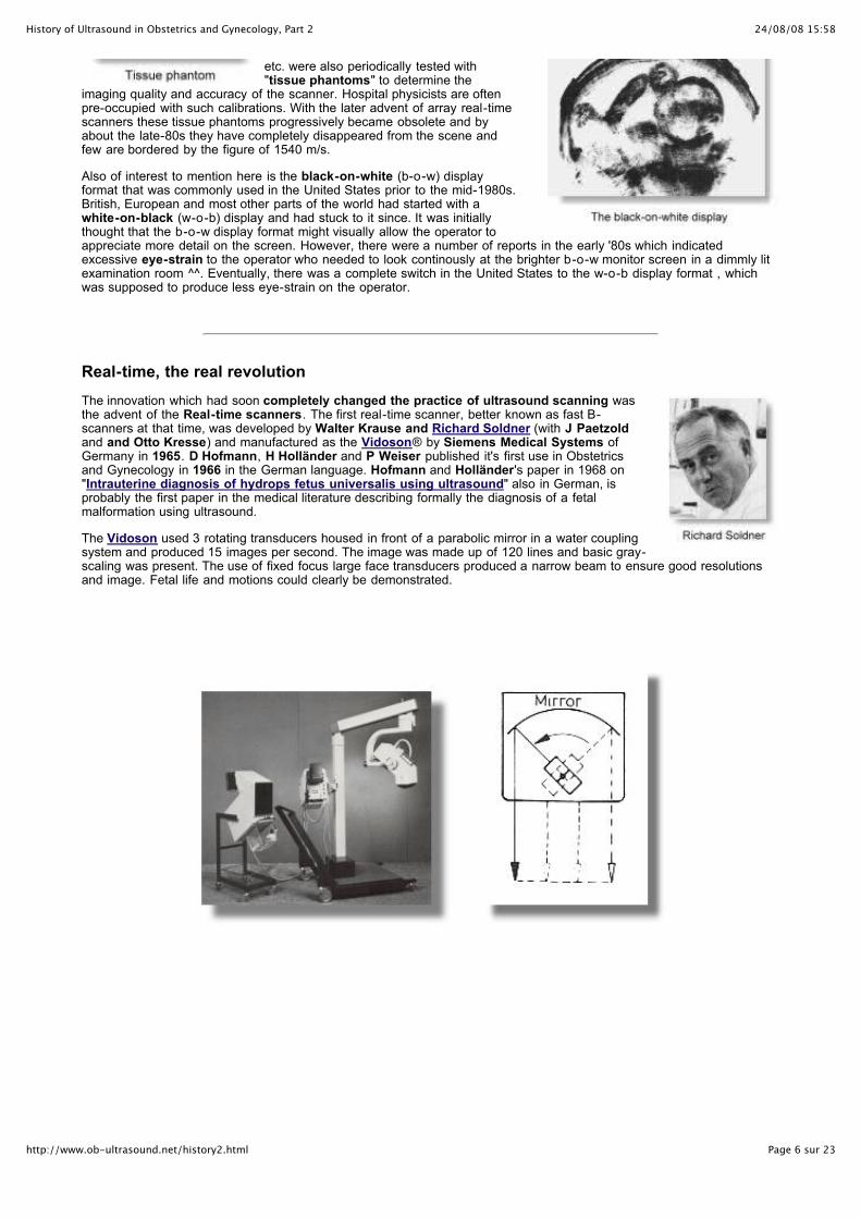
24/08/08 15:58History of Ultrasound in Obstetrics and Gynecology, Part 2
Page 6 sur 23http://www.ob-ultrasound.net/history2.html
etc. were also periodically tested with"tissue phantoms" to determine the
imaging quality and accuracy of the scanner. Hospital physicists are oftenpre-occupied with such calibrations. With the later advent of array real-timescanners these tissue phantoms progressively became obsolete and byabout the late-80s they have completely disappeared from the scene andfew are bordered by the figure of 1540 m/s.
Also of interest to mention here is the black-on-white (b-o-w) displayformat that was commonly used in the United States prior to the mid-1980s.British, European and most other parts of the world had started with awhite-on-black (w-o-b) display and had stuck to it since. It was initiallythought that the b-o-w display format might visually allow the operator toappreciate more detail on the screen. However, there were a number of reports in the early '80s which indicatedexcessive eye-strain to the operator who needed to look continously at the brighter b-o-w monitor screen in a dimmly litexamination room ^^. Eventually, there was a complete switch in the United States to the w-o-b display format , whichwas supposed to produce less eye-strain on the operator.
Real-time, the real revolution
The innovation which had soon completely changed the practice of ultrasound scanning wasthe advent of the Real-time scanners. The first real-time scanner, better known as fast B-scanners at that time, was developed by Walter Krause and Richard Soldner (with J Paetzoldand and Otto Kresse) and manufactured as the Vidoson® by Siemens Medical Systems ofGermany in 1965. D Hofmann, H Holländer and P Weiser published it's first use in Obstetricsand Gynecology in 1966 in the German language. Hofmann and Holländer's paper in 1968 on"Intrauterine diagnosis of hydrops fetus universalis using ultrasound" also in German, isprobably the first paper in the medical literature describing formally the diagnosis of a fetalmalformation using ultrasound.
The Vidoson used 3 rotating transducers housed in front of a parabolic mirror in a water couplingsystem and produced 15 images per second. The image was made up of 120 lines and basic gray-scaling was present. The use of fixed focus large face transducers produced a narrow beam to ensure good resolutionsand image. Fetal life and motions could clearly be demonstrated.

24/08/08 15:58History of Ultrasound in Obstetrics and Gynecology, Part 2
Page 7 sur 23http://www.ob-ultrasound.net/history2.html
The Vidoson*, its working mechanism and the resultant image of a fetal face and hand.The transducer housing is mounted on a mobile gantry and rigidly connected to the main console.
The scanning frequency was 2.25 MHz. Scaling and caliper functions were not present.
Hans Holländer, in his paper in 1968 demonstrated the usefulness of a 'real-time' scanner in the diagnosis of ovariantumors which were not spotted on pelvic examination. Malte Hinselmann, using the Vidoson, demonstrated in 1969 theuniversal visualization of fetal cardiac action from 12 weeks onwards. The Vidoson was popular in the ensuing 10 yearsor so and were used in many scientific work published from centers in Germany, France, Switzerland, Austria, Belgium,Italy and other European countries. The initial popularity was not based on its image resolution but rather its ability to allowthe operator to display and study movements, such as fetal cardiac motion, gross body movements and fetal breathingmovements (see also Part 3). In the International Symposium on Real-time ultrasound in Perinatal Medicine held inCharleroi, Belgium in 1978, most of the presentations were based on results from the Vidoson.
" ......... For almost ten years, real-time ultrasound has been used in many obstetrics departments. Bymeans of an apparatus which has since become technologically outdated many doctors, technicians andexpectant mothers had, at the time, the moving experience of being able to observe the living fetus. Thisseems to me to have been a psychological break-through. For the first time, the human eye pierced the'black box' of the womb...... Those who were present in obstetrics departments when this technique wasfirst used soon realized how indispensable it was proving to be in providing a valid means of observationof the fetus and its health, in ascertaining its age and studying its morphology and growth........ . Over thelast three years, the appearance of the multitransducer scanner has brought about substantial technicalprogress. At the same time, but quite independently of this, numerous studies on fetal breathingmovements, fetal behavior and neonatal cardiology were published ....... ." --- R. Chef, Maternité Reine-Astrid, Charleroi (Belgium), in the foreword to the Proceedings of theInternational Symposium on Real-time Ultrasound in Perinatal Medicine held in 1978 at Charleroi.
Read a history of the development of ultrasonography at Siemens, Germany.
Read also an article 40 Years of Realtime-Ultrasound Diagnostics by Hans Hollander.
James Griffith and Walter Henry at the National Institute of Health produced a mechanicaloscillating real-time scanning apparatus in 1973 which was capable of producing clear 30degree sectoral real-time images of good resolution. The scanner was essentially a motor-driven oscillating transducer coupled with a commercially available one-dimensionalechocardiograph (the SmithKline Eckoline 20). The 2-D scanning device was hailed as one ofthe most significant milestones in the development of echocardiography, and indeed in thedevelopment of sonography in general. Other mechanical systems published included anoscillating design with membrane-oil coupling from W N McDicken in Edinburgh (1974,produced commercially as the EMI® Emisonic 4260), a continuously rotating wheel with radially-mounted transducers from Hans Hendrik Holm and Allen Northeved in Denmark (1975), and asingle transducer direct-contact design from Reginald Eggleton in Indiana (1975). The design,which was supposed to have been modified from the mechanisms of an "electric toothbrush",was a commercial success. Toshiba®, in Japan produced their first prototype real-timemechanical sector scanner in 1975, the SSL-51H. A number of others were available commercially soon afterwards andsold well such as the circular rotating system Combison 100 from Kretztechnik® of Austria (1977), produced under theingenuity of Carl Kretz.
Although these have relatively heavy probes they produced outstanding real time resolution in the near and far field(because of highly focused beams resulting from the relatively large curvatured transducers and the lens apparatus) andwith much less image-degrading electronic noise that was associated with electronic scanners that soon became availableat around the same time.

24/08/08 15:58History of Ultrasound in Obstetrics and Gynecology, Part 2
Page 8 sur 23http://www.ob-ultrasound.net/history2.html
The large hand-held circular rotating transducer (Combison 100) from KretzTechnik® and the resultant sector image.
The transducer is connected to the main console by a flexible cable.
The concept of the multi-element linear electronic arrays was first described by WernerBuschmann in an ophthalmologic application in 1964 in East Berlin. His probe, fabricated incollaboration with Kretztechnik AG consisted of 10 small transducers mounted on an arc-shaped appartus to fit over the eye. Buschmann's transducer however, never became veryfunctional in a clinical setting and did not go into serial production. A number of similar designsfollowed on the same concept. Jean Perilhou and her group in France, working under theauspices of the Philips® Company, described a multi-element scanning array in 1967, althoughthey do not produce images in a real-time fashion. The real-time array concept was furtherexpanded by Nicolaas Bom at the Thoraxcenter, University Hospital, Erasmus University inRotterdam, the Netherlands. His initial design in 1971, which was described in his application fora Dutch patent, consisted of only 20 crystals (each 4mm x 10mm). The probe face was 66 mmlong and 10 mm wide and produced 20 scan lines. It operated at a frequency of 3.0 MHzsweeping at a frame rate of 150 frames/sec. The axial resolution was 1.25 mm while the beam width at 6 cm was 10 mm.This abeit simple and inadequate design at that time has evolved into the very sophisticated real-time scanners that arewidely available today.
In collaboration with cardiologist Paul Hugenholtz and local Dutch companyOrganon Teknika, they produced in 1972 the "Multiscan system", notably theearliest commercial linear array scanner in the world, mainly aimed at cardiacinvestigations.
The transducers operated at either 2.25 or 4.5 MHz, again with 20 crystals producing20 scan lines. The lateral resolution of this improved version at a dynamic range of10dB was 3.7 mm at 6 cm and 6 mm at 10 cm depth. It did not sell very well thoughbecause of its relatively primitive resolution and its inability to image abdominalstructures adequately.
In Japan, Rokuro Uchida at Aloka® (see alsoPart 1) had similar research on the arraytechnology in the late 1960s predating theirEuropean counterpart. In 1971 they published inJapanese (and presented at the Japan Society ofUltrasonics in Medicine) a system based on 200closely interspaced transducers. Electronic
switching and use of overlapping groups of 20 small elements yielded 2-D images with afield depth of 20 cm at a rate of 17/frames per second. The company produced their firstprototype linear array scanner in the same year. The model however, was notproduced commercially or given a model number. The first commercial linear arrayscanner from Aloka® only debuted in 1976. Toshiba® produced their first commercialreal-time linear array counterpart in the same year, the SSL-53H, aimed at abdominalapplications. Like the Aloka® this was a huge machine considering present dayfabrication standards.
Subsequent to Bom's work and theresearch in Rotterdam, LeandrePourcelot and Therese Planiol inTours, France was experimenting with more advanced segmentedsequence transducer-array scanning possibilities in 1972, in order to
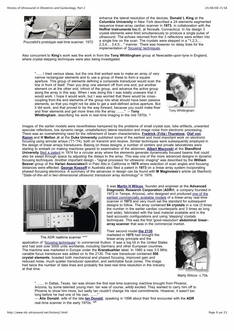
24/08/08 15:58History of Ultrasound in Obstetrics and Gynecology, Part 2
Page 9 sur 23http://www.ob-ultrasound.net/history2.html
enhance the lateral resolution of the devices. Donald L King at theColumbia University in New York described a 24-elements segmentedsequence linear-array cardiac scanner in 1973, in collaboration with theHoffrel Instruments Inc.®, at Norwalk, Connecticut. In his design 3crystal elements were fired simultaneously to produce a single pulse ofultrasound. The echoes returned from the 3 reflections were written intoa single line on the scan. The crystals were stepped in a "1,2,3,...2,3,4,... 3,4,5... " manner. There was however no delay lines for theimplementation of 'focusing' techniques.
Also concurrent to King's work was the work in from the Tony Whittingham group at Newcastle-upon-tyne in England,where crystal stepping techniques were also being investigated.
"...... I tried various ideas, but the one that worked was to make an array of verynarrow rectangular elements and to use a group of these to form a squareaperture. This group of elements defining a composite transducer would scan theline in front of itself. Then you drop one element off from one end, put anotherelement on at the other end, infront of the group, and advance the active groupalong the array in this way. When I was doing this I was totally unaware that itwould work. I hope it would work, but I was worried that there would be cross-coupling from the end elements of the group into what should have been passiveelements, so that you might not be able to get a well-defined active aperture. Butit did work, and that proved to be the way forward, because you could make finerand finer elements and get more lines into the array ........" -- TonyWhittingham, describing his work in real-time imaging in the mid 1970s. ^
Images of the earlier models were nevertheless hampered by the problems of small crystal size, lobe artifacts, unwantedspecular reflections, low dynamic range, unsatisfactory lateral resolution and image noise from electronic processing.There was an overwhelming need for the refinement of beam characteristics. Fredrick (Fritz) Thurstone, Olaf vonRamm and H Melton Jr at the Duke University published some of the earliest and most important work on electronicfocusing using annular arrays ('71-'74), both on transmit and receive. Similar techniques were subsequently employed inthe design of linear arrays transducers. Basing on these designs, a number of centers and private laboratories werestarting to embark on making machines geared to examination of the abdomen. Albert Macovski at the StandfordUniversity filed a patent in 1974 for a circular array where the elements generate dynamically focused beams that couldalso be swept through space by adjusting the delays to the array. This was one of the more advanced designs in dynamicfocusing techniques. Another important design - "signal processor for ultrasonic imaging" was described by the WilliamBeaver group of the Varian Associates® in Palo Alto in California in 1975 where selection of scan angles and focusingdistances were effected. George Kossoff in Australia also filed a patent in 1973 on a linear array system incoporatingphased-focusing electronics. A summary of the advances in design can be found with M Maginness's article (at Stanford)"State-of-the-art in two-dimensional ultrasonic transducer array technology" in 1976.
It was Martin H Wilcox, founder and engineer at the AdvancedDiagnostic Research Corporation (ADR®, a company founded in1972 in Tempe, Arizona), who designed and produced one of theearliest commercially available models of a linear-array real-timescanner in 1973 and very much set the standard for subsequentdesigns to follow. The array contained 64 crystals in a row (3 timesthe number in the earlier cardiac counterparts and 3 times as longand wide), fabricated with the best material available and in thebest accoustic configurations and using 'stepping' crystalstechniques. This was the first 'good-resolution' abdominal linear-array scanner that was in the commercial market.
Their second model the 2130marketed in 1975 had brought thelinear-array principle and the
application of 'focusing techniques' to commercial fruition. It was a big hit in the United Statesand had sold over 5000 units worldwide, including Germany and other European countries.The machine was marketed in Europe under the Kranzbuehler label. In 1980 a new 3.0 MHzvariable focus transducer was added on to the 2130. The new transducer contained 506crystal elements, boasted both mechanical and phased focusing, improved gain andreduced noise, much quieter transducer operation, and switchable focal zones. The imagehad twice the number of data lines and probably the best real-time resolution in the industryat that time.
" ....... In Dallas, Texas, Ian was shown the first real-time scanning machine brought from Phoenix,Arizona, by some talented young men. Ian was of course, wildly excited. They wanted to carry him off toPhoenix to show him more, but sadly Ian couldn't change his next commitments. However, it wasn't toolong before he had one of his own. ......." --- Alix Donald, wife of the late Ian Donald, speaking in 1998 about their first encounter with the ADR
real-time scanner in the early 1970s. ref.

24/08/08 15:58History of Ultrasound in Obstetrics and Gynecology, Part 2
Page 10 sur 23http://www.ob-ultrasound.net/history2.html
ADR® merged with ATL® (Advanced Technology laboratories®, see below) in 1984. ADR® produced the 2150 in 1980and the last model under the ADR label, the ADR 4000 in 1982.
Linear array and annular array technology had alsobeen heavily investigated by the Japanese since theearly 1970's. The country had been moving ahead verysuccessfully with innovative electronic engineering inmany domestic, commercial and professional sectors.Commercial linear array models from companies likeHitachi®, Toshiba®, and Aloka® soon began todominate the world market. Hitachi® also producedtheir first linear array scanner the EUB-10 in 1976,followed by the EUB-20 in 1977 and the EUB-22 in1979. The EUB-20Z produced in 1978 alreadyincorporated the world's first digital scan converter.
The Toshiba® SAL-10A and the more portable SAL-20A (pictured on the left)and SAL-30A, which were marketed in 1977, 1978 and 1979 respectively, and
the Aloka® SSD-202 (1979), SSD-203 (1980), SSD-240 (1981) and SSD-256 (1982) were popular and had found theirway into notable Institutions outside of Japan such as the King's College Hospital in London (Campbell), the Herlev(Gentofte) Hospital in Copenhagen (Holm), the Hospital Universitaire Brugman in Belgium (Levi) and were employedin many important early studies. The SAL-10A which was designed by acclaimed Japanese engineer Kazuhiro Iinuma,received many commendations. Other popular early choices included the Axiscan 5 (1976) and Abdoscan 5 (1979) fromRoche Kontron®, the Sono R from Philips® (1978), the RA-1 (1980) and the Imager 2300 (1981) from Siemens®, andthe LS 1500 (1981) from Picker/ Hitachi®. Aloka® scanners were marketed in the United States under the brand NarcoAir-Shield®. Diagnostic Sonar® Ltd, a company founded in 1975 in Edinburgh, Scotland produced the first electronicreal-time scanner, the System 85 in the United Kingdom in 1976.
Early scanner probe was bulky to fit on the abdomen ***Images from early real-time scanners
had obstrusive scan lines, low dynamic range and resolution.
Many of the early models typically had very large probes housing an array of some 64 transducer (crystal) elementsarranged in a linear row, and operating with sequential electronic switching or dynamic focusing. It was not until theearly 1980s that probe size had gotten smaller and image resolution significantly improved.
Read the short History of the development of Medical Ultrasonics in Japan for a chronology of Japanese
contributions to the development of ultrasound scanners.
At around the same time, steered-beam phased array transducers and annular array transducers with morecomplicated electronic circuitry were described, and had found their way into echocardiographic examinations because ofthe relatively small contact surface.
The phased-array scanning mechanism was first described by Jan C Somerat the University of Limberg in the Netherlands and in use from 1968, way

24/08/08 15:58History of Ultrasound in Obstetrics and Gynecology, Part 2
Page 11 sur 23http://www.ob-ultrasound.net/history2.html
ahead of its time and several years before the appearance of linear-arrayssystems. The principle of phased-arrays had probably been known muchearlier where the technique was engaged in underwater submarine warfareand hence the technology was kept confidential. Fredrick (Fritz) Thurstoneand Olaf von Ramm at the Duke University published one of the earliest andmost significant phased-array designs in 1974, which was incorporated into anumber of commercial sector-array scanners. Very sharp focusing over anextended range was obtained from annular arrays using focusing methods onboth transmit and receive. Other early significant contributors to the beam-former techniques included Albert Macovski at Standford University and
Samuel Maslak at Hewlett Packard®. Maslak later founded the Acuson Corporation (see also Part 3).
The Kossoff group in Australia had also made significant progress in the annular phased array transducer designs asearly as 1973 and the technology was incoporated into their water-bath scanner, the UI Octoson. In England, EMI®produced the Emisonic 4500, a phased-array sector scanner which was nevertheless expensive, electronically noisy andhad inferior resolution in the near fields. Early phased-arrays in the late 70s were all used in cardiac applications.Important manufacturers included Varian® and Irex®. In the first half of the 1980s, image quality in phased-arrays hadcontinued to improve and some outstanding designs had come from Irex® and later on Elscint® (Dynex) and HewlettPackard®. Despite the small probe size, phased-array sector scanners had never been popular with Obstetrical andGynecological examinations.
Compound static scanners continued it's tradition of being very huge bulkymachines, probably influenced by the design norms of other imagingmodalities such as tomographic x-ray machines and the bulky digitalelectronics housed in the console (see above on scan converters) before theimpact of the micro-processors. New static scanners which were in greatdemand and produced excellent images were still on the drawing board andproduction line in the early 1980s. It was believed that real-time scannerswould play only a complimentary role to static scanners in the assessmentof moving structures. These static machines however were starting to bereplaced or phased out at a rate that was faster than expected. There wasapparently little practical, economical or clinical advantage of these costlymachines over the more mobile and flexible electronic real-time scanners.
There were initially many who were so used to and skillful at operating thestatic machines that they were unhappy to switch over entirely to the real-timecounterparts. They were also anxious about the latter's limited field of view,poorer resolution and allegedly 'less accurate' on-screen measuring systemthat they have only started to get used to not too long ago. Static scannerswere not completely out of the scene until about 1985-86. The switch-over
had serious financial implications to some companies who had a large inventory of static scanners.
Read a commentary by Royal J Bartrum, Dartmouth Medical School, in the book "Real-time ultrasonography" in
1982, on the switch-over to the real-time scanner.
From Digital's PDP-11 to Intel's 8080 and beyond
The rapid reduction in the physical size of the machine console in the later half of the1970s (See Aloka® and Toshiba®'s early products above) was the direct result of theinvention of the microprocessor and the evolution of the minicomputer into themicrocomputer .
By the late 1960's, computers built from discretetransistors and simple integrated circuits (IC)already existed. The first practical IC wasfabricated in 1959 at Fairchild and TexasInstruments and Fairchild began its commercialmanufacture in 1961. As manufacturingtechnology evolved, more and more transistorswere put onto a single silicon chip with themaximum number of transistors per chip doublingevery year between 1961 and 1971. Theyprogressively became a device containing manycircuits and was called a LSI (Large Scale
Integration). The PDP-11 minicomputer from DEC® containing many ICs and LSIs,was used in many scanner consoles up to the late 1970s. The UNIBUS architecture

24/08/08 15:58History of Ultrasound in Obstetrics and Gynecology, Part 2
Page 12 sur 23http://www.ob-ultrasound.net/history2.html
used by the CPU in the PDP-11 was particularly suited to communicating withmemory and peripherals.
Towards the end of 1969 the structure of the smaller programmable calculator hademerged. Intel®, under contract from Japanese company Busicom for the design of asmall desktop programmable calculator, produced the world's first microprocessorchip the 4004 in 1971. In order to create a chip of such complexity, newsemiconductor design technologies had to be developed. The 4004 is considered thefirst general-purpose programmable microprocessor, even though it was only a 4-bitdevice. The original 4004 measured 1/8th of an inch long by 1/16th of an inch wideand contained 2,300 transistors. It ran at 108 KHz and executing 60,000 operations ina single second. It had about the same amount of computing power as the originalENIAC which weighed 30 tons, occupied 3,000 cubic feet of space and used 18,000vacuum tubes. Today's 64-bit microprocessors are still based on similar designs, withmore than 8.5 million transistors performing hundreds of millions of calculations eachsecond.
The 4004 was followed quickly by the 8-bit 8008microprocessor. The 8008, which contained 3300transistors, was originally intended for a CRTapplication and was developed concurrently with the4004. By using some of the production techniquesdeveloped for the 4004, Intel® was able tomanufacture the 8008 as early as March 1972. The 8-bit era between about 1975 and 1980 had a majorimpact on household computing and industry becausethe first few microprocessors were available at veryaffordable prices.
The 8008 microprocessor from Intel® was however relatively crude andunsophisticated. It had a poorly implemented interrupt mechanism and multiplexedaddress and data buses. The first really popular general-purpose 8-bit microprocessorwas Intel's 8080, in production in early 1974. This had a separate 8-bit data bus and16-bit address bus. It ran at 2 MHz with 6000 transistors. It has essentially ten timesthe performance of the 8008.
Shortly after the 8080 went into production, Motorola® created its own competitor,the 8-bit 6800, containing 4000 transistors and destined for use in automotive andindustrial applications . Although the 8080 and 6800 were broadly similar in terms ofperformance, they had rather different architectures. The 6800 was, to some extent,modeled on the PDP-11 and had much a cleaner architecture than the 8080. Othernewer processors followed and found their way into industrial operations includingmedical scanners and equipments.
Scanner engineering itself was soon in the hands of commercial companies rather than clinical personnel as advancedcomputer technologies were fiercely incoporated into each design to manipulate beam characteristics and signalprocessing to produce the best possible scan images. Apart from those mentioned above, other important earlymanufacturers of real-time equipments included Shimadsu® from Japan; EMI®, KretzTechnik AG, Bruel and Kjaer®,GEC® and Rohe® from Europe and Diasonics®, Dynex®, Ecoscan®, Elscint®, Hewlett-Packard®, Irex®, SKI®,Phosonic Searle®, Technicare® (acquired Unirad®) and Xonics® from the United States . The application ofultrasound in Obstetrics and Gynecology had since then undergone an explosive proliferation all over the world. By theearly 1980s there were over 45 large and small ultrasonic scanner manufacturers worldwide.
Further Improvement in performance aside from focusing the ultrasound beam was acheived largely through an increasein the number of transducer crystals (or channels, from 64 to 128), improvements in transducer crystal technology (goinginto broad-band and high dynamic range), increasing array aperture (more crystals firing in a single time-frame), fastercomputational capabilities, improving technical agorithms for focusing on receive (increasing the number of focal zonesalong the beam), incoporating automatic time-gain controls and progressively replacing analog portions of the signal pathto digital. It was perhaps regretable to see that British manufacturers have failed to keep up with developments made byother leaders in array technology, notably those from the United States and Japan. This was probably reflection of asimilar trend in other arenas of electronic and micro-processor development in these countries. It is also of interest to notethat the Siemens Vidoson® and the Octoson®from Australia both did not sell in North America at all. Both had thedisadvantage of being cumbersome when scanners from other manufacturers were rapidly getting better in resolution andmanuevability.

24/08/08 15:58History of Ultrasound in Obstetrics and Gynecology, Part 2
Page 13 sur 23http://www.ob-ultrasound.net/history2.html
In the early 1980s (around 1980-1985), many agreed that mechanical sector scanners (be it rotating,oscillating or wobbling designs) which employed relatively large area transducers produced better andless noisy images than electronic linear-array scanners. Shown here are very good images from SKI®(left), Diasonics® (middle) and ATL® (right) taken in 1981. The market in Obstetrics and Gynecologywas divided between the mechanical sector scanners and the linear-arrays until the second half of the1980s where both were replaced by convex sector-arrays.
Because of its smaller convex contact surface, the curvilinear or convexsector-array fits much better on the abdomen and allows for a wider field ofview than does the linear-array configuration. Work on the fabrication of anelectronic convex array had started in the late 1970s in the larger Japanesecompanies such as Hitachi® (publishing their convex attachment to their EUB-10A scanner in 1978); Aloka® (filing their patent on the convex scanner in1980), as well as in U. S. companies notably the North American Philips® andthe Picker Corporation®, who had filed their patents for convex arrays andprocessors in 1979 and 1980 respectively. The first commercially availableconvex array transducer apparently only debuted in 1983 in a scanner fromKontron Instruments® in Europe, the Sigma 20, which was designedespecially for use in Obstetrics and Gynecology. Hitachi® in Japan marketedtheir model EUB-40 with their new convex array later on in the same year.
Toshiba® introduced a similar array in 1985, in their new scanner model SAL-77A. Interestingly, the design actually replaced an earlier model (by only about 9 months) the SAL-90A which boasted anew "trapezoid" linear array in which the face of the transducer was flat but a trapezoid-shaped image was produced fromthe 128 transducer elements using phased electronics. American machines were apparently still using linear arrays by1985, although very shortly they were quickly replaced by the new convex configuration. By about 1987, convex arrays arestandard on every new scanner, whether or not it is configured for use in Obstetrics and Gynecology.
Model number of some of the scanners made after 1980 from important manufacturers are listed here with the year in
which they were marketed. Also view pictures of some of the early scanners.
Skin Coupling material for ultrasonic transmission has also switched from oil to a water-soluble (non cloth-staining) gel medium. One of the more well-known manufacturers wasthe Parker Laboratories® at New Jersey. Images are commonly recorded on "peel-apart" Polaroid® films (the Type 611 was most commonly used). Multi-formatradiographic films (6-9 images on one film) using dedicated video imagers soon becamemainstream with institutional users and thermal paper printers in the private practicemarket.
Worthy of mention here was the attempt in thelate 70s and early 80's to miniaturize scannersso that they could be portable and be used atthe bedside. Four such examples were the all-in-one MiniVisor® from Organon Teknika® inthe Netherlands, the Bion PSI-4000 from Bion Coporation® in Denver, theShimasonic SDL-30 from Shimadzu Corporation®, Japan, and the 210DX fromAloka®.
The Minivisor® (available from 1979) was a spin-off from Bom's laboratory. Itwas battery operated, shaped like a mushroom, had no wires and used a 2-inches display with an on-screen caliper system and digital readout. Thetransducer is fused to the bottom of the device similar to a 'large' fetal pulsedetector. Juiry Wladimiroff suggested in 1980 the device would be useful forroutine BPD screening. Nevertheless the popularity of these machines wereshort-lived for several important reasons pertaining at that time: The resolution
was unsatisfactory because of the available electronics. The images of the 'standard' and larger devices, as well as their

24/08/08 15:58History of Ultrasound in Obstetrics and Gynecology, Part 2
Page 14 sur 23http://www.ob-ultrasound.net/history2.html
overall 'portability' have seen rapid improvements round about the same time; and thirdly, real-time ultrasound has veryrapidly established itself as a definitive diagnostic entity and the concern for good image information appeared to overidethat of the extra portability.
The invention of the real-time scanner had enabled much more effective diagnosis of many fetal malformations and inparticular cardiac anomalies which hitherto was impossible to diagnose accurately. (see Part 3). Fetal sonography andprenatal diagnosis (a term which was only coined in the 1970s) had emerged as the 'new' science in Obstetrics and fetalmedicine.
John C. Hobbins at the Yale University, Connecticut and Stuart Campbell at the King'sCollege Hospital in London were among others, the two most important forerunners on eitherside of the Atlantic. Their centers have also become two of the most important teachingcenters in fetal sonography. Many of the research fellows and staff members that had comethrough Hobbins' department for example, have in time become celebrated names in the fieldof fetal sonography and prenatal diagnosis.
Other important early North American workers in fetal biometryincluded Peter Cooperberg, David Graham, Charles Hohler,Alfred Kurtz, Rudy Sabbagha and Roger Sanders. Their earlywork on the biparietal diameter was particularly notable,establishing charts for different populations, standardizingmeasuring methods and errors and comparing differences thatmay be present between measurements made on static and real-time equipments. (see also Part 3). The real-time scanner had soon enabled the accuratemeasurement of fetal limb bones that lead to the introduction of the important measurementof the fetal femur length by John Hobbins in 1979 for the evaluation of skeletal dysplasiafollowed by Gregory O'Brien and John Queenan who described it's use in fetal growthassessment. Phillipe Jeanty at Yale provided in 1984 measurement charts for all the fetallong bones.
Ultrasound guidance was started to be employed in procedures such as amniocentesis(Jens Bang and Allen Northeved 1972, Copenhagen), fetoscopy (John Hobbins andMaurice Mahoney, 1974) and transabdominal chorionic villus sampling (Steen Smidt-
Jensen and N Hahnemann, 1984, Copenhagen).
Read here a short history of Amniocentesis, fetoscopy and chorionic villus sampling.
Transvaginal scanners
Wild and Reid had invented and described the use of A-mode trans-vaginal and trans-rectal scanning transducers as early as 1955 (see Part1). Using a proprietary A-mode vaginal scanner from KretzTechnik in theearly 60s, Alfred Kratochwil in Austria had reported on fetal heartpulsation at slightly over 6 weeks menstrual age. He also developed athimble attachment transducer to facilitate vaginal sonography with pelvicexamination. Trans-vaginal sonograms were also reported by Lajos vonMicsky in New York around the same time, using innovative prototypeequipment that he devised. Other A-mode vaginal transducers weredeviced by Japanese researchers. With the advent of B-mode equipments,A-mode vaginal scanners had slowly disappeared from the market.
Shigemitsu Mizuno in Japan demonstrated in 1965 an endovaginal scanner forpelvic examination using a plan-position indication (PPI) B-mode format (the sameformat as an abdominal compound contact scanner). The device was mannuallyrotated and the resulting display was very similar to a circular military 'Radar"display. Used either transrectally or transvaginally, It was capable of producingsome meaningful pictures of the pelvic organs. Salvator Levi at Brussels was oneof the early advocates of Aloka® equipments and published on their transvaginalscanner as early as 1970. R A Khentov in the USSR and von Micsky in the UnitedStates also demonstrated vaginal and rectal scanning devices in the late 1960s.
Despite the advantages of transvaginal scans, its use had waited almost 20 years tobecome practical and popular in gynecological scanning. Athough the need andtechnology were there, the really practical real-time transvaginal scanning probewas not "invented" until 1985 when KretzTechnik® of Austria produced their firstreal-time mechanical vaginal sector scanner. The transducer had a scan angleof 240 degrees and was designed with the use for transvaginal ovum retrieval inmind, in collaborations with IVF pioneers Wilfried Feichtinger and Peter Kemeterin Austria. The coming of such mechanical devices was also in part the outcome ofadvances in microprocessor controller circuitry and ferro-magnetics resulting inmuch smaller and more efficient motors.

24/08/08 15:59History of Ultrasound in Obstetrics and Gynecology, Part 2
Page 15 sur 23http://www.ob-ultrasound.net/history2.html
Dutch manufacturer Philips® followed on with one ofthe earliest mechanical vaginal scanners in thesecond half of 1986. The probe was in the shape of a microphone with a roundishelongated head housing a 5MHz 13mm wobbler transducer. It could be retro-fitted ontotheir real-time scanner SDR 1550 which first debuted in 1985. Although they producedexcellent images compared to their abdominal counterpart, mechanical endovaginal designswere not favored by many ultrasound manufacturers, partly because of the vibration thatwas generated.
Mechanical designs were rapidly followed by electronic array versions which are ratherlike a reduced-size abdominal convex sector transducer that has appeared around thesame time from other manufacturers in Japan. ALOKA® produced an electronic sectorversion which could be retofitted onto their older model the SSD-256. GE MedicalSystems® produced their first endovaginal probe to fit their RT3200 in mid 1987. By 1988
most manufacturers had endovaginal options installed in their scanners.
The advent of tranvaginal scanning (at higher frequencies of 5 - 8 MHz and resulting in muchfiner resolution) had a significant impact on the diagnosis of gynecological and earlypregnancy pathologies. In particular the accurate recognition of fetal cardiac pulsations inmissed abortions was facilitated at an early gestational age of 6 weeks.
Non-palpable ovarian masses were often picked up by a vaginal scan. Transvaginalscanning has also progressively become standard practice in the management of infertilitypatients, in assessing follicular development and endometrial growth, as well as being anindispensible aid in the retrieval of oozytes from the ovaries. The assessment of ovarianfollicular development had before that been based on static and real-time abdominalultrasound, first popularised by B-Joachim Hackelöer and his group in Germany since 1977.(See also Part 3). And before that time, most clinicians did not envisage that cyclical changesin morphology and size of ovarian follicles can be so closely followed with ultrasonography.
M-mode and Doppler
The M-mode (time-motion) display was first described by Inge Edler andHellmuth Hertz in Lund, Sweden in 1954 using a modified metal-flaw detectorfrom Siemens® of Germany. They demonstrated the feasibility of recordingcardiac valvular motion ultrasonically. Sven Effert in Germany, who had beencollaborating with Hertz in some of his work, futher demonstrated the usefulnessof M-mode echocardiography, which had subsequently caught on as amainstay investigation in cardiology. Xin-Fang Wang first described in China in1964 the use of M-mode ultrasound in the study of fetal cardiac movements.Jens Bang and Hans Henrik Holm demonstrated fetal cardiac motion using M-mode from 10 weeks onward in 1968. In the same year Alfred Kratochwildescribed similar usefulness of detecting fetal cardiac motion by M-mode inpatients with threatened abortion. These were nevetheless 'blind' procedures.Hugh Robinson in Glasgow described with great success in 1972 the detectionof fetal cardiac motion at 7 weeks by A- and M-mode after locating the fetus with on B-mode ultrasound. In the sameyear Fred Winsberg in Canada described M-mode diagnosis of fetal cardiac anomaly.
The Doppler principle was first described over 100 years ago by Christian Andreas Doppler inAustria in 1842. Medical applications of the Ultrasonic doppler techniques were firstimplemented by Shigeo Satomura and Yasuhara Nimura at the Institute of Scientific andIndustrial Research in Osaka, Japan in 1955 for the study of cardiac valvular motion andpulsations of peripheral blood vessels.
The Satomura team which also included Ziro Kaneko, pioneered transcutaneous doppler flowmeasurements in 1959, several years ahead of the work at the University of Washington inSeattle.
It has also become known from the workof Ziro kaneko and Kanemasa Kato in
Satomura's laboratory in 1962 that blood flow can be detected bythe ultrasonic doppler method largely because blood itself consistsof a suspension of "uncorrelated point back-scatterers" (the redcells) with varying density and compressibility, and not because ofturbulence in the flow stream as was previously thought. In 1966,Kato and T Izumi pioneered the directional flow-meter using thelocal oscillation method where flow directions were detected and

24/08/08 15:59History of Ultrasound in Obstetrics and Gynecology, Part 2
Page 16 sur 23http://www.ob-ultrasound.net/history2.html
displayed. This was a breakthrough in doppler instrumentation asreverse flow in blood vessels could then be documented.
Compared to the western world, throughout the history of dopplerultrasound, Japanese research had played a leading and majorrole in its development. Much of the ground-breaking researchwere presented in national engineering journals and at the annual scientific meeting of the Japan Society of Ultrasonics inMedicine (in japanese) and were thus unknown to the west until a later date.
A brief history of the pioneering work in Doppler applications in Japan is described here.
In the United States, ground breaking work in Dopplerinstrumentation started at the University of Washingtonin Seattle from 1958 onwards led by Robert Rushmer, apediatrician and physiologist, who was determined tocharacterise cardiovascular functions in intact un-anaesthetised animals. He was soon joined by DeanFranklin, Dick Ellis and Donald Baker, engineers whowere recruited into Rushmer's cardiovascularinstrumentation development program. The teampioneered transcutaneous continuous-wave flowmeasurements and spectral analysis in 1963 and as aclinical application, surgeon D Eugene Strandnessundertook the clinical testing. He published the firsttranscutaneous vascular spectral flow signals in the
following year.
Although continuous wave doppler instruments were quite in place by the mid 1960s, for the next 10 years or so it's use inObstetrics and Gynecology was largely confined to that of a fetal pulse detector and continuous fetal heart ratemonitoring (see below). Gynecologist Wayne Johnson working with Rushmer reported the detection of fetal life at 12weeks gestation (from LMP) with Strandness' continuous wave instruments in 1965 (see also below). The continuouswave technology was transferred to Smith Kline Instruments® from the University of Washington and the companymanufactured the first Doptone in 1965. Other early commercial suppliers of continuous wave doppler devices in vascularapplications included Parks® Electronics Inc., Kay® Electric company (the SonaGraph), and Medasonics® Inc (theVersatone). An audio transcript of "Typical Doppler Sounds from peripheral arteries and veins" prepared by D EugeneStrandness in the late 1970s can be heard here.
The continuous wave Doppler method did not provide explicit information aboutthe distance between the ultrasonic transducer and the moving target. Furtherdevelopment led to the introduction of pulsed-doppler system by Baker in 1970,a concept based on the repetitive propagation of short ultrasound bursts andanalysis of the signal received at a preselected time delay with respect toemission (the sample volume). Baker also outlined a technique for determiningthe volume blood flow from Doppler velocity measurements. The team alsorecruited John Reid from Pennsylvania who complemented the flow designswith 2D and M-mode technology. New instruments basing on the ultrasounddoppler principles were developed. The first duplex pulsed-doppler scanner, amilestone development in ultrasound instrumentations was designed anddeveloped by Frank Barber, Baker, Reid and other colleagues in 1974. Theduplex scanner finally enabled 2D gray scale imaging to be used to guide theplacement of the ultrasonic beam for doppler signal acquisition.
" ....... The program was instrument and hardware orientated, because in those days, medical researchwas carried out in little the same way as wars are fought. Wars are fought according to the weapons youhave, and the rules of the game will be according to the weapons you have. I think a lot of researches arein the same way. If you have the tools that nobody else has you can create a new game that nobody elsecan play........ People saw this as an opportunity. ...... They saw an opportunity to get ahead of theircolleagues and publish ....... ." --- Donald Baker, speaking on his early work in doppler instrumentationsand his efforts in popularising them at ATL.
ATL® (Advanced Technology Laboratories) was founded in 1969 near Seattle, Washington, by a small group ofengineers developing marine electronic systems. Starting form 1973, technology developed at the University ofWashington Center for Bioengineering were transferred to ATL in the development of systems for diagnostic sonography.The first duplex pulsed-doppler scanner, the Mark1 (with pulsed-doppler module model 400) was released in 1974 forcardiac investigation. The later version 400B became part of the well-known ATL Mark V duplex scanner which debutedin 1978. Squibb Coporation® acquired ATL in 1980 and ADR in 1982. ADR was merged into ATL in 1984.
In England, pioneering work from Peter Wells in Bristol alsodemonstrated in 1969 that pulsed-doppler information may be obtainedthrough the combination of range-gating methodogy and continuous waveand 2D techniques. In the late 1960s the Okujima and Ohtsuki groupand the Ziro Kaneko group in Japan (after the death of ShigeoSatomura), and the Pierre Peronneau group in Paris were equally active
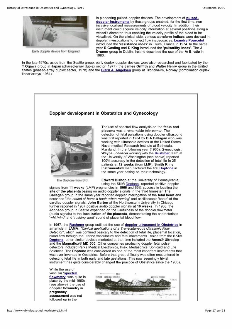
24/08/08 15:59History of Ultrasound in Obstetrics and Gynecology, Part 2
Page 17 sur 23http://www.ob-ultrasound.net/history2.html
in pioneering pulsed-doppler devices. The development of pulsed-doppler instruments by these groups enabled, for the first time, non-invasive localised measurements of blood velocity. In addition, theirinstrument could acquire velocity information at several positions along avessel's diameter, thus enabling the velocity profile of the blood to bevisualised. On the clinical side, various waveform indices were devised indoppler investigations to reflect flow inadequacies. Leandre Pourcelotintroduced the 'resistance index' in Tours, France in 1974. In the sameyear R Gosling and D King introduced the 'pulsatility index'. The JDrumm group in Dublin, Ireland described the use of the A/ B ratio in1980.
In the late 1970s, aside from the Seattle group, early duplex doppler devices were also researched and fabricated by theT Ogawa group in Japan (phased-array duplex sector, 1977), the James Griffith and Walter Henry group in the UnitedStates (phased-array duplex sector, 1978) and the Bjørn A. Angelsen group at Trondheim, Norway (combination duplexlinear arrays, 1981).
Doppler development in Obstetrics and Gynecology
The use of spectral flow analysis on the fetus andplacenta was a remarkable late-comer. Thedetection of fetal pulsations using doppler ultrasoundwas first reported in 1964 by D A Callagan who wasworking with ultrasonic devices at the United StatesNaval medical Research Institute at Bethesda,Maryland. In the following year (1965), GynecologistWayne Johnson working with the Rushmer team atthe University of Washington (see above) reported100% accuracy in the detection of fetal life in 25patients at 12 weeks (from LMP). Smith KlineInstruments® manufactured the first Doptone inthe same year basing on their technology.
Edward Bishop at the University of Pennsylvania,using the SKI® Doptone, reported positive doppler
signals from 11 weeks (LMP) pregnancies in 1966 and 65% success in locating thesite of the placenta basing on audio doppler signals in the third trimester. TheCallagan group in the same year reported doppler interrogation of the fetal heart anddescribed "the sound of horse's hoofs when running' and oscilloscopic 'beats' of thecardiac doppler signals. John Barton at the Northwestern University in Chicagofurther reported in 1967 positive audio doppler signals at 10 weeks . In 1968, theJohnson group in Seattle expanded on the usefulness of the doppler flowmeter(audio signals) to the localization of the placenta, demonstrating the characteristic'whirlwind ' and 'rushing wind' sound of placental blood flow.
In 1967, the Rushmer group outlined the use of doppler ultrasound in Obstetrics inan article in JAMA, "Clinical applications of a Transcutaneous Ultrasonic FlowDetector", which was confined basicaly to the detection of fetal life, placental location,blood flow through the uterine vasculature and fetal movements. Aside from the SKI®Doptone, other similar devices marketed at that time included the Ames® Ultradopand the Magnaflux® MD 500. Other companies producing doppler fetal pulsedetectors included Parks Medical Electronics, Imex, Medasonics, Sonicaid and LifeSciences. The Doptone was considered as one of the most important instruments thatwas ever invented in Obstetrics. Before that great difficulty was often encountered indetecting fetal life in both early and late gestations. This now seemingly trivialinstrument has quite considerably changed the practice of Obstetrics since the 1960s.
While the use ofvascular 'spectralflowmetry' was quite inplace by the mid-1960s(see above), the use ofdoppler flowmetry inpregnancyassessment was notfollowed up in the
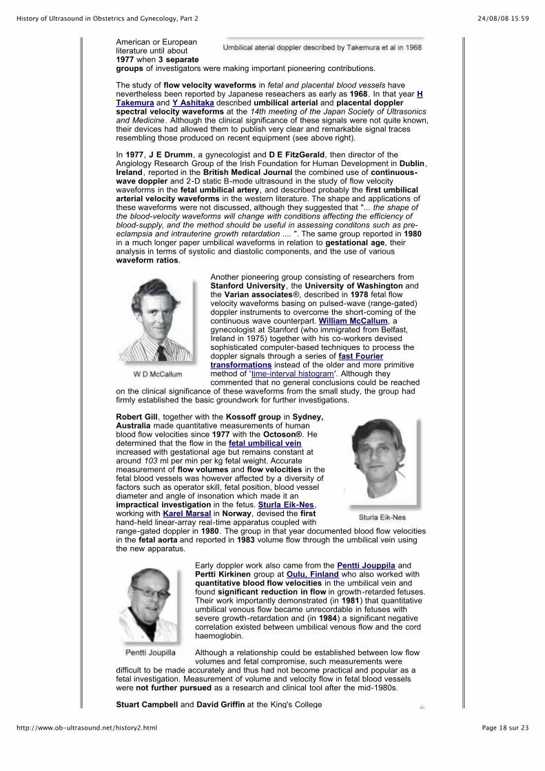
24/08/08 15:59History of Ultrasound in Obstetrics and Gynecology, Part 2
Page 18 sur 23http://www.ob-ultrasound.net/history2.html
American or Europeanliterature until about1977 when 3 separategroups of investigators were making important pioneering contributions.
The study of flow velocity waveforms in fetal and placental blood vessels havenevertheless been reported by Japanese reseachers as early as 1968. In that year HTakemura and Y Ashitaka described umbilical arterial and placental dopplerspectral velocity waveforms at the 14th meeting of the Japan Society of Ultrasonicsand Medicine . Although the clinical significance of these signals were not quite known,their devices had allowed them to publish very clear and remarkable signal tracesresembling those produced on recent equipment (see above right).
In 1977, J E Drumm, a gynecologist and D E FitzGerald, then director of theAngiology Research Group of the Irish Foundation for Human Development in Dublin,Ireland , reported in the British Medical Journal the combined use of continuous-wave doppler and 2-D static B-mode ultrasound in the study of flow velocitywaveforms in the fetal umbilical artery, and described probably the first umbilicalarterial velocity waveforms in the western literature. The shape and applications ofthese waveforms were not discussed, although they suggested that "... the shape ofthe blood-velocity waveforms will change with conditions affecting the efficiency ofblood-supply, and the method should be useful in assessing conditons such as pre-eclampsia and intrauterine growth retardation .... ". The same group reported in 1980in a much longer paper umbilical waveforms in relation to gestational age, theiranalysis in terms of systolic and diastolic components, and the use of variouswaveform ratios.
Another pioneering group consisting of researchers fromStanford University, the University of Washington andthe Varian associates®, described in 1978 fetal flowvelocity waveforms basing on pulsed-wave (range-gated)doppler instruments to overcome the short-coming of thecontinuous wave counterpart. William McCallum, agynecologist at Stanford (who immigrated from Belfast,Ireland in 1975) together with his co-workers devisedsophisticated computer-based techniques to process thedoppler signals through a series of fast Fouriertransformations instead of the older and more primitivemethod of 'time-interval histogram'. Although theycommented that no general conclusions could be reached
on the clinical significance of these waveforms from the small study, the group hadfirmly established the basic groundwork for further investigations.
Robert Gill, together with the Kossoff group in Sydney,Australia made quantitative measurements of humanblood flow velocities since 1977 with the Octoson®. Hedetermined that the flow in the fetal umbilical veinincreased with gestational age but remains constant ataround 103 ml per min per kg fetal weight. Accuratemeasurement of flow volumes and flow velocities in thefetal blood vessels was however affected by a diversity offactors such as operator skill, fetal position, blood vesseldiameter and angle of insonation which made it animpractical investigation in the fetus. Sturla Eik-Nes,working with Karel Marsal in Norway, devised the firsthand-held linear-array real-time apparatus coupled withrange-gated doppler in 1980. The group in that year documented blood flow velocitiesin the fetal aorta and reported in 1983 volume flow through the umbilical vein usingthe new apparatus.
Early doppler work also came from the Pentti Jouppila andPertti Kirkinen group at Oulu, Finland who also worked withquantitative blood flow velocities in the umbilical vein andfound significant reduction in flow in growth-retarded fetuses.Their work importantly demonstrated (in 1981) that quantitativeumbilical venous flow became unrecordable in fetuses withsevere growth-retardation and (in 1984) a significant negativecorrelation existed between umbilical venous flow and the cordhaemoglobin.
Although a relationship could be established between low flowvolumes and fetal compromise, such measurements were
difficult to be made accurately and thus had not become practical and popular as afetal investigation. Measurement of volume and velocity flow in fetal blood vesselswere not further pursued as a research and clinical tool after the mid-1980s.
Stuart Campbell and David Griffin at the King's College
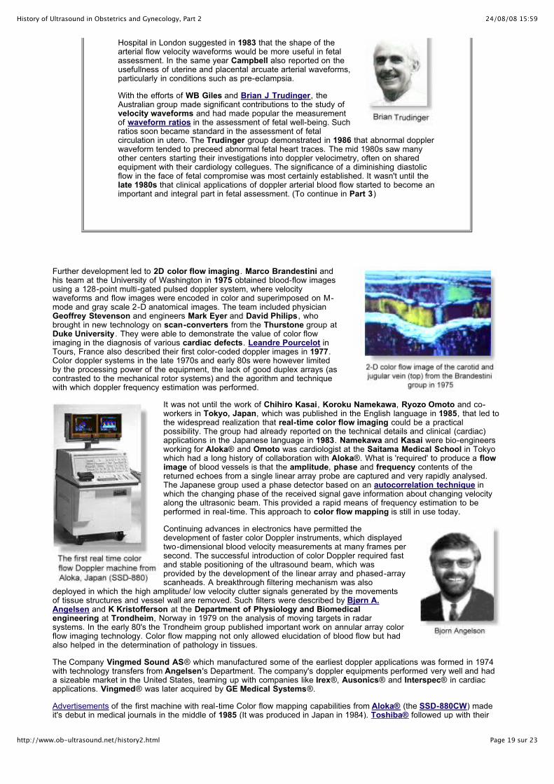
24/08/08 15:59History of Ultrasound in Obstetrics and Gynecology, Part 2
Page 19 sur 23http://www.ob-ultrasound.net/history2.html
Hospital in London suggested in 1983 that the shape of thearterial flow velocity waveforms would be more useful in fetalassessment. In the same year Campbell also reported on theusefullness of uterine and placental arcuate arterial waveforms,particularly in conditions such as pre-eclampsia.
With the efforts of WB Giles and Brian J Trudinger, theAustralian group made significant contributions to the study ofvelocity waveforms and had made popular the measurementof waveform ratios in the assessment of fetal well-being. Suchratios soon became standard in the assessment of fetalcirculation in utero. The Trudinger group demonstrated in 1986 that abnormal dopplerwaveform tended to preceed abnormal fetal heart traces. The mid 1980s saw manyother centers starting their investigations into doppler velocimetry, often on sharedequipment with their cardiology collegues. The significance of a diminishing diastolicflow in the face of fetal compromise was most certainly established. It wasn't until thelate 1980s that clinical applications of doppler arterial blood flow started to become animportant and integral part in fetal assessment. (To continue in Part 3)
Further development led to 2D color flow imaging. Marco Brandestini andhis team at the University of Washington in 1975 obtained blood-flow imagesusing a 128-point multi-gated pulsed doppler system, where velocitywaveforms and flow images were encoded in color and superimposed on M-mode and gray scale 2-D anatomical images. The team included physicianGeoffrey Stevenson and engineers Mark Eyer and David Philips, whobrought in new technology on scan-converters from the Thurstone group atDuke University. They were able to demonstrate the value of color flowimaging in the diagnosis of various cardiac defects. Leandre Pourcelot inTours, France also described their first color-coded doppler images in 1977.Color doppler systems in the late 1970s and early 80s were however limitedby the processing power of the equipment, the lack of good duplex arrays (ascontrasted to the mechanical rotor systems) and the agorithm and techniquewith which doppler frequency estimation was performed.
It was not until the work of Chihiro Kasai, Koroku Namekawa, Ryozo Omoto and co-workers in Tokyo, Japan, which was published in the English language in 1985, that led tothe widespread realization that real-time color flow imaging could be a practicalpossibility. The group had already reported on the technical details and clinical (cardiac)applications in the Japanese language in 1983. Namekawa and Kasai were bio-engineersworking for Aloka® and Omoto was cardiologist at the Saitama Medical School in Tokyowhich had a long history of collaboration with Aloka®. What is 'required' to produce a flowimage of blood vessels is that the amplitude, phase and frequency contents of thereturned echoes from a single linear array probe are captured and very rapidly analysed.The Japanese group used a phase detector based on an autocorrelation technique inwhich the changing phase of the received signal gave information about changing velocityalong the ultrasonic beam. This provided a rapid means of frequency estimation to beperformed in real-time. This approach to color flow mapping is still in use today.
Continuing advances in electronics have permitted thedevelopment of faster color Doppler instruments, which displayedtwo-dimensional blood velocity measurements at many frames persecond. The successful introduction of color Doppler required fastand stable positioning of the ultrasound beam, which wasprovided by the development of the linear array and phased-arrayscanheads. A breakthrough filtering mechanism was also
deployed in which the high amplitude/ low velocity clutter signals generated by the movementsof tissue structures and vessel wall are removed. Such filters were described by Bjørn A.Angelsen and K Kristofferson at the Department of Physiology and Biomedicalengineering at Trondheim, Norway in 1979 on the analysis of moving targets in radarsystems. In the early 80's the Trondheim group published important work on annular array colorflow imaging technology. Color flow mapping not only allowed elucidation of blood flow but hadalso helped in the determination of pathology in tissues.
The Company Vingmed Sound AS® which manufactured some of the earliest doppler applications was formed in 1974with technology transfers from Angelsen's Department. The company's doppler equipments performed very well and hada sizeable market in the United States, teaming up with companies like Irex®, Ausonics® and Interspec® in cardiacapplications. Vingmed® was later acquired by GE Medical Systems®.
Advertisements of the first machine with real-time Color flow mapping capabilities from Aloka® (the SSD-880CW) madeit's debut in medical journals in the middle of 1985 (It was produced in Japan in 1984). Toshiba® followed up with their

24/08/08 15:59History of Ultrasound in Obstetrics and Gynecology, Part 2
Page 20 sur 23http://www.ob-ultrasound.net/history2.html
SSH-65A later on in the same year. Asim Kurjak in Croatia, using the Aloka® machine was the earliest pioneer tointroduce the application of color flow doppler in fetal assessment, publishing his work in 1987. (See also Part 3).
Quantum Medical Systems®, Issaquah, Washington, a new company formed by a group ofengineers who left ATL, had started introducing the concept of real-time color dopplerimaging at the AIUM meeting in the fall of 1983. The first color images were shown at theRSNA (Radiological Society of North America) meeting in December 1984. The prototypemachine was tried out at several centers and one of the first papers was reported in theOctober 1985 meeting of the RSNA by Christopher Merritt 's group at Tulane University,New Orleans. Quantum® marketed their first machine, the QAD-1 in 1986 which producedsome very impressive real-time color flow images of the carotid and other arteries basing onthe newer array technology. They called it "AngioDynography" although the term had notsubsequently become popular. The transducer design and signal processing were described byAlfred Persson and Raymond Powis, one of Quantum 's founders in their article "Recentadvances in imaging and evaluation of blood flow using ultrasound" in 1986.
Color flow imaging made it's real impact in the United States in 1987 and in Europe in thefollowing year. ATL® after some re-organization, marketed its first color doppler machine, the
UltraMark 9 in 1988. Irex® did not make its own color doppler but marketed Aloka®'s SSD-880 in the united Statesinstead. Irex®, being acquired by the Johnson & Johnson Co®., and sold later to GE®, discontinued its line of duplexscanners. Quantum Medical Systems® was later acquired by Siemens® of Germany, who moved their mainoperations to Issaquah, Washington.
It was not until the early 1990's that the modality found it's way into the assessment of Gynecological and earlypregnancy abnormalities.
"Power doppler" or "Color power imaging" continued to develop in the 1990s. "Tissue doppler imaging" developedfurther from a revived concept with the arrival of better computational electronics. These developments had importantclinical impact on the diagnosis of malignant conditions where tissue vascularity is increased and on moving structuresother then blood flow (see Part 3).
Tissue Characterization
The ability to develop methods for signal processing of ultrasonic echoes from tissues which allow correlation of acousticparameters with tissue constituents had always been on the minds of early ultrasound inventors and reseachers such asJohn Wild, Tom Brown and Alfred Kratochwil. However, tissue characterization never became a fruitious area inultrasonic applications and had never caught on particularly in the field of Obstetrics and Gynecology. Texturecharacterization of human body tissues has never been easy to perform. Quantitative methods used included fractalmodels, multichannel methods, analysis of first and second-order statistics of texture, Gaussian Markov random fields,mean scatterer spacing and multiresolution analysis. Invented algorithms are complicated and often inefficient.
The success of tissue characterization in medical ultrasound, and particularly in Obstetrics and Gynecolgy, is largelyhampered by the presence of the inhomogeneous soft tissue medium and inconsistency of position between the tissueof interest and the insonifying ultrasound transducer (ie. abdominal wall etc.). As the acoustic wavefront propagatesthrough the inhomogeneous tissue, it is modified in amplitude and phase which often has a large and unpredictable effecton the output voltage of the receiving transducer. In addition, the tissue introduces attenuation, and multiple transmissionpaths through the soft tissue may create replicated echoes. Back-scattered signals are also degraded by speckle noise,acoustic shadowing, and system distortions present in all instrumentation. Important early researchers in this areaincluded Christopher Hill at Sutton, England; Frederick Kremkau at Yale, John Reid at Drexel and the Kossoff groupin Sydney. Up to this day, no effective technique for tissue characterization is in use in Obstetrics and Gynecology.
The safety of diagnostic ultrasound has received rigorous scrutiny since it'sinception, particularly when high power ultrasound had been used in the 1940s fordestructive and therapeutic purposes. Subsequent to a number of early reportswhich did not demonstrate any harmful effects of ultrasound insonation on humancells, Ian Donald, in co-operation with Malcolm Ferguson-Smith, director of thecytogenetic laboratory at the Queen Mother's Hospital conducted extensiveexperiments in 1967 to delineate possible harmful effects on high intensityultrasound on interphase and mitotic chromosomes and did not find any. Studies in1963 at the Juntendo Ultrasound Reasearch Center in Japan also did not revealany harmful effects on pregnant rats exposed at the maximum power of diagnosticequipments for 3 days after fertilization. Bertil Sunden in Lund, Sweden found noteratogenic effects in his thesis research in 1964. Other studies conducted inEngland by EI Kohorn in 1967 and John C. Hobbins in the United States also didnot show any appreciable cytological effects.
Important early researchers included Wesley Nyborg at the Pennsylvania StateUniversity and later at the University of Vermont; Paul Carson at the University ofColorado; Raymond Gramiak at the University of Rochester; William and Francis
Fry, Floyd Dunn and William O'Brien Jr. at the University of Illinois; Christopher Hill at Sutton, England; Marvin Ziskinin Philadelphilia; Masao Ide and Kazuo Maeda in Toyko; and Stanley Barnett and George Kossoff in Australia, to namejust a few. Much was done on the study of in-vitro effects of ultrasonic insonation at various intensities, their effects on
24/08/08 15:59History of Ultrasound in Obstetrics and Gynecology, Part 2
Page 21 sur 23http://www.ob-ultrasound.net/history2.html
heating, cavitaion and bubbles; their mechanisms of action, standardization of intensity measurements and drawing upvarious Guidelines on Biological Safety.
Researchers were also looking at immuno-suppresive effects, platelet aggregation, cell phagocytosis, DNA damage, fetalweight alterations and increase in sister chromatid exchanges in the presence of ultrasound insonation. LM Hellman'sstudy in 1970 on 400 newborns insonated before birth also reveal no increase in abnormalities in the infants. Many otherstudies followed. A Bioeffects committee was set up in the AIUM which reviews and monitors the world literature onultrasound bioeffects on a regular basis. Similar watch groups were set up by the EFSUMB and the WFUMB. A definiteharmful effect at diagnostic levels of ultrasound cannot be confirmed. Such findings give further impertus to the rapiddevelopment of ultrasonography in Obstetrics, where concerns for bioeffects safety was obvious.
In the 1980s and 90s, new studies emerged looking at the long term effects in children exposed prenatally to ultrasound.No definite adverse effects could be determined in the long term. Other activities in the United States are described here.The Bioeffects Committee of the AIUM made the following statement in 1982 and again in 1997:
" No confirmed biological effects on patients or instrument operators caused by exposure at intensitiestypical of present diagnostic ultrasound instruments have ever been reported. Although the possibilityexists that such biological effects may be identified in the future, current data indicate that the benefits topatients of the prudent use of diagnostic ultrasound outweigh the risks, if any, that may be present. "
It was widely held that the clinical use of ultrasound over the years has not established any adverse effect arising from itsexposure. While randomized clinical studies are the most rigorous method for assessing potential adverse effects ofdiagnostic ultrasound, studies using this methodology show no evidence of any effect on birthweight in humans. Otherepidemiologic studies have also shown no causal association of diagnostic ultrasound with any of the adverse fetaloutcomes studies (AIUM statements). The World Federation, European Federation and the Australasian Society also heldsimilar views. These activities and statements played an important role in the proliferation of ultrasound use inObstetrics as any slightest harmful effect would likely have constituted a major deterrant.
Read the article: "Assessing the Risks for Modern Diagnostic Ultrasound Imaging" by Dr William O'Brien, Jr. ^
An important article on the Historical Development of Bioeffects Assessment can be found here.
Read also the article: "History of the American Institute of Ultrasound in Medicine's efforts to keep Ultrasound
safe" by Wesley Nyborg
....... Image quality of real-time ultrasound scanners made steady improvements during the mid 1980's to early 90'ssecondary to the increasing versatility and affordability in microprocessor technology. Nev ertheless it was not until theearly to mid 1990's that more substantial enhancements in image quality were seen (see Part 3) .......
....... The use of ultrasonography continued to boom into the 1980s. According to statistics from the Bureau ofRadiological Health Surveys (FDA), in the United States, the percentage of hospitals using ultrasound for datingincreased from 35% in 1976 to 97% in 1982 .......
....... The World Federation of Ultrasound in Medicine and Biology (WFUMB) 1988 Meetingin Washington was preceeded by a two day Symposium on the History of Ultrasound.This event was the culmination of the sustained efforts of the AIUM Archives Committee,chaired by Barry Goldberg. Of the 500 or so delegates about 200 were recognised aspioneers. During the awards ceremony a further 40 pioneers all over the world wererecognised In Memorium. The British Medical Association (BMUS) held a similar parallelevent in Glasgow in the same year. In both meetings there were exhibition of manyhistorical ultrasound instruments .......
....... Stuart Campbell with his committee of international luminaries soon started theInternational Society of Ultrasound in Obstetrics and Gynecology (ISUOG) in 1990and held it's first world congress in the following year. He also became the founding editorof the Society's official journal: Utrasound in Obstetrics and Gynecology .......
Go to [ Part 3 ]
A list of the Landmark references : (in the order as they appeared in the text)
Gottesfeld, K.R., Thompson, H.E., Homles, J.H., Taylor, E.S. (1966) Ultrasonic placentography - a new method for placental localization. Am. J.Obstet. Gynecol. 96:538-547.Donald, I. Abdulla, U. (1967) Further advances in ultrasonic diagnosis. Ultrasonics 5:8-12.MacVicar. J. and Donald, I. (1963) Sonar in the diagnosis of early pregnancy and its complications. Br. J. Obstet. Gynaecol. 70:387-395.Willocks, J. (1962) The use of ultrasonic cephalometry. Proc. Roy. Soc. Med. 55:640.Campbell, S. (1968) An improved method of fetal cephalometry by ultrasound. Br. J. Obstet. Gynaecol. 75:568-576.Gottesfeld, K.R.(1970) The ultrasonic diagnosis of intrauterine fetal death. Am. J. Obstet. Gynecol. 108:623.
24/08/08 15:59History of Ultrasound in Obstetrics and Gynecology, Part 2
Page 22 sur 23http://www.ob-ultrasound.net/history2.html
Donald, I. Morley, P. Barnett, E.(1972) The diagnosis of blighted ovum by sonar. J Obstet. Gynaecol. Br. Commonw. 79:4 304-10.Robinson, H. P. (1973) Sonar measurement of fetal crown-rump length as means of assessing maturity in the first trimester of pregnancy. Br.Med. J. 4:28-31.Bishop, E.H. (1966) Obstetric uses of the ultrasonic motion scanner. Am. J. Obstet. Gynecol. 96:863.Carlsen, E.E. (1973) Gray scale ultrasound. J. Clin. Ultrasound. 1:193.Kossoff, G., Garrett, W.J., and Radovanovich, G. (1973) gray scale echography in Obstetrics and Gynaecology. Commonwealth AcousticLaboratories, Report No. 59, Sydney, Australia.Kossoff, G., Garrett, W.J., and Radovanovich, G. (1974) gray scale echography in Obstetrics and Gynaecology. Australas. Radiol. 18:62-111.Kossoff, G., Carpenter, D., Robinson, D., and Garrett, W.J. (1975) A new multi -transducer water coupling echoscope. Pro 2nd Eurp Congr onUltrasonics in Medicine. Kazner, E., Muller, H.R. and de Vlieger, M., eds., Munich. Abstract no. 17.Taylor, K.J.W. and McCready, V.R.(1976) A clinical evaluation of gray scale ultrasonography. Br. J. Radiol. 49:244-252.Taylor, K.J.W., Carpenter, D.A., Hill, C.R. and McCready, V.R.(1976) gray scale ultrasound imaging. The anatomy and pathology of the liver.Radiology 119:415-423.Donald, I. (1975) gray scale imaging in Sonar. Scot. Med. J. 20:177.Campbell, S and Wilkin, D. (1975) Ultrasonic measurement of the fetal abdomen circumference in the estimation of fetal weight. (1975) Br. J.Obstet. Gynaecol. 82:687-689.Robinson, D.E. and Kossoff, G. (1979) Computer processing of line mode echogram data. Proc WFUMB meeting, Miyazaki, Japan.Hofmann, D., Höllander, H.J. and Weiser, P. (1966) Neue Moglichkeiten der Ultraschalldiagnostik in der Gynakologie und Geburtshilfe. Fortschr.Med. 84:689-693.Krause, W. and Soldner, R. (1967) Ultrasonic imaging technique (B scan) with high image rate for medical diagnosis. Electromedica 4:1-5.Hofmann, D and Hollander, H.J. (1968) Intrauterine diagnosis of hydrops fetus universalis using ultrasound. Zbl. Gynaekol. 90:667-669.Griffith, J.M. and Herny, W.L. (1974) A sector scanner for real -time two-dimensional echocardiography. Circulation 49:1147.Hofmann, D., Hollander, H.J. (1978) The application of the Vidoson Ultrasonic imaging unit in Gynaecology and Obstetrics. Electromedica 4:103-105.Uchida, R., Hagiwara, Y. and Irie, T. (1971) Electro-scanning ultrasonic diagnostic equipment. Jap Med El. 58:1971.Bom, N., Lance, C.T., Honkoop J. and Hugenholtz, P.G. (1971) Ultrasonic Viewer for cross-sectional analyses of moving cardiac structures.BioMed Eng. 6:500.Bom, N.,Lance, C.T., Van Zweten, G.,Kloster, F.E. and Roelandt, J. (1973) Multiscan Echocardiography 1. Technical description. Circulation48:1066-1074.Abbowit, S.H., Jennett, R.J., Langhead, M.K., (1975) Assessment of fetal viability with multi -element real -time scanning systems. Pro 2nd EuropCongr on Ultrasonics in Medicine. Munich. Abstract no. 219.King D. L. (1973) Real-time cross-sectional ultrasonic imaging of the heart using a linear-array multi -element transducer. J Clin. Ultrasound1:196.Pourcelot, L., Pottier, J.M., Berson, M., Paniol, T.H. (1975) Fast ultrasonic imaging system (USABEL). Pro 2nd Europ Congr on Ultrasonics inMedicine. Munich. Abstract no.20.O'Brien, G.D., Queenan, J.T. (1981) Growth of the ultrasound fetal femur length during normal pregnancy. Am J Obstet Gynaecol. 141:833.Somer, J.C. (1968) Instantaneus and continuous pictures obtained by a new two-dimensional scan technique with a stationary transducer.Proceedings in Echo-encephalography.E. Kazner, W. Schiefer and K. Zulch., Eds. 234-236.von Ramm, O. T. and F. L. Thurstone. (1976) Cardiac imaging using a phased array system (I. System design). Circulation 53(2): 258-262.Kemeter P, Feichtinger W (1986) Trans-vaginal oocyte retrieval using a trans-vaginal sector scan probe combined with an automated puncturedevice. Hum Reprod 1:21-4.Feichtinger W, Kemeter P (1986) Transvaginal sector scan sonography for needle guided transvaginal follicle aspiration and other application ingynaec, routine and research. Fertil. Steril. 45; 722-725. Hackeloer B. J. (1978) Ultrasonic demonstration of follicular development. lancet i: 941.Edler, I. and Hertz, C.H. (1954) The use of Ultrasonic reflectoscope for the continuous recording of the movements of heart walls. K. Fysiogr.Sallsk. Lund. Forh. 24:40.Effert, S., Erkens, H. and Grosse-Brockhoff, F. (1957) Uber die Anwendung des Ultraschall - Echoverfahren in der Hertzdiagnostik. Deutsch MedWschr. 82:1253.Winsberg F. (1972) Echocardiography of the fetal and newborn heart. Invest Radiol 7:152-8.Baker, D.W. and Watkins, D. (1967) A phase coherent pulse doppler system for cardio-vascular measurement. Proc. 20th Ann. Conf. Eng. Med.Biol. 27:2.Reid, J.M., Spencer, M.P. (1972) Ultrasonic Doppler technique for imaging blood vessels. Science. 1972 16 176:40 1235-6 Barber, F.E., Baker, D.W., Nation, A.W.C. et al. (1974) Ultrasonic duplex echo-doppler scanner. IEEETrans Biomed Eng. 21:109.Barber FE, Baker DW, Strandness DE Jr, et al. (1974) Duplex Scanner II: for simultaneous imaging of artery tissues and flow. 1974 UltrasonicsSymposium Proceedings, IEEE Press, 744-748.Peronneau, P.A. and Leger, F. (1969) Doppler Ultrasonic pulsed blood flowmeter. Proc. 8th Int. Conf. Med. Biol. Eng. 10.Wells, P.N.T. (1969) A range-gated ultrasonic Doppler system. Med. Biol. Eng. 7:641-652.Johnson, W. L., Stegall, H.F., Lein, J.N. and Rushmer, R.F. (1965) Detection of fetal life in early pregnancy with an ultrasonic Doppler flowmeter.Obstet. Gynecol. 26:305.Ashitaka Y, Murachi K, Takemura H, et al. (1968) Analysis of the fetal heart valve with the fetal electro-cardiogram, phonocardiogram anddoppler signals. Proceedings of the 14th Scientific Meeting of the Japan Society of Ultrasonics in Medicine. 29-30. (in Japanese).FitsGerald, D.E, Drumm J.E. (1977) Non-invasive measurement of human fetal circulation using ultrasound: a new method. Br Med Journal.2:1450-1451.McCallum W.D., Williams C.S., Napel S. and Daigle R. E. (1978) Fetal blood velocity waveforms. Am. J. Obstet. Gynecol. 132:425:429.Gill R.W. Quantitative blood flow measuement in deep-lying vessels using pulsed Doppler with the Octoson. (1978) in White, D., and Lyons, E.A.Ultrasound in Medicine, vol 4, p.341.Eyer M.K., Brandestini M.A., Phillips D.J., Baker D.W. (1981) Color digital echo/Doppler image presentation. Ultrasound Med Biol 1981 7:1 21-31Omoto, R., Namekawa, K., Kasai, C. (1983) A prototype device incorporating a new thechnology for visualizing intracardiac flow.Namekawa K, Kasai C, Tsukamoto M, Koyano A. (1983) Realtime bloodflow imaging system utilizing auto-correlation techniques. Ultrasound MedBiol 1983 Suppl 2: 203-8Omoto, R., Yokote, Y., Takamoto, S. et al . (1983) Clinical significance of newly developed real -time intracardiac two-dimensional blood flowimaging system (2-D doppler). Jpn Circ J. 47:191.Kasai, C., Namekawa, K., Koyano. A. et al (1985) Realtime two-dimensional blood flow imaging using autocorrelation technique. IEEE. Trans.Sonics. Ultrason. 32:460.Angelsen, B. A. J. and Kristoffersen, K., (1979) On ultrasonic MTI measurement of velocity profiles in blood flow," IEEE Trans. on BiomedicalEng., vol. BME-26, pp. 665-671, 12.Angelsen, B. A. J, Kristoffersen, K, Torp, H, A., Brubakk, O, Sæther, O, and Myhre, H. O., (1987) Imaging of Flow in Peripheral Vessels with anAnnular Array Colour Flow Mapper in 1st Workshop internazionale di emodinamica vascolare pp. 3 - 18, Bologna.Persson, A.V., Powis R.L. (1986) Recent advances in imaging and evaluation of blood flow using ultrasound. Med Clin North Am. 70, 6:1241.
ref "Looking at the Unborn: Historical Aspects of Obstetric Ultrasound" - Witness seminar transcript. Wellcome Witness to Twentieth CenturtyMedicine. E M Tansey, D A Christie, eds. January 2000.Horace Thompson's image courtesy of the AIUM.George Kossoff's picture courtesy of Professor Salvator Levi.** Courtesy of KretzTechnik®, Zipf, Austria.*** Scottish machine, images reproduced with permission from Dr. RG Law, from his book 'Ultrasound in Clinical Obstetrics', John Wright andSons Ltd, Bristol, 1980.**** Image courtesy of Dr. Eric Blackwell, reproduced with permission.
24/08/08 15:59History of Ultrasound in Obstetrics and Gynecology, Part 2
Page 23 sur 23http://www.ob-ultrasound.net/history2.html
*^* From "Medical Diagnostic Ultrasound: A Retrospective on its 40th Anniversary", reproduced with permission from Dr. Barry Goldberg.A-scope image courtesy of the Department of Ultrasonics, Polish Academy of Science. ^ quoted from "Looking at the Unborn: Historical sapects of Obstetrical Ultrasound" - Wellcome Witnesses to Twentieth Century Medicine Vol 5,page 39.^^ See Craig M. Sonography: an occupational health hazard. JDMS 1985;Vol. 1 No. 3:121-126. Image of Aloka's 1971 real -time scanner courtesy of Aloka Co.Dr. Kato and Dr. Takemura's spectral traces are from the article: "Introduction of the Ultrasonic Doppler Technique in medicine: A historicalperspective" by Yasuhara Nimura which appeared in the journal of Medical Ultrasound 1998, 6:5-13.Prefessor Joupilla's image courtesy of Dr. Aydin Tekay at the Oulu University.- It is not possible to include all the names who have contributed significantly to the advancement of Obstetrical and Gynecological sonography,some who may have been less well-known than the others and some who may not have published so extensively in the English language.Apologies are extended to those whose contribution has not been fully credited in this article.
All original contents Copyright © 1998-2001 Joseph SK Woo. All Rights Reserved.
Search this site
Back to [ Part 1 ]
Move on to [ Part 3 ]
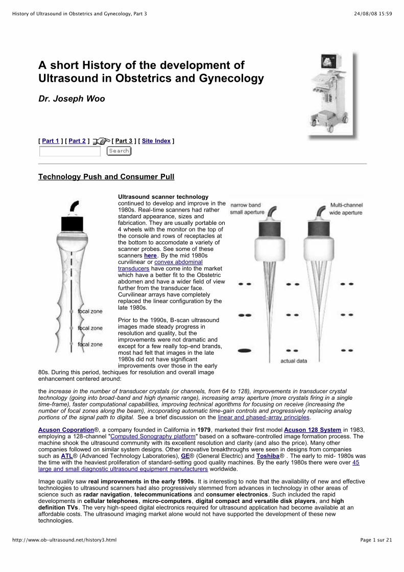
24/08/08 15:59History of Ultrasound in Obstetrics and Gynecology, Part 3
Page 1 sur 21http://www.ob-ultrasound.net/history3.html
A short History of the development ofUltrasound in Obstetrics and Gynecology
Dr. Joseph Woo
[ Part 1 ] [ Part 2 ] [ Part 3 ] [ Site Index ]
Technology Push and Consumer Pull
Ultrasound scanner technologycontinued to develop and improve in the1980s. Real-time scanners had ratherstandard appearance, sizes andfabrication. They are usually portable on4 wheels with the monitor on the top ofthe console and rows of receptacles atthe bottom to accomodate a variety ofscanner probes. See some of thesescanners here. By the mid 1980scurvilinear or convex abdominaltransducers have come into the marketwhich have a better fit to the Obstetricabdomen and have a wider field of viewfurther from the transducer face.Curvilinear arrays have completelyreplaced the linear configuration by thelate 1980s.
Prior to the 1990s, B-scan ultrasoundimages made steady progress inresolution and quality, but theimprovements were not dramatic andexcept for a few really top-end brands,most had felt that images in the late1980s did not have significantimprovements over those in the early
80s. During this period, techiques for resolution and overall imageenhancement centered around:
the increase in the number of transducer crystals (or channels, from 64 to 128), improvements in transducer crystaltechnology (going into broad-band and high dynamic range), increasing array aperture (more crystals firing in a singletime-frame), faster computational capabilities, improving technical agorithms for focusing on receive (increasing thenumber of focal zones along the beam), incoporating automatic time-gain controls and progressively replacing analogportions of the signal path to digital. See a brief discussion on the linear and phased-array principles.
Acuson Coporation®, a company founded in California in 1979, marketed their first model Acuson 128 System in 1983,employing a 128-channel "Computed Sonography platform" based on a software-controlled image formation process. Themachine shook the ultrasound community with its excellent resolution and clarity (and also the price). Many othercompanies followed on similar system designs. Other innovative breakthroughs were seen in designs from companiessuch as ATL® (Advanced Technology Laboratories), GE® (General Electric) and Toshiba® . The early to mid- 1980s wasthe time with the heaviest proliferation of standard-setting good quality machines. By the early 1980s there were over 45large and small diagnostic ultrasound equipment manufacturers worldwide.
Image quality saw real improvements in the early 1990s. It is interesting to note that the availability of new and effectivetechnologies to ultrasound scanners had also progressively stemmed from advances in technology in other areas ofscience such as radar navigation , telecommunications and consumer electronics. Such included the rapiddevelopments in cellular telephones, micro-computers, digital compact and versatile disk players, and highdefinition TVs. The very high-speed digital electronics required for ultrasound application had become available at anaffordable costs. The ultrasound imaging market alone would not have supported the development of these newtechnologies.

24/08/08 15:59History of Ultrasound in Obstetrics and Gynecology, Part 3
Page 2 sur 21http://www.ob-ultrasound.net/history3.html
(Model number of scanners made after 1980 from important manufacturers are listed here with the year in which theywere marketed).
The new developments in the 1990s which has lead to some real enhancement in image quality and resolution
include:
1. The entire signal processing chain becomes digital. The entire signal chain which includes:
[ the transducer ] --> [ beamformer ] --> [ signal processor ] --> [ scan converter ] --> [ Monitor ]
all operate under digital electronics.
Previously the beamformer (employinganalog delay lines) and the signalprocessing stages are usually analog intheir operation. The digital change-overwas based on the very powerfulcomputer platforms that were onlyavailable after the mid 1990s. Theprocessor in the newer highendmachines has the power equivalent of
roughly 40 Pentium tm processors,executing some 20 to 30 billionoperations per second. Most of theprocessing are also programmablesoftware-based rather than hardware-based and allow for much more versatilityand finer adjustments in the manipulationof beam signals. Signals from and to thetransducer elements are digitized beforeany signal processing, which is one ofthe most important advancement inultrasound technology in the 90s. It opened the venue for dealing with some of more difficult areas inultrasound physics.Superfast digital beamformers allow for many times the number of focal points along the beam andproduce microfine focal points on receive to the size of a screen pixel. Digital beamforming also reducesnoise in the signal processing by several hundred folds producing a much cleaner picture.
2. Extensive use of refined broad-band wide aperture transducers, improving both definition oftissue textures and dynamic range. With wide aperture transducers, transmit and receive apodization alsoallowed for the electronic reduction of the lateral array elements (sidelobes). In the early 1990s there wasmuch improvements in transducer material design and fabrication technology allowing for higher frequencytransducers, improved sensitivity and contrast resolution. The number of channels in high-end systemswent up to 256 and more recently to 512 and 1024 (2-D arrays) in several high-end systems allowing forextremely wide aperture on transmission and reception. In ultrasound physics, the lateral resolution is theproduct of the wavelength and the f-number. The f-number equals the depth of the returning echodivided by the aperture of the beam. (the aperture of the beam is the width of the number ofsimultaneous firing transducer elements in the array, that means the larger the aperture the moreelements are fired simultaneously). Therefore lateral resolution will be best (smallest) if there is a largeaperture and short wavelength (higher frequency).
Too large an aperture will slow the frame rate considerably and requires very fast computation and parallelprocessing. This has been made possible with the more recent digital electronics and the very powerfulsuper-processors (see above). Many slightly older ultrasound systems are capable of using low f-numberson reception at an affordable cost. However, they often employed large f-numbers on transmit in order tocover a large area. Significant improvement in lateral resolution requires low f-numbers both on transmitand receive. With the new 'very wide' aperture beamformer (often up to 128 channels), the transmit andreceive f-numbers are lowered. The resulting improvements in lateral resolution can be as much as 4times.
3. The phase data in returning ultrasound echoes, inaddition to the amplitude data are processed in what isknown as coherent image processing. The techniqueproduced twice the amount of data from which to createultrasound images of high resolution. The frame rate isalso increased. The late 1990s has also seen transducerdeveloping into 2D arrays which is made up of largenumber of elements arranged in rows and columnsacross the face of the transducer. Focusing occurs in twodirections which produced a finer and clearer definition inboth planes eliminating artifacts from adjacent tissue planes which may produce the partial volume effect.
4. The advent of tissue harmonic imaging. The technology, which has emerged as a major imagingtrend in the last 4 years of the 1990s, made used of the generation of harmonic frequencies as anultrasound wave propagates through tissue, dramatically reducing near field and side lobe artifacts. In anutshell, tissue harmonic Imaging made use of lower frequency sound waves to improve penetration,

24/08/08 15:59History of Ultrasound in Obstetrics and Gynecology, Part 3
Page 3 sur 21http://www.ob-ultrasound.net/history3.html
while receiving and processing only the higher frequency echoes produced by the body's inherentharmonic characteristics. This process can reduce clutter and improve image clarity significantly. Asultrasound waves propagate through tissue, there is non-linearities in sound propagation that graduallychange the shape of the wave, a shape change that can only result from the development of harmonicfrequencies within the wave. There are no harmonic frequencies present at the transducer face. Theydevelop gradually as the wave propagates through tissue, and so in the near field there is very littleharmonic energy available for reflection from tissue. Since the near field is a source of much of the artifactin the ultrasound image, selective display of harmonic energy will show dramatically less near-fieldartifact. The strength of the harmonic energy generated is proportional to the square of the energy in thefundamental wave. Most of the harmonic energy results from the strongest part of the beam, and weakerportions of the beam (side lobes, for example) generate relatively little harmonic energy. selectiveharmonic imaging will yield a dramatically cleaner contrast between adjacent tissue structures. It is asonologist dream come true. The black is black and the white is white. Much of the fuzzy picture noisethat have made diagnosis difficult are gone.
The development of harmonic imaging would not have been possible until the late 1990s as there must beexcellent beam linearity on transmission and super sensitivity and dynamic range on receive to display theharmonic energy without an unacceptable amount of noise, as the harmonic signals are always much lessin amplitude than the original fundamental signal. There must also be a very selective and fast digital filterwithin the receiver, to exclude the large percentage of the fundamental signal. Harmonic imaging isparticularly useful in obese patients.
From left to right: Changes in image quality from 1985, 1990 to 1995 respectively.There were improvements in spatial and contrast resolution, background noise reduction,
dynamic range, and near and far field visualization.

24/08/08 15:59History of Ultrasound in Obstetrics and Gynecology, Part 3
Page 4 sur 21http://www.ob-ultrasound.net/history3.html
More significant improvements came after the mid-1990's. This image from ATL® * demonstrating fetal spine and cord.
Ultrasound scanners came into different categories according to their performance and price.From the early 1980s, scanners have started to move into clinics and private offices and there isa trend to decentralise ultrasound services all over the world. Acceptance and demand from thelay public have also increased exponentially coupled with increased utilization by various medicalspecialties and sub-specialties. Standards and quality of scans became an emerging problemnot seen in other areas of medical imaging, where Radiogists received the relevant training andunderwent apppropriate examinations before running the service. Obstetricians were simplyusing the scanner probe as a torch to "look inside" the uterus. Standards varied and mis-diagnosis was not uncommon. Obstetricians and Gynecologists took on the fact that they are themore suitable persons to do the scans as compared to their radiological colleagues. Specialtraining centers and accreditation boards were gradually set up by the health authorities in theUnited States, Australia, Europe and other countries.
In the United States, an impetus for the development of ultrasonographywas that medical insurance agencies such as "Blue Cross" had started re-imbursing ultrasonographic examinations since 1966. Medical sonographers also took up much ofthe responsibilities of the scans and sonography itself was recognised as a separate profession bythe American Medical Association in 1974, the first sonography program being accredited in 1981 bythe Joint Review Committee for Education in Diagnostic Medical Sonography ( the JRC-DMS).The American Registry of Diagnostic Medical Sonographers was founded in 1974 by theSociety of Diagnostic Medical Sonographers (SDMS). Joan Baker was the first Chairperson. TheAIUM gradually went beyond developing guidelines and established an ultrasound practiceaccreditation program. Since 1996, this voluntary program has accredited over several hundredultrasound practices. To achieve accreditation in obstetrics-gynecology, physicians andsonographers must interpret a minimum of 170 ultrasound scans per year. In Australia , the Council
of the Australian Society (ASUM) awards the Diploma of Diagnostic Ultrasound and the Diploma of MedicalUltrasound to medical practitioners and sonographers who sucessfully pass the examination.
And in both a 'Consumer pull' and 'Technology Push' situation the diagnostic application of ultrasound in the field ofObstetrics and Gynecology continued to expand into new horizons. In 1975, that is before the advent of real-timeequipments, in the United States there were only 5 legitimate indications in obstetric sonography: ! measurement of thebiparietal diameter (and other dating purposes), ! determining amniotic fluid volume, ! diagnosis of early pregnancyfailure, ! evaluation of multiple gestations and ! placental localisation. The indications have since the early 80's expandedinto at least 2 dozen, including most notably the accurate evaluation of fetal growth and the diagnosis of fetalmalformations.
Fetal biometry developed and 'flourished ' in the1980s as accurate fetal measurements do notrequire the prerequsite of very high resolutionequipments. At least two dozen measurementswere "invented" to assess gestational age and fetal

24/08/08 15:59History of Ultrasound in Obstetrics and Gynecology, Part 3
Page 5 sur 21http://www.ob-ultrasound.net/history3.html
size, each claiming their unique usefulness.Nevertheless by the mid 1980s only a fewparameters were considered as standardmeasurements and ones that had "stood the test oftime". These include the crown-rump length (CRL),the biparietal diameter (BPD), the headcircumference (HC), the femur length (FL) and theabdominal circumference (AC). Many othermeasurements were considered useful only insituations where fetal dysmorphology was inquestion.
Take for example the BPD, at least 200 charts are inused in the 1990s in different parts of the world(information supplied by commercial ultrasoundscanner venders who set up the charts for theirclients). Early workers in the United States who havepublished extensively on fetal biometry include RudySabbagha at the Northwestern University, Chicago,Alfred Kurtz at the Thomas Jefferson University,John Hobbins at Yale, Charles Hohler in Phoenix, Arizona, Peter Cooperberg at the University of British Columbia,Canada, David Graham and Roger Sanders at Johns Hopkins, Baltimore and Frank Hadlock and Russell Deter at theBaylor College of Medicine, Houston, Texas. There are others from Britain and Europe like Hansmann, Jouppila, Kurjakand Levi . Fetal biometry was explored from many different perspectives and in different populations.
The abdominal circumference measurement which was described by Campbell and Wilkin in 1975 remained themainstay measurement in the evaluation of fetal growth and nutrition. The assessment of gestational age andintrauterine growth retardation using ultrasonic parameters was the subject of a huge number of research papers.Fetal growth analysis and charting were also performed on desktop personal computers (PC) using commercial or home-made proprietory softwares.
! Stuart Campbell published the first BPD chart in 1971. Since then, Charts and Tableshad become an important and integral part of Obstetric practice, at which Obstetricians
and Gynecologists were slowly getting used to. !
Estimation of in-utero fetal weight basing on the combination of the biparietal diameter and the thoracic circumferencewas first reported in 1965 by the Thompson group in Denver, Colorado. He reported an accuracy of within 300 grams in66% of the weight estimates. This was followed by work from Garrett in Australia, Hansmann in Germany and Campbellin England. In 1977 the Hobbins' group at Yale published one of the most important papers in fetal biometry,"Estimation of fetal weight by computer-assisted analysis of fetal dimensions" which had started in the next 10 to15 years, an almost non-stopping search all over the world for computer-generated models of fetal weight determinationbasing on multiple fetal parmeters.
Similarily the craving to produce normograms forincremental growth of every 'measurable' part of thefetal body never stopped. One can find gestational-age normograms for parameters such as theBinocular diameter (Mayden K et al, 1982, Jeanty Pet al, 1982), thigh circumference (Deter et al, 1983),clavicular length (Yarkoni et al, 1985), fractionalspine length (Li DF et al, 1986), fetal foot length(Mercer BM et al, 1987) or fetal ear length (BirnholzJC et al, 1988); and charts for parameters such ascerebral ventricular width, cardiac chamberdimensions, chest circumference, limb lengths,renal and calyceal sizes that were of great value in
the assessment of specific fetal anomalies. In 1987 E Albert Reece at Yaledemonstrated the usefulness of the trans-cerebellar diameter as a growth-independent parameter to assess gestational age.
Among many others, the comprehensive normogramsof Frank Hadlock and Russell Deter from the BaylorCollege of Medicine, Houston, Texas were widely used.The group had in particular incorporated the femur length measurement into the calculationsand popularised the concept of limb length/ trunk circumference ratios in the assessment of fetalgrowth.

24/08/08 15:59History of Ultrasound in Obstetrics and Gynecology, Part 3
Page 6 sur 21http://www.ob-ultrasound.net/history3.html
In the last 40 years or so in the development of ultrasonic fetal biometry, there has been mucheffort among the obstetric and ultrasound community to devise and develop fetal measurementsthat would be able to distinguish between fetuses that are small because of nutritional reasonsand those fetuses that are "by nature" small. Head, limb versus abdomen ratios were oncethought of be of promise but as it was noted that the growth-retardation process also affectedfetal head and limb growth in varying degrees, the value of these ratios did not stand up to their
initial expectations. Up to this day, there is still not one or several size measurement parameter in combination that canunequivocally diagnose growth retardation in the fetus when a woman is seen for the first time in the later part ofpregnancy.
Visualization of the the fetal yolk sac with the real-time scan was first described by Eric Sauerbrei and PeterCooperberg in Vancouver, Canada in 1980. It is interesting to note that perhaps because of its size (and hence thedifficulty to visualize with existing equipments at that time) its significance and usefulness in early pregnancy failures wasnot discussed until much later in the second half of the 1980s.
The diagnosis of fetal malformations obviously received the enormous attentionthat was deserved and findings of many abnormalities diagnosable by ultrasoundhave been described.
Ian Donald included a case of hydrocephaly in one of his early "introduction"ultrasound papers in 1961, which demonstrated "tissue interface within the body byultrasonic echo sounding". In Bertil Sunden 's thesis in 1964 there was descriptionof the diagnosis of anencephaly in the third trimester using on the bi-stableDiasonograph. In 1968 D Hofmann and Hans Hollander in Germany reported on9 cases of 'hydrops fetus universalis' diagnosed with the Vidoson (see Part 2)and William Garrett in Sydney reported the diagnosis by ultrasound of a fetuswith polycystic kidneys using the CAL echoscope in 1970 (see also Part 1).These two papers were probably the two earliest papers describing formally thediagnosis of a congenital anomaly using ultrasound. Both reports were about casesin the third trimester and resulted in fetal death.
The diagnosis and management of a 17 weeks anencephaly was reported as early as 1972 by Stuart Campbell usingstatc B-mode equipment. This was followed by the diagnosis of spina bifida in 1975. Both reports had appeared aslandmark papers in the Lancet. They were the first cases of such conditions in which a correct diagnosis by ultrasoundhad effectively led to a termination of pregnancy. Manfred Hansmann in Bonn, Germany and John Hobbins at yalewere among others, early pioneers in the ultrasonographic diagnosis of fetal malformations. With the advent of better real-time scanners, many more malformations were diagnosed, abeit in the late second trimester when fetal organs becomemore discernible on the scans. A review published in 1981 (Stephenson and weaver) reported that around 90 differentfetal malformations had been diagnosed by ultrasound.
Common anomalies that were considered "straight forward" to diagnose at that timeincluded anencephaly, hydrocephaly, exomphalos, duodenal atresia, polycystickidneys, hydrops fetalis and limb dysplasias. More difficult areas for diagnosis ofmalformations were the fetal face , the fetal extremities and the fetal heart. Thediagnostic accuracy progressively improved with more experience and betterresolution machines. With the advent of the newer high-resolution scanners andthe transvaginal transducer the diagnosis of these and other more subtle conditionswere achieved, and particularly at an earlier gestation, moving from the thirdtrimester of pregnancy to the second and later on to the first trimester in the latterhalf of the 1990's. Fetal trisomies, spina bifida and the more subtle cardiacanomalies were among the many examples. So-called soft signs andsonographic markers for chromosomal anomalies (see below) were started tobe described.
! The ability to recognise and follow up in utero of all these malformations by ultrasoundhas opened up further the entire avenue of "Prenatal Diagnosis" and has markedly
enhanced and pushed forward the study of congenital abnormalities among obstetricians,pediatricians, geneticist, pathologist and other allied specialties. All of a sudden,
obstetricians started to learn about so many congenital malformations that they have noteven heard of. !
The diagnosis of fetal cardiac malformations gained footin the early 1980s. Pioneers included the Wladimiroffgroup in Rotterdam, the Netherlands; the Hobbins group(Charles Kleinman, Greggory Devore, Joshua Copel,Peter Grannum .... ) at Yale; Lindsey Allan at Guy'sHospital, London (now in New York); L W Lange, DavidSahn at Portland, Oregon; Kathryn Reed at Tucson,Arizona and Beryl Benacerraf at Harvard.

24/08/08 15:59History of Ultrasound in Obstetrics and Gynecology, Part 3
Page 7 sur 21http://www.ob-ultrasound.net/history3.html
The first real-time fetal heart images and quantitative datawere published by the Lange, Sahn and Reed group inTuscon, Arizona in 1980. Allan published her
echo/anatomical correlates in the same year. Allan, a pediatric cardiologist,described systematically real-time normal and abnormal ultrasonic anatomy ofthe fetal heart which laid the foundation for subsequent studies. Using ultrasonic equipment available in the early 1980sand much painstaking enthusiasm and skill, she had very importantly shown that real-time cross-sectional study anddiagnosis of fetal cardiac anomalies in utero in the second trimester was a distinct possibility and would no doubtdevelop into a distinctive diagnostic science.
The improvements in diagnostic capabilitiesthat came with the 'new technology'scanners in the 90's had tremendousimpact on fetal cardiac diagnosis. Theusefulness of direct doppler interrogation offetal intracardiac flow was firstdemonstarted in 1985 by Dev Maulik andNavin Nanda (Professor of Cardiology) atthe University of Alabama. Allan and Reedfollowed up with more publications. TheMaulik group further demonstrated thevalue of color doppler in fetal cardiac
studies in 1986. Greg Devore soon popularised the use of dopplercolor flow mapping in the assessment of fetal cardiacmalformations and particularly in a screening situation in 1987.The use of color doppler has become indispensible in the diagnosisof more complicated cardiac malformations. By the late 1990s, thediagnostic accuracy of the nature of complex cardiac malformationsin utero can be as high as 95 percent.
Read also: the American College of Cardiology Position Statement on Doppler Echocardiography in the Human
Fetus prepared by Charles Kleinman, James Huhta, and Norman Silverman which covers some of the historicalaspects of fetal doppler echocardiography.
The accuracy of diagnosing fetal malformations in a "high-risk" populations have been variously reported in the early1980s right up to the early 1990s. These included some classic reports from Kurjak in Croatia (1980), Campbell andPearce in London (1983), Gembruch and Hansmann in Germany (1984), Sabbagha in the U.S. (1985) and a goodnumber of other authors. Diagnostic accuracies in "targeted" scans progressively increased with improvement ininstrumentations and accumulation of knowledge about the ultrasound characteristics of the various anomalies.
With improved resolution in the new equipment, diagnosis of fetal cardiac anomalies have moved into the late firsttrimester. All the improvements in machinery and earlier detection of abnormal structures in the fetus have neverthelessbrought along with it "false positives" and difficult-to-be-sure-what-will-happen diagnosis that generate much undueanxiety in patients. Such has far-reaching effects on a woman's perception of child-bearing. Researchers try very hard todetermine the course of events for a particularly abnormality or the implication and prognosis of a certain finding onultrasound examination so that proper counselling can be done to alleviate anxiety and uncertainty from the diagnosis.
"Textbooks" in Obstetrical and Gynecological sonography had emerged outof necessity because of the large amount of information that had becomeavailable and as reference materials of those sitting for examinations. Bookshad gone from being just overviews and atlases to systematic discussions ofultrasound techniques and findings. Notably the books by Peter Callen at theUniversity of California, San Francisco (1983) and Sanders/James at JohnsHopkins were popular and represented two of the earliest standard textbooksin the field. Multiple authorship contributed to the excellence of these texts.Many other texts followed, some devoted mainly to special topics such asfetal anomalies or doppler ultrasound.
The advent of the real-time scanners also prompted research into body movements andbreathing movements of the fetus The study of fetal breathing movements (FBM) was firstsuggested by Geoffrey Dawes and K Boddy at the Nuffield Institute of Medical Research, OxfordUniveristy, England, in the early '70s, in that the presence or absence of breathing movements,theiir amplitude and intervals will be indicative of fetal well-being. Much research into these areascame from the Karel Marsal group at the University Hospital at Malmo, Sweden, theTchobroutsky group at the Maternite de Port-Royal, paris, the Wladimiroff group at Rotterdam,and the Brian Trudinger group in Australia. all of them having switched to the use of real-timeapparatus in the early to mid '70s. Wladimiroff demonstrated in 1977 that maternal hyperventilationdecreased fetal breathing. The quantitative documentations of fetal breathing movements howeverrequire elaborate equipments, and was very time-consuming, so much so it would be difficult to be

24/08/08 15:59History of Ultrasound in Obstetrics and Gynecology, Part 3
Page 8 sur 21http://www.ob-ultrasound.net/history3.html
incoporated into clinical practice. The results also have wide overlap between positives and negatives. The advent of real-time had also raised hopes of being able to study physiological responses , sensations and behavior in the fetus.Again the assessment were time consuming and results were often equivocal, which made them unsuitable as clinicaltests. Jason Birnholz at Harvard published several pioneering papers in these areas including the assessment of fetalmovement patterns as a possible means of defining neurological developmental milstones in-utero and the developmentof fetal eye movements and possible 'dream states' in the feus.
The study of FBM and FM patterns did not gain further popularity although the semi-quantitative counting of FBMs and thedocumentation of 'adequate" fetal body movements were popularised further in fetal biophysical profile scores madepopular by Frank Manning and Lawrence Platt in Winnipeg and Los Angeles, who had started studies into FBMs roundabout the same time as their European counterparts. In 1980, Manning and Platt reported on the important finding that areactive cardiotocographic finding was just as predictive as the presence of FBMs or total fetal movements.
Spectral doppler or doppler velocimetry (thestudy of doppler waveforms), had evolved tobecome a standard tool in the late 1980's in theassessment of fetal wellbeing and compromise.(See Part 2 for more of its early development).
Its advent in Obstetrics in the mid 1980s hadfascinated (or perhaps misguided) many as thenew promise in fetal assessment. The absenceand reversal of end-diastolic flow in theumbilical arteries in severely compromisedfetuses were striking demonstration of fetalpathophysiology , so was the finding of clear and
unequivocal increase in diastoic flow in the middle cerebral arterialwaveforms in mounting fetal hypoxia. It has also become clear thatumbilical doppler velocimetry does not correlate with fetal weight in utero,nor is useful as a screening procedure.
Stuart Campbell's group at the King's College Hospital in London reported in 1983 theevaluation of utero-placental flow velocity waveforms in compromised pregnancies withduplex doppler, and described the 'frequency index profile'. In 1984, P Reuwer in Utrecht, theNetherlands first discussed the ominous significance of absent end-diastolic flow in theumbilical artery. Further work from the Campbell group, including work from Gerald Hackettand T Cohen-Overbeek in 1986 and 1987 clearly demonstrated the prognostic significance ofabsent end-diastolic flow in the fetal descending aorta. In 1986, Brian Trudinger in Australiademonstrated that abnormal doppler waveform patterns tended to preceed abnormalcardiotocographic traces. In the same year the Wladimiroff group reported the value of middlecerebral artery waveforms in the assessment of severely compromised fetuses. Sanjay Vyasworking at King's College Hospital in England described the use of renal artery waveforms in1989. The value of fetal Venous blood flow in the assessment of fetal compromise was firstsuggested by Torvid Kiserud in Bergen, Norway in 1991. Giuseppe Rizzo at the Universita diRoma Tor Vergata in Italy furthered expounded the usefulness of the ductal venus velocimetry
in fetal acidemia and cardiac decompensation. In 1987, Asim Kurjak introduced the use of color flow doppler in fetalassessment.
Doppler ultrasound became a standard and indispensible tool in the evaluation of progressive fetal anoxia (the umbilicalartery), compensation and decompensation (the middle cerebral artery), acidosis and progression to cardiac failureand emminent fetal death (the ductus venosus). It has also been employed in the assessment of women at risk of pre-eclampsia and utero-placental arterial compromise, leading to early and effective therapeutic intervention. It is ofinterest to note that historically, these velocimetric parmeters have appeared each a number of years apart with increasingsophistication of the apparatus. By the beginning of the 1990's, most mid- to highend ultrasonic equipments hadincoporated duplex doppler as standard facility. In the mid 90's, color flow mapping had also found its way into mostmid- and highend machines. Aside from aiding cardiac diagnosis (see above), impressive flow images were oftenpopularly reported such as those found in Vasa Previa and the fetal Circle of Willis.
! Doppler velocimetry is not only a fascinating demonstration of fetal physiology thatprovides a vital assessment of fetal well-being, but will likely change the concept ofroutine antenatal care by picking up placental insufficiency at the earliest stage. !
As early as 1967, and basing on theVidoson, Hofmann and Hollander inGermany had discussed the importance ofplacental localization using ultrasoundbefore amniocentesis. Jens Bang andAllen Northeved in Copenhagen describedultrasound-guided amniocentesis in 1972.In the mid 1970s to early 80s, geneticamniocentesis was largely performedunder static B-scan ultrasound guidance. Anultrasound scan was performed to locate a

24/08/08 15:59History of Ultrasound in Obstetrics and Gynecology, Part 3
Page 9 sur 21http://www.ob-ultrasound.net/history3.html
feasible pocket of amniotic fluid before atap, which was done basically without actually seeing the needle tip going into the fluid pocket. With the advent of real-time scanners, a small number of centers had in the late 1970s started to perform amniocentesis by simultaneouslyvisualizing the puncture needle tip on the scanner monitor. One such pioneer was the Birnholz group at Harvard whoused an early phased array for the purpose. Needle-guide adapters soon became available from ultrasoundmanufacturers which could be coupled to the linear array or phased array sector probes where the needle passed througha fixed path either parallel or at an angle to the ultrasonic beam. These were cumbersome to use however, particularly ina busy setting. They also had serious problem of keeping the equipment sterile. The adapters may also increase the riskof traumatisation as it did not allow for the 'desired' and sensitive placement of needles.
Many centers started to do it freehand with anassistent holding onto the transducer probethat was commonly wrapped in a sterileadhesive drape. In 1984, WolfgangHolzgreve in Basel, Switzerland described alarge series of over 3000 'freehand'amniocentesis with low complication rate.Similar experience was also reported byLawrence Platt in Los Angeles, who
emphasised on the need for the transducer probe to be manipulated by the sameoperator which resulted in better hand-eye co-ordination. In the following year,Roberto Romero at Yale formally described the single operator two-handstechnique in amniocentesis and the reduction in the number of multiple taps and bloody taps associated with theprocedure. Most centers soon adopted this single operator technique, which had become popular because of itconvenience and effectiveness. Chorionic Villus sampling (CVS) also relied heavily on sonographic guidance. Z Kazyand his group in the USSR reported in 1980 fetal sexing and enzyme assay on chorion biopsies taken at 6 -12 weeks'gestation, using either an endoscopic or ultrasound-guided approach. RH Ward in London and Bruno Brambati in Milanboth reported transcervical CVS under ultrasonic guidance in 1983. Brambati reported the success rate of obtainingchorionic villi rose from 75% without ultrasound to 96% with ultrasound guidance. Danish investigators Steen Smidt-Jensen and N Hahnemann first described the ultrasound-guided transabdominal approach in 1984. Other adjunctiveultrasonic techniques were reported by the Brambati group and the Golbus group in San Francisco in 1985.
The Beryl Benacerraf group at Harvard (se below) reported the feasibility of early amniocentesis (11-14 weeks) in1988. In 1990, the same group reported an early fetal loss rate of over 2.3%. Several important reviews in the mid-90sconfirmed this high incidence of fetal loss. The practice has for this reason not gained general acceptance.
Read here a short history of Amniocentesis, fetoscopy and chorionic villus sampling.
Other interventional intra-uterine diagnostic and therapeutic procedures alsostarted to catch on. After fetoscopy, ultrasound-guided pure fetal bloodcordocentesis was pioneered in France in 1983 by Fernand Daffos. Purefetal blood was aspirated in-utero at around 18 weeks from the umbilicalvein near the placental insertion of the cord using a twenty gauge needleunder ultrasound guidance. Their group reported the first case ofHaemophilia A diagnosed in-utero by this method. The procedure was alsopopularised around the same time in England by the Stuart Campbell andCharles Rodeck group at King's College Hospital. The Hobbins group atYale described their technique in 1985 and called the procedurepercutaneous umbilical blood sampling (PUBS). This replaced bloodsampling via fetoscopy which the group had pioneered in 1974. Kypros
Nicolaides at King's developed the single operator two-hands method and became a leading figure in cordocentesisexploring many important aspects of fetal physiology and pathophysiology. With the advent of color flow mapping, thetechnique has become even more accessible. In 1988, Umberto Nicolini, working with Charles Rodeck at the QueenCharlotte's Maternity Hospital in London, first described fetal blood sampling from the intrahepatic portion of the umbilicalvein in the fetus, as an alternative procedure in cases where cord needling was unsuccessful. In the late 1980s fetoscopyhas mainly been reserved for tissue or organ sampling, and fetal blood sampling are always done via the ultrasonic-guidance needle procedure. The commonest indication of fetal blood sampling has evolved to become one of quickconfirmation of abnormal karyotype in the 22-24 weeks fetus, when a chromosomal abnorrmality has been suggestedon ultrasound scan.
Various in-utero drainage procedures under ultrasound guidance were described in the late 1980s such as the drainage ofChylothorax and hydronephrosis, and the treating of fetuses by transfusion into the umbilcal vessels. Selectivereduction of the number of fetuses in cases of triplet or quadruplet pregnancies was first described by Richard Berkowitzin New York in 1988, using intra-cardiac potassium chloride injections given under ultrasonic control. Vesico-amnioticshunt placement was described by the Mitchell Golbus group (with Michael Harrison, Roy Filly , Peter Callen .... ) inSan Francisco in 1982. The group became one of the most important forerunners in fetal surgery and continued tomake many new fetal surgical innovations. In that year, they published their multicenter classic paper "Fetal Surgery1982" in the New England Journal of Medicine.
Read here a short history of Amniocentesis, fetoscopy and chorionic villus sampling.
With improved resolution from transvaginal sonography,Mark Cullen at Yale first reported in 1990 a large seriesof congenital anomalies detected in the first trimester

24/08/08 15:59History of Ultrasound in Obstetrics and Gynecology, Part 3
Page 10 sur 21http://www.ob-ultrasound.net/history3.html
using transvaginal ultrasound, and pointed out theimportance of a good understanding of normal embryonicdevelopment in such diagnosis. Many studies followedaround the same time reporting on the usefulness of thefirst trimester transvaginal scan in the evaluation of fetalanomalies. Moshe Bronshtein, working in Haifa, Israel,described extensively since the early 1990s results oftransvaginal sonography in the first trimester. A similar
report to Cullen's also appeared in 1990. Ilan E. Timor-Tritsch, working in Israeland later on at New York University, followed on with many reports on fetalanatomy and anomalies systematically studied using high resolution transvaginaltransducers in the first trimester, opening up convincingly a new area in fetal ultrasound diagnosis, that of "sono-embryology". Timor-Tritsch was also credited for organizing the first three transvaginal ultrasound courses in theUnited States.
As ultrasound became a widely available and popular investigation, it contributed heavily to several population screeningprograms that took place between the late 70's and the 90's. The first was the Maternal serum alpha-feto protein(MSAFP) screening programs for the detection of neurotube defects (NTD). It started in the United Kingdom in the late1970s, notably in parts of Scotland, where the prevalence of NTDs was high.
In 1972, David Brock and R Sutcliffe measured the AFP values in the amniotic fluid of 31 pregnancies withanencephaly and 6 pregnancies with spina bifida. All of the cases of anencephaly and most of those with spina bifidademonstrated markedly elevated AFP levels. This was a landmark 'discovery' in the history of prenatal diagnosis. In 1974,Nick Wald and co-workers at the University of London reported in the Lancet maternal serum AFP levels in 7pregnancies with open neural tube defects which were significantly higher than that in 14 other controls. This led to theidea of measuring MSAFP in screening for NTDs. The 19-centers U.K. Collaborative study in 1977 subsequentlydemonstrated the utility of this test for prospective open neural tube defect screening. Similar results were arrived at inlarge-scale studies in the United States in 1979. By 1984, MSAFP screening had also become part of standardantenatal care in the U.S.
As scanner resolution and sonography skill improved, ultrasound gradually replaced amniocentesis in the diagnosis ofscreened-positive cases. By the mid-1990s amniocentesis is often not performed in patients with elevated MSAFP levels.Either a positive or negative diagnosis is made basing solely on ultrasound findings.
The second was the routine fetal scan at 20 weeks which had progressively become anintegral part of antenatal care in the early 1990's.
In the late 1970s a number of large scale population ultrasound screening studies havebeen described variously from London, Germany, Brussels, Sweden, Norway, Finland and othercountries in Europe and in the United States. At least 20 similar large-scales studies werereported up to the year 1990.
There is apparently a distinct difference in the attitude towards routine ultrasound screeningbetween the Europeans and the Americans. Routine screening scans were introduced inGermany in 1980, in Norway in 1986 and in Icelend in 1987. The scans basically try to date thepregnancy, exclude twins and detect any fetal malformations that may be present. In the U.S.,routine scans in pregnancy has however been looked upon with much controversy and theircost-effectiveness and validity in improving 'quantifiable' perinatal outcome has not been
firmly established. Nevertheless, those who argue for a routine scan would claim that parents have a natural desire toknow if any fetal congenital anomaly is present or if there is any health compromise in the fetus. Being able to reassure theparents is a natural part of prenatal care.
Two important large scale studies emerged to address the issue: the RADIUS study (Routine Antenatal DiagnosticImaging with Ultrasound) with a cohort of 15,000 low-risk pregnancies in the United States in 1993 and the EurofetusStudy in Europe in 1997 in which 200,000 low-risk pregnant women in 60 hospitals had obstetric ultrasound examinationsperformed in centers proficient in prenatal diagnosis. Salvator Levi was the Project leader of the Eurofetus StudyGroup and a strong proponent for routine screening. A 61% overall detection rate of structural anomalies in the Eurofetusstudy contrasted sharply with the 35% overall detection rate in the RADIUS study.
In order to address the conflicting data and conflicting opinions on this topic, a conference was held at The RockefellerUniversity in New York City, in June 1997, sponsored by the New York Academy of Sciences. Over 150 scientists andclinicians participated in the meeting, with highly informative presentations and discussions.
The following conclusion was made:
" .... In summary .... we have tried to put together comprehensive state-of-the-art information on the routine obstetric ultrasound controversy. Ourconclusion .... is that routine obstetric ultrasound is warranted for allpregnancies, but only if it is performed in a quality manner. Althoughthere is still scientific and economic controversy about our conclusion, wewould argue that, at a minimum, there is an ethical obligation to presentthe option of an 18-22-week routine obstetric ultrasound examination in

24/08/08 15:59History of Ultrasound in Obstetrics and Gynecology, Part 3
Page 11 sur 21http://www.ob-ultrasound.net/history3.html
clinical centers in which quality ultrasound is available. We hope that ourefforts will move public policy in this direction and encourage furtherdiscourse on this most important topic in contemporary obstetrics." ---Preface, Ann NY Acad Sci 1998 847: 1-ix.
A discussion on the findings of these two important studies can also be found
here.
In any case, by the late 1990s and the turn of the century, an ultrasoundexamination for each and every pregnancy at 20 weeks has become quitestandard practice worldwide, as long as facilities are available. As the skill ofultrasonographers and equipment improved the diagnostic accuracies of theexaminations also improved. This improvement is also brought on by the largenumber of ultrasound training courses throughout the world. Some, like the fetalechocardiography course at Guy's Hospital in London has been shown toeffectively improve the pick-up rate of congenital heart defects in certain parts of the United Kingdom. Many other centersin the United States, Europe and the United Kingdom, also run well-attended and effective courses in the prenatalultrasound diagnosis of congenital anomalies. Guidelines formulated by accreditation bodies also have a positive effecton ensuring quality of the examinations.
! The standard 20 weeks fetal examination could have been the single most importantdevelopment in the practice of Obstetric ultrasound in the last 40 years, ever since the
day of the biparietal diameter. It was a development that has culminated fromdevelopments in skill, training, practice, attitudes, machinery and administration. !
The third was the screening for chromosomal abnormalites, notably Down syndrome, whichbecame popular in the mid 1990's. Screening basing on multiple biochemical parameters in thelow-risk population had started around 1990 in some centers and because of its relativeconvenience had soon caught on and become standard antenatal care in many parts of the world.
Screening basing on ultrasonic parameters had not become popular until the mid-1990s.Measurement of the nuchal translucency, which was first described by Beryl Benacerraf and hergroup at the Harvard Medical School in 1985 formed the basis of such screening. Working withfetuses between 15 and 20 weeks of gestation her group discovered good correlation between athickened nuchal skin fold that was above 5mm and the presence of Down syndrome in the fetus.Benacerraf had also earlier on published biometric parameters (shorter femurs and humerus, and
decreased femur length/ biparietal diameter ratios) as markers for the diagnosis of Down syndrome which formed one ofthe earliest observations and endeavours for an "indirect" diagnosis of chromosomal anomalies. Endocardial cushiondefect, atrial and ventricular septal defects, omphaloceole, pyelectasis, choroid plexus cyst, echogenic cardiac foci,echogenic bowels, hypoplasia of the middle phalanx of the fifth digit and simian creases were considered as other'indirect' or 'soft' signs of fetuses with Down syndrome.
Benacerraf had on top of other things brought to the attention of researchers and clinicians the necessity and feasibilityof visualising 'small ' abnomalites in the fetus and had in so doing pushed scanning skills, machine resolution andoperator patience to the limit. Anomalies such as club foot, early hydrocephalus, phalangeal abnormalities, facial clefts ....are some of these examples. In a contribution to a book on prenatal diagnosis Benacerraf wrote in 1989^:
"..... Although many fetal anomalies -- such as anencephaly, hydrocephalus, and anterior abdominal wallabnormalities -- can now be diagnosed sonographically even in the second trimester, more refineddiagnosis involve examination of the face and extremities. It is not sufficient, however, to diagnoseisolated cleft lip and palate or a clubfoot. Rather, the presence of these lesions should stimulate theultrasonographer to seek a pattern and fit the pieces of the puzzles together in order to recognize thesyndromes associated with chromosomal anomalies ......"
The reason for the seemingly 'late' popularity of 'ultrasonic' screening was probablytwofold. Much time was spend initially on making the measurement at 16-19 weeks(following important work from Benacerraf, James Crane, Hélène Grandjean andothers) and not at 11-14 weeks as practised now (see below). Measurement of thenuchal fold at this latter gestation is demanding on operator skill and machineresolution and is also error prone.
With improved resolution of ultrasound scanners, better understanding of fetal patho-physiology and more emerging data, ultrasonic screening for nuchal fold thicknesshas moved from between 16 and 19 weeks (using a cutoff level of around 6mm) tobetween 11 and 14 weeks (using a cutoff of around 3mm) in the first trimester.Kypros Nicolaides and his group at King's published the landmark paper in 1992 inthe British Medical Journal, where the measurement of nuchal translucencybetween 11 and 14 weeks was used to screen for Down syndrome. Hedemonstrated the importance of likelihood ratios in the detection. The group later onturned out some of the most important data regarding the appication of nuchal translucency measurements includingrisk estimates and the quantitization of the measurement into gestational-age related multiples of the median (MoM).

24/08/08 15:59History of Ultrasound in Obstetrics and Gynecology, Part 3
Page 12 sur 21http://www.ob-ultrasound.net/history3.html
Read also: A discussion on the Nuchal Translucency from an online book by Kypros Nicolaides , NJ Sebire and
RJM Snijders.
In Gynecology, ultrasound has started as a diagnostic tool in the differentiationand assessment of solid, cystic or mixed masses in the pelvis. Even in the late1970s (still very much a static-B era), it has already become a well-establishedand indispensibe tool in the evaluation of a variety of pelvic pathologies. In 1976,the B-J Hackelöer and Hansmann group in Germany, basing on the static B-scan, reported on the successful monitoring of follicular size and number inpatients undergoing ovulation induction. A 'very-full' bladder was a pre-requisitefor good visualization of the ovaries. Follicular growth was noted to be linear andis around 1-4mm/day in the pre-ovulatory time period and follicles ovulate whenthey reach 15 to 28 mm in diameter. Other morphological parameters were alsodescribed. Ultrasound monitoring was 'formally' introduced into ovulation inductionprograms in 1979. By about 1980-82 there were a number of important reportsattesting to the usefulness of abdominal ultrasound in the assessment of folliculardevelopment and ovulation. In 1982, the Colm O'Herlihy, Lachlan deCrespigny and Hugh Robinson group at the Royal Women's Hospital inMelbourne, Australia, published on important follicular size criterion and protocols
for ovulation inductions. Other important early work had also come from the Joupilla group in Finland, the Lopata groupin Melbourne, the Queenan and O'Brien group in England and the Fleischer group in Tennessee. Transvaginalscanners replaced the abdominal counterparts after they became available in the mid 1980's. The addition ofendometrial evaluation using transvaginal scanning enhanced diagnostic accuracies in the mangement of ovulationinduction cycles. Follicular and edometrial sonography, although tremendously useful when used in combination withestrogen assays was unable to ro predict ovulation and avoid multiple pregnancies.
Vaginal sonography had also become indispensible in the evaluation of non-palpable masses, ascites, uterine andcervical leisions, early pregnancies and the localization of IUCDs . It's value as a tool in the diagnosis of ectopicpregnancies and ovarian and edometrial cancers was extensively re-evaluated in the late 1980's and later on in theearly 90's with addition of transvaginal color flow imaging (see below). As mentioned the greatest development oftransvaginal imaging in the late 1980s has been in assisted reproduction, where all aspects of diagnosis andmanagement are incomplete without a vaginal scan. From initial assessment for pelvic pathologies to surveillance ofovarian follicles and edometrial responses with or without medications, to ovum retrieval in In-vitro fertilization/ embryotransfer cycles, vaginal sonography had become essential and indispensible. The diagnosis of ectopic pregnanciescontinued to be a challenge, despite better machines and the transvaginal approach. A number of authors such asRoberto Romero at Yale devised diagnostic criterion for making a diagnosis, combining the use of sonography, HCGlevels and color doppler assessment, which allowed a vast majority of the diagnosis to be made.
Interventional sonography in gynecology dated back to the early 1970s when Hans HenrikHolm described percutaneous puncture of ovarian tumours in 1972. They performed over 500procedures with very few serious complications. It was not until 1982 that David Graham andRoger C Sanders at the Johns Hopkins Hospital, Baltimore, revisited the idea of transvaginalaspiration of pelvic masses under transabdominal ultrasound guidance. There was a necessity todevelop similar techniques for the retrieval of follicles in IVF programmes which has hitherto beenachieved only through laparoscopy. Susan Lenz and JG Lauritsen at the University HospitalRigshospitalet in Copenhagen described percutaneous transabdominal - transvesical aspiration ofovarian follicles in 1981 and 1982 which showed for the first time that ovum retrieval can beperformed as an ultrasound-guided and out-patient procedure.
Transvaginal ovum retrieval under abdominal ultrasound guidance wasfurther described by Norbert Gleicher in Chicago in 1983 in a letter to theLancet and several months later by the P Dellenbach group in Shiltigheim,France in 1984. They reported for the first time successful pregnancies (5out of 30 patients) using this technique. They further reported on favorableresults in 1985 in more than 100 cases of oozyte retreival using this'transabdominal scan - transvaginal puncture ' method.
The advantage of this technique is that the ovariesare more accessible and the procedure is safer andrelatively pain free. More importantly, the procedure isrepeatable on an out-patient basis, and dramatically cuts down the cost of the IVF procedure. Thetrue impact on ovum pickup came with the appearance of the mechanical transvaginal sectorscanner from Kretztechnik in 1985 when Wilfried Feichtinger and Peter Kemeter in Austriadescribed its use in transvaginal aspiration of ovarian follicles for IVF. Since then, ovum retrieval hadsteadfastly become an outpatient routine compared to just a few years ago when it was done as alaparoscopic procedure under general anaesthesia. The technique has also found its way into manyultrasound-guided interventional procedures in gynaecology (refer to Part 2).
! In another consumer-pull and technology-push situation, ovum retrieval hasinterestingly gone from a transabdominal scan - transabdominal puncture approach to a
transabdominal - transvaginal approach and further onto the universally accepted

24/08/08 15:59History of Ultrasound in Obstetrics and Gynecology, Part 3
Page 13 sur 21http://www.ob-ultrasound.net/history3.html
transvaginal - transvaginal approach.!
Campbell's group at Kings (with John Parsons) was among one of the earliest pioneering groups to have set up an out-patient oocyte retrieval service in IVF. In 1989 Campbell's department was the first to publish on a large scale screeningproject for ovarian cancer using abdominal ultrasound over a span of 5 years. Their results showed a low positivepredictive value. Subsequent to this Paul DePriest's group in Kentucky published in 1993 results in ovarian cancerscreening using the vaginal approach which arrived at similarily low postitive predictive values. Morphological scoringsystems to improve the diagnosis of ovarian cancer in ultrasound-detected pelvic masses were described by severalworkers, notably Ilan E. Timor-Tritsch and AM Sassone in 1991. Results of studies appeared to indicate that manywomen would undergo surgical procedures to diagnose relatively few cancers. It has so far not been convincinglydemonstrated that screening will reduce morbidity or mortality from ovarian cancer or for that matter improve the healthstatus of women. Bengt Karlsson and Seth Granberg in Helsinki, Finland reported in 1994 the use of endometrialthickness measurement (over 5mm) to predict endometrial cancer.
The application of doppler ultrasound in gynecologydid not appear until the mid 1980's when KennethTaylor at Yale described blood flow in the ovarianand uterine arteries in 1985 and Asim Kurjak inCroatia pioneered the use of transvaginal colordoppler in the assessment of the pelvic circulation in1989. Kurjak was the founder of the Ian Donald Inter-University School of Medical Ultrasound inDubronik, one of the largest and most importantUltrasound schools in the world. The Coatian groupcontinued to contribute heavily to the applications oftrans-vaginal color doppler in Obstetrics and
Gynecology.
It should be mentioned here that the use of transvaginal 2-D, doppler andcolor doppler ultrasound almost started around the same time in the late1980s, and one finds the introduction of vaginal doppler studies almost coincided with the first reports on vaginalsonography. Work also came out from the United States from the Arthur Fleischer group in Nashville, Tennessee in theearly 1990s on ovarian tumour vascularity using transvaginal color doppler. The group had around the same timepublished other important work on transvaginal ultrasound in gynecology. Color flow imaging of Intra-follicular blood flowand impedance in the uterine arteries during assisted reproductive cycles further added to the diagnostic capabilities ofvaginal doppler ultrasound.
The work of Tom Bourne and the group at Kings also confirmed the usefulness of color dopplerin refining the prediction of ovarian cancer in ovarian masses, and in a screening setting. Theirgroup had also made exhaustive investigations into the use of transvaginal color doppler in thediagnosis of a variety of gynecological conditions. They have also documented the usefullness ofperiovulatory blood flow in ovarian and uterine arteries in the management of assistedreproductive cycles.
In Color power imaging (Power doppler), red or blue luminosity is used to indicate the power oramplitude of the blood flow signal. The process is more sensitive than color velocity imaging. Thedisplay of color from image areas with low amplitude echoes can by adjustment be inhibited andonly high amplitude echoes are displayed and color coded according to their power or velocity. Theprocess has been called "Tissue doppler imaging" by W N McDicken in England in 1992. This
was expounded in 1994 by K Miyatake and M Yamagishi in Japan in the evaluation of left ventricular wall motion. Thedevelopment had come about with the availability of more powerful electronics. Further developments had also led to thedegree of tumor vascularization being quantitatively estimated. The approach has received much attention from thegynecology sector in the investigation of pelvic malignancies.
Three-dimensional ultrasound comes of age. Visualization of the fetus in 3-D has always been on the minds of manyinvestigators, including Tom Brown in Glasgow in the early 1970s, who had developed an elaborate Multiplanar scannerin 1973, under the Sonicaid Ltd®. With improvements in ultrasonic and computer technology, work on three-dimensional visualization began to appear in the early 1980's. Some basic computer algorithms came from the group atStanford (JF Brinkley, WD McCallum and others) and also from the Holm group at Gentofte, Denmark. Other workcame from the domain of cardiologists where initial efforts were directed to acertaining the volume of cardiac chambers.Real-time scanner probes mounted on articulated arms were often employed where positions of the probe can beaccurately determined. The principle has always been to stack successive parallel image sections together with theirpositional information into a computer.
Kazunori Baba at the Institute of Medical Electronics,University of Tokyo, Japan, first reported on a 3-Dultrasound system in 1984 and succeeded in obtaining 3-Dfetal images by processing the raw 2-D images on a mini-computer in 1986. Their setup was reported in the ActaObstetrica et Gynaecologica Japonica. Baba, with Kazuo

24/08/08 15:59History of Ultrasound in Obstetrics and Gynecology, Part 3
Page 14 sur 21http://www.ob-ultrasound.net/history3.html
Satoh and Shoichi Sakamoto at the Saitama MedicalCenter described the improved equipments in 1989 inwhich they used a traditional real-time convex array probefrom an Aloka SSD280 scanner mounted on the position-sensing arm of a static compound scanner (Aloka M8U-10C). The images obtained were processed on elaboratecomputer systems (see picture with description below).
This approach successfully produced 3-D images of the fetus which were nevertheless inferior to that produced onconvenional 2-D scanners. At the same time, to generate each 3-D image it took on an average some 10 minutes for datainput and reconstruction making the setup impractical for routine clinical use. Baba published in 1992 in the Japaneselanguage the first book on ultrasonography in Obstetrics and Gynecology which contained chapters on 3-D ultrasound. Inthe mid 1990s, Baba collaborated with ALOKA® with technology developed at the Biomedical Engineering Department ofthe Tokyo University, and was a driving force in the development of commercial 3-D ultrasound technology in Japan.
Kazunori Baba's 3-D setup in the mid 1980s. A linear array probe was mounted on an articulated arm forposition sensing. On the right is the computer setup for making the calculations. (click on the picture for
larger view and description)
Another group at the Columbia University led by Donald King described in 1990 other approaches and computeralgorithms for 3-D spatial registration and display of position and orientation of real-time ultrasound images. HC Kuo, FMChang and CH Wu at the National Cheng Kung University Hospital in Taiwan, Republic of China, reported in 1992 3-Dvisualization of the fetal face, cerebellum, and cervical vertebrate using a the Combison 330 from Kretztechnik®, Zipf,Austria. The Combison 330 which appeared in 1989, was the first commercial 3-D scanner in the market. The Taiwanesegroup were also the first to describe 3-D visualisation of the fetal heart in the same year although at that time they wereonly able to image static parts in 3-D.
In 1987, the Center for Emerging CardiovascularTechnologies at Duke University started a project todevelop a real-time volumetric scanner for imaging theheart. In 1991 they produced a matrix array scannerthat could image cardiac structures in real-time and 3-D.In 1994, Olaf von Ramm, Stephen Smith and theirteam produced an improved scanner that could providegood resolution down to 20 centimeters. The teamdeveloped state-of-the-art "Medical Ultrasound imaging"integrated circuits (MUsIC) which were capable ofprocessing signals from multiple real-time phased-arrayimages. The microprocessors were developed incollaboration with the Volumetric Medical Imaging Inc.at Durham, North Carolina. The MUsIC 3.2, a 40MHz
1.2! chip completed in 1994, was the basis for the beam-former in the world's first electronically steered matrix-array 3-D ultrasound imager. This became available commercially from Volumetric Medical Imaging, Inc. in 1997.
The matrix-array transducer, which steered the ultrasound beam in three dimensions, contained 2,000 elements in which512 were used for image formation. The beam-former produced 4,096 lines running at 30 frames per second. Thisrequired as much ultrasound signal processing power as eight top-end 2-D systems, running on microprocessors that
execute instructions 30 times the speed of a typical 2 GHz Pentium tm. Due to the relatively small size of the 2-D matrixarray probe, it is more suited to cardiac examination rather than for the abdomen. The apparatus is also costly toproduce and poses problem in manufacturing and in image quality due to the large amount of crystals andinterconnections.
Other pioneering investigators included Ian Kelly and John Gardener at the MiddlessexHospital in London and the Sturla Eik-Nes group at Tronheim, Norway, using equipmentsfrom Vingmed® which were partly developed at the University's bio-engineering department(see Part 2) . They were able to demonstrate early gestational age fetuses with theirapparatus. Wilfried Feichtinger at the University of Vienna, Austria reported images of 10
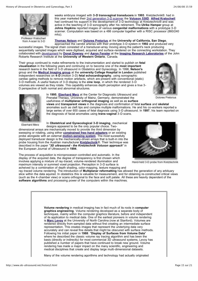
24/08/08 15:59History of Ultrasound in Obstetrics and Gynecology, Part 3
Page 15 sur 21http://www.ob-ultrasound.net/history3.html
weeks embryos imaged with 3-D transvaginal transducers in 1993. Kretztechnik®. had inthis year marketed their 2nd generation 3-D scanner the Voluson 530D. Alfred Kratochwilhad continued his support in the development of 3-D technology at Kretztechnic® and wasactive in the teaching of 3-D sonography after his retirement. The Ulrike Hamper group atJohns Hopkins reported images of various congenital malformations with a prototype 3-Dscanner. Computation was based on a 486 computer together with a RISC processor (860/240mhz).
Thomas Nelson and Dolores Pretorius at the University of California, San Diego,approached the carotid arteries with their prototype 3-D system in 1992 and produced very
successful images. The signal chain consisted of a transducer-array moving along the patient's neck producingsequentially sampled images which were digitised, acquired and surface-rendered on the connecting workstation. Theycollaborated with development by Donal Downey and Aaron Fenster at the Imaging Research Laboratories of the JohnRobarts Research Institute at the University of Western Ontario, Canada.
Their group continued to make refinements to the instrumentation and started to publish on fetalvisualization in the following years and continuing on to become one of the most importantresearch teams in the field of 3-D ultrasound in Obstetrics and Gynecology. In 1996, Nelson'sgroup and the Medical Imaging group at the university College Hospital in London publishedindependent researches on 4-D (motion 3-D) fetal echocardiography, using sonographiccardiac gating methods to remove motion artefacts, which are present with conventional (static)3-D methods. A useful feature of 3-D display is the cine loop, in which the rendered 3-Dvolumes are viewed as they rotate. This capability enhances depth perception and gives a true 3-D perspective of both normal and abnormal structures.
In 1995, Eberhard Merz at the Center for Diagnostic Ultrasound andPrenatal Therapy, University of Mainz, Germany, demonstrated theusefulness of multiplanar orthogonal imaging as well as as surfaceviews and transparent views in the diagnosis and confirmation of fetal surface and skeletalanomalies such as cleft lips and complex multiple malformations. He and his co-workers reported alarge series of over 600 cases of fetal diagnosis using 3-D ultrasound. In 1997, his team reported onthe diagnosis of facial anomalies using trans-vaginal 3-D scans.
In Obstetrical and Gynecological 3-D imaging, mechanicaldesigns appeared to be the only popular choice. Two-
dimensional arrays are mechanically moved to provide the third dimension bysweeping or rotating, using either constrained free-hand adapters or an existingprobe alongside with an external motion-sensing system. The most sucessfullydeployed transducer design is the mechanically-driven arrays that is built-in into theprobe housing from the Austrian manufacturer Kretztechnik®. Their technique wasdescribed in the paper "3D ultrasound - the Kretztechnik Voluson approach" inthe European Journal of Ultrasound in 1994.
The process of acquisition is microprocessor-controlled and automatic. In thedisplay of the acquired data, the degree of transparency is first chosen whichinvolves applying a mixture of ray-traced, volume-rendered illumination andmaximum intensity or summed voxel projection. Perception in 3-D surface isacheived by a combination of depth shading, color-mapping, texture mapping andray-traced volume rendering. The introduction of Multiplanar reformatting has allowed the generation of any arbituaryslice within the data aquired. In obstetrics this is valuable for measurement, and for obtaining re-constructed critical views(such as the 4-chamber view) or scans orthogonal to the face and soft palate. All these are heavily dependent of thesoftware algorithms and processing power of the computers within the machines.
Volume rendering in medical imaging has in fact much of its roots in computergraphics engineering. Volume rendering developed as a separate body oftechniques, mainly within the computer graphics literature, before and independentof its application to medical data. One of the earliest pioneers in volume renderingis Marc Levoy at the University of North Carolina (now at Stanford). Volumes arerendered directly from sampled data without first creating an intermediate surfacerepresentation. This creates images that represent the underlying data veryaccurately and can reveal fine details that might be obscured with surface methods.Following his initial paper in 1988: "Display of Surfaces from Volume Data"where he described the classic volume ray tracing algorithm and has been thebasis (directly or indirectly) for most commercial 3D ultrasound systems, Levoy haspublished a number of papers that have continued to break new ground. Volumerendering has made a major impact on the many scientific, engineering andmedical disciplines that create and display large multi-dimensional datasets.
Many of the volume rendering agorithms and technology had actually originated

24/08/08 15:59History of Ultrasound in Obstetrics and Gynecology, Part 3
Page 16 sur 21http://www.ob-ultrasound.net/history3.html
from computer scientists at the filmmaking company Pixar Animation Studios,famous for its 3D computer animated films! Initial volume rendering techniques andagorithms weres "invented" by company founders Robert Drebin, LorenCarpenter, and Pat Hanrahan. The algorithm embodied three key ideas:Directional shading based on the gradient in a volume, digital compositing tocombine the slices of a volume, and Image warping, also applied to a volume.
Gradient shading of volumes first appeared in a 1986 paper by Karl-HeinzHoehne, hamburg, who called it graylevel gradient shading. Compositing can betraced through Thomas Porter and Tom Duff's 1984 paper to Edwin Catmull andAlvy Ray Smith's invention of the alpha channel in the mid-70's. Image warping isa special case of texture mapping, which dates back to Edwin Catmull's 1974 PhDthesis at the University of Utah.
Volume rendering approximates the passage of light through a participating media.In this respect, James Blinn's 1982 paper on clouds and dusty surfaces, JimKajiya's 1984 paper on volume densities, and Holly Rushmeier's 1987 paper withKen Torrance on zonal radiosity must also be regarded as formative. Thisrelationship between volume rendering and light transport was pointed out to thevolume rendering community by Wolfgang Krueger. Another key pioneer wasGabor Herman at the City University of New York, who in a 1979 paper with KsunKao Liu proposed using cube-shaped voxels to display computed tomographydata.^^
(From citations by Dr. Marc Leroy, Stanford University. Excerpted with permission).
Medison®, which had acquired Kretztechnik® in 1996 continued to produce more advancedversions of the Voluson series of scanners that produced some of the best 3-D images in themarket. Bernard Benoit in Nice, France working in collaboration with Kretztechnik®,published some of the earliest, most stunning and convincing 3-D images in the mid- 1990susing prototype Voluson scanners (see picture of cleft lip on the left). His pictures had beenresponsible for drawing the attention of many to this new scanning modality.
In the second half of the 1990's at least twenty other important centers worldwide wereembarking on distinctive laboratory and clinical research into 3-D ultrasound, usually backed bywork done at their own university's medical physics and bio-engineering departments, orcommercial enterprises. Many of the innovations relied heavily on software programming. 3-Dultrasound technology would not have been a viable commercial proposition if not for the factthat computer technology was in the mid to late 1990s at a stage where the computations canbe done with staggering speed and at an acceptable cost.
In November 1996, with technical assistance from Takashi Okai and Shiro Kozuma fromALOKA®, Kazunori Baba published in the Lancet their initial experience with real-time processable 3-D, which used a simpler algorithm compared to conventional 3-Drendering. Fetal surfaces are demonstrated in near real-time imaging basing on simple'accoustic impedance thresholding' to identify fetal surfaces in the amniotic fluid.Image quality was very high and required less expensive computers to make thecalculations. However the viewing direction is limited to that of the probe so that adesirable direction may not always be possible. Sufficient amniotic fluid is alsoprerequisite to a good scan. Similarily image clarity depends on the difference betweenthe accoustic impedance of the fetal skin and that of amniotic fluid which made goodimages difficult to obtain before 20 weeks. Aloka® adapted the technology into theircommercial scanners.
Other early manufacturers of 3-D systems included ATL®,Tom-Tec Imaging Systems®, GE-Vingmed®, 3D-EchoTech®, and Life Imaging Systems Inc.®. The first English textbook on 3-D ultrasound inObstetrics and Gynecology came out in 1996 which was edited by Kazunori Baba and DavorJurkovic, King's College Hospital, London.
Eberhard Merz hosted the First World Congress on 3-D Ultrasound in Obstetrics andGynecology in Mainz in 1997. Many important teething issues surrounding the new practice of3-D sonography were discussed. In 1999 the 3D Focus group was formed by the ISUOG tolook after matters concerning the practice and education of 3-D ultrasound in Obstetrics andGynecology.
The increasing availabililty of 3-D ultrasound has resulted largely fromthe rapid advancement in computer technology and the decreasing

24/08/08 15:59History of Ultrasound in Obstetrics and Gynecology, Part 3
Page 17 sur 21http://www.ob-ultrasound.net/history3.html
cost of micro-processor electronics. The benefits that 3-D has boughtto ultrasound diagnosis has quickly become a matter for debate. Theimportant advantages of 3-D over conventional 2-D ultrasound as it isat the turn of the millenium are its ability to enhance maternal-fetalbonding, improved comprehension of certain fetal anomalies byparents, Improved recognition and better confirmation of certainanomalies such as cleft lips, polydactyl, micrognathia, malformed ears,club foot, vertebral malformations and other anomalies appearing onthe 'exterior ' of the fetus, consequent to the benefit of volume andsurface rendering. The development of transvaginal 3-D probeshave further enhanced its value in the early diagnosis of congenitalmalformations.
In a recent article by Asim Kurjak and his team, "Three-dimensionalsonography in prenatal diagnosis: a luxury or a necessity?" (Journal ofPerinatology, issue 3, 2000), he concluded,
".... the main advantages of three-dimensional ultrasound inperinatal medicine and antenatal diagnosis include scanning in the coronal plane, improved assessmentof complex anatomic structures, surface analysis of minor defects, volumetric measuring of organs,"plastic" transparent imaging of fetal skeleton, spatial presentation of blood flow arborization and, finally,storage of scanned volumes and images. It is our decided opinion that three-dimensional sonography hasgained a valuable place in prenatal diagnosis, becoming a necessity for every modern perinatal unit .... ".
Stuart Campbell at the St. Georges Hospital in London was one of the earlyproponents for the 3-D scan to be an important catalyst for mothers to bond totheir babies. What are known as 're-assurance scans ' and the perhapsmisnamed 'entertainment scans ' have started to develop. The attraction of beingable to look at the face of your baby before birth was enthusiastically reported inlay parenting and health magazines. Manufacturers had adopted anunprecedented "profit marketing" strategy to advertise to providers and "reversemarketing" strategy to advertise to consumers, particularly after the arrival ofthe 4-D (dynamic or motion 3-D) machines.
Barbara Maier and his group (Horst Steiner, AlfStaudach etc.) in a study in Salzburg in 1996 reportedthat mothers are more incentive to endure pregnancy-
related difficulties, reduced anxiety, and improved capacity to cope. Pretorius reported in thesame year that improved bonding between the mother and fetus could motivate mothers torefrain from smoking and other harmful behaviors during pregnancy.
Usefulness has been reported for calculating volumes of thegestational sac, fetal lungs and heart from second trimester to term,placental volume, liver volume, and thigh and abdominal volume for theestimation of fetal weight. Andreas Lee with the Kratochwil groupevaluated the accuracy of fetal weight estimation using 3-D abdominaland thigh volumes and reported in 1994 similar mean errors as compared to standard 2-Devaluations. Fong-Ming Chang 's group at the National Cheng Kung University Hospital inTaiwan, Republic of China reported in 1997 the feasibility of predicting birth weight by upper armvolume measured in 3-D. In 1998, Harm-Gerd Blaas at Tronheim, Norway published 3-Dstudies of embryos that were less than 10mm and further expanded the usefulness andimportance of 3-D sonography as an in-vivo research tool in fetal embryology. Transvaginal 3-Dwork on early fetal anatomy and malformations had also come out of Ilan Timor-Tritsch's centerin New York. 3-D power doppler in the study of placental blood circulation was first described in
1996 by C J Ritchie in a proprietary setup at the Bioengineering Department of the University of Washington, Seattle.Dolores Pretorius published on its usefulness and techniques in 1998. The Croatian group led by Asim Kurjak andSanja Kupesic expounded this new diagnostic entity further. Their book "Three-Dimensional Power Doppler inObstetrics and Gynecology " was published in 2000.
In gynecological applications, Davor Jurkovic at Kings convincinglydemonstrated in 1995 the usefulness of 3-D ultrasound in accuratelydifferentiating uterine anomalies such as bicornuate uterus and septateuteri. Similarily the assessment of the endometrial cavity with 3-Dsonohysterography and characterization of endometrial masses,adhesions, tubo-ovarian masses, hydrosalpinges, ovarian cysts, smallintraovarian tumors and mullerian anomalies have all been quickly andconvincingly demonstrated. Diagnostic accuracy of malignant ovariantumors can be up to 100 percent as reported by Bonilla-Musoles in1995, who also demonstrated the value of 3-D examination over theconvention 2-D transvaginal scans and 2-D sonohysterography in thediagnosis of endometrial lesions. 3-D color power doppler is valuablefor visualization of intra-tumor flow and thus is useful in evaluating in particular cervical
carcinomas and ovarian carcinomas. It is envisaged that the investigation will lead to greater appreciation of tumorangiogenesis. Another potential benefit of 3-D ultrasound lies in data documentation, storage, and networking. Digitally

24/08/08 15:59History of Ultrasound in Obstetrics and Gynecology, Part 3
Page 18 sur 21http://www.ob-ultrasound.net/history3.html
saved volumes of patient data can be readily transferred to a remote site for interpretation or second-opinion consultation. How much these all add up to make 3-D ultrasound cost-effective and an indispensible tool in Obstetrics andGynecology will remain to be seen.
Epilogue
The evolution of diagnosticultrasonography has been thecombined efforts of physicists,mechanical, electrical and bio-medicalengineers, computer technologists,clincians, sonographers, researchers,university and governmentadministrators as well as adventurousand perceptive commercial enterprises.
I particularly salute the ingeniousengineers and physicists throughoutthe history of the development as theyare the true heroes behind the entire'scientific' advance in ultrasonic imaging.Without them the innovative ideas of the brightest clinicians cannot be put into action.Developments in echocardiography, neurosonography, ophthalmology and breastechography have all supplemented the advancement in ultrasound instrumentations andmethodologies in Obstetrical and Gynecological sonography. The first linear-arrays forexample were invented for the purpose of ophthalmologic and cardiac investigations.Ultrasonography has very quickly become the single most important diagnosticinvestigation in the field of Obstetrics and the healthcare for women.
Interestingly, diagnostic medical ultrasound had evolved from technology used inmapping waves through liquid (the sonar), through air (the radar) and through solids(the metal-flaw detector). The A-scan which had evolved from the sonar and earlymetal-flaw detectors would not have a lasting impact on clinical medicine withoutevolving into the B-scan which had it's origin in the military radar. The A-scan did notprovide sufficiently accurate, reproducible and interpretable information to allow a firmdiagnosis to be made, particularly in Gynecology. The bistable B-scan would not haveadvanced to become a respectable diagnostic tool as it is now, without the developmentof the scan-converter and gray-scaling. The gray scale compound static scanner,with the incorporation of progressive electronic and computer technology available in thelate 1970s had establish itself as a genuine stand-alone clinical diagnostic tool, providinghitherto unavailable information to the clinician regarding a particular disease condition. Howry's original concept of deriving clear outline anatomical pictures by selectivelyrecording larger echoes from major interfaces and suppressing any other small echoeswas completely reversed in later developments, where attempts are made to detect thesmallest echoes in the presence of noise and displaying them in finer spatial detail andecho amplitudes.
The arrival of the real-time scanners have added further impetus to ultrasoundtechniques and had established ultrasonography as the most important imagingmodality in Obstetrics and Gynecology.The concept of the transvaginal scanner was in situ in the early 1950's but was unableto make any real headway until the appearance of sophisticated mechanical andelectronic sectoral real-time vaginal scanners in the mid 1980's. Ovum retrieval has forexample, interestingly gone from a transabdominal scan - transabdominal punctureapproach to a transabdominal - transvaginal approach and further onto the universallyaccepted transvaginal - transvaginal approach.
A 'technology push' situation further evolved when enhancement in diagnosticcapabilities of scanners was propelled by the almost explosive advancements inelectronic and microprocessor technology, occurring most significantly in the 1980s and90s. The advent of ultrasonography in Obstetrics has also 'created' the new specialtycalled Prenatal diagnosis that has developed by leaps and bounds since it's earlyconception. Ultrasound has markedly enhanced and pushed forward the study ofcongenital abnormalities among obstetricians, pediatricians, geneticist, pathologist andother allied specialties. All of a sudden, obstetricians started to learn about so manycongenital malformations that they have not even heard of.

24/08/08 15:59History of Ultrasound in Obstetrics and Gynecology, Part 3
Page 19 sur 21http://www.ob-ultrasound.net/history3.html
On the other hand, every single measurable parts of the fetus has been measured andtheir changes throughout gestation documented. It is of interest to note that historicallythe 4 basic fetal measurements, namely the BPD, the CRL, the AC and the FL hadevolved successively at different time periods (' 62/' 68, ' 73, ' 75, ' 80 respectively) eachbeing brought on by technical developments in ultrasound instrumentations at that time(B-scan, gray-scale, real-time). Stuart Campbell published the first BPD chart in 1971.Since then, charts and tables had become an important and integral part of Obstetricpractice, at which Obstetricians and Gynecologists were slowly getting used to.
Fetal malformations were diagnosed with increasing accuracy and at an earliergestation. Bold, daring and visionary clinicians and researchers invented newinterventional techniques that work under the guidance of ultrasound to diagnose fetaldisease and gynecological conditions. Doppler devices moved progressively fromdepicting flow velocity waveforms to color flow mapping, power doppler anddoppler tissue imaging. Velocimetric parameters of the umbilical artery, the middlecerebral artery and the ductus venosus had made their appearance one after anotheragain subsequent to progressive developments in the imaging apparatus. 3-Dultrasound made the scene in the late 1980's and further revolutionarised sonography inobstetrics and gynecology.
It is regretable that John Wild's original conception of precise quantitative detection ofcancer echoes with ultrasound also had not materialize to the initial expectations. Sowas the application of tissue characterization in the specialty. On the other hand, all theimprovements in machinery and earlier detection of abnormal structures in the fetus havenevertheless brought along with it "false positives" and difficult-to-be-sure diagnosis thatcould generate much undue anxiety in patients. Such could only have far-reachingeffects on a woman's perception of child-bearing.
I am still finding it awsome to imagine the "wire-frame" images of Douglass Howry andIan Donald could have now become "photo-realistic", and between all these are theunfolding magnificence of the invention and genius of science and man's endeavour tofind-out and to perfect. From the detection of life to the measuring of fetal sizes; fromthe determination of morphological normality to the evaluation of circulatory and growthdynamics, all have been making profound changes to the entire concept of routineantenatal care and Obstetric practice.
J. W.
A list of the Landmark references : (in the order as they appeared in the text)
Shepard MJ, Richards VA, et al . (1982) An evaluation of two equations for predicting fetal weight by ultrasound. Am J Obstet Gynecol 142:47. Donald I., Brown TG. (1961). Demonstration of tissue interfaces within the body by ultrasonic echo sounding. Br. J. Radiol. 34: 539-546. Sunden B. (1964). On the diagnostic value of ultrasound in obstetrics and gynaecology. Acta Obstet. Gynecol. Scand. 43(Suppl. 6). Garrett WJ, Grunwald G, Robinson DE. (1970). Prenatal diagnosis of fetal polycystic kidney by ultrasound. Aust. N. Z. J. Obstet. Gynaecol 10: 7-9.Allan LD, Tynan MJ, Campbell S, et al . (1980) Echocardiographic and anatomical correlates in the fetus. Br Heart J 44:444.Maulik D, Nanda NC, Moodley S, Saini VD, Thiede HA (1985) Application of Doppler echocardiography in the assessment of fetal cardiacdisease. Am J Obstet Gynecol 151:7 951-7. Maulik D, Nanda NC, Hsiung MC, Youngblood JP. (1986) Doppler color flow mapping of the fetal heart. Angiology 37:9 628-32.Kurjak A, Kirkinen P, Latin V, Rajhvajn B. (1980). Diagnosis and assessment of fetal malformations and abnormalities by ultrasound. J. Perinat.Med. 8: 219-235.Campbell S, Pearce JM. (1983). Ultrasound visualization of congenital malformations. Br. Med. J. 39: 322-331. Gembruch U, Hansmann M. (1984). Gezielte sonographische Ausshlussdiagnostik fetaler feblbildungen in risikogruppen. Gynäkologe 17: 19-32. Sabbagha RE, Sheikh Z, Tamura RK et al . (1985). Predictive value, sensitivity, and specificity of ultrasonic targeted imaging for fetal anomaliesin gravid women at high-risk for birth-defects. Am. J. Obstet. Gynecol. 152: 822-827.DeVore GR, Horenstein J, Siassi B, Platt LD. (1987) Fetal echocardiography. VII. Doppler color flow mapping: a new technique for the diagnosisof congenital heart disease. Am J Obstet Gynecol 156:5 1054.Wladimiroff JW, Tonge HM, Stewart PA. (1986) Doppler ultrasound assessment of cerebral blood flow in the human fetus. Br J Obstet Gynaecol93:5 471.Vyas S, Nicolaides KH, Campbell S . (1989) Renal artery flow-velocity waveforms in normal and hypoxemic fetuses. Am J Obstet Gynecol 161:1168.Kiserud T, Eik-Nes SH, Blass HG, et al . (1991) Ultrasonographic velocimetry of the fetal ductus venosus. Lancet 338:8780 1412.Rizzo G, Capponi A, Ardini D, et al . (1994) Ductus Venosus celocity waveforms in appropriate and small for gestational age fetuses. Early HumDev 39:1 15. Birnholz, J., Stephens, J. C. and Faria, M. (1978). Fetal Movement Patterns: A Possible Means of Defining Neurologic Developmental Milestonesin Utero. American J. Roentology 130: 537-540.Birnholz, JC. (1981). The Development of Human Fetal Eye Movement Patterns. Science 213: 679-681. Busnel, Marie-Claire, Granier -Deberre,C. and Lecanuet, J. P.(1992). Fetal Audition. Annals of the New York Academy of Sciences 662:118-134.Hofman D, Mast H and Hollander HJ. (1967) The importance of placenta localization by means of ultrasonics for amniocentesis. GeburtshFrauenheilk. 27: 1199. Kazy Z, Stygar AM, Bakharev VA (1980) Chorionic biopsy under immediate realtime (ultrasonic) control. Orv Hetil 121:2765.Kazy Z, Rozovsky IS, Bakharev VA (1982) Chorionic biopsy in early pregnency. A method of early prenatal diagnosis for inherited disorders.Prenat Diagn. 2:39.Ward RHT, Model B, Petrou M, et al (1983) Method of sampling chorionic villi in first trimester of pregnancy under guidance of real timeultrasound. Br Med J 286:1542

24/08/08 15:59History of Ultrasound in Obstetrics and Gynecology, Part 3
Page 20 sur 21http://www.ob-ultrasound.net/history3.html
Smidt -Jensen S, Hahnemann N, Jensen PKA, et al (1984) Experience with find needle biopsy in the first trimester -an alternative toamniocentesis. Clin Genet 26:272.Daffos, F., Cappella-Pavlovsky, M. and Forestier, F. (1983). Fetal blood sampling via the umbilical cord using a needle guided by ultrasound.Report of 66 cases. Prenat. Diagn., 3,271-7Daffos F, Kappela Pavlosvky M, Forestier F. (1985) Fetal blood sampling during pregnancy with use of a needle guieded by ultrasound : a studyof 606 consecutive cases. Am J Obstet Gynecol 153: 655-60.Nicolaides, K.H., Soothill, P.W., Rodeck, C.H. and Campbell, S . (1986). Ultrasound-guided sampling of umbilical cord and placental blood toassess fetal wellbeing. Lancet, 1,1065-7Benacerraf BR, Greene MF, Saltzman DH, Barss VA, Penso CA, Nadel AS, Heffner LJ, Stryker JM, Sandstrom MM, Frigoletto FD (1988). Earlyamniocentesis for prenatal cytogenetic evaluation. Radiology 169:3 709-10.Nicolini U, Santolaya J, Ojo OE, Fisk NM, Hubinont C, Tonge M, Rodeck CH. (1988) The fetal intrahepatic umbilical vein as an alternative to cordneedling for prenatal diagnosis and therapy. Prenat Diagn 8:9 665-7.Harrison MR, Filly RA, Golbus MS, Berkowitz RL, Callen PW, Canty TG, Catz C, Clewell WH, Depp R, Edwards MS, Fletcher JC, Frigoletto FD,Garrett WJ, Johnson ML, Jonsen A, De Lorimier AA, Liley WA, Mahoney MJ, Manning FD, Meier PR, Michejda M, Nakayama DK, Nelson L,Newkirk JB, Pringle K, Rodeck C, Rosen MA, Schulman JD (1982). N Engl J Med 1982 Dec 23 307:26 1651-2Cullen MT, Green J, Whetham J, Salafia C, Gabrielli S, Hobbins JC (1990) Transvaginal ultrasonographic detection of congenital anomalies in thefirst trimester. Am J Obstet Gynecol 163(2):466.Cullen MT, Reece EA, Whetham J, Hobbins JC. (1990) Embryoscopy: description and utility of a new technique. Am J Obstet Gynecol 162(1):82.Rottem, S., Bronshtein, M. (1990). Transvaginal sonographic diagnosis of congenital anomalies between 9 weeks and 16 weeks, menstrual age,J. Clin. Ultrasound, 18, 307-314. Bronshtein. M., Blumenfeld, Z., Drugan, E., Ophir, H., Yoffe, N., Peer, E., Brandes, J.M. (1991). Detection of fetal cardiac malformations bytransvaginal sonography in the first and early second trimester. Am. J. Obstet. Gynecol., 1 (Supp 1), 85.Benacerraf BR, Barss VA, Laboda LA. (1985) A sonographic sign for the detection in the second trimester of the fetus with Down's syndrome.Am J Obstet Gynecol 15 151:8 1078.Goswamy RK, Campbell S, Chamberlain J. (1986) Screening for ovarian cancer. IARC Sci Publ 76 305.Bourne TH, Campbell S, Reynolds KM, et al . (1993) Screening for early familial ovarian cancer with transvaginal ultrasonography and colourblood flow imaging. BMJ 306:6884 1025.Holm H.H (1972) Ultrasound as a guide in percutaneous puncture procedures. Ultrasonic 10:83, 1972. Graham D, Sanders RC (1982) Ultrasound guided transvaginal aspiration biopsy of pelvic muscles. J. Ultrasound in Med. 1:279. Lenz S (1981) Collection of Human Oocytes in IVF by Ultrasonically guided follicular puncture. Lancet I, 1163. Lenz S, Lauritsen JG (1982) Ultrasonically guided percutaneous aspiration of human follicles under local anesthesia: a new method of collectingoocytes for in vitro fertilization. Fertil Steril Dec 38:6 673-7.Gleicher N (1983) Egg retrieval for IVF by sonographically controlled vaginal culdocentesis. Lancet II, 508-509. Dellenbach P, Nisand I, Moreau L, Feger B, Plumere C, Gerlinger P, Brun B, Rumpler Y (1984) Transvaginal, sonographically controlled ovarianfollicle puncture for egg retrieval. Lancet 1:8392 1467.Dellenbach P, Nisand I, Moreau L, Feger B, Plumere C, Gerlinger P (1985) Transvaginal sonographically controlled follicle puncture for oocyteretrieval. Fertil Steril 1985 44:656-62.Kemeter P, Feichtinger W (1986) Trans-vaginal oocyte retrieval using a trans-vaginal sector scan probe combined with an automated puncturedevice. Hum Reprod 1:21-4.Feichtinger W, Kemeter P (1986) Transvaginal sector scan sonography for needle guided transvaginal follicle aspiration and other application ingynaec, routine and research. Fertil. Steril. 45; 722-725. Timor-Tritsch, I.E., Monteagudo. A., Peisner, D.B (1992). High frequency transvaginal sonographic examination for the potential malformationassessment of the 9 week to 14 week fetus, J. Clin. Ultrasound., 20, 231-238. Taylor KJ, Burns PN, Woodcock JP, Wells PN. (1985) Blood flow in deep abdominal and pelvic vessels: ultrasonic pulsed-Doppler analysis.Radiology 154:2 487.Kurjak A, Zalud I, Jurkovi D, Alfirevi Z, Miljan MT. (1989) Transvaginal color Doppler for the assessment of pelvic circulation. Acta Obstet GynecolScand 68:2 131.Brinkley JF, SK Muramatsu, Wd McCallum, RL Popp. (1982) n vitro evaluation of ultrasonic three dimensional imaging and volume system.Ultrasonic Imaging 4:126.Fredfelt KE, HH Holm, JF Pedersen. (1984). Three dimensional ultrasonic scanning. Acata Radiol Diagn 25: 237.Moritz WE, Pearlman AS, McCabe DH, et al . (1983) An ultrasonic technique for imaging the ventricle in three dimensions and calculating itsvolume. IEEE Trans Biomed Eng 30:8 482.Baba K, Satoh K, Sakamoto S, et al,. (1989) Development of an ultrasonic system for three-dimensional reconstruction of the fetus. J PerinatMed 17:1 19.King DL, King DL Jr, Shao MY. (1990) Three-dimensional spatial registration and interactive display of position and orientation of real -timeultrasound images. J Ultrasound Med 9:9 525.Kuo HC, Chang FM, Wu CH, et al . (1992) The primary application of three-dimensional ultrasonography in obstetrics. Am J Obstet Gynecol166:3 880.Kelly IM, Gardener JE, Brett AD, et al ,. (1994) Three-dimensional US of the fetus. Work in progress. Radiology 192:1 253.Feichtinger W. (1993) Transvaginal three-dimensional imaging. Ultrasound Obstet Gynecol. 3:375.Hamper UM, Trapanotto V, Sheth S, et al,. (1994) Three-dimensional US: preliminary clinical experience. Radiology 191:2 397.Pretorius DH, Nelson TR, (1991) 3-Dimensional Ultrasound Imaging in Patient Diagnosis and Management: The Future. Ultrasound ObstetGynecol 1(6) 381.Pretorius DH, Nelson TR, Jaffe JS. (1992) 3-D Sonographic Analysis Based on Color Flow Doppler and Gray Scale Image Data: A PreliminaryReport. J Ultrasound Med 11:225.Nelson TR, Pretorius DH. (1992) 3-Dimensional Ultrasound of Fetal Surface Features. Ultrasound Obste Gynecol 2:166.Nelson TR and Elvins TT. (1993) Visualization of 3D Ultrasound Data. IEEE Computer Graphics and Applications 13(6):50.Nelson TR, Pretorius, DH. (1993) The Future of Three-Dimensional Ultrasound. Diagn Imaging Technol Report 2:173.Pretorius DH and Nelson TR. (1994) Prenatal Visualization of Cranial Sutures and Fontanelles with Three-Dimensional Ultrasonography. JUltrasound Med 13:871.Pretorius DH and Nelson TR. (1995) Fetal Face Visualization Using Three-Dimensional Ultrasonography. J Ultrasound Med 14:349.Nelson TR, Pretorius DH, Sklansky M, Hagen-Ansert S. (1996) Three-dimensional echocardiographic evaluation of fetal heart anatomy andfunction: acquisition, analysis and display. J Ultrasound Med 15: 1.Deng J, Gardener JE, Rodeck CH, Lees WR . (1996) Fetal echocardiography in three and four dimensions. Ultrasound Med Biol 1996; 22: 979.Kirbach D, TA Whittingham. (1994) 3D ultrasound-the Kretztechnik Voluson approach. Eur J Ultrasound I 85.Blaas HG, Eik-Nes SH, Berg S, et a l. (1998) In-vivo three-dimensional ultrasound reconstructions of embryos and early fetuses. Lancet 352:91351182.Merz E, Bahlmann F, Weber G. (1995) Volume scanning in the evaluation of fetal malformations: a new dimension in prenatal diagnosis.Ultrasound Obstet Gynecol 5(4):222.Merz E, Weber G, Bahlmann F, Miric-Tesanic D. (1997) Application of transvaginal and abdominal three-dimensional ultrasound for the detectionor exclusion of malformations of the fetal face. Ultrasound Obstet Gynecol 9(4):237.Merz E, Bahlmann R, Weber G. (1995) Volume scanning in the evaluation of fetal malformations: a new dimension in prenatal diagnosis.Ultrasound Obstet Gynecol 5:222-227.Merz E, Bahlmann R, Weber G. Macchiella D. (1995) Three-dimensional ultrasonography in prenatal diagnosis. J Perinat Med 23:213-222.Maier B, Steiner H, Wieneroither H, Staudach A. (1997) The psychological impact of three-dimensional fetal imaging on the fetomaternalrelationship. In: Baba K, Jurkovic D, eds. Three-dimensional ultrasound in obstetrics and gynecology. New York: Parthenon, p. 67-74.Pretorius DH, House M, Nelson TR, et al . (1995) Evalustion of normal and abnormal lips in fetuses: comparison between three- and two-dimensional sonography. AJR 165:1233-1237.Pretorius DH, Nelson TR, et al . (1995) Fetal face visualization using three-dimensional ultrasonography. J Ultrasound Med 14:349-356.Pretorius DH. (1996) Maternal smoking habit modification via fetal visualization. University of California tobacco related disease research program.Annual report to the California State Legislature, 1996:76.
24/08/08 15:59History of Ultrasound in Obstetrics and Gynecology, Part 3
Page 21 sur 21http://www.ob-ultrasound.net/history3.html
Lee W, Comstock CH, Kirk J, et al . Birthweight prediction by three-dimensional ultrasonographic volumes of the fetal thigh and abdomen. JUltrasound Med 16:799-805.Liang RI, Chang FM, Yao BL, et al . (1997) Predicting birth weight by upper-arm volume with use of three-dimensional ultrasonography. ObstetGynecol 177(3):632.Ritchie CJ, Edwards WS, Mack LA, Cyr DR, Kim Y. (1996) Three-dimensional ultrasonic angiography using power-mode Doppler. Ultrasound MedBiol 22:277-286.
Pixel focusing image courtesy of Medison ®.* Copyrighted ATL®, reproduced with permission.** 3-D image and image of the hand-held probe courtesy of Dr. Bernard Benoit. Reproduced with permission.^ Dr. William O'Brien Jr., Professor, Bioaccoustic Research Laboratory, Department of Electrical and Computer Engineering, University of Illinois.*** Color doppler image courtesy of Dr. Greggory Devore. Reproduced with permission.Dr. Susan Lenz's image courtesy of Dr. Lenz. Reproduced with permission.Picture of parenting magazines coutesy of Medison®.Dr. Baba's images of his setup courtesy of Dr. Baba.^ In Diagnosis and Therapy of Fetal Anomalies. John C Hobbins and Beryl R Benacerraf (eds), Churchill Livingstone, 1989.^^ Excepted with permission from the homepage of Dr. Marc Leroy, Stanford University: Citation; SIGGRAPH 1996 Computer GraphicsAchievement Award- It is not possible to include all the names who have contributed significantly to the advancement of Obstetrical and Gynecological sonography, some who may have been less well-known than the others and some who may not have published so extensively in the English language. Apologies are extended to those whose contribution has not been fully credited in this article.
All original contents Copyright © 1998-2001 Joseph SK Woo. All Rights Reserved.
Search this site
Back to [ Part 1 ] and [ Part 2 ]