histological classification of canine ovarian cyst types
TRANSCRIPT
Original ArticleJ Vet Sci 2018, 19(6), 725-734ㆍhttps://doi.org/10.4142/jvs.2018.19.6.725 JVS
Received 10 May 2018, Revised 31 Aug. 2018, Accepted 17 Sep. 2018*Corresponding author: Tel: +49-5513921760; Fax: +49-5513933381; E-mail: [email protected] of Veterinary Scienceㆍⓒ 2018 The Korean Society of Veterinary Science. All Rights Reserved.This is an Open Access article distributed under the terms of the Creative Commons Attribution Non-Commercial License (http://creativecommons.org/licenses/ by-nc/4.0) which permits unrestricted non-commercial use, distribution, and reproduction in any medium, provided the original work is properly cited.
pISSN 1229-845XeISSN 1976-555X
Histological classification of canine ovarian cyst types with reference to medical history
Yvonne Knauf1,2,*, Kernt Köhler3, Sascha Knauf4, Axel Wehrend1
1Clinic for Obstetrics, Gynecology and Andrology of Large and Small Animals with Veterinary Ambulance, 3Institute of Veterinary Pathology, Justus-Liebig-University, D 35392 Giessen, Germany2Department of Animal Sciences, Georg-August-University, D 37077 Goettingen, Germany4Department of Infection Biology, Work Group Neglected Tropical Diseases, German Primate Center, Leibniz-Institute for Primate Research, D 37077 Goettingen, Germany
Ovaries of 21 bitches presented with gynecopathies were surgically removed and histologically examined. Standard histological, as well as immunohistochemical, classification of 193 cystic structures resulted in the classification of 72 cysts of subsurface epithelial structures (SES), 61 follicular cysts (FCs), 38 cystic rete ovarii (CRO), 13 lutein cysts (LCs), and 9 non-classifiable cysts (NCCs). In addition to the histological classification, results were interpreted according to subject medical history, clinical examination outcome, and macroscopic observations during ovariohysterectomy. Dogs with ovarian cysts (OCs) and associated reproductive perturbations were mostly nulliparous, of large breed, and had an average of 9.5 ± 3 years. Prolonged or shortened inter-estrus intervals of past heats, however, seemed to be relatively low-risk factors for the development of OCs in dogs. Furthermore, we provide histological observations of a rarely seen canine LC including a degenerated oocyte in the central cavity.
Keywords: dogs, histology, immunohistochemistry, medical history taking, ovarian cysts
Introduction
Ovarian cysts (OCs) of clinical relevance are commonly found in domestic animals [16] in which they cause gynecopathies [2,18,28] and hyperestrogenism [15]. OCs can originate from different ovarian structures and their development, frequency, and size varies across species [16]. Histology is used to distinguish different types of OCs [10,15]. The most common types are follicular cysts (FCs), cysts of subsurface epithelial structure (SES), cystic rete ovarii (CRO), lutein cysts (LCs), and cystic corpora lutea (CCL) [10,15,16,29]. While OCs and related gynecopathies are well characterized in sows [28] and cattle [2], the association of OC morphology based on histology and immunohistochemistry in relation to the clinical manifestation of the patient has rarely been investigated in dogs [4,6]. In particular, it should be noted, that histologically evaluated LCs in dogs have not been fully documented in recent reports [4].
The present study investigated advanced histology features of canine OCs in relationship to corresponding clinical manifestations that were observed when the bitch was presented in our clinic. It
was hypothesized that multiple cyst types occur simultaneously on a single ovary in bitches having clinically manifest gynecopathies, and that it is possible to determine LCs histologically in bitches with gynecopathies.
Materials and Methods
Ethical statementThe study is in accordance with German legal and ethical
requirements for appropriate animal procedures. Good veterinary practice was applied to all procedures whenever animals were handled. The aims of this study were explained to all pet owners. All experimental procedures were approved by the Ethics Committee of Regierungspraesidium Gießen Germany (approval No. V 54-19c20 15h02Gl18/14kTV13/2017).
BitchesA total of 21 bitches of different ages and breeds were
presented to the Clinic for Obstetrics, Gynecology and Andrology of Large and Small Animals, Justus-Liebig-
726 Yvonne Knauf et al.
Journal of Veterinary Science
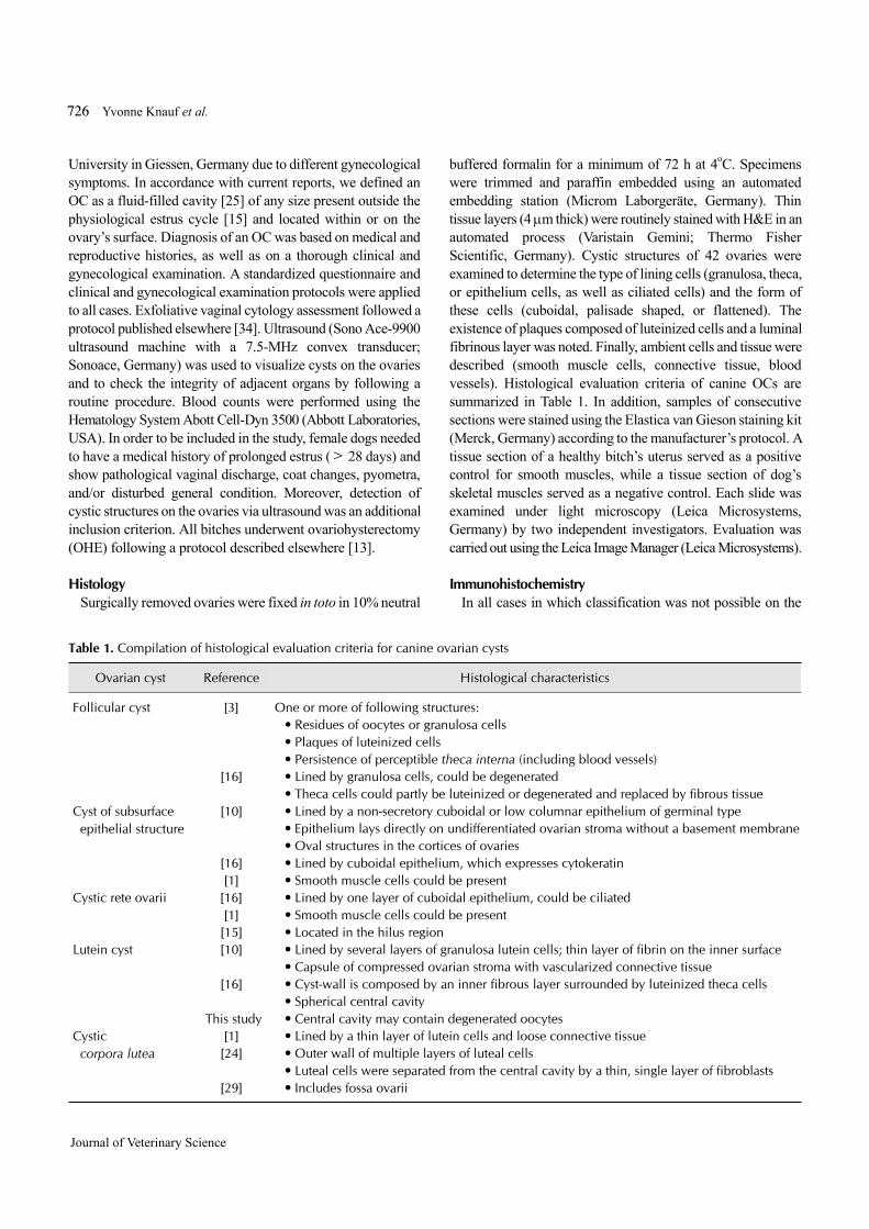
Table 1. Compilation of histological evaluation criteria for canine ovarian cysts
Ovarian cyst Reference Histological characteristics
Follicular cyst [3] One or more of following structures:• Residues of oocytes or granulosa cells• Plaques of luteinized cells• Persistence of perceptible theca interna (including blood vessels)
[16] • Lined by granulosa cells, could be degenerated• Theca cells could partly be luteinized or degenerated and replaced by fibrous tissue
Cyst of subsurface epithelial structure
[10] • Lined by a non-secretory cuboidal or low columnar epithelium of germinal type• Epithelium lays directly on undifferentiated ovarian stroma without a basement membrane• Oval structures in the cortices of ovaries
[16] • Lined by cuboidal epithelium, which expresses cytokeratin[1] • Smooth muscle cells could be present
Cystic rete ovarii [16] • Lined by one layer of cuboidal epithelium, could be ciliated[1] • Smooth muscle cells could be present[15] • Located in the hilus region
Lutein cyst [10] • Lined by several layers of granulosa lutein cells; thin layer of fibrin on the inner surface• Capsule of compressed ovarian stroma with vascularized connective tissue
[16] • Cyst-wall is composed by an inner fibrous layer surrounded by luteinized theca cells• Spherical central cavity
This study • Central cavity may contain degenerated oocytesCystic
corpora lutea[1] • Lined by a thin layer of lutein cells and loose connective tissue[24] • Outer wall of multiple layers of luteal cells
• Luteal cells were separated from the central cavity by a thin, single layer of fibroblasts[29] • Includes fossa ovarii
University in Giessen, Germany due to different gynecological symptoms. In accordance with current reports, we defined an OC as a fluid-filled cavity [25] of any size present outside the physiological estrus cycle [15] and located within or on the ovary’s surface. Diagnosis of an OC was based on medical and reproductive histories, as well as on a thorough clinical and gynecological examination. A standardized questionnaire and clinical and gynecological examination protocols were applied to all cases. Exfoliative vaginal cytology assessment followed a protocol published elsewhere [34]. Ultrasound (Sono Ace-9900 ultrasound machine with a 7.5-MHz convex transducer; Sonoace, Germany) was used to visualize cysts on the ovaries and to check the integrity of adjacent organs by following a routine procedure. Blood counts were performed using the Hematology System Abott Cell-Dyn 3500 (Abbott Laboratories, USA). In order to be included in the study, female dogs needed to have a medical history of prolonged estrus (> 28 days) and show pathological vaginal discharge, coat changes, pyometra, and/or disturbed general condition. Moreover, detection of cystic structures on the ovaries via ultrasound was an additional inclusion criterion. All bitches underwent ovariohysterectomy (OHE) following a protocol described elsewhere [13].
HistologySurgically removed ovaries were fixed in toto in 10% neutral
buffered formalin for a minimum of 72 h at 4oC. Specimens were trimmed and paraffin embedded using an automated embedding station (Microm Laborgeräte, Germany). Thin tissue layers (4 m thick) were routinely stained with H&E in an automated process (Varistain Gemini; Thermo Fisher Scientific, Germany). Cystic structures of 42 ovaries were examined to determine the type of lining cells (granulosa, theca, or epithelium cells, as well as ciliated cells) and the form of these cells (cuboidal, palisade shaped, or flattened). The existence of plaques composed of luteinized cells and a luminal fibrinous layer was noted. Finally, ambient cells and tissue were described (smooth muscle cells, connective tissue, blood vessels). Histological evaluation criteria of canine OCs are summarized in Table 1. In addition, samples of consecutive sections were stained using the Elastica van Gieson staining kit (Merck, Germany) according to the manufacturer’s protocol. A tissue section of a healthy bitch’s uterus served as a positive control for smooth muscles, while a tissue section of dog’s skeletal muscles served as a negative control. Each slide was examined under light microscopy (Leica Microsystems, Germany) by two independent investigators. Evaluation was carried out using the Leica Image Manager (Leica Microsystems).
ImmunohistochemistryIn all cases in which classification was not possible on the
Histology of ovarian cysts in bitches 727
www.vetsci.org
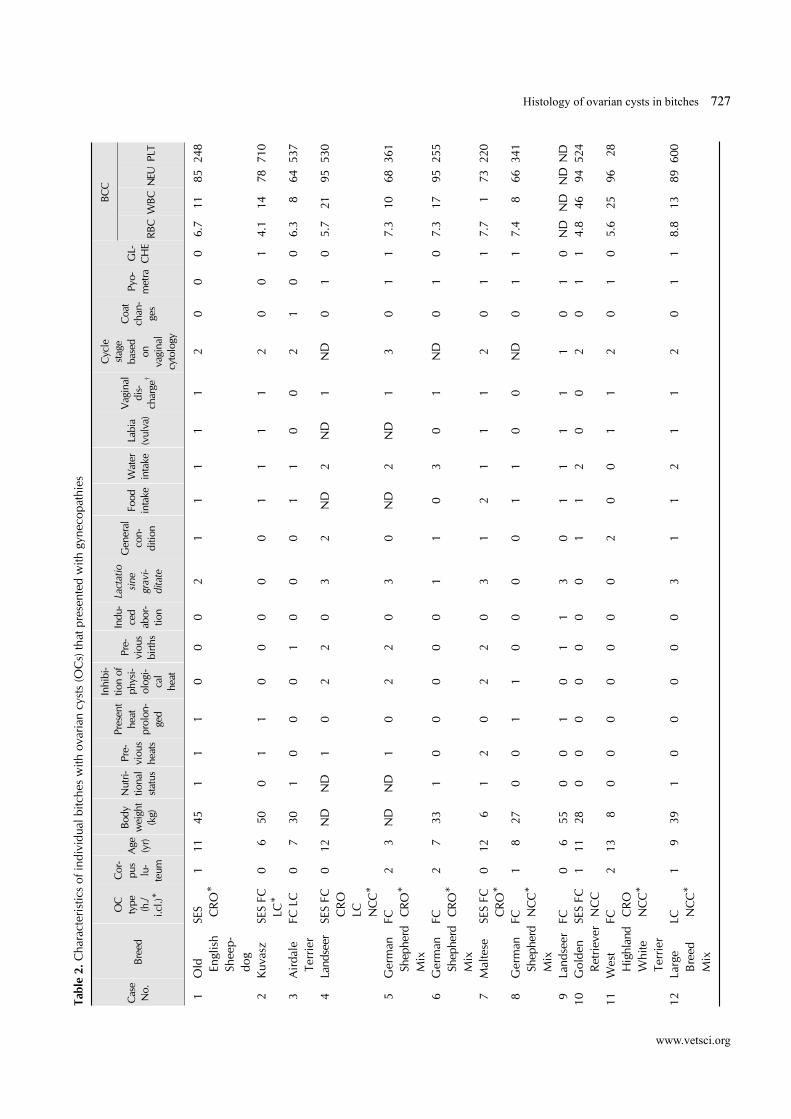
Tabl
e 2.
Cha
ract
eris
tics
of in
divi
dual
bitc
hes
with
ova
rian
cyst
s (O
Cs)
that
pre
sent
ed w
ith g
ynec
opat
hies
Cas
e N
o.Br
eed
OC
ty
pe
(h./
i.cl.)
*
Cor
-pu
s lu
-te
um A
ge
(yr)
Body
w
eigh
t (k
g)
Nut
ri-tio
nal
stat
us
Pre-
viou
s he
ats
Pres
ent
heat
pr
olon
-ge
d
Inhi
bi-
tion
of
phys
i-ol
ogi-
cal
heat
Pre-
viou
s bi
rths
Indu
-ce
d ab
or-
tion
Lact
atio
si
ne
grav
i-di
tate
Gen
eral
co
n-di
tion
Food
in
take
Wat
er
inta
ke
Labi
a (v
ulva
)
Vag
inal
di
s-ch
arge
†
Cyc
le
stag
e ba
sed
on
vagi
nal
cyto
logy
Coa
t ch
an-
ges
Pyo-
met
raG
L-C
HE
BCC
RBC
WBC
NEU
PLT
1O
ld
Engl
ish
Shee
p-do
g
SES CRO
*1
1145
11
10
00
21
11
11
20
00
6.7
1185
248
2Ku
vasz
SES
FC
LC*
0 6
500
11
00
00
01
11
12
00
14.
114
7871
0
3A
irda
le
Terr
ier
FC L
C0
730
10
00
10
00
11
00
21
00
6.3
864
537
4La
ndse
erSE
S FC
C
RO
LC
N
CC
*
012
ND
ND
10
22
03
2N
D2
ND
1N
D0
10
5.7
2195
530
5G
erm
an
Shep
herd
M
ix
FC CRO
*2
3N
DN
D1
02
20
30
ND
2N
D1
30
11
7.3
1068
361
6G
erm
an
Shep
herd
Mix
FC CRO
*2
733
10
00
00
11
03
01
ND
01
07.
317
9525
5
7M
alte
seSE
S FC
C
RO*
012
61
20
22
03
12
11
12
01
17.
7 1
7322
0
8G
erm
an
Shep
herd
Mix
FC NC
C*
1 8
270
01
10
00
01
10
0N
D0
11
7.4
866
341
9La
ndse
erFC
0 6
550
01
01
13
01
11
11
01
0N
DN
DN
DN
D10
Gol
den
Ret
rieve
rSES
FC
NC
C1
1128
00
00
00
01
12
00
20
11
4.8
4694
524
11W
est
Hig
hlan
dW
hite
Te
rrie
r
FC CR
O
NC
C*
213
80
00
00
00
20
01
12
01
05.
625
96 2
8
12La
rge
Bre
ed
Mix
LC NC
C*
1 9
391
00
00
03
11
21
12
01
18.
813
8960
0
728 Yvonne Knauf et al.
Journal of Veterinary Science
Tabl
e 2.
Con
tinue
d
Cas
e N
o.Br
eed
OC
ty
pe
(h./
i.cl.)
*
Cor
-pu
s lu
-te
um A
ge
(yr)
Body
w
eigh
t (k
g)
Nut
ri-tio
nal
stat
us
Pre-
viou
s he
ats
Pres
ent
heat
pr
olon
-ge
d
Inhi
bi-
tion
of
phys
i-ol
ogi-
cal
heat
Pre-
viou
s bi
rths
Indu
-ce
d ab
or-
tion
Lact
atio
si
ne
grav
i-di
tate
Gen
eral
co
n-di
tion
Food
in
take
Wat
er
inta
ke
Labi
a (v
ulva
)
Vag
inal
di
s-ch
arge
†
Cyc
le
stag
e ba
sed
on
vagi
nal
cyto
logy
Coa
t ch
an-
ges
Pyo-
met
raG
L-C
HE
BCC
RBC
WBC
NEU
PLT
13La
rge
Bre
ed
Mix
FC CRO
*0
227
02
12
20
30
12
ND
ND
30
00
ND
ND
ND
ND
14Po
inte
r M
ixSE
S FC
C
RO
1 9
270
00
00
01
11
11
02
01
1N
DN
DN
DN
D
15Bo
uvie
r D
e Fl
ande
rs
FC*
012
400
00
01
00
02
20
12
01
16.
2 9
87N
P
16M
ediu
m
Bre
ed
Mix
SES FC
*2
14N
DN
D2
02
20
31
22
11
30
10
ND
ND
ND
ND
17M
alte
seC
RO1
14 3
00
00
00
00
12
11
20
11
6.8
994
429
18La
brad
or
Mix
FC2
727
00
10
00
10
02
11
30
11
6.9
2696
258
19La
brad
or
Ret
rieve
rCRO
2 8
371
10
20
00
10
21
12
01
13.
964
9627
9
20G
olde
n R
etrie
verFC
110
381
01
00
03
11
21
12
11
15.
510
9658
1
21G
erm
an
Shep
herd
SES
FC
LC
NC
C
210
270
01
00
00
01
11
12
00‡
18.
2 6
8136
0
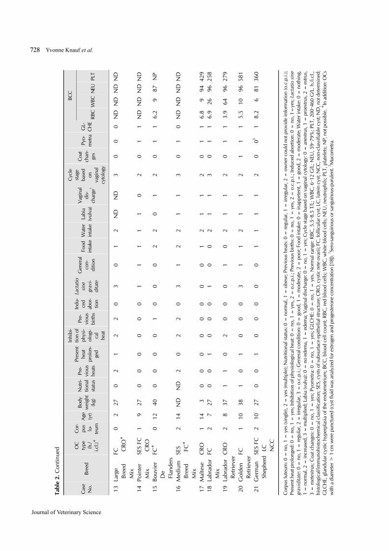
Cor
pus l
uteu
m: 0
= n
o, 1
= y
es (s
ingl
e), 2
= y
es (m
ultip
le);
Nut
ritio
nal s
tatu
s: 0
= n
orm
al, 1
= o
bese
; Pre
viou
s he
ats:
0 =
regu
lar,
1 =
irre
gula
r, 2
= o
wne
r cou
ld n
ot p
rovi
de in
form
atio
n (o
.c.p
.i.);
Pres
ent h
eat p
rolo
nged
: 0 =
no,
1 =
yes
; Inh
ibiti
on o
f phy
siol
ogic
al h
eat:
0 =
no,
1 =
yes
, 2 =
o.c
.p.i.
; Pre
viou
s birt
hs: 0
= n
o, 1
= y
es, 2
= o
.c.p
.i.; I
nduc
ed a
borti
on: 0
= n
o, 1
=ye
s; L
acta
tio si
ne
grav
idita
te: 0
= n
o, 1
= re
gula
r, 2
= ir
regu
lar,
3 =
o.c
.p.i.
; Gen
eral
con
ditio
n: 0
= g
ood,
1 =
mod
erat
e, 2
= p
oor;
Food
inta
ke: 0
= in
appe
tent
, 1 =
goo
d, 2
= m
oder
ate;
Wat
er in
take
: 0 =
not
hing
, 1
= n
orm
al, 2
= in
crea
sed,
3 =
mul
tiplie
d; L
abia
(vul
va):
0 =
no
edem
a, 1
= e
dem
a; V
agin
al d
isch
arge
: 0 =
no,
1 =
yes
; Cyc
le st
age
base
d on
vag
inal
cyt
olog
y: 0
= a
nest
rus,
1 =
pro
estru
s, 2
= e
stru
s,
3 =
met
estru
s; C
oat c
hang
es: 0
= n
o, 1
= y
es; P
yom
etra
: 0 =
no,
1 =
yes
; GLC
HE:
0 =
no,
1 =
yes
. Nor
mal
rang
e: R
BC, 5
.5–8.
5 T/
L; W
BC, 6
–12 G
/L; N
EU, 5
9–79%
; PLT
, 200
–460
G/L
. h./i
.cl.,
hi
stol
ogic
al/im
mun
ohis
toch
emic
al c
lass
ifica
tion;
SES
, cys
ts o
f sub
surfa
ce e
pith
elia
l stru
ctur
e; C
RO, c
ystic
rete
ova
rii; F
C, f
ollic
ular
cys
t; LC
, lut
ein
cyst
; NC
C, n
on-c
lass
ifiab
le c
yst;
ND
, not
det
erm
ined
; G
LCH
E, g
land
ular
cys
tic h
yper
plas
ia o
f the
end
omet
rium
; BC
C, b
lood
cel
l cou
nt; R
BC, r
ed b
lood
cel
ls; W
BC, w
hite
blo
od c
ells
; NEU
, neu
troph
ils; P
LT, p
late
lets
; NP,
not
pos
sibl
e. *
In a
dditi
on: O
Cs
with
a d
iam
eter
> 1 c
m w
ere
punc
ture
d (c
yst f
luid
was
ana
lyze
d fo
r est
roge
n an
d pr
oges
tero
ne c
once
ntra
tion
[18]
). † Se
ro-s
angu
inou
s or
san
guin
ous-
puru
lent
. ‡ Muc
omet
ra.
Histology of ovarian cysts in bitches 729
www.vetsci.org
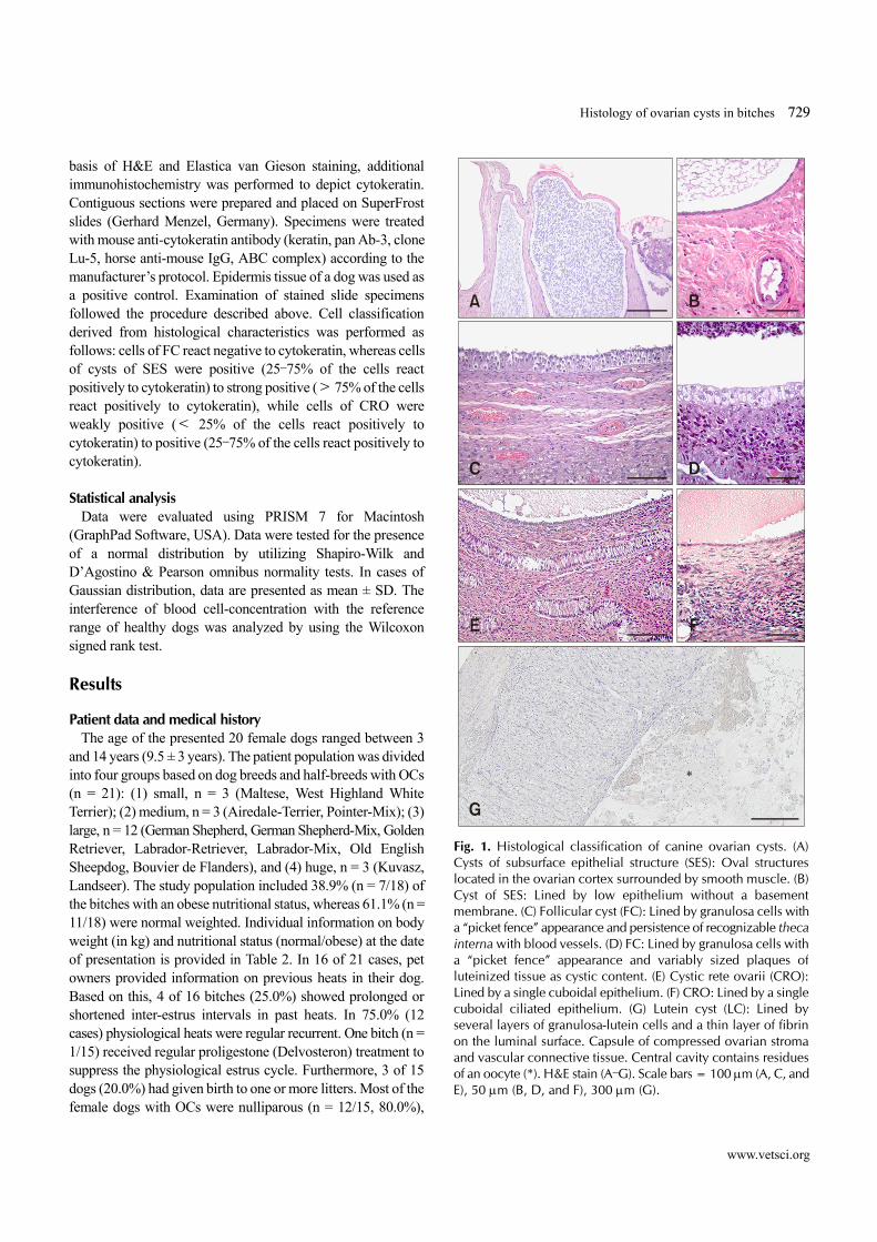
Fig. 1. Histological classification of canine ovarian cysts. (A) Cysts of subsurface epithelial structure (SES): Oval structures located in the ovarian cortex surrounded by smooth muscle. (B)Cyst of SES: Lined by low epithelium without a basement membrane. (C) Follicular cyst (FC): Lined by granulosa cells witha “picket fence” appearance and persistence of recognizable thecainterna with blood vessels. (D) FC: Lined by granulosa cells witha “picket fence” appearance and variably sized plaques of luteinized tissue as cystic content. (E) Cystic rete ovarii (CRO): Lined by a single cuboidal epithelium. (F) CRO: Lined by a singlecuboidal ciliated epithelium. (G) Lutein cyst (LC): Lined by several layers of granulosa-lutein cells and a thin layer of fibrin on the luminal surface. Capsule of compressed ovarian stroma and vascular connective tissue. Central cavity contains residuesof an oocyte (*). H&E stain (A–G). Scale bars = 100 m (A, C, andE), 50 m (B, D, and F), 300 m (G).
basis of H&E and Elastica van Gieson staining, additional immunohistochemistry was performed to depict cytokeratin. Contiguous sections were prepared and placed on SuperFrost slides (Gerhard Menzel, Germany). Specimens were treated with mouse anti-cytokeratin antibody (keratin, pan Ab-3, clone Lu-5, horse anti-mouse IgG, ABC complex) according to the manufacturer’s protocol. Epidermis tissue of a dog was used as a positive control. Examination of stained slide specimens followed the procedure described above. Cell classification derived from histological characteristics was performed as follows: cells of FC react negative to cytokeratin, whereas cells of cysts of SES were positive (25–75% of the cells react positively to cytokeratin) to strong positive (> 75% of the cells react positively to cytokeratin), while cells of CRO were weakly positive (< 25% of the cells react positively to cytokeratin) to positive (25–75% of the cells react positively to cytokeratin).
Statistical analysisData were evaluated using PRISM 7 for Macintosh
(GraphPad Software, USA). Data were tested for the presence of a normal distribution by utilizing Shapiro-Wilk and D’Agostino & Pearson omnibus normality tests. In cases of Gaussian distribution, data are presented as mean ± SD. The interference of blood cell-concentration with the reference range of healthy dogs was analyzed by using the Wilcoxon signed rank test.
Results
Patient data and medical historyThe age of the presented 20 female dogs ranged between 3
and 14 years (9.5 ± 3 years). The patient population was divided into four groups based on dog breeds and half-breeds with OCs (n = 21): (1) small, n = 3 (Maltese, West Highland White Terrier); (2) medium, n = 3 (Airedale-Terrier, Pointer-Mix); (3) large, n = 12 (German Shepherd, German Shepherd-Mix, Golden Retriever, Labrador-Retriever, Labrador-Mix, Old English Sheepdog, Bouvier de Flanders), and (4) huge, n = 3 (Kuvasz, Landseer). The study population included 38.9% (n = 7/18) of the bitches with an obese nutritional status, whereas 61.1% (n = 11/18) were normal weighted. Individual information on body weight (in kg) and nutritional status (normal/obese) at the date of presentation is provided in Table 2. In 16 of 21 cases, pet owners provided information on previous heats in their dog. Based on this, 4 of 16 bitches (25.0%) showed prolonged or shortened inter-estrus intervals in past heats. In 75.0% (12 cases) physiological heats were regular recurrent. One bitch (n = 1/15) received regular proligestone (Delvosteron) treatment to suppress the physiological estrus cycle. Furthermore, 3 of 15 dogs (20.0%) had given birth to one or more litters. Most of the female dogs with OCs were nulliparous (n = 12/15, 80.0%),
730 Yvonne Knauf et al.
Journal of Veterinary Science
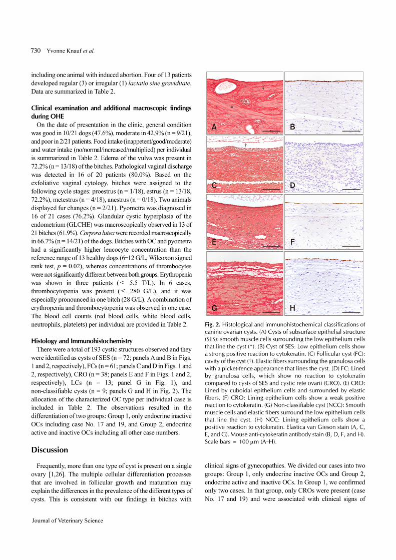
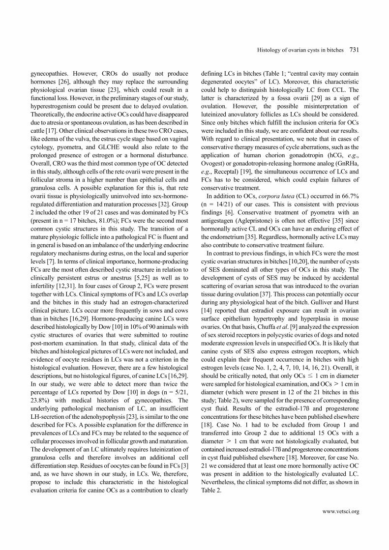
Fig. 2. Histological and immunohistochemical classifications of canine ovarian cysts. (A) Cysts of subsurface epithelial structure (SES): smooth muscle cells surrounding the low epithelium cellsthat line the cyst (*). (B) Cyst of SES: Low epithelium cells showa strong positive reaction to cytokeratin. (C) Follicular cyst (FC):cavity of the cyst (†). Elastic fibers surrounding the granulosa cellswith a picket-fence appearance that lines the cyst. (D) FC: Linedby granulosa cells, which show no reaction to cytokeratin compared to cysts of SES and cystic rete ovarii (CRO). (E) CRO: Lined by cuboidal epithelium cells and surrounded by elastic fibers. (F) CRO: Lining epithelium cells show a weak positive reaction to cytokeratin. (G) Non-classifiable cyst (NCC): Smooth muscle cells and elastic fibers surround the low epithelium cells that line the cyst. (H) NCC: Lining epithelium cells show a positive reaction to cytokeratin. Elastica van Gieson stain (A, C, E, and G). Mouse anti-cytokeratin antibody stain (B, D, F, and H).Scale bars = 100 m (A–H).
including one animal with induced abortion. Four of 13 patients developed regular (3) or irregular (1) lactatio sine graviditate. Data are summarized in Table 2.
Clinical examination and additional macroscopic findings during OHE
On the date of presentation in the clinic, general condition was good in 10/21 dogs (47.6%), moderate in 42.9% (n = 9/21), and poor in 2/21 patients. Food intake (inappetent/good/moderate) and water intake (no/normal/increased/multiplied) per individual is summarized in Table 2. Edema of the vulva was present in 72.2% (n = 13/18) of the bitches. Pathological vaginal discharge was detected in 16 of 20 patients (80.0%). Based on the exfoliative vaginal cytology, bitches were assigned to the following cycle stages: proestrus (n = 1/18), estrus (n = 13/18, 72.2%), metestrus (n = 4/18), anestrus (n = 0/18). Two animals displayed fur changes (n = 2/21). Pyometra was diagnosed in 16 of 21 cases (76.2%). Glandular cystic hyperplasia of the endometrium (GLCHE) was macroscopically observed in 13 of 21 bitches (61.9%). Corpora lutea were recorded macroscopically in 66.7% (n = 14/21) of the dogs. Bitches with OC and pyometra had a significantly higher leucocyte concentration than the reference range of 13 healthy dogs (6–12 G/L, Wilcoxon signed rank test, p = 0.02), whereas concentrations of thrombocytes were not significantly different between both groups. Erythropenia was shown in three patients (< 5.5 T/L). In 6 cases, thrombocytopenia was present (< 280 G/L), and it was especially pronounced in one bitch (28 G/L). A combination of erythropenia and thrombocytopenia was observed in one case. The blood cell counts (red blood cells, white blood cells, neutrophils, platelets) per individual are provided in Table 2.
Histology and ImmunohistochemistryThere were a total of 193 cystic structures observed and they
were identified as cysts of SES (n = 72; panels A and B in Figs. 1 and 2, respectively), FCs (n = 61; panels C and D in Figs. 1 and 2, respectively), CRO (n = 38; panels E and F in Figs. 1 and 2, respectively), LCs (n = 13; panel G in Fig. 1), and non-classifiable cysts (n = 9; panels G and H in Fig. 2). The allocation of the characterized OC type per individual case is included in Table 2. The observations resulted in the differentiation of two groups: Group 1, only endocrine inactive OCs including case No. 17 and 19, and Group 2, endocrine active and inactive OCs including all other case numbers.
Discussion
Frequently, more than one type of cyst is present on a single ovary [1,26]. The multiple cellular differentiation processes that are involved in follicular growth and maturation may explain the differences in the prevalence of the different types of cysts. This is consistent with our findings in bitches with
clinical signs of gynecopathies. We divided our cases into two groups: Group 1, only endocrine inactive OCs and Group 2, endocrine active and inactive OCs. In Group 1, we confirmed only two cases. In that group, only CROs were present (case No. 17 and 19) and were associated with clinical signs of
Histology of ovarian cysts in bitches 731
www.vetsci.org
gynecopathies. However, CROs do usually not produce hormones [26], although they may replace the surrounding physiological ovarian tissue [23], which could result in a functional loss. However, in the preliminary stages of our study, hyperestrogenism could be present due to delayed ovulation. Theoretically, the endocrine active OCs could have disappeared due to atresia or spontaneous ovulation, as has been described in cattle [17]. Other clinical observations in these two CRO cases, like edema of the vulva, the estrus cycle stage based on vaginal cytology, pyometra, and GLCHE would also relate to the prolonged presence of estrogen or a hormonal disturbance. Overall, CRO was the third most common type of OC detected in this study, although cells of the rete ovarii were present in the follicular stroma in a higher number than epithelial cells and granulosa cells. A possible explanation for this is, that rete ovarii tissue is physiologically uninvolved into sex-hormone- regulated differentiation and maturation processes [32]. Group 2 included the other 19 of 21 cases and was dominated by FCs (present in n = 17 bitches, 81.0%); FCs were the second most common cystic structures in this study. The transition of a mature physiologic follicle into a pathological FC is fluent and in general is based on an imbalance of the underlying endocrine regulatory mechanisms during estrus, on the local and superior levels [7]. In terms of clinical importance, hormone-producing FCs are the most often described cystic structure in relation to clinically persistent estrus or anestrus [5,25] as well as to infertility [12,31]. In four cases of Group 2, FCs were present together with LCs. Clinical symptoms of FCs and LCs overlap and the bitches in this study had an estrogen-characterized clinical picture. LCs occur more frequently in sows and cows than in bitches [16,29]. Hormone-producing canine LCs were described histologically by Dow [10] in 10% of 90 animals with cystic structures of ovaries that were submitted to routine post-mortem examination. In that study, clinical data of the bitches and histological pictures of LCs were not included, and evidence of oocyte residues in LCs was not a criterion in the histological evaluation. However, there are a few histological descriptions, but no histological figures, of canine LCs [16,29]. In our study, we were able to detect more than twice the percentage of LCs reported by Dow [10] in dogs (n = 5/21, 23.8%) with medical histories of gynecopathies. The underlying pathological mechanism of LC, an insufficient LH-secretion of the adenohypophysis [23], is similar to the one described for FCs. A possible explanation for the difference in prevalences of LCs and FCs may be related to the sequence of cellular processes involved in follicular growth and maturation. The development of an LC ultimately requires luteinization of granulosa cells and therefore involves an additional cell differentiation step. Residues of oocytes can be found in FCs [3] and, as we have shown in our study, in LCs. We, therefore, propose to include this characteristic in the histological evaluation criteria for canine OCs as a contribution to clearly
defining LCs in bitches (Table 1; “central cavity may contain degenerated oocytes” of LC). Moreover, this characteristic could help to distinguish histologically LC from CCL. The latter is characterized by a fossa ovarii [29] as a sign of ovulation. However, the possible misinterpretation of luteinized anovulatory follicles as LCs should be considered. Since only bitches which fulfill the inclusion criteria for OCs were included in this study, we are confident about our results. With regard to clinical presentation, we note that in cases of conservative therapy measures of cycle aberrations, such as the application of human chorion gonadotropin (hCG, e.g., Ovogest) or gonadotropin-releasing hormone analog (GnRHa, e.g., Receptal) [19], the simultaneous occurrence of LCs and FCs has to be considered, which could explain failures of conservative treatment.
In addition to OCs, corpora lutea (CL) occurred in 66.7% (n = 14/21) of our cases. This is consistent with previous findings [6]. Conservative treatment of pyometra with an antigestagen (Aglepristone) is often not effective [35] since hormonally active CL and OCs can have an enduring effect of the endometrium [35]. Regardless, hormonally active LCs may also contribute to conservative treatment failure.
In contrast to previous findings, in which FCs were the most cystic ovarian structures in bitches [10,20], the number of cysts of SES dominated all other types of OCs in this study. The development of cysts of SES may be induced by accidental scattering of ovarian serosa that was introduced to the ovarian tissue during ovulation [37]. This process can potentially occur during any physiological heat of the bitch. Gulliver and Hurst [14] reported that estradiol exposure can result in ovarian surface epithelium hypertrophy and hyperplasia in mouse ovaries. On that basis, Chuffa et al. [9] analyzed the expression of sex steroid receptors in polycystic ovaries of dogs and noted moderate expression levels in unspecified OCs. It is likely that canine cysts of SES also express estrogen receptors, which could explain their frequent occurrence in bitches with high estrogen levels (case No. 1, 2, 4, 7, 10, 14, 16, 21). Overall, it should be critically noted, that only OCs ≤ 1 cm in diameter were sampled for histological examination, and OCs > 1 cm in diameter (which were present in 12 of the 21 bitches in this study; Table 2), were sampled for the presence of corresponding cyst fluid. Results of the estradiol-17ß and progesterone concentrations for these bitches have been published elsewhere [18]. Case No. 1 had to be excluded from Group 1 and transferred into Group 2 due to additional 15 OCs with a diameter > 1 cm that were not histologically evaluated, but contained increased estradiol-17ß and progesterone concentrations in cyst fluid published elsewhere [18]. Moreover, for case No. 21 we considered that at least one more hormonally active OC was present in addition to the histologically evaluated LC. Nevertheless, the clinical symptoms did not differ, as shown in Table 2.
732 Yvonne Knauf et al.
Journal of Veterinary Science
Based on previous reports, the age of female dogs with OCs varies between 1 and 18 years [10,11,22,31]. This was confirmed by our results in which bitches had an average lifetime of 9.5 ± 3 years. However, we were unable to confirm that the prevalence of OCs is in direct proportion to dog age, as reported by Marino et al. [22]. Since life expectancy of different dog breeds varies, there is a tendency of increasing OC prevalence in dogs over the age of six. A relationship between obesity and OC incidence has been reported in human [21], mouse [27], and rat [36], but investigations into that association in dogs are lacking. Our study population included 38.9% (n = 7/18) overweight bitches, whereas 61.1% (n = 11/18) were normal weighted. A nutritional assessment was carried out during the dogs’ first clinical examination and followed generally accepted protocols. However, based on our results we can only speculate that, with an increase in adipose tissue, the risk of developing OCs also increases in dogs; further investigations are needed. Most female dogs with OCs were nulliparous (n = 12/15, 80.0%), which supports findings reported elsewhere [6,10].
In Germany and other prosperous societies, most dogs are well integrated into households and are not allowed to breed. Under the assumption that most female dogs in Germany are nulliparous, it is not surprising that a similar situation was reflected in our OC study population. Studies that include free-ranging stray bitches may derive different results. Referring to the medical and reproductive histories of our bitches, there was no correlation between previous treatment of bitches with hormones to suppress their physiological estrus cycle or induce abortion and the prevalence of OCs. But, under the assumption that treatment with hormones can promote OC formation [5,8,11], we recommend further investigations, including studies with standardized control and patient groups. Prolonged or shortened inter-estrus intervals of the past heats seem to be relatively low-risk factors for the development of OCs. Pet owners reported irregular past heats in 25% of the study animals, which did not reveal any gynecopathies. This could be due to the spontaneous healing of endocrine active OCs, as has been described in cattle [17].
The leading symptom in the bitches with OCs in our study was prolonged vaginal discharge (16 of 20 examined cases, 80.0%), which was consistent with a previous report on bitches with OC syndrome [6]. The most common OC-accompanying diagnosis was pyometra in 16 of 21 cases (76.2%). In this study population, the endometrium of 11 bitches (68.8%) also showed GLCHE. Another dog had a GLCHE accompanied by a mucometra. Overall, 13 bitches with OCs had GLCHE, which is indicative of longer-term exposure of the endometrium to estrogen and progesterone. Due to the association of OCs with pyometra in the majority of animals in this study (n = 16/21, 76.2%), we qualified the statement of an estrogen-induced leukocytosis with neutrophilia. Blood parameters of bitches
with OCs should be separately evaluated from those in bitches with OCs and pyometra due to in utero inflammatory processes obscuring the changes. Our results indicated leukocytosis without accompanying pyometra in one of four dogs with OC. OCs may trigger hyperestrogenism through the non-regulated release of steroid sex hormones. However, dogs are estrogen sensitive [29]. An estrogen-induced myelotoxic effect on the hematopoietic system can lead to anemia and a loss of condition [25,30,33]. In our study, we found one bitch with a thrombocyte level clearly below the physiological range (28 G/L), and that level in five further animals was in the lower part of the normal range (280–200 G/L). Only in one case (No. 19), was this linked with an erythropenia (3.9 T/L) and a loss of condition. Despite the low number of animals in this study, we suggest, that hemograms are not useful as a proxy for the presence of hyperestrogenism. It should, however, be emphasized that a thorough clinical examination must include a hemogram in order to exclude life-threatening conditions in bitches with gynecopathies and to determine the most appropriate treatment approach (conservative or surgical).
Supporting our hypothesis that multiple cyst types occur simultaneously on a single ovary in bitches having clinically manifest gynecopathies, we observed more than one type of OC on single ovaries isolated from bitches with clinical signs of gynecopathies. Despite the relatively small study population (n = 21) we identified four of five OC types with the occurrence of LCs being higher than that reported previously. Because we surprisingly could not detect a clinical difference between dogs with endocrine active and endocrine inactive OCs at the time of histological classification, our results indicate that every OC-affected bitch should be considered as an individual case. Therefore, further investigations are needed to identify the primary factors that trigger ovarian dysfunction and to better understand the complex pathophysiology of OC development.
Acknowledgments
The authors wish to thank J. Blad-Stahl, F. Kotarski, and S. Heerdt of the clinical laboratory for their help. We thank M. Bleyer, K. Mätz-Rensing, and E. Gruber-Dujardin from the Pathology Unit of the German Primate Center for their assistance in the preparation of Figures 2 and 3. We would also like to acknowledge H. Bostedt for his encouragement.
Conflict of Interest
The authors declare no conflicts of interest.
References
1. Akihara Y, Shimoyama Y, Kawasako K, Komine M, Hirayama K, Kagawa Y, Omachi T, Matsuda K, Okamoto M,
Histology of ovarian cysts in bitches 733
www.vetsci.org
Kadosawa T, Taniyama H. Immunohistochemical evaluation of canine ovarian cysts. J Vet Med Sci 2007, 69, 1033-1037.
2. Amweg AN, Rodríguez FM, Huber E, Marelli BE, Salvetti NR, Rey F, Ortega HH. Role of glucocorticoids in cystic ovarian disease: expression of glucocorticoid receptor in the bovine ovary. Cells Tissues Organs 2016, 201, 138-147.
3. Andersen AC, Simpson ME. Pathology of the ovary and genital tract. In: Andersen AC, Simpson ME (eds.). The Ovary and Reproductive Cycle of the Dog (Beagle). pp. 246-272, Geron-X, Los Altos, 1973.
4. Arlt SP, Haimerl P. Cystic ovaries and ovarian neoplasia in the female dog – a systematic review. Reprod Domest Anim 2016, 51 (Suppl 1), 3-11.
5. Arlt SP, Spankowsky S, Heuwieser W. Follicular cysts and prolonged oestrus in a female dog after administration of a deslorelin implant. N Z Vet J 2011, 59, 87-91.
6. Bostedt H, Jung C, Wehrend A, Boryzcko Z. [Clinical and endocrinological findings of bitches with ovarian cyst syndrome]. Schweiz Arch Tierheilkd 2013, 155, 543-550. German.
7. Bostedt H, Tammer I, Hecker BR. Infertility in the breeding bitch: a short review. Tierärztl Prax 1999, 27, 179-185.
8. Bowen RA, Olson PN, Young S, Withrow SJ. Efficacy and toxicity of tamoxifen citrate for prevention and termination of pregnancy in bitches. Am J Vet Res 1988, 49, 27-31.
9. Chuffa LG, Lupi Júnior LA, da Maia Lima AF. Sex steroid receptors and apoptosis-related proteins are differentially expressed in polycystic ovaries of adult dogs. Tissue Cell 2016, 48, 10-17.
10. Dow C. Ovarian abnormalities in the bitch. J Comp Pathol 1960, 70, 59-69.
11. Ervin E, Homans P. Giant ovarian cyst. Compend Contin Educ Pract Vet 1986, 8, 698-700.
12. Fontbonne A. Infertility in the bitch. In: Proceedings of the 31th World Small Animal Veterinary Association (WSAVA) Congress; 11 October 2006, Prague, Czech Republic.
13. Groeger S, Weiss R, Trasch K, Wehrend A. Significance of the bacteriological results from vaginal swabs taken from bitches with overt pyometra. Kleintierpraxis 2007, 52, 426-428.
14. Gulliver LS, Hurst PR. Repeat estradiol exposure differentially regulates protein expression patterns for estrogen receptor and E-cadherin in older mouse ovarian surface epithelium: implications for replacement and adjuvant hormone therapies? Steroids 2012, 77, 674-685.
15. Johnston SD, Kustritz MV, Olson PS. Disorders of the canine ovary. In: Canine and Feline Theriogenology. pp. 193-205, Saunders, Philadelphia, 2001.
16. Kennedy PC, Cullen JM, Edwards JF, Goldschmidt MH, Larsen S, Munson L, Nielsen S. Cysts in and around the ovary. In: Kennedy PC (ed.). Histological Classification of Tumors of the Genital System of Domestic Animals, Vol. IV. 2nd ed. pp. 29-31, Armed Forces Institute of Pathology, Washington, 1998.
17. Kesler DJ, Garverick HA. Ovarian cysts in dairy cattle: a review. J Anim Sci 1982, 55, 1147-1159.
18. Knauf Y, Bostedt H, Failing K, Knauf S, Wehrend A. Gross pathology and endocrinology of ovarian cysts in bitches. Reprod Domest Anim 2014, 49, 463-468.
19. Knauf Y, Failing K, Knauf S, Wehrend A. [Treatment of bitches with ovarian cysts using human chorionic gonadotropin-releasing hormone analogue. A case series of 30 bitches]. Tierarztl Prax Ausg K Kleintiere Heimtiere 2013, 41, 93-100. German.
20. Knauf Y, Wehrend A. [Ovarian cysts in the bitch]. Tierarztl Prax Ausg K Kleintiere Heimtiere 2010, 38, 333-340. German.
21. Lecke SB, Mattei F, Morsch DM, Spritzer PM. Abdominal subcutaneous fat gene expression and circulating levels of leptin and adiponectin in polycystic ovary syndrome. Fertil Steril 2011, 95, 2044-2049.
22. Marino G, Mannarino C, Di Prima ML, Rizzo S, Zanghi A. Stromal cysts in the canine ovary. In: Proceedings of the 27th Meeting of the European Society of Veterinary Pathology and European College of Veterinary Pathologists; 9-12 September 2009, Olsztyn–Kraków, Poland.
23. McEntee K. Cysts in and around the ovary. In: Reproductive Pathology of Domestic Mammals. pp. 52-67, Academic Press, San Diego, 1990.
24. Miller DM, McCrory VS, Anderson WI. Polycystic ovarian tissue in a spayed bitch. Mod Vet Pract 1983, 64, 749.
25. Olson PN, Wrigley RH, Husted PW, Bowen RA, Nett TA. Persistent estrus in the bitch. In: Ettinger SJ (ed.). Textbook of Veterinary Internal Medicine, Vol. 2. pp. 1792-1796, WB Saunders, Philadelphia, 1989.
26. Ortega-Pacheco A, Segura-Correa JC, Jimenez-Coello M, Linde Forsberg C. Reproductive patterns and reproductive pathologies of stray bitches in the tropics. Theriogenology 2007, 67, 382-390.
27. Radavelli-Bagatini S, Blair AR, Proietto J, Spritzer PM, Andrikopoulos S. The New Zealand obese mouse model of obesity insulin resistance and poor breeding performance: evaluation of ovarian structure and function. J Endocrinol 2011, 209, 307-315.
28. Sant’Ana F, Reis Junior JL, Blume GR, Gimeno EJ, Rey F, Ortega HH. Immunohistochemical expression of growth factors in the follicular wall of normal and cystic ovaries of sows. Reprod Domest Anim 2015, 50, 327-332.
29. Schlafer DH, Miller RB. Pathology of the ovary (nondevelopmental lesions). In: Maxie MG, Jubb KVF, Kennedy PC, Palmer N (eds.). Jubb, Kennedy, and Palmer’s Pathology of Domestic Animals, Vol. 3. pp. 431-563, Elsevier Saunders, Philadelphia, 2007.
30. Schwarz H, Geyer S, Rüsse M, Hänichen T. [Intoxication caused by estrogen administration in the bitch]. Tierarztl Prax 1982, 10, 393-402. German.
31. Shille VM, Calderwood-Mays MB, Thatcher MJ. Infertility in a bitch associated with short interestrus intervals and cystic follicles: a case report. J Am Anim Hosp Assoc 1984, 20, 171-176.
32. Songsasen N, Fickes A, Pukazhenthi BS, Wildt DE. Follicular morphology, oocyte diameter and localisation of fibroblast growth factors in the domestic dog ovary. Reprod Domest Anim 2009, 44 (Suppl 2), 65-70.
33. Suttorp M, Hoffmann B, Sippell WG. Prevention of oestradiol-associated toxicosis in a dalmatian by early intervention with granulocyte colony-stimulating factor. Vet Rec 2002, 151, 244-245.
734 Yvonne Knauf et al.
Journal of Veterinary Science
34. Tammer I, Blendinger K, Sobiraj A, Bostedt H. [The use of exfoliative vaginal cytology for the gynecological evaluation of the bitch]. Tierarztl Prax 1994, 22, 199-207. German.
35. Trasch K, Wehrend A, Bostedt H. Follow-up examinations of bitches after conservative treatment of pyometra with the antigestagen aglepristone. J Vet Med A Physiol Pathol Clin Med 2003, 50, 375-379.
36. Volk KM, Pogrebna VV, Roberts JA, Zachry JE, Blythe SN,
Toporikova N. High-fat, high-sugar diet disrupts the preovulatory hormone surge and induces cystic ovaries in cycling female rats. J Endocr Soc 2017, 1, 1488-1505.
37. Weiss E. Weibliche Geschlechtsorgane. In: Dahme E, Hafner-Marx A (eds.). [Grundriss der Speziellen Pathologischen Anatomie der Haustiere, Vol. 6]. pp. 213- 232, Enke, Stuttgart, 2007. German.