histological, histomorphometrical and histochemical
TRANSCRIPT
Int.J.Curr.Microbiol.App.Sci (2018) 7(3): 1477-1491
1477
Original Research Article https://doi.org/10.20546/ijcmas.2018.703.176
Histological, Histomorphometrical and Histochemical Studies on the
Large Intestine of Uttara Fowl
K. Pandit1*
, B.S. Dhote1, D. Mahanta
1, S. Sathapathy
2,
S. Tamilselvan1, M. Mrigesh
1 and S. Mishra
3
1Department of Veterinary Anatomy, C.V.A.Sc., GBPUAT, Pantnagar – 263 145,
Uttarakhand, India 2Department of Veterinary Anatomy and Histology, C.V.Sc. and A.H., OUAT,
Bhubaneswar – 751003, Odisha, India 3Division of Veterinary Pathology, ICAR-Indian Veterinary Research Institute,
Izzatnagar – 243 122, Bareilly, U.P, India
*Corresponding author
A B S T R A C T
Introduction
Uttara fowl is a well adopted bird found in the
taral region of Uttarakhand (Kaur et al., 2010).
Their germplasm is unexplored and possess
various potential genes responsible for
survival in rough conditions of hilly terrain of
Uttarakhand. This breed has evolved through
natural selection in indigenous agro-ecological
conditions and is well adapted to the local
environment. It has low cholesterol content in
their blood which makes it suitable for heart
patients and obese people. It has high glucose
level in its blood due to more energy
requirement at high altitude, more activity and
more flying tendency than commercial birds
(Kaur, 2007). Fundamentals for the production
performance in a poultry flock are their
genetic merit, management and health. The
study of avian gastrointestinal tract anatomy is
still in its infancy and needs to be further
explored as it plays a vital role in feed
International Journal of Current Microbiology and Applied Sciences ISSN: 2319-7706 Volume 7 Number 03 (2018) Journal homepage: http://www.ijcmas.com
The present study was conducted on 24 Uttara fowl. The histological, histomorphometrical
and histochemical features with their age related changes in large intestine were studied.
The surface epithelium in both caeca and colorectum was lined by simple columnar
epithelium with interspersed goblet cells. Finger like villous projections were seen in the
tunica mucosa. PAS- Alcian blue reaction was showed by all the segments of caecum as
well as colorectum. Presence of Elastic fibres around the blood vessels, between muscle
layers of tunica muscularis and lamina propria was found in caecum and colorectum of
112 days old bird. Masson’s trichrome stain showed presence of collagen fibres in tunica
serosa, between muscle fibres, in core of villi and around intestinal glands. Gomori’s stain
showed presence of reticular fibers, which was maximum in 112 days old bird. Argentaffin
cells were present in epitheliums of intestinal gland and villi mostly in contact with
basement membrane.
K e y w o r d s Histology,
Histomorphometry,
Histochemistry, Large
intestine, Uttara fowl
Accepted:
12 February 2018
Available Online: 10 March 2018
Article Info
Int.J.Curr.Microbiol.App.Sci (2018) 7(3): 1477-1491
1478
utilization which ultimately affects the overall
productivity of the bird. There are many
nutritional reports on the avian intestinal
system but very little is known about the
detailed anatomy of large intestine (Bayer et
al., 1981; Turk, 1982). Different fields of
veterinary and animal sciences are dependent
on the anatomy and histology of body organs
(Argenzio, 1980). The large intestine due to its
lympahatic tissue aggregates in the colon and
caeca has a major immunological role
(Ushakumary S. et al., 2002). Caecal
functioning is still only partly understood
(McNab, 1973; Braun and Duke 1989).
However, research of many decades has
revealed its role in energy balance and
osmoregulation which involves absorption of
material from the caecum. Caeca may also
serve as the site for several different functions,
especially digestion of small food particles,
absorption of nutrients, production of
immunoglobulins, utilization and absorption
of water and metabolism of uric acid into
amino acids. Caecotomy has shown to
increase the water intake and increased
excretion of water in faeces (Son et al., 2000)
Reports are available on the histological
structure and age related changes in certain
organs of digestive system of guinea fowl,
Japanese quail, and Kadaknath fowl
(Sivagnanam et al., 2004; Venkatasan et al.,
2005 and Vaish et al., 2006). Samte (2008)
carried out Gross morphometric, light and
electron microscopic studies on the large
intestine of Kadaknath fowl. Persual of
literature revealed limited information
pertaining to the large intestine of Uttara fowl.
The histological structure of digestive tract
differs from species to species among birds
depending upon their nature of feed (Marshall,
1960). Keeping in view the above
consideration, the present study was
conducted to study detailed histology,
histomorphometry and histochemistry of large
intestine of Uttara fowl.
Materials and Methods
Experimental birds
To carry out study on structural organization
of the large intestine of Uttara fowl, a total of
twenty-four birds were purchased from
Instructional Poultry Farm, G.B Pant
University of Agriculture and Technology,
Pantnagar. All the birds were vaccinated
against Newcastle disease and Infectious
Bursal disease with primary (for both) and
booster (for Newcastle disease) doses.
Experimental design
Based on age, the birds were divided into four
group viz. day old, 7, 28 and 112 days old
birds with six birds in each age group. On
each observation day, six birds were utilized
for gross morphometrical features of the large
intestine, from these birds tissue samples were
preserved for histological,
histomorphometrical and histochemical
studies.
Histological studies
After the organs were carefully dissected out,
the gross measurements were taken and then
cross sections of around 4-5 mm were cut
using BP blade and the samples were then
immediately washed using normal saline
solution gently to remove the fecal matter.
After washing the tissues were immediately
fixed in10% neutral buffered formalin. After
48 hours of fixation the tissues were washed
overnight under running tap water in tissue
cassettes.
To remove water from the tissues, ascending
grades of alcohol (viz. 50%, 70%, 80%, 90%
and three changes in 100%) one hour each
were used. Xylene-I and xylene-II, one hour
each was used as a clearing agent, to clear
alcohol from dehydrated tissues and makes the
Int.J.Curr.Microbiol.App.Sci (2018) 7(3): 1477-1491
1479
tissue translucent. After clearing with xylene,
the tissues samples were immersed in paraffin
bath I, II and III for 1 hour each at 62°C to
ensure the penetration of paraffin into tissues
as well as for the complete removal of clearing
reagent. The tissues were oriented in melted
paraffin, in order to make paraffin blocks.
Soon after embedding, tissues in molds were
kept overnight for cooling so that cassettes
separate out from mold then the sections were
trimmed until we start getting tissue sections.
Blocks are then kept at -20 degree Celsius for
hardening.
Sections were cut 4-5µ thickness in Leica
microtome, Japan and stained with
Hematoxylin and Eosin for general
histoarchitecture, Masson’s Trichome stain for
collagen fibres, Weigerts-iron hematoxylin
stain for elastic fibres gomori’s reticular stain
or reticular fibres Bancroft et al., (2013).
Thereafter, the stained tissue sections were
examined under Nikon Microscope and
photomicrography was performed with eclipse
Ci-L/S microscope.
Histomorhmetrical parameters
Micrometry of stained histological section was
done after calibration with ocular to stage
micrometer scale (Culling, 1969).
Parameters that taken for
histomormphometrical studies were thickness
of tunica mucosa, tunica submucosa, tunica
muscularis, tunica serosa, epithelial cell
height, nucleus size and villi height and width.
Histochemical studies
The histochemical studies were conducted by
using PAS technique for carbohydrate (Mc
Manus, 1946), Alcian blue for
mucosubstances (Luna, 1972), Masson-
hamperl for argentaffin/chromaffin cells
(Singh, 1964) and modified Geimsa method
for chromaffin cell granules (Geimsa, 1902).
Results and Discussion
Histological studies
The large intestine (caecum and colorectum)
of Uttara fowl comprised of four basic tunics
or layers namely tunica mucosa, tunica
submucosa, tunica muscularis and tunica
serosa from within to outwards. Similar
observations have also been reported by
Nickel et al., (1977), Ushakumary et al.,
(1994), Sisson and Grossman (1953), Samte
(2008) and Nasrin et al., (2012). The tunica
mucosa layer possessed considerable
complexity, being thrown into well-developed
folds. It comprised of innermost lamina
epithelialis resting on connective tissue layer,
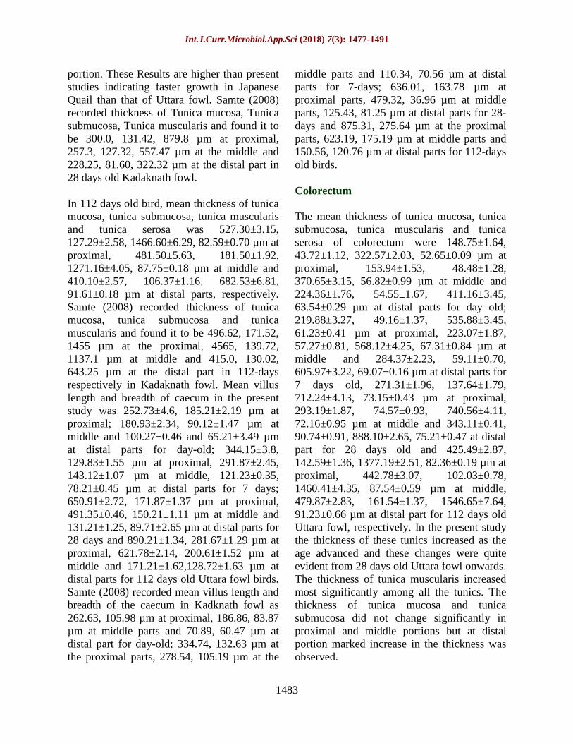
the lamina propria. The surface lining
epithelium comprised of tall columnar cells
and mucus secreting goblet cells (Fig. 1) as
reported by Aitken (1958), Nickel et al.,
(1977) and Usha Kumary et al., (1994). The
lamina propria consisted of loose connective
tissue fibres infiltrated with large number of
lymphocytes. The lymphoid tissues were
found to be present in mucosa, submucosa and
core of villi. These findings are in line with
the findings of Majeed et al., (2009), Samte
(2008) and Nasrin et al., (2012). The lamina
muscularis mucosae in both caeca and
colorectum consisted of thin layer of muscles
which extended to the core of villus in the
mucosal folds. The tunica submucosa was
seen as a thin layer and was more or less
absent in young ages. Small blood vessels
were occasionally seen in the submucosa.
Tunica muscularis was made up of two layers,
inner circular and an outer longitudinal muscle
layer. The outermost layer was tunica serosa,
which was made up of loose connective tissue.
Samte (2008) also reported in Kadaknath fowl
that a thin layer of lamina muscularis mucosae
is present having only a few bundles of muscle
fibres which extends to the core of villi and
mucosal folds in caecum as well as rectum.
The height of the villi was more in proximal
part compared to middle part of the caecum
Int.J.Curr.Microbiol.App.Sci (2018) 7(3): 1477-1491
1480
within the same group while the villi were
short, blunt and very few in number in the
distal part but large mucosal folds were
present. The height and breadth of the villi
increased in both proximal and middle parts as
the age advanced. The increase in the height
and width of villi at proximal part may be for
better absorption of nutrients. These findings
are in accordance with Samte (2008).
Caecum
The wall of caecum consisted of all the tunics
i.e. tunica mucosa, tunica submucosa, tunica
muscularis and tunica serosa and caecum was
divided into proximal, middle and distal
portions in all studied age groups as observed
by Nasrin et al., (2012), Majeed et al., (2009),
Samte (2008), Dellmann and Eurell (1998).
The surface epithelial lining consisted of
simple columnar cells and goblet cells. The
lamina propria, submucosa and core of villi
consisted of loose connective tissue fibres
infiltrated with large number of lymphocytes.
Similar observations were recorded by Majeed
et al., (2009), Nasrin et al., (2012) in chicken
and Samte (2008) in Kadaknath fowl. An
aggregation of lymphocytes forming a nodular
appearance was seen towards the base of
crypts of Lieberkuhn in the lamina propria in
28 and 112 days old birds but they were not
noted in other studied age groups. The
lymphatic nodules enclosed by a capsule were
observed only in case of distal portion of
caecum in 28 and 112 days old birds only.
Rest of the portions of caeca as well as
colorectum showed lymphatic aggregations
without presence of connective tissue capsule
around them. Samte (2008) reported presence
of lymphatic nodules only in 112 days old
Kadaknath fowl and did not mention any
finding regarding encapsulated lymphatic
aggregations. The lamina muscularis in both
right and left caeca consisted of thin layer of
muscles which extended to the core of the
villus and the mucosal folds as observed by
Samte (2008). The tunica muscularis consisted
of an inner circular coat which was well
developed but the outer longitudinal layer was
thin and hardly appreciable in day old and 7
days old bird. The tunica serosa was also
present and consisted of a loose connective
tissue (Fig. 2).
The structure of proximal caeca resembles
with the structure of jejunum giving indication
of same functionality this finding is supported
by Fenna and Boag (1974). The proximal part
of caecum consisted of prominent villi. The
villi being well developed both in length and
breadth but the crypts of Liberkuhn were short
and rounded. Nasrin et al., (2012), Majeed et
al., (2009) and Samte (2008) also reported the
presence of long villi in the proximal portion
of caecum. Longer villi are interdigitating and
opening is narrow, giving it a mesh like
appearance hence it may function as a filter
for large course particles entering into caeca.
This finding is in accordance with Ferrer et
al., (2008). Large numbers of goblet cells
were present in epithelium but they did not
outnumber the columnar cells in caecum. This
finding is in accordance with Samte (2008),
Dellmann and Eurell (1998). The lamina
muscularis mucosa was poorly developed and
consisted of only a few bundles of muscle
fibres. The tunica submucosa was present as a
thin layer. The proximal portion of caecum is
composed of thicker tunica muscularis layer
than middle and distal portions of caecum
making its wall thicker. The tunica muscularis
was composed of a thin, outer longitudinal
layer and inner circular layer which was
thicker than outer longitudinal layer. Calhoun
(1954) in chicken reported that the tunica
muscularis thickness and arrangement varies
greatly within the entire length of the
intestinal tracts. He reported that the proximal
part of the caecum has tunica muscularis with
thick inner circular and thin outer longitudinal
muscle layer. The middle part of caecum had
shorter but broader villi, mucosal folds were
Int.J.Curr.Microbiol.App.Sci (2018) 7(3): 1477-1491
1481
seen. The tunica submucosa was present as a
thin layer below lamina muscularis mucosa
but thickening of tunica submucosa was seen
at the regions where plica was present. Plicae
are well developed folds of the mucus
membrane. Only a thin layer of the lamina
muscularis mucosa was observed in the distal
part of caecum which enters core of villi. Size
of tunica muscularis externa increased in the
middle portion of caecum. These are similar
with Dellmann and Eurell (1998) and Samte
(2008). In present study middle part of the
caecum was showing distinct inner circular
and outer longitudinal layer in chicks but
definite arrangement of these layers was lost
in older birds similar observations were
recorded by Calhoun (1954).
The distal part of caecum was found to have
large mucosal folds and the villi were short,
straight and very few in numbers. This portion
normally possessed the largest diameter of the
whole organ, but the lamina muscularis
mucosa, tunica submucosa and the tunica
mucularis were very similar to preceding
region. Samte (2008) reported that the distal
portion of caecum contains mucosal folds and
villi are very short. Dellman and Eurell (1998)
reported that prominent villi were observed in
the proximal portion, shorter and broader in
middle portion and the distal portion was
having short villi or devoid of villi. Nodular
type of lymphoid aggregation was seen in the
lamina propria of caecum of 28 days and 112
days old Uttara fowl but their presence was
not noted in other age groups. Nickel et al.,
(1977) stated that the intestinal mucosa is rich
in lymphoreticular tissue which is arranged
diffusely or in follicles and even extends into
the villi. They also reported that the lymphoid
tissue is particularly abundant in the caeca.
Majeed et al., (2009) reported the maximum
numbers of lymphatic nodules were present in
the proximal portion of caecum then middle
and distal portions contains least number of
lymphatic nodules while he did not find any
significant difference in the length and width
of lymphatic nodules in different portions of
caecum. The lymphatic aggregation in the
proximal portion of caeca forms a nodular
structure, caecal tonsil which may act as
immunological surveillance against foreign
microorganisms entering caeca with urine as a
result of reverse peristalsis same was reported
by Getty (1975). Myentric plexus was
observed in the outer longitudinal layer of
tunica muscularis. Neuroendocrine cells with
large round nuclei were observed in the crypts
of lieberkuhn. Similar findings were reported
by Samte (2008).
Colorectum
The wall of the colorectum at proximal,
middle and distal portions consisted of tunica
mucosa, tunica submucosa, tunica muscularis
and tunica serosa in the entire studied group.
The surface epithelial lining consisted of
simple columnar cells with goblet cells. The
columnar cells tend to be obscured by large
number of distended goblet cells. Aitkin
(1958) and Samte (2008) reported similar
results in broiler, native fowl and Kadaknath
fowl respectively. The lamina propria
consisted of loose connective tissue fibres and
was infiltrated with large number of
lymphocytes. Nodular type of lymphoid
aggregation was seen in the lamina propria of
colon of 112 days old Uttara fowl but their
presence was not noted in other age groups.
The lamina muscularis of colorectum
consisted of thin layer of muscles which
extended to the core of villus in the mucosal
folds.
The tunica submucosa was seen as a thin layer
of connective tissue. Tunica muscularis was
divided into tunica muscularis interna and
externa. The tunica serosa was also present
and consisted of a layer of loose connective
tissue.
Int.J.Curr.Microbiol.App.Sci (2018) 7(3): 1477-1491
1482
The tunica muscularis consists of an inner
circular layer which was well developed but
the outer longitudinal layer was thin. Present
results corresponds to the results of Sissons
and Grossman (1953) who reported that the
tunica muscularis is well developed in colon
with the inner circular layer thicker than the
outer longitudinal layer. The thickness of
tunica muscularis was found to increase
caudally in the present studies. Ushakumary et
al., (1994) had reported similar observations
in quail. The tunica mucosa of colorectum was
thrown into villi. Sisson and Grossman (1953)
and Nickel et al., (1977) in domestic fowl and
Ushakumary et al., (1994) in quail reported
the presence of villi in colon. In the proximal
part of colon, villi were branched and
digitated, few unbranched individual villus
were also seen. The middle part of villi was
digitated and branched similar to proximal
part. In the distal part of colorectum there
were large mucosal folds showing branching.
The unbranched individual villi were also
observed in between large mucosal folds in
the distal region. However, among different
age groups studied, the branched villi and
mucosal folds were lesser in day old Uttara
fowl. Ushakumary et al., (1994) reported that
histological differences exist along the length
of colon in initial and terminal portions in
quail. They also observed that initial part of
the colon presents branched and digitated villi
while the terminal part of colon had a large
mucosal fold with small villi. They also
observed that the branched villi and the
mucosal folds were lesser in the day old birds
These observations corresponds to present
studies contrary to these findings Sisson and
Grossman (1953) reported that the villi in
colon were short in chicken.
Histomorphometry
Caecum
The thickness of tunica mucosa, tunica
submucosa, tunica muscularis of caecum were
105.95±1.36 (Table 1), 44.40±1.29,
351.64±3.33 (Table 2), 52.73±0.93 µm at
proximal, 86.19±1.85, 29.31±0.82,
306.52±2.78, 52.87±0.05 µm at middle and
57.29±1.92, 23.66±0.99, 212.44±4.19,
57.18±0.76 µm at distal parts, respectively in a
day old bird. Samte (2008) reported mean
thickness of tunica mucosa, tunica submucosa
and tunica muscularis of caecum as 98.22,
76.07, 311.25 µm at proximal part, 76.07,
49.8, 286.35 µm at the middle and 69.172,
47.02, 127.27 µm at the distal part in day old
kadaknath fowl birds. In a 7 days old bird,
mean thickness of tunica mucosa, tunica
submucosa, tunica muscularis and tunica
Serosa were 275.90±1.59, 65.23±1.37,
486.49±3.45, 60.15±0.41 µm at proximal,
160.39±3.12, 41.89±0.59, 456.24±3.78,
62.13±0.18 µm at middle and 135.9±1.76,
30.54±2.86, 300.52±3.06, 65.17±0.19 µm at
distal parts, respectively. Hamedi et al., (2013)
recorded mucosal, submucosal and muscular
width in 10 day old Japanese Quail as 0.40,
0.09 and 0.06 mm in proximal portion 0.85,
0.06 and 0.88 mm respectively for middle
portion and 0.05, 0.04 and 0.6 mm at distal
portion. Samte (2008) reported the mean
thickness of Tunica Mucosa, Submucosa and
muscularis as 250.37, 85.77, 450 µm at the
proximal, 222.72, 73.32, 301.57 µm at the
middle and 208.87, 56.72, 201.97 µm at the
distal part in 7 days old Kadaknath fowl birds.
In 28 days old bird, mean thickness of tunica
mucosa, tunica submucosa, tunica muscularis
and tunica serosa was 320.82±3.04,
114.50±1.71, 884.55±3.23, 65.93±0.72 µm at
proximal, 270.13±3.97, 88.58±1.83,
848.41±1.62, 71.35±0.01 µm at middle and
254.25±3.97, 86.28±0.95, 436.96±5.04,
75.09±0.76 µm at distal parts, respectively.
Hamedi et al., (2013) recorded width of
mucosa, submucosa and muscularis in 30 day
old Japanese Quail as 0.45, 0.12 and 0.12 mm
respectively at proximal end 0.89, 0.06 and
0.88 mm respectively at middle portion 1.38,
0.06 and 1.27 mm respectively at distal
Int.J.Curr.Microbiol.App.Sci (2018) 7(3): 1477-1491
1483
portion. These Results are higher than present
studies indicating faster growth in Japanese
Quail than that of Uttara fowl. Samte (2008)
recorded thickness of Tunica mucosa, Tunica
submucosa, Tunica muscularis and found it to
be 300.0, 131.42, 879.8 µm at proximal,
257.3, 127.32, 557.47 µm at the middle and
228.25, 81.60, 322.32 µm at the distal part in
28 days old Kadaknath fowl.
In 112 days old bird, mean thickness of tunica
mucosa, tunica submucosa, tunica muscularis
and tunica serosa was 527.30±3.15,
127.29±2.58, 1466.60±6.29, 82.59±0.70 µm at
proximal, 481.50±5.63, 181.50±1.92,
1271.16±4.05, 87.75±0.18 µm at middle and
410.10±2.57, 106.37±1.16, 682.53±6.81,
91.61±0.18 µm at distal parts, respectively.
Samte (2008) recorded thickness of tunica
mucosa, tunica submucosa and tunica
muscularis and found it to be 496.62, 171.52,
1455 µm at the proximal, 4565, 139.72,
1137.1 µm at middle and 415.0, 130.02,
643.25 µm at the distal part in 112-days
respectively in Kadaknath fowl. Mean villus
length and breadth of caecum in the present
study was 252.73±4.6, 185.21±2.19 µm at
proximal; 180.93±2.34, 90.12±1.47 µm at
middle and 100.27±0.46 and 65.21±3.49 µm
at distal parts for day-old; 344.15±3.8,
129.83±1.55 µm at proximal, 291.87±2.45,
143.12±1.07 µm at middle, 121.23±0.35,
78.21±0.45 µm at distal parts for 7 days;
650.91±2.72, 171.87±1.37 µm at proximal,
491.35±0.46, 150.21±1.11 µm at middle and
131.21±1.25, 89.71±2.65 µm at distal parts for
28 days and 890.21±1.34, 281.67±1.29 µm at
proximal, 621.78±2.14, 200.61±1.52 µm at
middle and 171.21±1.62,128.72±1.63 µm at
distal parts for 112 days old Uttara fowl birds.
Samte (2008) recorded mean villus length and
breadth of the caecum in Kadknath fowl as
262.63, 105.98 µm at proximal, 186.86, 83.87
µm at middle parts and 70.89, 60.47 µm at
distal part for day-old; 334.74, 132.63 µm at
the proximal parts, 278.54, 105.19 µm at the
middle parts and 110.34, 70.56 µm at distal
parts for 7-days; 636.01, 163.78 µm at
proximal parts, 479.32, 36.96 µm at middle
parts, 125.43, 81.25 µm at distal parts for 28-
days and 875.31, 275.64 µm at the proximal
parts, 623.19, 175.19 µm at middle parts and
150.56, 120.76 µm at distal parts for 112-days
old birds.
Colorectum
The mean thickness of tunica mucosa, tunica
submucosa, tunica muscularis and tunica
serosa of colorectum were 148.75±1.64,
43.72±1.12, 322.57±2.03, 52.65±0.09 µm at
proximal, 153.94±1.53, 48.48±1.28,
370.65±3.15, 56.82±0.99 µm at middle and
224.36±1.76, 54.55±1.67, 411.16±3.45,
63.54±0.29 µm at distal parts for day old;
219.88±3.27, 49.16±1.37, 535.88±3.45,
61.23±0.41 µm at proximal, 223.07±1.87,
57.27±0.81, 568.12±4.25, 67.31±0.84 µm at
middle and 284.37±2.23, 59.11±0.70,
605.97±3.22, 69.07±0.16 µm at distal parts for
7 days old, 271.31±1.96, 137.64±1.79,
712.24±4.13, 73.15±0.43 µm at proximal,
293.19±1.87, 74.57±0.93, 740.56±4.11,
72.16±0.95 µm at middle and 343.11±0.41,
90.74±0.91, 888.10±2.65, 75.21±0.47 at distal
part for 28 days old and 425.49±2.87,
142.59±1.36, 1377.19±2.51, 82.36±0.19 µm at
proximal, 442.78±3.07, 102.03±0.78,
1460.41±4.35, 87.54±0.59 µm at middle,
479.87±2.83, 161.54±1.37, 1546.65±7.64,
91.23±0.66 µm at distal part for 112 days old
Uttara fowl, respectively. In the present study
the thickness of these tunics increased as the
age advanced and these changes were quite
evident from 28 days old Uttara fowl onwards.
The thickness of tunica muscularis increased
most significantly among all the tunics. The
thickness of tunica mucosa and tunica
submucosa did not change significantly in
proximal and middle portions but at distal
portion marked increase in the thickness was
observed.
Int.J.Curr.Microbiol.App.Sci (2018) 7(3): 1477-1491
1484
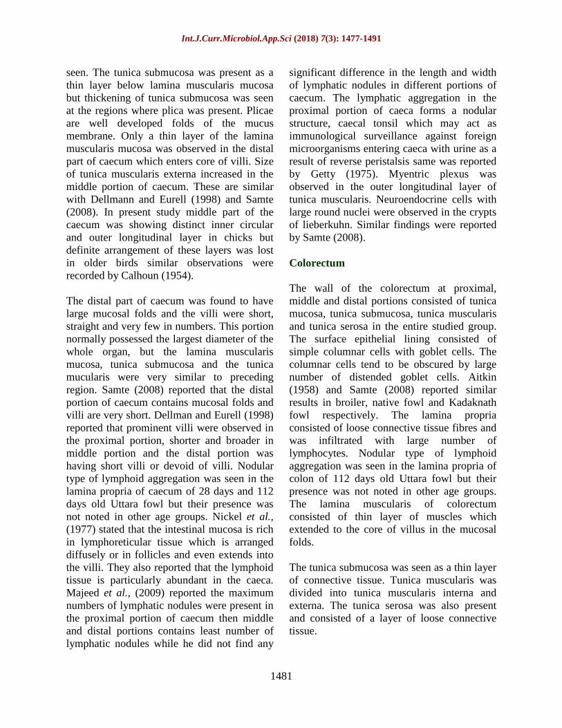
Table.1 Mean thickness of tunica mucosa of various segments of large intestine (µm)
Various
segments
Age Group
Day 1 Day 7 Day 28 Day 112
Caecum
Proximal portion 105.95±1.36 275.90±1.59 320.82±3.04 527.30±3.15
Middle portion 86.19±1.85 160.39±3.12 270.13±3.97 481.50±5.63
Distal portion 57.29±1.92 135.9±1.76 254.25±3.97 410.10±2.57
Colorectum Proximal portion 148.75±1.64 219.88±3.27 271.31±1.96 425.49±2.87
Middle portion 153.94±1.53 223.07±1.87 293.19±1.87 442.78±3.07
Distal portion 224.36±1.76 284.37±2.23 343.11±6.41 479.87±2.83
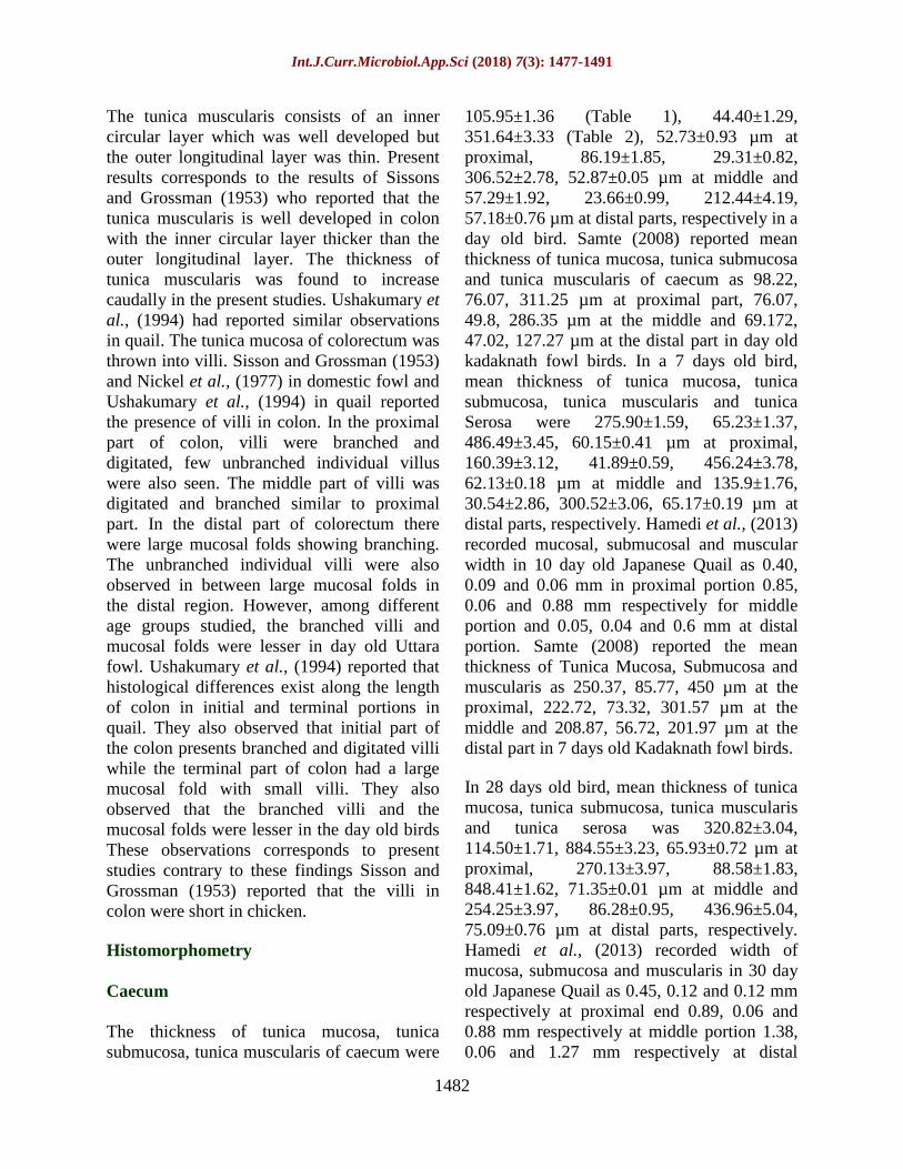
Table.2 Mean thickness of tunica muscularis of various segments of large intestine (µm)
Various
segments
Age Group
Day 1 Day 7 Day 28 Day 112
Caecum
Proximal portion 351.64±3.33 486.49±3.45 884.55±3.23 1466.60±6.29
Middle portion 306.52±2.78 456.24±3.78 848.41±1.62 1271.16±4.05
Distal portion 212.44±4.19 300.52±3.06 436.96±5.04 682.53±6.81
Colorectum Proximalportion 322.57±2.03 535.88±3.64 712.24±4.13 1377.19±2.51
Middle portion 370.65±3.15 568.12±4.25 740.56±4.11 1460.41±4.35
Distal portion 411.16±3.45 605.97±3.22 888.10±2.65 1546.65±7.64
Fig.1 Photomicrograph showing goblet cells (G), columnar cells (cc) in the villi of 112 days old
bird. (H&E X1000)

Int.J.Curr.Microbiol.App.Sci (2018) 7(3): 1477-1491
1485
Fig.2 Photomicrograph showing long villi (V), crypts of lieberkuhn (CL), intestinal glands (IG),
tunica muscularis (TM) and tunica serosa (TS) in proximal caecum of day old bird. (H&E X100)
Fig.3 Photomicrograph showing network of reticular fibres (arrow) in lamina propria of
proximal caecum in 28 day old caecum (Gomori’s stain X400)
Int.J.Curr.Microbiol.App.Sci (2018) 7(3): 1477-1491
1486
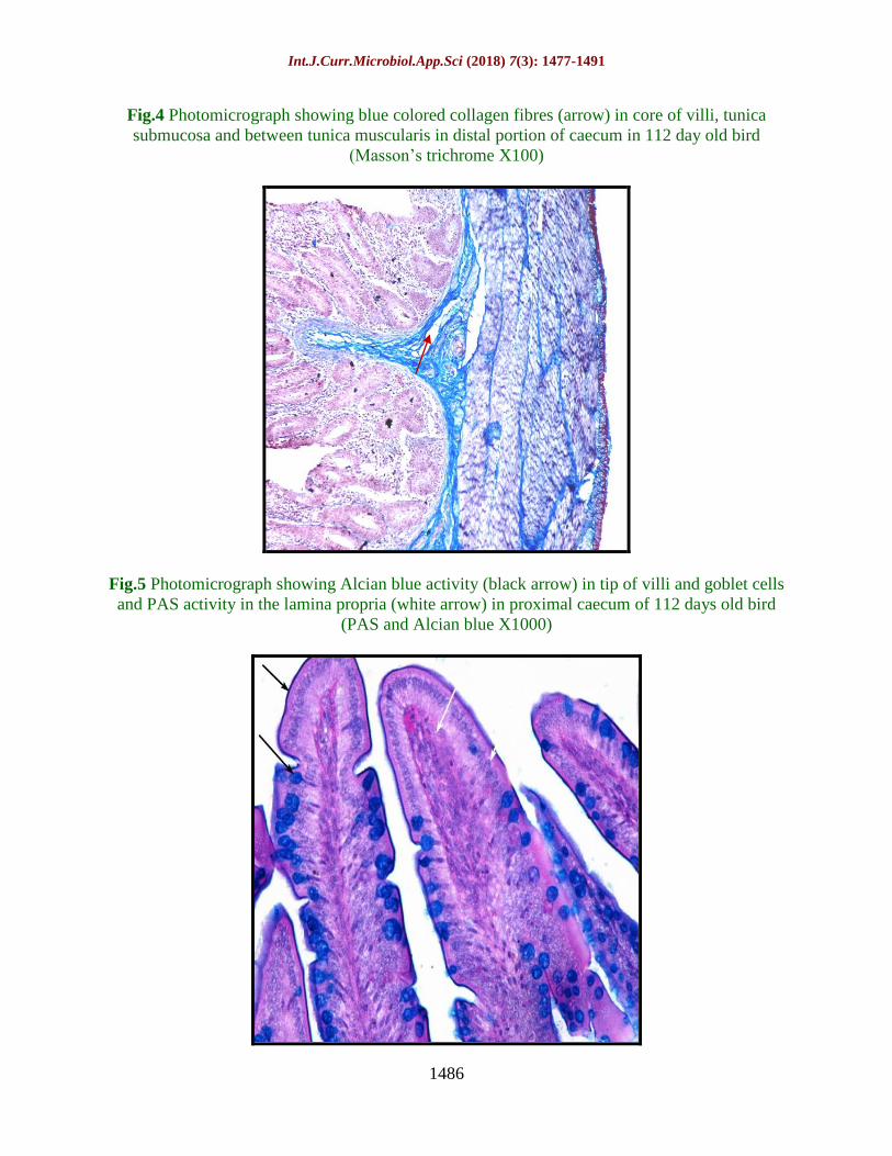
Fig.4 Photomicrograph showing blue colored collagen fibres (arrow) in core of villi, tunica
submucosa and between tunica muscularis in distal portion of caecum in 112 day old bird
(Masson’s trichrome X100)
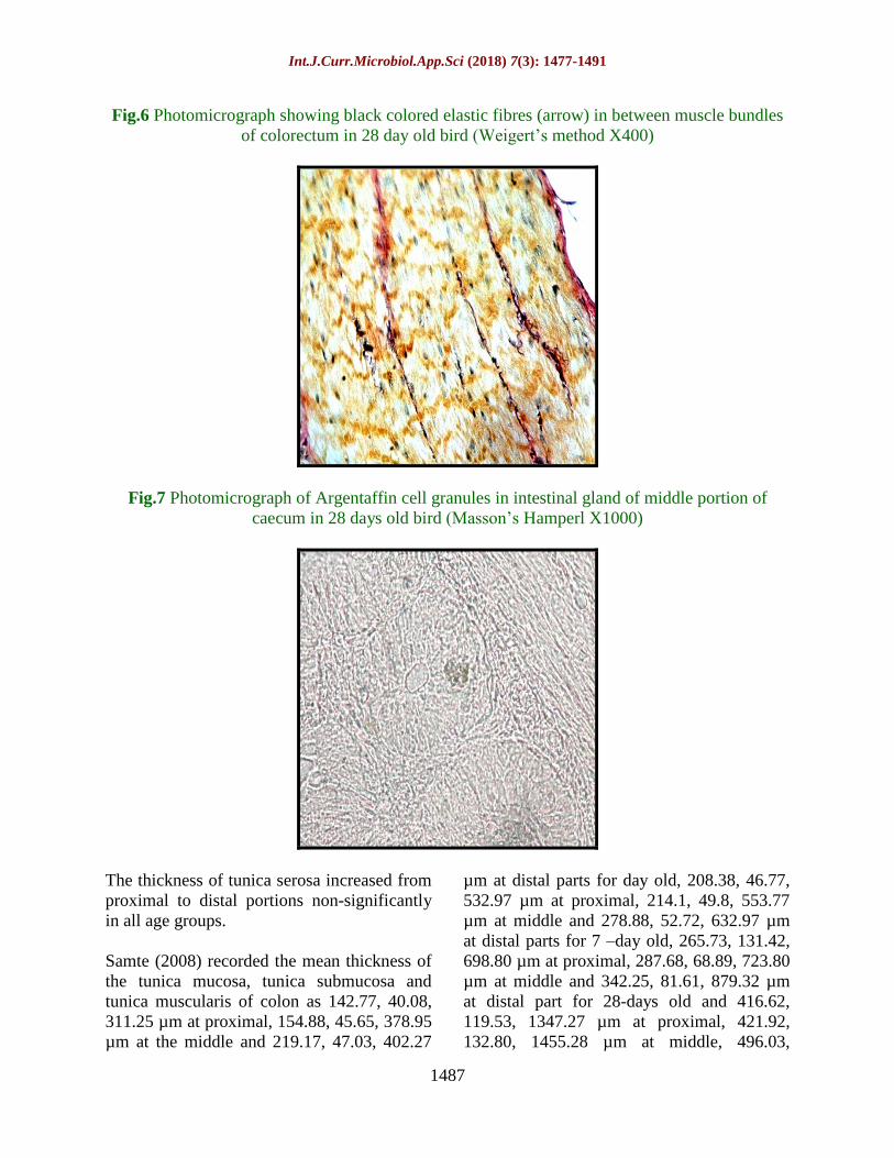
Fig.5 Photomicrograph showing Alcian blue activity (black arrow) in tip of villi and goblet cells
and PAS activity in the lamina propria (white arrow) in proximal caecum of 112 days old bird
(PAS and Alcian blue X1000)
Int.J.Curr.Microbiol.App.Sci (2018) 7(3): 1477-1491
1487
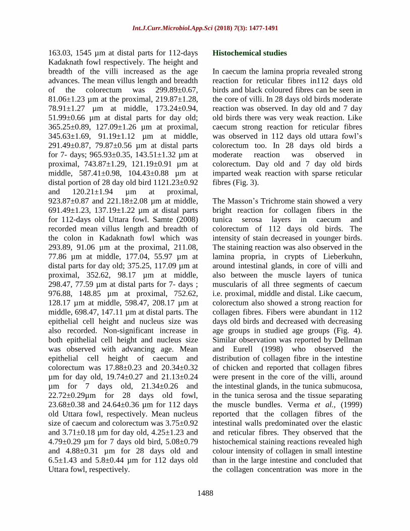
Fig.6 Photomicrograph showing black colored elastic fibres (arrow) in between muscle bundles
of colorectum in 28 day old bird (Weigert’s method X400)
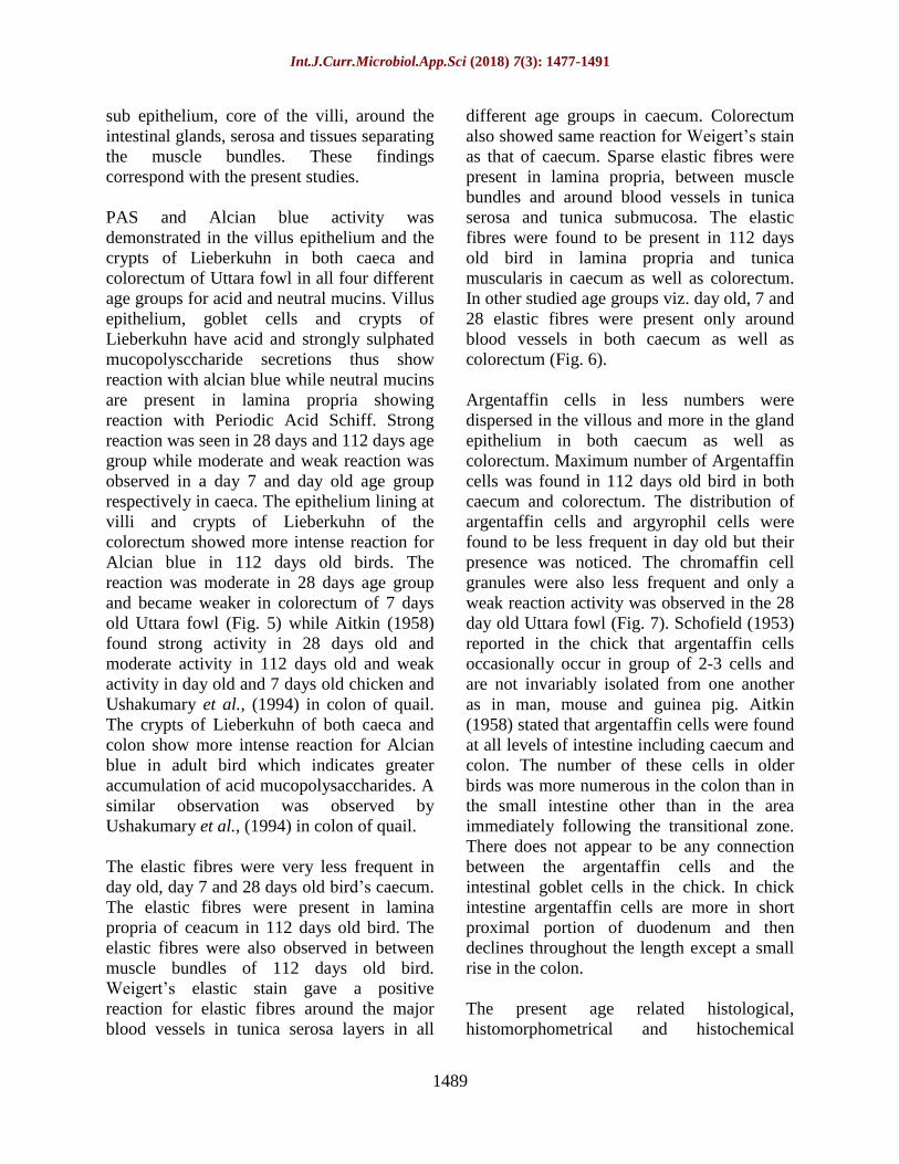
Fig.7 Photomicrograph of Argentaffin cell granules in intestinal gland of middle portion of
caecum in 28 days old bird (Masson’s Hamperl X1000)
The thickness of tunica serosa increased from
proximal to distal portions non-significantly
in all age groups.
Samte (2008) recorded the mean thickness of
the tunica mucosa, tunica submucosa and
tunica muscularis of colon as 142.77, 40.08,
311.25 µm at proximal, 154.88, 45.65, 378.95
µm at the middle and 219.17, 47.03, 402.27
µm at distal parts for day old, 208.38, 46.77,
532.97 µm at proximal, 214.1, 49.8, 553.77
µm at middle and 278.88, 52.72, 632.97 µm
at distal parts for 7 –day old, 265.73, 131.42,
698.80 µm at proximal, 287.68, 68.89, 723.80
µm at middle and 342.25, 81.61, 879.32 µm
at distal part for 28-days old and 416.62,
119.53, 1347.27 µm at proximal, 421.92,
132.80, 1455.28 µm at middle, 496.03,
Int.J.Curr.Microbiol.App.Sci (2018) 7(3): 1477-1491
1488
163.03, 1545 µm at distal parts for 112-days
Kadaknath fowl respectively. The height and
breadth of the villi increased as the age
advances. The mean villus length and breadth
of the colorectum was 299.89±0.67,
81.06±1.23 µm at the proximal, 219.87±1.28,
78.91±1.27 µm at middle, 173.24±0.94,
51.99±0.66 µm at distal parts for day old;
365.25±0.89, 127.09±1.26 µm at proximal,
345.63±1.69, 91.19±1.12 µm at middle,
291.49±0.87, 79.87±0.56 µm at distal parts
for 7- days; 965.93±0.35, 143.51±1.32 µm at
proximal, 743.87±1.29, 121.19±0.91 µm at
middle, 587.41±0.98, 104.43±0.88 µm at
distal portion of 28 day old bird 1121.23±0.92
and 120.21±1.94 µm at proximal,
923.87±0.87 and 221.18±2.08 µm at middle,
691.49±1.23, 137.19±1.22 µm at distal parts
for 112-days old Uttara fowl. Samte (2008)
recorded mean villus length and breadth of
the colon in Kadaknath fowl which was
293.89, 91.06 µm at the proximal, 211.08,
77.86 µm at middle, 177.04, 55.97 µm at
distal parts for day old; 375.25, 117.09 µm at
proximal, 352.62, 98.17 µm at middle,
298.47, 77.59 µm at distal parts for 7- days ;
976.88, 148.85 µm at proximal, 752.62,
128.17 µm at middle, 598.47, 208.17 µm at
middle, 698.47, 147.11 µm at distal parts. The
epithelial cell height and nucleus size was
also recorded. Non-significant increase in
both epithelial cell height and nucleus size
was observed with advancing age. Mean
epithelial cell height of caecum and
colorectum was 17.88±0.23 and 20.34±0.32
µm for day old, 19.74±0.27 and 21.13±0.24
µm for 7 days old, 21.34±0.26 and
22.72±0.29µm for 28 days old fowl,
23.68±0.38 and 24.64±0.36 µm for 112 days
old Uttara fowl, respectively. Mean nucleus
size of caecum and colorectum was 3.75±0.92
and 3.71±0.18 µm for day old, 4.25±1.23 and
4.79±0.29 µm for 7 days old bird, 5.08±0.79
and 4.88±0.31 µm for 28 days old and
6.5±1.43 and 5.8±0.44 µm for 112 days old
Uttara fowl, respectively.
Histochemical studies
In caecum the lamina propria revealed strong
reaction for reticular fibres in112 days old
birds and black coloured fibres can be seen in
the core of villi. In 28 days old birds moderate
reaction was observed. In day old and 7 day
old birds there was very weak reaction. Like
caecum strong reaction for reticular fibres
was observed in 112 days old uttara fowl’s
colorectum too. In 28 days old birds a
moderate reaction was observed in
colorectum. Day old and 7 day old birds
imparted weak reaction with sparse reticular
fibres (Fig. 3).
The Masson’s Trichrome stain showed a very
bright reaction for collagen fibers in the
tunica serosa layers in caecum and
colorectum of 112 days old birds. The
intensity of stain decreased in younger birds.
The staining reaction was also observed in the
lamina propria, in crypts of Lieberkuhn,
around intestinal glands, in core of villi and
also between the muscle layers of tunica
muscularis of all three segments of caecum
i.e. proximal, middle and distal. Like caecum,
colorectum also showed a strong reaction for
collagen fibres. Fibers were abundant in 112
days old birds and decreased with decreasing
age groups in studied age groups (Fig. 4).
Similar observation was reported by Dellman
and Eurell (1998) who observed the
distribution of collagen fibre in the intestine
of chicken and reported that collagen fibres
were present in the core of the villi, around
the intestinal glands, in the tunica submucosa,
in the tunica serosa and the tissue separating
the muscle bundles. Verma et al., (1999)
reported that the collagen fibres of the
intestinal walls predominated over the elastic
and reticular fibres. They observed that the
histochemical staining reactions revealed high
colour intensity of collagen in small intestine
than in the large intestine and concluded that
the collagen concentration was more in the
Int.J.Curr.Microbiol.App.Sci (2018) 7(3): 1477-1491
1489
sub epithelium, core of the villi, around the
intestinal glands, serosa and tissues separating
the muscle bundles. These findings
correspond with the present studies.
PAS and Alcian blue activity was
demonstrated in the villus epithelium and the
crypts of Lieberkuhn in both caeca and
colorectum of Uttara fowl in all four different
age groups for acid and neutral mucins. Villus
epithelium, goblet cells and crypts of
Lieberkuhn have acid and strongly sulphated
mucopolysccharide secretions thus show
reaction with alcian blue while neutral mucins
are present in lamina propria showing
reaction with Periodic Acid Schiff. Strong
reaction was seen in 28 days and 112 days age
group while moderate and weak reaction was
observed in a day 7 and day old age group
respectively in caeca. The epithelium lining at
villi and crypts of Lieberkuhn of the
colorectum showed more intense reaction for
Alcian blue in 112 days old birds. The
reaction was moderate in 28 days age group
and became weaker in colorectum of 7 days
old Uttara fowl (Fig. 5) while Aitkin (1958)
found strong activity in 28 days old and
moderate activity in 112 days old and weak
activity in day old and 7 days old chicken and
Ushakumary et al., (1994) in colon of quail.
The crypts of Lieberkuhn of both caeca and
colon show more intense reaction for Alcian
blue in adult bird which indicates greater
accumulation of acid mucopolysaccharides. A
similar observation was observed by
Ushakumary et al., (1994) in colon of quail.
The elastic fibres were very less frequent in
day old, day 7 and 28 days old bird’s caecum.
The elastic fibres were present in lamina
propria of ceacum in 112 days old bird. The
elastic fibres were also observed in between
muscle bundles of 112 days old bird.
Weigert’s elastic stain gave a positive
reaction for elastic fibres around the major
blood vessels in tunica serosa layers in all
different age groups in caecum. Colorectum
also showed same reaction for Weigert’s stain
as that of caecum. Sparse elastic fibres were
present in lamina propria, between muscle
bundles and around blood vessels in tunica
serosa and tunica submucosa. The elastic
fibres were found to be present in 112 days
old bird in lamina propria and tunica
muscularis in caecum as well as colorectum.
In other studied age groups viz. day old, 7 and
28 elastic fibres were present only around
blood vessels in both caecum as well as
colorectum (Fig. 6).
Argentaffin cells in less numbers were
dispersed in the villous and more in the gland
epithelium in both caecum as well as
colorectum. Maximum number of Argentaffin
cells was found in 112 days old bird in both
caecum and colorectum. The distribution of
argentaffin cells and argyrophil cells were
found to be less frequent in day old but their
presence was noticed. The chromaffin cell
granules were also less frequent and only a
weak reaction activity was observed in the 28
day old Uttara fowl (Fig. 7). Schofield (1953)
reported in the chick that argentaffin cells
occasionally occur in group of 2-3 cells and
are not invariably isolated from one another
as in man, mouse and guinea pig. Aitkin
(1958) stated that argentaffin cells were found
at all levels of intestine including caecum and
colon. The number of these cells in older
birds was more numerous in the colon than in
the small intestine other than in the area
immediately following the transitional zone.
There does not appear to be any connection
between the argentaffin cells and the
intestinal goblet cells in the chick. In chick
intestine argentaffin cells are more in short
proximal portion of duodenum and then
declines throughout the length except a small
rise in the colon.
The present age related histological,
histomorphometrical and histochemical
Int.J.Curr.Microbiol.App.Sci (2018) 7(3): 1477-1491
1490
features of large intestine of Uttara fowl
would immensely help in developing a
baseline data for further studies in this field.
Acknowledgement
The authors are very much grateful to the
Dean, CVASc., GBPUAT, Pantnagar and In-
Charge, Electron Microscopy Lab, GBPUAT,
Pantnagar for providing necessary facilities in
carrying out the research work in time. The
authors are thankful to the Indian Council of
Agricultural Research, New Delhi (ICAR-
JRF) for providing the financial assistance.
References
Aitkin, R.N.C. 1958. A histochemical study
of the stomach and intestine of the
chicken. J. Anat. 92: 453-466.
Argenzio, R. A. 1980. Pathophysiology of
diarrhoea. Veterinary Gastroenterology.
Lea and Febiger publication Ltd.,
Philadelphia, USA. Pp. 172-198.
Bancroft, J.D., Layton, C. and Suvarna, S.K.
2013. Bancroft’s theory and practice of
histological techniques.
Bayer, R. C., Rittenburg, J. H., Bird, F. H.,
Chawan, C. B., and Allen, M. 1981.
Influence of short term fasting on
chicken alimentary canal mucosa.
Poultry Science. 60: 1293-1302.
Braun, E. J., and Duke, G. E. 1989. Function
of the avian cecum. J. Exp. Zool. Suppl.
(USA). Pp. 16-22.
Calhoun, M. L. 1954. Microscopic anatomy
of the digestive system of chicken.
Ames, Iowa: Iowa State College Press.
Pp:1-108.
Culling, C. F. A. 1969. In “Handbook of
Histopatological technique (including
museum technique).” 2: 228 – 238.
Dellman and Eurell, J. 1998. Textbook of
veterinary histology. 5th Edition,
Williams & Wilkins, a waverly
company, Baltimore, Philadelphia,
London.
Fenna, L. and Boag, D.A. 1974. Filling and
emptying of galliform caecum.
Canadian Journal of Zoology 52: 537-
540.
Ferrer, R., Jana, M. P. and Durfort, M. M.
2008. Morphological study of the cecal
epithelium of chicken. Brit. Poultry Sci.
32 (4):679-691.
Getty, R. 1975. Sisson and Grossman’s the
Anatomy of the Domestic Animals, 5th
Edition. vol. 2. WB Saunders,
Philadelphia, PA.
Giemsa, G. 1902. The azure dyes: Their
purification and physiochemical
properties. Zentralblatt fiir
Bakteriologie Parasitenkunde
infectienskrankherten and Hygiene. 31:
429.
Hamedi, H., Abdel-Wahab El-Ghareeb,
Zaher, M., AbuAmod, F. 2013.
Anatomical, Histological, Histo
chemical adaptations of the avian
elementary canal to their food habbits.
IJSER. 4(10).
Kaur, N. 2007. Studies on morphological,
haemato-biochemical and production
traits in local hill fowl of Pithoragarh
region. Ph.D. Thesis. G.B.P.U.A&T,
Pantnagar.
Kaur, N., Kumar, S., Singh, B., Pandey, A.K.
and Somvanshi, S.P.S. 2010.
Morphological characterization of
feathered shank local hill fowl of
Central Himalayan Region of India.
Indian J. Anim. Sci. 80(9): 934-936.
Luna. L.G. 1972. Manual of Histological
Staining Methods of the Armed Forces
Institute of Pathology. The Blakiston
Division, McGraw Hill Book Co. N.Y.
Pp. 162, 163 and 164.
Majeed, M. F., F.S. Al- Asadi, F. S., Al.
Nassir, A. N., Rahi, E. H. 2009. The
morphological and histological study of
Int.J.Curr.Microbiol.App.Sci (2018) 7(3): 1477-1491
1491
the caecum in broiler chicken. Bas J Vet
Res. 8:19-25.
Marshall, A.J. 1960. Biology and comparative
physiology of birds. Vol. I. Academic
Press, New York and London.
McManus, J.F.A. 1946. Histological
demonstration of mucin after periodic
acid. Nature. 158: 202.
McNab, J. M. 1973. The avian caeca: a
review. Worlds Poult Sci J. 29:251-263.
Nasrin, M., Siddiqi, M. N. H., Masum, M. A.
and Wares, M. A. 2012. Gross and
histological studies of digestive tract of
broiler chicken during post natal growth
and development. J. Bangladesh Agril.
Univ. 10(1): 69-77.
Nickel, R., Schummer, A. and Seiferle, E.
1977. Anatomy of the domestic birds.
Verlag Paul Parey.
Samte, L. 2008. Gross morphometric, Light
and electron microscopic studies on the
large intestine of Kadaknath fowl.
Master’s dissertation G.B.P.U.A. & T.
Pantnagar.
Schofield, G. 1953. The argentaffin and
mucous cells of the small and large
intestine of the mouse. Acta. Anat. 18:
256-272.
Singh, I. 1964. On argyrophile and argentaffin
reactions in individual granules of
enterochromaffin cells of the human
gastro-intestinal tract. J. Anat., Lond. Pp
497-500.
Sisson, S. and Grossman, J. D. 1953. The
Anatomy of the Domestic Animals.
Philadelphia: WB Saunders Company
pp. 940-941.
Sivagnanam, S., Ramesh, G., Basha, S. H.
and Ushakumary 2004. Histological
studies of liver in Guinea Fowl. Indian
J. Vet. Anat. 16:21-25
Son, J. H., Karasawa, Y., and Nahm, K. H.
2000. Effect of caecectomy on growth,
moisture in excreta, gastrointestinal
passage time and uric acid excretion in
growing chicks. Brit. Poultry Sci. 41(1):
72-74.
Turk, D. E. 1982. The anatomy of the avian
digestive tract as related to feed
utilization. Poultry Science. 61: 1225-
1244.
Ushakumary, S., Geetha Ramesh and
Vijayaraghavan, C. 2002. Lymphoid
aggregation in caecum and colon of
Japanese Quail. Indian Journal of
Veterinary Anatomy. 14:16-21.
Vaish, M.K., Parmer, M.L., Taluja, J.S. and
Vaish Rakhi. 2006. Histological
observations on the large intestine of
posthatch Kadaknath Fowl. In
“Souvenir and Abstracts” xx Annual
Convention of Indian Association of
Veterinary Anatomistsn and National
Symposium held on 27-29 Jan., 2006 at
Jabalpur. Pp.54.
Venkatesan, S., Ramesh, G. and
Vijayaragavan, C. 2005. Age related
changes in histomorphology of the
spleen of the Japanese Quail.
IJVA.17:19-23.
Verma, D., Malik, M.R., Shrivastava, A. M.
and Parmar, M. L. 1999. Histogenesis
of intestinal villi in fowl (Gallus
domesticus). Indian J. Anim. Sci.
69(11): 902-904.
How to cite this article:
Pandit, K., B.S. Dhote, D. Mahanta, S. Sathapathy, S. Tamilselvan, M. Mrigesh and Mishra, S.
2018. Histological, Histomorphometrical and Histochemical Studies on the Large Intestine of
Uttara Fowl. Int.J.Curr.Microbiol.App.Sci. 7(03): 1477-1491.
doi: https://doi.org/10.20546/ijcmas.2018.703.176