histone h3 variant regulates rna polymerase ii transcription
TRANSCRIPT
RESEARCH ARTICLE
Histone H3 Variant Regulates RNAPolymerase II Transcription Termination andDual Strand Transcription of siRNA Loci inTrypanosoma bruceiDavid Reynolds1, Brigitte T. Hofmeister2, Laura Cliffe1, Magdy Alabady3,T. Nicolai Siegel4, Robert J. Schmitz5, Robert Sabatini1*
1 Department of Biochemistry and Molecular Biology, University of Georgia, Athens, Georgia, United Statesof America, 2 Institute of Bioinformatics, University of Georgia, Athens, Georgia, United States of America,3 Department of Plant Biology, University of Georgia, Athens, Georgia, United States of America,4 Research Center for Infectious Diseases, University of Wuerzburg, Wuerzburg, Germany, 5 Department ofGenetics, University of Georgia, Athens, Georgia, United States of America
AbstractBase J, β-D-glucosyl-hydroxymethyluracil, is a chromatin modification of thymine in the
nuclear DNA of flagellated protozoa of the order Kinetoplastida. In Trypanosoma brucei, J isenriched, along with histone H3 variant (H3.V), at sites involved in RNA Polymerase
(RNAP) II termination and telomeric sites involved in regulating variant surface glycoprotein
gene (VSG) transcription by RNAP I. Reduction of J in T. brucei indicated a role of J in the
regulation of RNAP II termination, where the loss of J at specific sites within polycistronic
gene clusters led to read-through transcription and increased expression of downstream
genes. We now demonstrate that the loss of H3.V leads to similar defects in RNAP II termi-
nation within gene clusters and increased expression of downstream genes. Gene dere-
pression is intensified upon the subsequent loss of J in the H3.V knockout. mRNA-seq
indicates gene derepression includes VSG genes within the silent RNAP I transcribed telo-
meric gene clusters, suggesting an important role for H3.V in telomeric gene repression and
antigenic variation. Furthermore, the loss of H3.V at regions of overlapping transcription at
the end of convergent gene clusters leads to increased nascent RNA and siRNA produc-
tion. Our results suggest base J and H3.V can act independently as well as synergistically
to regulate transcription termination and expression of coding and non-coding RNAs in
T. brucei, depending on chromatin context (and transcribing polymerase). As such these
studies provide the first direct evidence for histone H3.V negatively influencing transcription
elongation to promote termination.
PLOS Genetics | DOI:10.1371/journal.pgen.1005758 January 21, 2016 1 / 25
a11111
OPEN ACCESS
Citation: Reynolds D, Hofmeister BT, Cliffe L,Alabady M, Siegel TN, Schmitz RJ, et al. (2016)Histone H3 Variant Regulates RNA Polymerase IITranscription Termination and Dual StrandTranscription of siRNA Loci in Trypanosoma brucei.PLoS Genet 12(1): e1005758. doi:10.1371/journal.pgen.1005758
Editor: Luisa Figueiredo, Universidade de LisboaInstituto de Medicina Molecular, PORTUGAL
Received: June 30, 2015
Accepted: December 1, 2015
Published: January 21, 2016
Copyright: © 2016 Reynolds et al. This is an openaccess article distributed under the terms of theCreative Commons Attribution License, which permitsunrestricted use, distribution, and reproduction in anymedium, provided the original author and source arecredited.
Data Availability Statement: All sequencing datadiscussed in this publication have been deposited inNCBI’s Gene Expression Omnibus and areaccessible through GEO Series accession numberGSE70229 and GSE69929.
Funding: This work was supported by the NationalInstitutes of Health [grant number AI109108](to RS);and funding by the Young Investigator Program of theResearch Center of Infectious Diseases (ZINF) of theUniversity of Wuerzburg, Germany; GermanResearch Foundation DFG [SI 1610/2-1] and Human
Author Summary
Trypanosoma brucei is an early-diverged parasitic protozoan that causes African sleepingsickness in humans. The genome of T. brucei is organized into polycistronic gene clustersthat contain multiple genes that are co-transcribed from a single promoter. Because of thisgenome arrangement, it is thought that all gene regulation in T. brucei occurs after tran-scription at the level of RNA (processing, stability, and translation). We have recentlydescribed the presence of a modified DNA base J and variant of histone H3 (H3.V) at tran-scription termination sites within gene clusters where the loss of base J leads to read-through transcription and the expression of downstream genes. We now find that H3.Valso promotes termination prior to the end of gene clusters, thus regulating the transcrip-tion of specific genes. Additionally, H3.V inhibits transcription of siRNA producing loci.Our data suggest H3.V and base J are utilized for regulating gene expression via terminat-ing transcription within polycistronic gene arrays and regulating the synthesis of siRNAsin trypanosomes. These findings significantly expand our understanding of epigenetic reg-ulatory mechanisms underlying transcription termination in eukaryotes, including diver-gent organisms that utilize polycistronic transcription, providing the first example of ahistone variant that promotes transcription termination.
IntroductionKinetoplastids are early-diverged protozoa that include the human parasites Trypanosoma bru-cei, Trypanosoma cruzi, and Leishmania major, which cause African sleeping sickness, Chagasdisease, and leishmaniasis, respectively. The genomes of kinetoplastids are arranged into longgene clusters, or polycistronic transcription units (PTUs), which are transcribed by RNA poly-merase (RNAP) II [1–3]. RNAP II transcription initiation and termination occurs at regionsflanking PTUs called divergent strand switch regions (dSSRs) and convergent strand switchregions (cSSRs), respectively [4]. Pre-messenger RNAs (mRNA) are processed to maturemRNA with the addition of a 5’ spliced leader sequence through trans-splicing, followed by 3’polyadenylation [5–10]. The arrangement of genes into PTUs has led to the assumption thattranscription is an unregulated process in these eukaryotes and a model in which gene regula-tion occurs strictly post-transcriptionally [11, 12]. However, specific chromatin marks havebeen characterized at sites of transcription initiation and termination, including histone vari-ants and modified DNA base J, which could function to regulate polycistronic transcriptionand gene expression [13–17].
Base J, β-D-glucosyl-hydroxymethyluracil, is a modified DNA base consisting of O-linkedglycosylation of thymine in the genome of kinetoplastids and closely related unicellular flagel-lates [18, 19]. Whilst J is largely a telomeric modification, it is also found internally withinchromosomes at RNAP II transcription initiation and termination sites [13, 20–25]. Asreviewed in Borst and Sabatini (2008), analysis of RNAP I transcribed telomeric polycistronicunits in T. brucei led to the discovery of base J [20, 26]. Regulation of the ~15 telomeric variantsurface glycoprotein expression sites (VSG ESs) allows the parasite to evade the host immunesystem in a process called antigenic variation [27, 28]. Although the genome of T. brucei hasover 1,000 VSG genes, only one VSG is expressed at a given time. This is achieved through reg-ulated transcription of the telomeric ESs, only one of which is productively transcribed at anytime. The association of the modified base with silent ESs in the bloodstream life-cycle stage ofthe parasite has led to the hypothesis that J plays a role in the regulation of antigenic variation.However, no direct evidence has been provided.
H3.V Regulates Transcription Termination in T. brucei
PLOSGenetics | DOI:10.1371/journal.pgen.1005758 January 21, 2016 2 / 25
Frontier Science Program (to TNS); funding by UGAOffice of the Vice President for Research (to RJS).The funders had no role in study design, datacollection and analysis, decision to publish, orpreparation of the manuscript.
Competing Interests: The authors have declaredthat no competing interests exist.
Base J is synthesized in a two-step pathway in which a thymidine hydroxylase, JBP1 orJBP2, hydroxylates thymidine residues at specific positions in DNA to form hydroxymethylur-acil, followed by the transfer of glucose to hydroxymethyluracil by the glucosyltransferase, JGT[26, 29, 30]. JBP1 and JBP2 belong to the TET/JBP subfamily of dioxygenases, which requireFe2+ and 2-oxoglutarate for activity [31–34]. The synthesis of base J can be inhibited by com-petitive inhibition of the thymidine hydroxylase domain of JBP1 and JBP2 by dimethyloxalyl-glycine (DMOG), a structural analog of 2-oxoglutarate [31, 35]. Removal of both JBP1 andJBP2 or the JGT also results in T. brucei cells devoid of base J [29–31, 36].
The co-localization of base J with modified and variant histones at dSSRs and cSSRs sug-gested a functional role of modified DNA in the regulation of RNAP II transcription [13]. Ourwork in T. cruzi described a unique role of J in regulating RNAP II transcription initiation,where the loss of base J resulted in the formation of more active chromatin, increased RNAP IIrecruitment and increased PTU transcription rate [24, 37]. Recent studies have described a rolefor base J regulating RNAP II termination in T. brucei and Leishmania. van Luenen et al.(2012) found that reduction of base J in L. tarentolae is associated with the generation of RNAsdownstream of the cSSR that are antisense to the genes on the opposing gene cluster [25].Reduction of base J in L.major resulted in similar defects [35]. Strand-specific RT-PCR detec-tion of the nascent transcript confirmed that the J-dependent generation of RNAs downstreamof the cSSR is due to read-through transcription at cSSR termination sites. In contrast, loss of Jin T. brucei failed to indicate any defect in termination at cSSRs [35]. However, we localizedbase J at sites within PTUs where the loss of J led to read-through transcription and upregu-lated expression of downstream genes. Therefore, base J is required for RNAP II termination inboth Leishmania and T. brucei, but to different degrees and at different locations. In L.major, Jregulates termination at the end of each PTU to prevent read-through transcription and thegeneration of RNAs antisense to the genes on the opposing PTU. In contrast, although termi-nation occurs at the end of each PTU in T. brucei in a J-independent manner, J-dependent ter-mination within a PTU allows developmentally regulated expression of downstream genes.
The core histones H2A, H2B, H3 and H4, package DNA into nucleosomes and represent acritical component of higher order chromatin. All core histones have variant counterparts.Although histone post-translational modifications (PTMs) and their impact on transcriptionhave been well documented, less is known about the role of histone variants in the regulationof transcription [38]. The most understood are variants of H2A and H3. Several variants ofH2A exist, including H2A.Z, H2A.B, and macroH2A. Both H2A.Z and H2A.B are associatedwith transcriptional activation [39–41]. Knockdown of H2A.Z inhibits transcriptional activa-tion [42–44]. Consistent with this, and perhaps the most direct evidence of a transcriptionalrole of a histone variant, H2A.Z positively correlates with rates of RNAP II elongation, suchthat the reduction of H2A.Z increases RNAP II stalling [40]. Presumably, the nucleosomedestabilizing effect of H2A.Z [45] leads to more accessible DNA at promoter regions for tran-scription factor binding, as well as promoting RNAP II elongation through gene bodies. LikeH2A.Z, H2A.B is enriched at promoter regions and its reduction largely results in the downre-gulation of gene expression [39, 46]. In contrast, macroH2A is enriched at transcriptionallyrepressed regions [47] and its reduction results in increased gene expression in an unknownmechanism [48]. Several H3 variants have also been characterized, including H3.3 andCENP-A, both of which are found in most eukaryotes including plants, mammals, and yeast.H3.3 differs from canonical H3 by only 4–5 amino acids and is found predominately at activelytranscribed genes, forming more accessible nucleosomes [49–51]. H3.3 also provides a genomestabilization function at repetitive regions such as telomeres and centromeres [49, 52–55].Recent studies have implicated H3.3 in the maintenance of a repressed chromatin structure[56–58]. Evidence in mouse embryonic stem cells indicates H3.3 is enriched at lowly
H3.V Regulates Transcription Termination in T. brucei
PLOSGenetics | DOI:10.1371/journal.pgen.1005758 January 21, 2016 3 / 25
transcribed developmentally regulated genes where it promotes polycomb repressive complex2 activity, which catalyzes the formation of the repressive modification H3K27me3 [57, 58].These findings suggest H3.3 maintains the promoters of developmentally regulated genes in arepressed, but transcriptionally “poised” state important for proper differentiation. H3.3 hasalso been implicated in the maintenance of H3K9me3 at endogenous retroviral elements inmouse embryonic stem cells [56]. Presence of H3.3 (and H3K9me3) at endogenous retroviralelements repressed retrotransposition and expression of adjacent genes [56]. The centromericspecific histone variant has a well-characterized role in kinetochore formation, but its role inthe regulation of transcription, if any, remains unknown [59]. Overall, although much progresshas been achieved in the characterization of histone variants, few studies have revealed a directlink between histone variant function and transcriptional regulation.
The J-independent nature of termination at cSSRs in T. brucei led us to characterize the roleof H3.V in regulating RNAP II termination. H3.V and base J co-localize at RNAP II termina-tion sites in T. brucei, including cSSRs and PTU internal termination sites [13, 14]. H3.V andbase J also co-localize at telomeric repeats involved in regulating RNAP I transcription of theVSG expression sites [14, 60]. T. bruceiH3.V shares 45% sequence identity with canonical H3,much of the sequence divergence lying within the N-terminus, outside of the histone folddomain. H3.V appears to be unique to kinetoplastids [14, 60] and aside from its localization totermination sites and telomeres, very little is known about H3.V and its potential role in theregulation of transcription termination. We demonstrate here that, similar to phenotypes asso-ciated with the loss of J, loss of H3.V leads to defects in RNAP II termination within gene clus-ters and increased expression of downstream genes. Interestingly, many of the gene expressionchanges in the H3.V knockout (KO) are further increased upon the subsequent loss of base J,suggesting that J and H3.V have independent but overlapping roles in regulating transcriptiontermination in T. brucei. Although the loss of H3.V from cSSRs did not indicate any termina-tion defects leading to transcription of the opposing strand of the adjacent convergent genecluster, it does lead to increased generation of small interfering RNAs (siRNAs) that map toregions of overlapping transcription. Analysis of nascent RNA suggests this is due to increasedtranscription of the dual strand siRNA loci at cSSRs. We also detect increased expression ofVSGs from silent VSG ESs in theH3.V KO, indicating H3.V can act independently in regulat-ing telomeric repression and antigenic variation. Overall these findings provide the first knownexample of a histone H3 variant that functions as a repressive chromatin mark to promotetranscription termination, in this case repressing both mRNAs and non-coding RNAs.
Results
H3.V regulates RNAP II transcription at cSSRsThe co-localization of base J and H3.V at RNAP II termination sites in T. brucei prompted usto examine the role of H3.V in transcription termination. High-throughput sequencing ofsmall RNAs has been shown previously to reveal transcription termination sites in trypanoso-matids, as reflected in RNA degradation products [25, 35]. The reduction of base J in L.majorby treatment with DMOG resulted in the production of antisense small RNAs correspondingto genes in the opposing PTU due to read-through transcription at cSSRs [35]. In contrast, wefound no evidence of termination defects in T. brucei at cSSRs following DMOG treatment andthe complete loss of J. Antisense small RNAs, indicative of read-through transcription at cSSRsinto the downstream PTU, were not increased following the loss of J [35]. We now show thatthe loss of H3.V also does not result in read-through transcription at cSSRs. No significantchanges in antisense small RNAs corresponding to read-through transcription at cSSRs intothe downstream PTU were detected by small RNA-seq in the H3.V KO compared to wild type
H3.V Regulates Transcription Termination in T. brucei
PLOSGenetics | DOI:10.1371/journal.pgen.1005758 January 21, 2016 4 / 25
(WT) T. brucei (Fig 1A) (small RNA sequencing data discussed in this publication have beendeposited in NCBI’s Gene Expression Omnibus and are accessible through GEO Series acces-sion number GSE70229). Subsequent loss of base J in theH3.V KO parasites, via DMOG treat-ment, also failed to uncover any defect. These results suggest H3.V is not required to preventRNAP II read-through transcription at the end of convergent gene arrays in T. brucei.
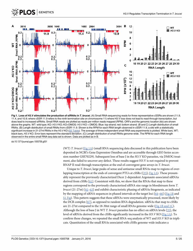
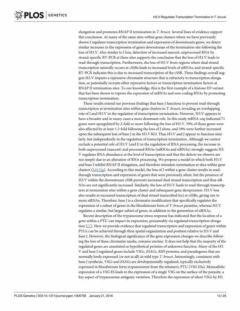
Unique to T. brucei, large peaks of sense and antisense small RNAs map to regions of over-lapping transcription at the ends of convergent PTUs at cSSRs ([35]; Fig 1A). These presum-ably represent the previously characterized Dicer 2-dependent Argonaute-associated siRNAsderived from cSSRs [61]. Consistent with this, we show that the RNAs that map to theseregions correspond to the previously characterized siRNA size range in bloodstream form T.brucei (21-27nt) [61–63] and exhibit characteristic phasing of siRNAs biogenesis, as indicatedby the mapping of siRNA sequences in phased intervals at the target loci [64–66] (Fig 1B andS1 Fig). This pattern suggests that these siRNAs were enzymatically processed, most likely bythe DCR complex [67], as opposed to random RNA degradation. siRNAs that map to cSSRsare 21-27nt compared to the 18-30nt range of small RNAs genome-wide (Fig 1B and 1C).Although the loss of base J in WT T. brucei parasites has no effect on siRNA levels [35], thelevel of siRNAs derived from the cSSRs significantly increased in theH3.V KO (Fig 1A). Toconfirm these changes, we repeated the small RNA-seq analysis of WT andH3.V KO in tripli-cate. Quantitation of the small RNAs associated with cSSRs genome-wide indicates a
Fig 1. Loss of H3.V stimulates the production of siRNAs in T. brucei. (A) Small RNA-sequencing reads for three representative cSSRs are shown (11.9,11.4, and 10.9; where cSSR 11.9 refers to the ninth termination site on chromosome 11) where H3.V loss does not lead to read-through transcription, butdoes lead to increased siRNAs. Small RNA reads are plotted as reads per million reads mapped (RPM). ORFs and the genomic location (kb) are shownabove the graphs. WT: wild type; KO: H3.V KO; KO+DMOG: H3.V KO + DMOG. Blue: top strand; red: bottom strand. (B and C) Length distribution of smallRNAs. (B) Length distribution of small RNAs from cSSR 11.9. Shown is the RPM for each RNA length observed in cSSR 11.9, a site with a statisticallysignificant increase in 21-27nt RNAs in theH3.V KO (S2 Table). The average of three independent small RNA-seq experiments is plotted. White bars, WT;black bars,H3.V KO. Error bars represent the standard deviation. (C) Length distribution of small RNAs genome-wide. The RPM for each RNA lengthobserved in the entire small RNA-seq data set is shown. Data are plotted as in B.
doi:10.1371/journal.pgen.1005758.g001
H3.V Regulates Transcription Termination in T. brucei
PLOSGenetics | DOI:10.1371/journal.pgen.1005758 January 21, 2016 5 / 25
statistically significant increase in 21-27nt RNAs at 30 out of 72 cSSRs in theH3.V KO (Fig 1Band S2 Table). This can be visualized by specifically mapping the siRNA size range of smallRNAs (S1A Fig). The increase in siRNAs is not restricted to cSSRs, but occurs at all previouslycharacterized siRNA generating regions of the T. brucei genome [61, 62]; including the SLACSand ingi retrotransposable elements, CIR147 centromeric repeats, and inverted repeats (S2Fig). These regions are also enriched with H3.V [14, 68].
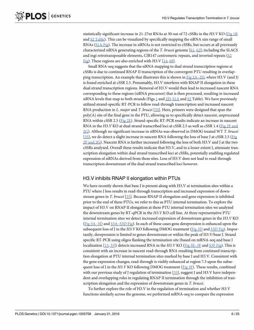
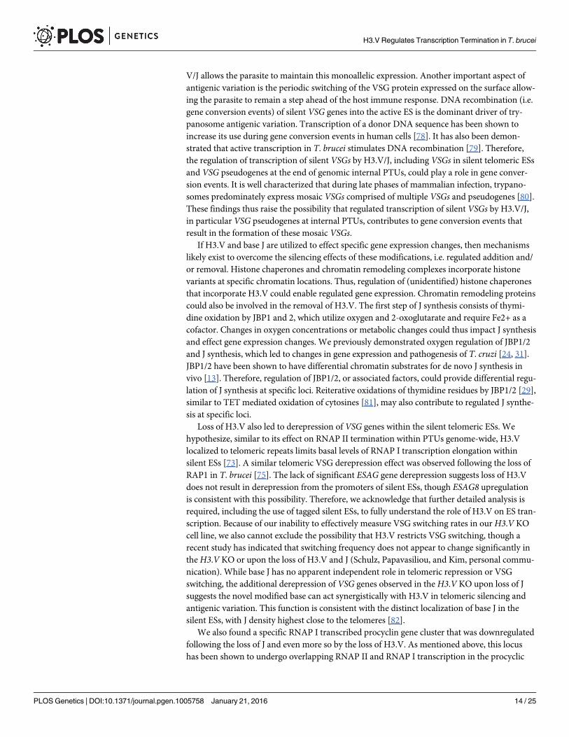
Small RNA-seq suggests that the siRNA mapping to dual strand transcription regions atcSSRs is due to continued RNAP II transcription of the convergent PTU resulting in overlap-ping transcription. An example that illustrates this is shown in Fig 2A–2D, where H3.V (and J)is found enriched at cSSR 2.5. Presumably, H3.V interferes with RNAP II elongation in thesedual strand transcription regions. Removal of H3.V would then lead to increased nascent RNAcorresponding to these regions (siRNA precursor) that is then processed, resulting in increasedsiRNA levels that map to both strands (Figs 1 and 2D, S1A and S2 Table). We have previouslyutilized strand-specific RT-PCR to follow read-through transcription and increased nascentRNA production in L.major and T. brucei [35]. Here, primers were designed that span thepoly(A) site of the final gene in the PTU, allowing us to specifically detect nascent, unprocessedRNA within cSSR 2.5 (Fig 2E). Strand-specific RT-PCR results indicate an increase in nascentRNA in theH3.V KO at dual strand transcribed loci at cSSR 2.5 as well as cSSR 1.4 (Fig 2F and2G). Although no significant increase in siRNAs was observed in DMOG treated WT T. brucei[35], we do detect a slight increase in nascent RNA following the loss of base J at cSSR 2.5 (Fig2F and 2G). Nascent RNA is further increased following the loss of both H3.V and J at the twocSSRs analyzed. Overall these results indicate that H3.V, and to a lesser extent J, attenuate tran-scription elongation within dual strand transcribed loci at cSSRs, potentially enabling regulatedexpression of siRNAs derived from these sites. Loss of H3.V does not lead to read-throughtranscription downstream of the dual strand transcribed loci however.
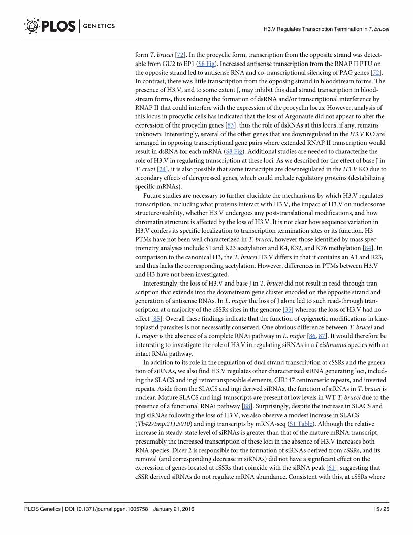
H3.V inhibits RNAP II elongation within PTUsWe have recently shown that base J is present along with H3.V at termination sites within aPTU where J loss results in read-through transcription and increased expression of down-stream genes in T. brucei [35]. Because RNAP II elongation and gene expression is inhibitedprior to the end of these PTUs, we refer to this as PTU internal termination. To explore theimpact of H3.V on RNAP II elongation at these PTU internal termination sites we analyzedthe downstream genes by RT-qPCR in the H3.V KO cell line. At three representative PTUinternal termination sites we detect increased expression of downstream genes in theH3.V KO(Fig 3A–3D and S3A–S3D Fig). In each of these cases gene derepression is enhanced upon thesubsequent loss of J in theH3.V KO following DMOG treatment (Fig 3D and S3D Fig). Impor-tantly, derepression is limited to genes downstream or within the peak of H3.V/base J. Strandspecific RT-PCR using oligos flanking the termination site (based on mRNA-seq and base Jlocalization [13, 35]) detects increased RNA in theH3.V KO (Fig 3E–3F and S3F Fig). This isconsistent with an increase in nascent read-through RNA resulting from continued transcrip-tion elongation at PTU internal termination sites marked by base J and H3.V. Consistent withthe gene expression changes, read-through is visibly enhanced at region 7.3 upon the subse-quent loss of J in theH3.V KO following DMOG treatment (Fig 3F). These results, combinedwith our previous study of J regulation of termination [35], suggest J and H3.V have indepen-dent and overlapping roles in regulating RNAP II termination through the inhibition of tran-scription elongation and the expression of downstream genes in T. brucei.
To further explore the role of H3.V in the regulation of termination and whether H3.Vfunctions similarly across the genome, we performed mRNA-seq to compare the expression
H3.V Regulates Transcription Termination in T. brucei
PLOSGenetics | DOI:10.1371/journal.pgen.1005758 January 21, 2016 6 / 25
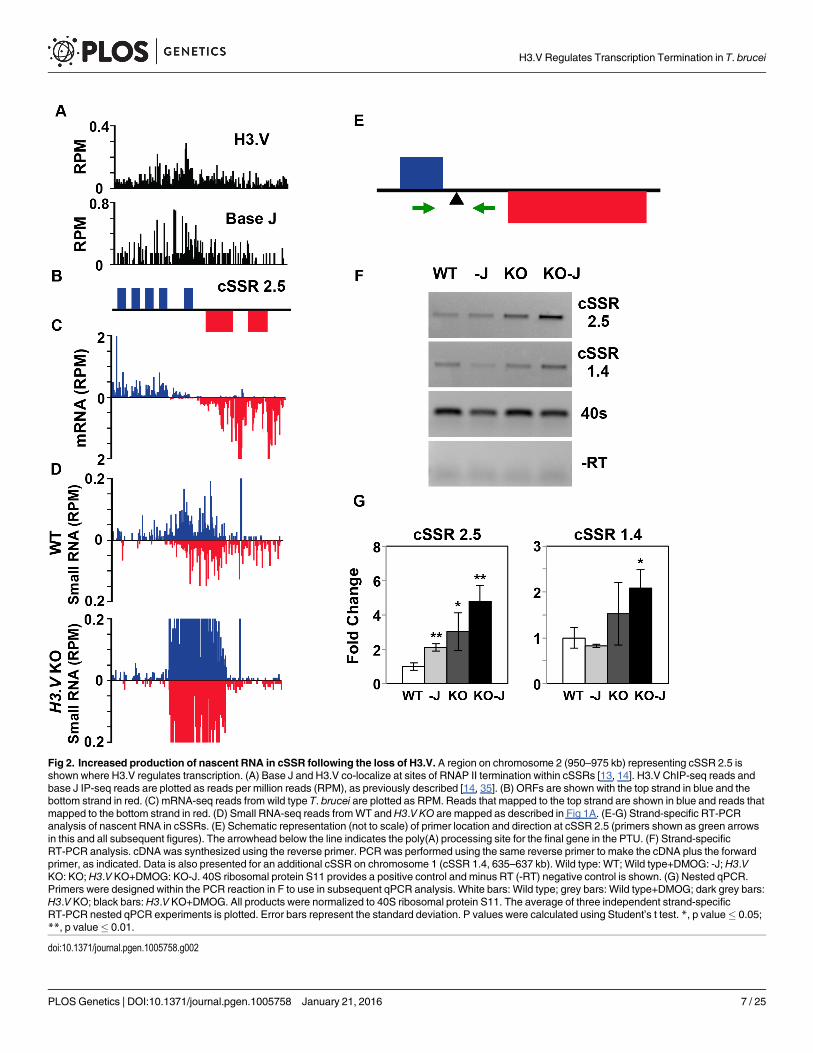
Fig 2. Increased production of nascent RNA in cSSR following the loss of H3.V. A region on chromosome 2 (950–975 kb) representing cSSR 2.5 isshown where H3.V regulates transcription. (A) Base J and H3.V co-localize at sites of RNAP II termination within cSSRs [13, 14]. H3.V ChIP-seq reads andbase J IP-seq reads are plotted as reads per million reads (RPM), as previously described [14, 35]. (B) ORFs are shown with the top strand in blue and thebottom strand in red. (C) mRNA-seq reads from wild type T. brucei are plotted as RPM. Reads that mapped to the top strand are shown in blue and reads thatmapped to the bottom strand in red. (D) Small RNA-seq reads fromWT andH3.V KO are mapped as described in Fig 1A. (E-G) Strand-specific RT-PCRanalysis of nascent RNA in cSSRs. (E) Schematic representation (not to scale) of primer location and direction at cSSR 2.5 (primers shown as green arrowsin this and all subsequent figures). The arrowhead below the line indicates the poly(A) processing site for the final gene in the PTU. (F) Strand-specificRT-PCR analysis. cDNA was synthesized using the reverse primer. PCR was performed using the same reverse primer to make the cDNA plus the forwardprimer, as indicated. Data is also presented for an additional cSSR on chromosome 1 (cSSR 1.4, 635–637 kb). Wild type: WT; Wild type+DMOG: -J; H3.VKO: KO; H3.V KO+DMOG: KO-J. 40S ribosomal protein S11 provides a positive control and minus RT (-RT) negative control is shown. (G) Nested qPCR.Primers were designed within the PCR reaction in F to use in subsequent qPCR analysis. White bars: Wild type; grey bars: Wild type+DMOG; dark grey bars:H3.V KO; black bars:H3.V KO+DMOG. All products were normalized to 40S ribosomal protein S11. The average of three independent strand-specificRT-PCR nested qPCR experiments is plotted. Error bars represent the standard deviation. P values were calculated using Student’s t test. *, p value� 0.05;**, p value� 0.01.
doi:10.1371/journal.pgen.1005758.g002
H3.V Regulates Transcription Termination in T. brucei
PLOSGenetics | DOI:10.1371/journal.pgen.1005758 January 21, 2016 7 / 25
profiles of WT, WT+DMOG, H3.V KO andH3.V KO+DMOG cells (GEO accession numberGSE69929). This led to the detection of 153 mRNAs that are increased at least 2-fold in one ormore of the treatments (S1 Table). Many of the gene expression changes have been confirmedby RT-qPCR (S4 Fig and below). Consistent with our previous mRNA-seq results, in WT cellstreated with DMOG we observe similar increases in the expression of genes downstream ofbase J (and H3.V), which we previously demonstrated is caused by an RNAP II transcriptiontermination defect within a PTU [35]. However, we now see that a significant number of genesdownstream of J/H3.V within other PTUs are upregulated following the loss of H3.V, and thatmany are further increased following the subsequent loss of J (Fig 4, S1 and S4 Tables). In theH3.V KO we identified 71 genes that are upregulated (Fig 5A and S1 Table). Although many ofthese genes are not increased by 2-fold in the WT+DMOG condition, some respond at leastslightly to the loss of J in WT cells: 28 of the 71 genes upregulated in theH3.V KO are also
Fig 3. Decreased efficiency of RNAP II termination and increased gene expression following the loss of histone H3.V. A region on chromosome 3(617–670 kb) representing cSSR 3.3 and chromosome 7 (453–525 kb) representing cSSR 7.3 is shown where H3.V regulates transcription of a cluster ofgenes. (A-C) Base J and H3.V co-localize at sites of RNAP II termination within a PTU. H3.V ChIP-seq reads and base J IP-seq reads, ORFs, and mRNA-seq reads from wild type T. brucei are plotted for cSSR 3.3 (left) and cSSR 7.3 (right) as described in Fig 2. (D) RT-qPCR analysis of genes numberedaccording to the ORFmaps above in panel B. As described in Fig 2G, white bars: Wild type; grey bars: Wild type+DMOG; dark grey bars:H3.V KO; blackbars:H3.V KO+DMOG. Transcripts were normalized against 40S ribosomal protein S11, and are plotted as the average and standard deviation of threereplicates. P values were calculated using Student’s t test. *, p value� 0.05; **, p value� 0.01. The silent gene cluster at cSSR 7.3 consists of nine highlysimilar retrotransposon hot spot protein genes, therefore the primers used to analyze gene 2 also amplify the additional upstream genes. (E and F) Strand-specific RT-PCR analysis of read-through transcription of the two cSSRs analyzed in A-D. Above each panel is a schematic representation (not to scale) ofprimer location and direction at a transcription termination site (TTS). The vertical arrow indicates the proposed TTS as described in the text [35]. The longsolid arrow indicates the direction of transcription and the dashed arrow indicates read-through transcription past the TTS. cDNA was synthesized using thereverse primer (relative to transcription). PCR was performed using the same reverse primer to make the cDNA plus the forward primer, as indicated. 40Sribosomal protein S11 provides a positive control and a minus RT (-RT) negative control is shown.
doi:10.1371/journal.pgen.1005758.g003
H3.V Regulates Transcription Termination in T. brucei
PLOSGenetics | DOI:10.1371/journal.pgen.1005758 January 21, 2016 8 / 25
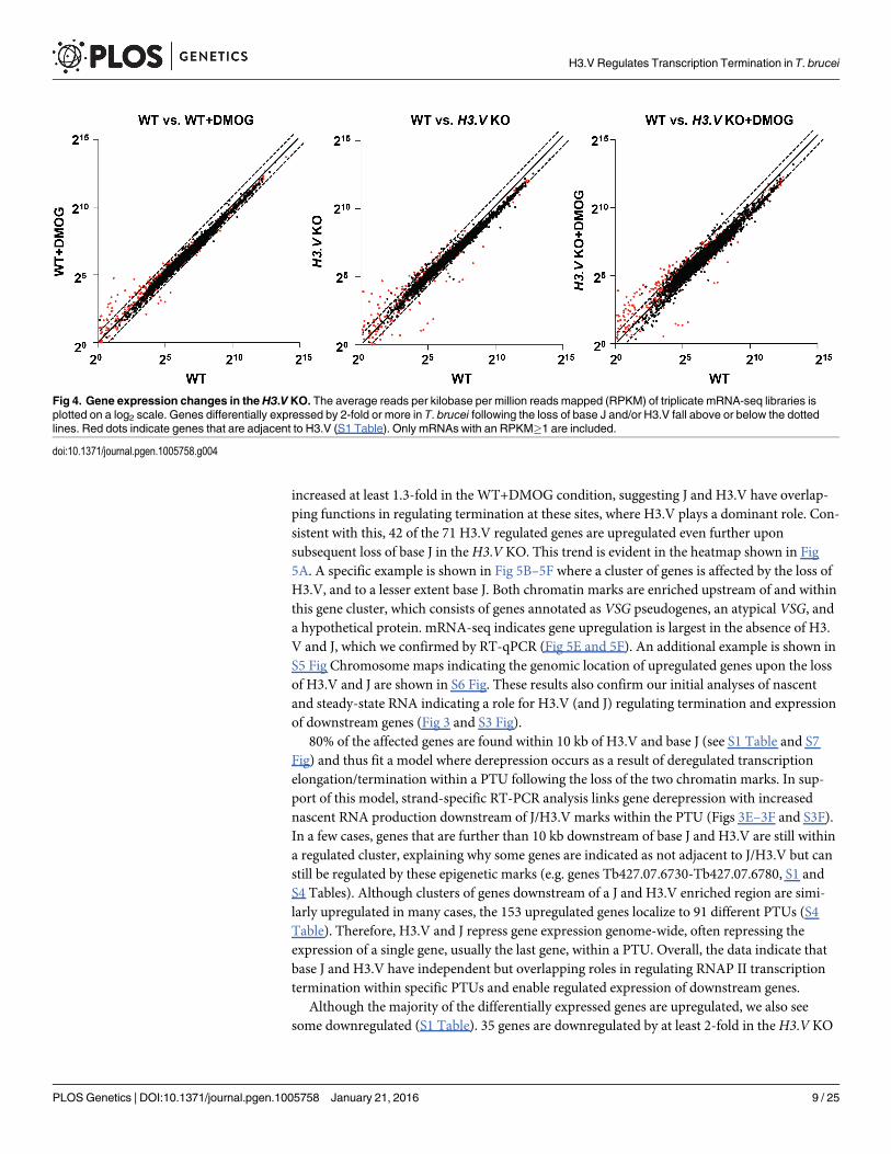
increased at least 1.3-fold in the WT+DMOG condition, suggesting J and H3.V have overlap-ping functions in regulating termination at these sites, where H3.V plays a dominant role. Con-sistent with this, 42 of the 71 H3.V regulated genes are upregulated even further uponsubsequent loss of base J in theH3.V KO. This trend is evident in the heatmap shown in Fig5A. A specific example is shown in Fig 5B–5F where a cluster of genes is affected by the loss ofH3.V, and to a lesser extent base J. Both chromatin marks are enriched upstream of and withinthis gene cluster, which consists of genes annotated as VSG pseudogenes, an atypical VSG, anda hypothetical protein. mRNA-seq indicates gene upregulation is largest in the absence of H3.V and J, which we confirmed by RT-qPCR (Fig 5E and 5F). An additional example is shown inS5 Fig Chromosome maps indicating the genomic location of upregulated genes upon the lossof H3.V and J are shown in S6 Fig. These results also confirm our initial analyses of nascentand steady-state RNA indicating a role for H3.V (and J) regulating termination and expressionof downstream genes (Fig 3 and S3 Fig).
80% of the affected genes are found within 10 kb of H3.V and base J (see S1 Table and S7Fig) and thus fit a model where derepression occurs as a result of deregulated transcriptionelongation/termination within a PTU following the loss of the two chromatin marks. In sup-port of this model, strand-specific RT-PCR analysis links gene derepression with increasednascent RNA production downstream of J/H3.V marks within the PTU (Figs 3E–3F and S3F).In a few cases, genes that are further than 10 kb downstream of base J and H3.V are still withina regulated cluster, explaining why some genes are indicated as not adjacent to J/H3.V but canstill be regulated by these epigenetic marks (e.g. genes Tb427.07.6730-Tb427.07.6780, S1 andS4 Tables). Although clusters of genes downstream of a J and H3.V enriched region are simi-larly upregulated in many cases, the 153 upregulated genes localize to 91 different PTUs (S4Table). Therefore, H3.V and J repress gene expression genome-wide, often repressing theexpression of a single gene, usually the last gene, within a PTU. Overall, the data indicate thatbase J and H3.V have independent but overlapping roles in regulating RNAP II transcriptiontermination within specific PTUs and enable regulated expression of downstream genes.
Although the majority of the differentially expressed genes are upregulated, we also seesome downregulated (S1 Table). 35 genes are downregulated by at least 2-fold in theH3.V KO
Fig 4. Gene expression changes in theH3.V KO. The average reads per kilobase per million reads mapped (RPKM) of triplicate mRNA-seq libraries isplotted on a log2 scale. Genes differentially expressed by 2-fold or more in T. brucei following the loss of base J and/or H3.V fall above or below the dottedlines. Red dots indicate genes that are adjacent to H3.V (S1 Table). Only mRNAs with an RPKM�1 are included.
doi:10.1371/journal.pgen.1005758.g004
H3.V Regulates Transcription Termination in T. brucei
PLOSGenetics | DOI:10.1371/journal.pgen.1005758 January 21, 2016 9 / 25
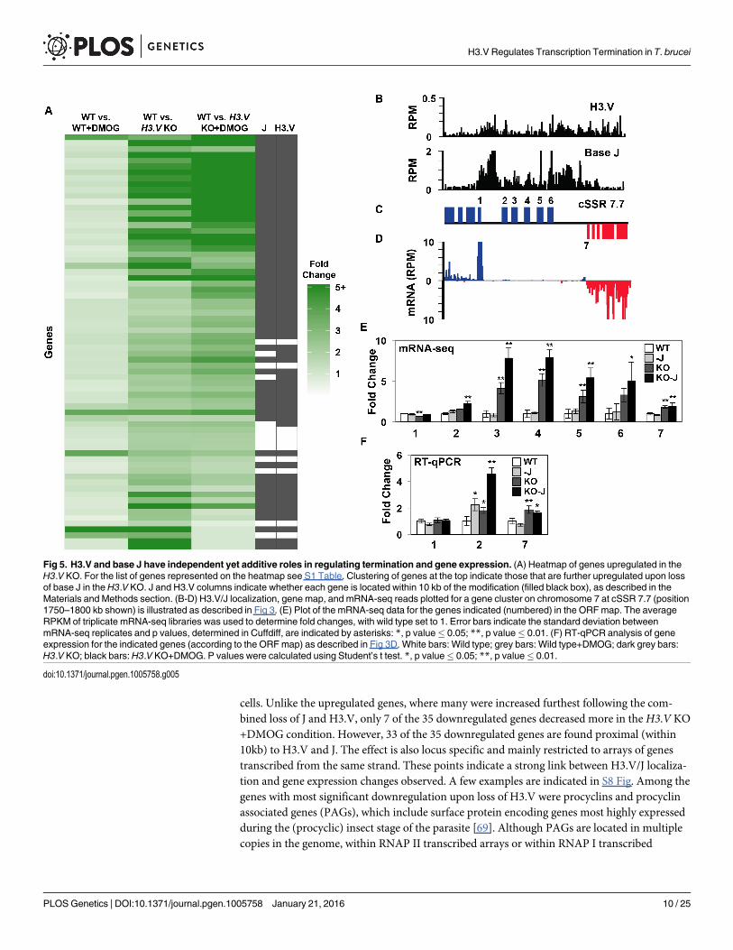
cells. Unlike the upregulated genes, where many were increased furthest following the com-bined loss of J and H3.V, only 7 of the 35 downregulated genes decreased more in the H3.V KO+DMOG condition. However, 33 of the 35 downregulated genes are found proximal (within10kb) to H3.V and J. The effect is also locus specific and mainly restricted to arrays of genestranscribed from the same strand. These points indicate a strong link between H3.V/J localiza-tion and gene expression changes observed. A few examples are indicated in S8 Fig. Among thegenes with most significant downregulation upon loss of H3.V were procyclins and procyclinassociated genes (PAGs), which include surface protein encoding genes most highly expressedduring the (procyclic) insect stage of the parasite [69]. Although PAGs are located in multiplecopies in the genome, within RNAP II transcribed arrays or within RNAP I transcribed
Fig 5. H3.V and base J have independent yet additive roles in regulating termination and gene expression. (A) Heatmap of genes upregulated in theH3.V KO. For the list of genes represented on the heatmap see S1 Table. Clustering of genes at the top indicate those that are further upregulated upon lossof base J in theH3.V KO. J and H3.V columns indicate whether each gene is located within 10 kb of the modification (filled black box), as described in theMaterials and Methods section. (B-D) H3.V/J localization, gene map, and mRNA-seq reads plotted for a gene cluster on chromosome 7 at cSSR 7.7 (position1750–1800 kb shown) is illustrated as described in Fig 3. (E) Plot of the mRNA-seq data for the genes indicated (numbered) in the ORFmap. The averageRPKM of triplicate mRNA-seq libraries was used to determine fold changes, with wild type set to 1. Error bars indicate the standard deviation betweenmRNA-seq replicates and p values, determined in Cuffdiff, are indicated by asterisks: *, p value� 0.05; **, p value� 0.01. (F) RT-qPCR analysis of geneexpression for the indicated genes (according to the ORFmap) as described in Fig 3D. White bars: Wild type; grey bars: Wild type+DMOG; dark grey bars:H3.V KO; black bars:H3.V KO+DMOG. P values were calculated using Student’s t test. *, p value� 0.05; **, p value� 0.01.
doi:10.1371/journal.pgen.1005758.g005
H3.V Regulates Transcription Termination in T. brucei
PLOSGenetics | DOI:10.1371/journal.pgen.1005758 January 21, 2016 10 / 25
procyclin arrays [69, 70], the PAGs downregulated following the loss of H3.V (PAG1, PAG2,PAG4 and PAG5) are specifically arranged in an RNAP I transcribed array. For example, thereare two PAG2 genes located on chromosome 10, one in an RNAP II transcribed PTU and theother in the RNAP I transcribed procyclin locus. The only gene that is significantly downregu-lated when H3.V is deleted is the one within the RNAP I procyclin locus. A similar locus spe-cific alteration of PAG expression was seen upon the depletion of histone H1 [71].Interestingly, the PAGs within this locus undergo overlapping RNAP II and RNAP I transcrip-tion, i.e. continued RNAP II transcription of the upstream opposing PTU produces antisensePAG RNAs [72]. This suggests a possible mechanism of PAG (and procyclin) downregulationresulting from increased formation of dsRNAs upon the loss of H3.V (see discussion). Simi-larly, other downregulated genes are arranged in opposing transcriptional genes pairs whereextended RNAP II transcription would result in dsRNA for each mRNA (S8 Fig). We also iden-tified 25 genes that are downregulated specifically in theH3.V KO+DMOG condition, though22 of these genes are not located near H3.V or J. We therefore assume many of these changesare an indirect effect of genes that are upregulated in this cell line. For example, we have dem-onstrated that the genome-wide increase in RNAP II transcription in T. cruzi results in a globalincrease in gene expression that includes proteins that degrade specific mRNAs [24].
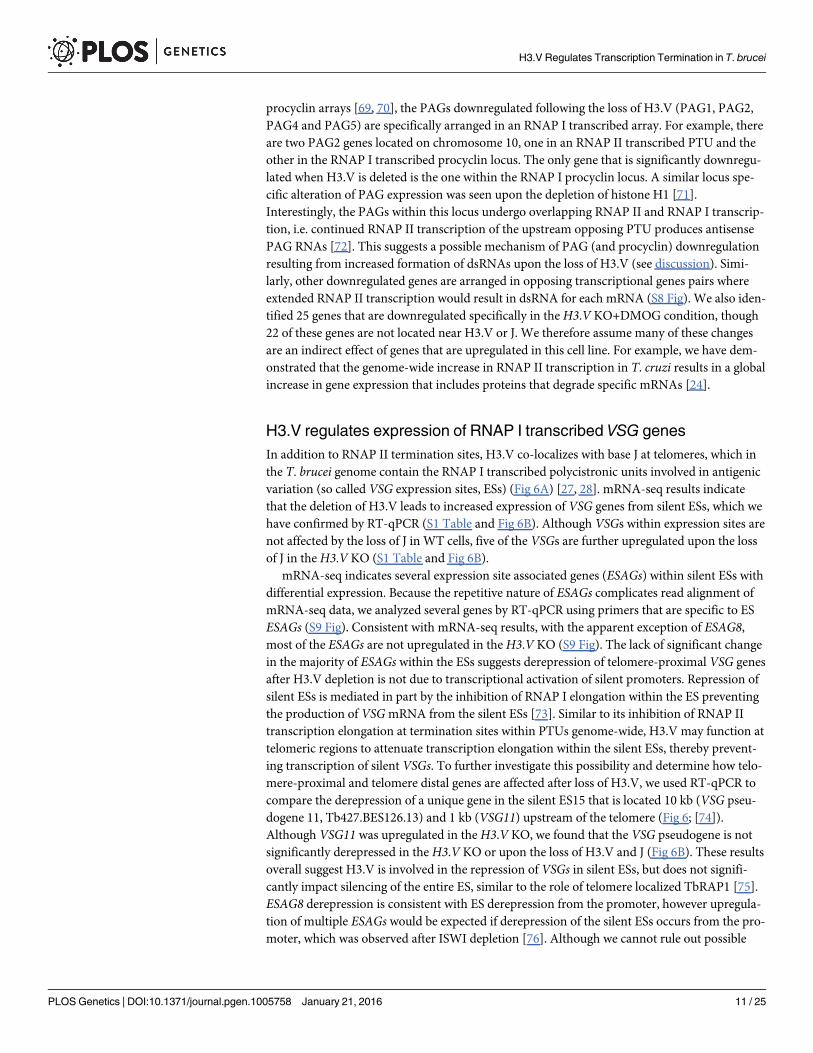
H3.V regulates expression of RNAP I transcribed VSG genesIn addition to RNAP II termination sites, H3.V co-localizes with base J at telomeres, which inthe T. brucei genome contain the RNAP I transcribed polycistronic units involved in antigenicvariation (so called VSG expression sites, ESs) (Fig 6A) [27, 28]. mRNA-seq results indicatethat the deletion of H3.V leads to increased expression of VSG genes from silent ESs, which wehave confirmed by RT-qPCR (S1 Table and Fig 6B). Although VSGs within expression sites arenot affected by the loss of J in WT cells, five of the VSGs are further upregulated upon the lossof J in theH3.V KO (S1 Table and Fig 6B).
mRNA-seq indicates several expression site associated genes (ESAGs) within silent ESs withdifferential expression. Because the repetitive nature of ESAGs complicates read alignment ofmRNA-seq data, we analyzed several genes by RT-qPCR using primers that are specific to ESESAGs (S9 Fig). Consistent with mRNA-seq results, with the apparent exception of ESAG8,most of the ESAGs are not upregulated in theH3.V KO (S9 Fig). The lack of significant changein the majority of ESAGs within the ESs suggests derepression of telomere-proximal VSG genesafter H3.V depletion is not due to transcriptional activation of silent promoters. Repression ofsilent ESs is mediated in part by the inhibition of RNAP I elongation within the ES preventingthe production of VSGmRNA from the silent ESs [73]. Similar to its inhibition of RNAP IItranscription elongation at termination sites within PTUs genome-wide, H3.V may function attelomeric regions to attenuate transcription elongation within the silent ESs, thereby prevent-ing transcription of silent VSGs. To further investigate this possibility and determine how telo-mere-proximal and telomere distal genes are affected after loss of H3.V, we used RT-qPCR tocompare the derepression of a unique gene in the silent ES15 that is located 10 kb (VSG pseu-dogene 11, Tb427.BES126.13) and 1 kb (VSG11) upstream of the telomere (Fig 6; [74]).Although VSG11 was upregulated in theH3.V KO, we found that the VSG pseudogene is notsignificantly derepressed in theH3.V KO or upon the loss of H3.V and J (Fig 6B). These resultsoverall suggest H3.V is involved in the repression of VSGs in silent ESs, but does not signifi-cantly impact silencing of the entire ES, similar to the role of telomere localized TbRAP1 [75].ESAG8 derepression is consistent with ES derepression from the promoter, however upregula-tion of multiple ESAGs would be expected if derepression of the silent ESs occurs from the pro-moter, which was observed after ISWI depletion [76]. Although we cannot rule out possible
H3.V Regulates Transcription Termination in T. brucei
PLOSGenetics | DOI:10.1371/journal.pgen.1005758 January 21, 2016 11 / 25
alterations in VSG switching in theH3.V KO (see discussion), the data here suggest H3.V isinvolved in repressing RNAP I transcription of VSGs within the silent ESs as well as RNAP IItranscription of VSG genes within genome internal PTUs.
DiscussionH3.V is a kinetoplastid-specific H3 variant and appears to be the only H3 variant found inthese early-diverged eukaryotes. The T. bruceiH3.V shares only 45% sequence identity withthe canonical H3 [60]. Although H3.V localizes to centromeres, it is not essential for viabilityand does not contain sequence variations common to all identified centromeric H3 variants[14, 60, 68]. Aside from its localization to RNAP II termination sites and telomeres, the func-tional significance of H3.V and its potential role in the regulation of RNAP II termination hasbeen unexplored. We have demonstrated that H3.V negatively regulates transcription
Fig 6. H3.V regulates VSG gene expression from silent telomeric bloodstream expression sites. (A) A schematic diagram of the silent ES15 (not toscale). The box with stripes represents the 70 bp repeats. Numbers indicate ESAG genes. Grey box represents the VSG pseudogene 11 (Tb427.BES126.13). (B-C) mRNA-seq and RT-qPCR analysis of the indicated VSG genes in silent expression sites. As described in Figs 3D and 5E, white bars:Wild type; grey bars: Wild type+DMOG; dark grey bars: H3.V KO; black bars:H3.V KO+DMOG. For mRNA-seq analysis, p values determined by Cuffdiff areindicated by asterisks: *, p value� 0.05; **, p value� 0.01. For RT-qPCR analysis, p values were calculated using Student’s t test. *, p value� 0.05; **, pvalue� 0.01.
doi:10.1371/journal.pgen.1005758.g006
H3.V Regulates Transcription Termination in T. brucei
PLOSGenetics | DOI:10.1371/journal.pgen.1005758 January 21, 2016 12 / 25
elongation and promotes RNAP II termination in T. brucei. Several lines of evidence supportthis conclusion. At many of the same sites within gene clusters where we have previouslyshown J regulates transcription termination and expression of downstream genes, we detectsimilar increases in the expression of genes downstream of the termination site following theloss of H3.V. Also similar to J loss, detection of increased nascent, unprocessed RNA bystrand-specific RT-PCR at these sites supports the conclusion that the loss of H3.V leads toread-through transcription. Furthermore, the loss of H3.V from regions where dual strandtranscription naturally occurs at cSSRs leads to increased levels of siRNAs, and strand-specificRT-PCR indicates this is due to increased transcription of the cSSR. These findings overall sug-gest H3.V imparts a repressive chromatin structure that is refractory to transcription elonga-tion, or potentially recruits other repressive factors or transcription termination factors atRNAP II termination sites. To our knowledge, this is the first example of a histone H3 variantthat has been shown to repress the expression of mRNAs and non-coding RNAs by promotingtranscription termination.
These results extend our previous findings that base J functions to prevent read-throughtranscription at termination sites within gene clusters in T. brucei, revealing an overlappingrole of J and H3.V in the regulation of transcription termination. However, H3.V appears tohave a broader and in many cases a more dominant role. In this study mRNA-seq indicated 71genes were upregulated by 2-fold or more following the loss of H3.V. 39% of those genes werealso affected by at least 1.3-fold following the loss of J alone, and 59% were further increasedupon the subsequent loss of base J in theH3.V KO. Thus H3.V and J appear to function simi-larly, but independently in the regulation of transcription termination. Although we cannotexclude a potential role of H3.V (and J) in the regulation of RNA processing, the increase inboth unprocessed (nascent) and processed RNAs (mRNAs and siRNAs) strongly suggests H3.V regulates RNA abundance at the level of transcription and that the defects we observe arenot simply due to an alteration of RNA processing. We propose a model in which both H3.Vand base J inhibit RNAP II elongation, and therefore stimulate termination at sites within geneclusters (S10 Fig). According to this model, the loss of J within a gene cluster results in read-through transcription and expression of genes that were previously silent, but the presence ofH3.V within the downstream cSSR prevents increased dual strand transcription and thus siR-NAs are not significantly increased. Similarly, the loss of H3.V leads to read-through transcrip-tion at termination sites within a gene cluster and subsequent gene derepression. H3.V lossalso results in increased transcription of dual strand transcribed loci at cSSRs, giving rise tomore siRNAs. Therefore, base J is a chromatin modification that specifically regulates theexpression of a subset of genes in the bloodstream form of T. brucei parasites, whereas H3.Vregulates a similar, but larger subset of genes, in addition to the generation of siRNAs.
Recent description of the trypanosome stress response has indicated that the location of agene within a PTU can impact its expression, presumably via regulated transcription elonga-tion [77]. Here we provide evidence that regulated transcription and expression of genes withinPTUs can be achieved through their spatial organization and position relative to H3.V andbase J. However, the biological significance of the gene expression changes we describe follow-ing the loss of these chromatin marks, remains unclear. It does not help that the majority of theregulated genes are annotated as hypothetical proteins of unknown function. Many of the H3.V and base J regulated genes include VSGs, ESAGs, RHS proteins, and pseudogenes that arenormally lowly expressed (or not at all) in wild type T. brucei. Interestingly, consistent withbase J synthesis, VSGs and ESAGs are developmentally regulated, typically exclusivelyexpressed in bloodstream form trypanosomes from the telomeric PTU (VSG ESs). Monoallelicexpression of a VSG ES leads to the expression of a single VSG on the surface of the parasite, akey aspect of trypanosome antigenic variation. Therefore the repression of silent VSGs by H3.
H3.V Regulates Transcription Termination in T. brucei
PLOSGenetics | DOI:10.1371/journal.pgen.1005758 January 21, 2016 13 / 25
V/J allows the parasite to maintain this monoallelic expression. Another important aspect ofantigenic variation is the periodic switching of the VSG protein expressed on the surface allow-ing the parasite to remain a step ahead of the host immune response. DNA recombination (i.e.gene conversion events) of silent VSG genes into the active ES is the dominant driver of try-panosome antigenic variation. Transcription of a donor DNA sequence has been shown toincrease its use during gene conversion events in human cells [78]. It has also been demon-strated that active transcription in T. brucei stimulates DNA recombination [79]. Therefore,the regulation of transcription of silent VSGs by H3.V/J, including VSGs in silent telomeric ESsand VSG pseudogenes at the end of genomic internal PTUs, could play a role in gene conver-sion events. It is well characterized that during late phases of mammalian infection, trypano-somes predominately express mosaic VSGs comprised of multiple VSGs and pseudogenes [80].These findings thus raise the possibility that regulated transcription of silent VSGs by H3.V/J,in particular VSG pseudogenes at internal PTUs, contributes to gene conversion events thatresult in the formation of these mosaic VSGs.
If H3.V and base J are utilized to effect specific gene expression changes, then mechanismslikely exist to overcome the silencing effects of these modifications, i.e. regulated addition and/or removal. Histone chaperones and chromatin remodeling complexes incorporate histonevariants at specific chromatin locations. Thus, regulation of (unidentified) histone chaperonesthat incorporate H3.V could enable regulated gene expression. Chromatin remodeling proteinscould also be involved in the removal of H3.V. The first step of J synthesis consists of thymi-dine oxidation by JBP1 and 2, which utilize oxygen and 2-oxoglutarate and require Fe2+ as acofactor. Changes in oxygen concentrations or metabolic changes could thus impact J synthesisand effect gene expression changes. We previously demonstrated oxygen regulation of JBP1/2and J synthesis, which led to changes in gene expression and pathogenesis of T. cruzi [24, 31].JBP1/2 have been shown to have differential chromatin substrates for de novo J synthesis invivo [13]. Therefore, regulation of JBP1/2, or associated factors, could provide differential regu-lation of J synthesis at specific loci. Reiterative oxidations of thymidine residues by JBP1/2 [29],similar to TET mediated oxidation of cytosines [81], may also contribute to regulated J synthe-sis at specific loci.
Loss of H3.V also led to derepression of VSG genes within the silent telomeric ESs. Wehypothesize, similar to its effect on RNAP II termination within PTUs genome-wide, H3.Vlocalized to telomeric repeats limits basal levels of RNAP I transcription elongation withinsilent ESs [73]. A similar telomeric VSG derepression effect was observed following the loss ofRAP1 in T. brucei [75]. The lack of significant ESAG gene derepression suggests loss of H3.Vdoes not result in derepression from the promoters of silent ESs, though ESAG8 upregulationis consistent with this possibility. Therefore, we acknowledge that further detailed analysis isrequired, including the use of tagged silent ESs, to fully understand the role of H3.V on ES tran-scription. Because of our inability to effectively measure VSG switching rates in ourH3.V KOcell line, we also cannot exclude the possibility that H3.V restricts VSG switching, though arecent study has indicated that switching frequency does not appear to change significantly intheH3.V KO or upon the loss of H3.V and J (Schulz, Papavasiliou, and Kim, personal commu-nication). While base J has no apparent independent role in telomeric repression or VSGswitching, the additional derepression of VSG genes observed in the H3.V KO upon loss of Jsuggests the novel modified base can act synergistically with H3.V in telomeric silencing andantigenic variation. This function is consistent with the distinct localization of base J in thesilent ESs, with J density highest close to the telomeres [82].
We also found a specific RNAP I transcribed procyclin gene cluster that was downregulatedfollowing the loss of J and even more so by the loss of H3.V. As mentioned above, this locushas been shown to undergo overlapping RNAP II and RNAP I transcription in the procyclic
H3.V Regulates Transcription Termination in T. brucei
PLOSGenetics | DOI:10.1371/journal.pgen.1005758 January 21, 2016 14 / 25
form T. brucei [72]. In the procyclic form, transcription from the opposite strand was detect-able from GU2 to EP1 (S8 Fig). Increased antisense transcription from the RNAP II PTU onthe opposite strand led to antisense RNA and co-transcriptional silencing of PAG genes [72].In contrast, there was little transcription from the opposing strand in bloodstream forms. Thepresence of H3.V, and to some extent J, may inhibit this dual strand transcription in blood-stream forms, thus reducing the formation of dsRNA and/or transcriptional interference byRNAP II that could interfere with the expression of the procyclin locus. However, analysis ofthis locus in procyclic cells has indicated that the loss of Argonaute did not appear to alter theexpression of the procyclin genes [83], thus the role of dsRNAs at this locus, if any, remainsunknown. Interestingly, several of the other genes that are downregulated in theH3.V KO arearranged in opposing transcriptional gene pairs where extended RNAP II transcription wouldresult in dsRNA for each mRNA (S8 Fig). Additional studies are needed to characterize therole of H3.V in regulating transcription at these loci. As we described for the effect of base J inT. cruzi [24], it is also possible that some transcripts are downregulated in theH3.V KO due tosecondary effects of derepressed genes, which could include regulatory proteins (destabilizingspecific mRNAs).
Future studies are necessary to further elucidate the mechanisms by which H3.V regulatestranscription, including what proteins interact with H3.V, the impact of H3.V on nucleosomestructure/stability, whether H3.V undergoes any post-translational modifications, and howchromatin structure is affected by the loss of H3.V. It is not clear how sequence variation inH3.V confers its specific localization to transcription termination sites or its function. H3PTMs have not been well characterized in T. brucei, however those identified by mass spec-trometry analyses include S1 and K23 acetylation and K4, K32, and K76 methylation [84]. Incomparison to the canonical H3, the T. bruceiH3.V differs in that it contains an A1 and R23,and thus lacks the corresponding acetylation. However, differences in PTMs between H3.Vand H3 have not been investigated.
Interestingly, the loss of H3.V and base J in T. brucei did not result in read-through tran-scription that extends into the downstream gene cluster encoded on the opposite strand andgeneration of antisense RNAs. In L.major the loss of J alone led to such read-through tran-scription at a majority of the cSSRs sites in the genome [35] whereas the loss of H3.V had noeffect [85]. Overall these findings indicate that the function of epigenetic modifications in kine-toplastid parasites is not necessarily conserved. One obvious difference between T. brucei andL.major is the absence of a complete RNAi pathway in L.major [86, 87]. It would therefore beinteresting to investigate the role of H3.V in regulating siRNAs in a Leishmania species with anintact RNAi pathway.
In addition to its role in the regulation of dual strand transcription at cSSRs and the genera-tion of siRNAs, we also find H3.V regulates other characterized siRNA generating loci, includ-ing the SLACS and ingi retrotransposable elements, CIR147 centromeric repeats, and invertedrepeats. Aside from the SLACS and ingi derived siRNAs, the function of siRNAs in T. brucei isunclear. Mature SLACS and ingi transcripts are present at low levels in WT T. brucei due to thepresence of a functional RNAi pathway [88]. Surprisingly, despite the increase in SLACS andingi siRNAs following the loss of H3.V, we also observe a modest increase in SLACS(Tb427tmp.211.5010) and ingi transcripts by mRNA-seq (S1 Table). Although the relativeincrease in steady-state level of siRNAs is greater than that of the mature mRNA transcript,presumably the increased transcription of these loci in the absence of H3.V increases bothRNA species. Dicer 2 is responsible for the formation of siRNAs derived from cSSRs, and itsremoval (and corresponding decrease in siRNAs) did not have a significant effect on theexpression of genes located at cSSRs that coincide with the siRNA peak [61], suggesting thatcSSR derived siRNAs do not regulate mRNA abundance. Consistent with this, at cSSRs where
H3.V Regulates Transcription Termination in T. brucei
PLOSGenetics | DOI:10.1371/journal.pgen.1005758 January 21, 2016 15 / 25
we detect increased siRNAs we do not observe significant decreases in mRNAs from genes thatoverlap the siRNA peak. Therefore the function, if any, of cSSR derived siRNAs remainsunknown.
In summary, we have provided evidence for the connection between a histone H3 variant andtranscription termination for the first time. These findings highlight the importance of chroma-tin modifications in the regulation of transcription termination, particularly in early-divergedeukaryotes with unique polycistronic transcription. These findings also have direct implicationsfor a strictly post-transcriptional model of gene expression regulation in kinetoplastids.
Materials and Methods
Parasite cell cultureWT and H3.V KO bloodstream form T. brucei 221a cell lines of strain 427 were cultured inHMI-9 medium as described previously [89]. The bloodstream form T. brucei H3.V KO cellline, generated by deleting both H3.V alleles by homologous recombination [60], was providedby George Cross. DMOG treatment of cells was performed by supplementing media with1mMDMOG for 5 days as described previously [35].
Strand-specific RNA-seq library constructionSmall RNA-sequencing was performed using two different methods. The analysis of WT, H3.VKO, and H3.V KO+DMOG (Figs 1A, 2D and S2 Fig) was performed as previously described[35]. Briefly, small RNAs were isolated from T. brucei (5x10⁷ cells) using a Qiagen miRNeasykit according to the manufacturer’s instructions. The small RNA-seq libraries were preparedusing approximately 250ng small RNA by Vertis Biotechnology AG, Germany. The small RNAsample was poly(A)-tailed using poly(A) polymerase. Then, the 5'PPP and cap structures wereremoved using tobacco acid pyrophosphatase (TAP, Epicentre). Afterwards, an RNA adapterwas ligated to the 5'-monophosphate of the RNA. First-strand cDNA synthesis was performedusing an oligo(dT)-adapter primer and the M-MLV reverse transcriptase. The resultingcDNAs were PCR-amplified to about 10–20 ng/μL using a high fidelity DNA polymerase. ThecDNAs were purified using the Agencourt AMPure XP kit (Beckman Coulter Genomics).Quality and concentration of all libraries was determined by capillary electrophoresis and highthroughput sequencing was performed on a HiSeq2000 (Illumina). Sequencing reads weremapped to the T. brucei reference genome using Bowtie2 version 2.2.3 with local sensitivemode, all other parameters default, [90] and further processed using Samtools 1.2 [91]. Readsshorter than 18 bp were discarded before mapping. Genome and gene annotations of strain427 version 6.0 were downloaded from EuPathDB [92] and used as the reference in all smallRNA-seq analyses. RPM were calculated using a window size of 101 bp and a step size of 101bp. Total sequence reads and overall alignment rate for all RNA-seq libraries discussed in thispublications are listed in S3 Table.
Small RNA-sequencing of the triplicate analysis of WT and H3.V KO (Figs 1B, 1C, S1A andS2 Tables) was performed in a similar manner. Briefly, total RNA was isolated from log phaseT. brucei cultures (5x10⁷ cells) using Trizol according to the manufacturer’s instructions. Thesmall RNA-seq libraries were prepared using approximately 250ng total RNA using the Illu-mina-compatible NEBNext small RNA library preparation kit following the manufacturer pro-tocol (New England Biolabs). Quality and concentration of all libraries was determined using aBioanalyzer 2100 (Agilent). Libraries were pooled using equi-molar amounts and sequencedon a NextSeq500 (Illumina). Both library construction and sequencing were done at the Geor-gia Genomics Facility (GFF). Small RNA reads were quality and adapter trimmed using Cuta-dapt [93] and reads shorter than 18 nucleotides were discarded. Reads were mapped to the T.
H3.V Regulates Transcription Termination in T. brucei
PLOSGenetics | DOI:10.1371/journal.pgen.1005758 January 21, 2016 16 / 25
brucei reference genome using Bowtie2 version 2.2.3 with the following parameters “-a -D 10-R 5 -N 1 -L 15 -i S,1,0.50”[90] and further processed using Samtools 1.2 [91], BEDTools [94],and custom scripts. RPM shown in Fig 1B and 1C were calculated by dividing the total numberof reads in each size class by the total million reads mapped. For Fig 1B, only reads that mappedto the dual strand transcribed region on cSSR 11.9 (3826–3835 kb) were included, whereas Fig1C includes all mapped reads. Differential expression analysis on small RNA-seq read countdata (WT versusH3.V KO) was performed using EdgeR (S2 Table). Significance testing waspairwise using Fisher’s Exact test. Significance was assessed in both the total small RNA-seqreads and in the 21-27nt reads.
For the mRNA-seq, total RNA was isolated from log phase T. brucei cultures (5x10⁷ cells)using Trizol. 12 mRNA-seq libraries were constructed (triplicate WT, WT+DMOG, H3.V KO,andH3.V KO+DMOG) using Illumina TruSeq Stranded RNA LT Kit following the manufac-turer’s instructions with limited modifications. The starting quantity of total RNA was adjustedto 1.3 μg, and all volumes were reduced to a third of the described quantity. High throughputsequencing was performed at the Georgia Genomics Facility (GFF) on a NextSeq500 (Illu-mina). Raw reads from mRNA-seq were first trimmed using Trimmomatic version 0.32 [95].The single-end reads were trimmed for TruSeq3 adapters; leading and trailing bases with qual-ity less than 15 and reads with average quality less 20 were removed. Finally, any reads shorterthan 50 base pair were discarded. Remaining reads were locally aligned to the T. brucei Lister427 version 9.0 genome, from EuPathDB [92], using Bowtie2 version 2.2.3 [90]. All settingswere default except specifying sensitive local and further processed with Samtools 1.2 [91].Transcript abundances were computed using the Cufflinks suite version 2.2.1 [96]. For individ-ual replicates, Cuffnorm was used with the library type fr-firststrand flag and the T. bruceiLister 427 version 9.0 annotation (downloaded from EuPathDB [92]). To estimate gene expres-sion levels for a condition, replicates were used together and analyzed by Cuffdiff with the T.brucei Lister 427 version 9.0 annotation. Default parameters were used except specifying librarytype fr-firststrand. All p values reported here, determined by Cuffdiff, reflect the FDR-adjustedp value. Correlation coefficients for mRNA-seq replicates of WT, WT+DMOG, andH3.V KOwere all greater than 0.96 and H3.V KO+DMOG replicates were greater than 0.91. To expressthe transcripts levels for individual mRNA encoding genes as shown in S1 Table, we deter-mined the number of reads per kilobase per million reads (RPKM) [97]. Briefly, we countedthe number of reads mapped to all annotated transcriptomic features (e.g. mRNA) on the samestrand (i.e. sense) and opposite strand (i.e. antisense). Both the sense and antisense read num-bers were normalized by length of the feature (in kilobase) and the total number of reads (inmillions) mapped to non-structural RNAs in the corresponding library (i.e. number of mappa-ble reads excluding rRNA and tRNA reads). mRNA-seq data shown in Figs 2C, 3C, 5D, S3Cand S5C are from our previously published dataset [35] and are consistent with mRNA-seqperformed in this study (see S3 Table for the RNA-seq datasets used in each figure). Geneswere considered adjacent to base J and/or H3.V if the gene, according to the T. brucei Lister427 annotation, overlapped within 10,000 base pairs upstream or downstream of the modifica-tion. All J IP-seq and H3.V ChIP-seq data shown here are from previously published work [13,14]. Fold changes for the heatmaps were computed as (RPKMvar + pseudocount) / (RPKMwt
+ pseudocount), where pseudocount = 0.5. Once all fold changes were computed, any foldchange value above five was set equal to five to improve visualization.
Strand-specific RT-PCR analysis of read through transcriptionTotal RNA was isolated using the hot phenol method, as described previously [98]. To ensurecomplete removal of contaminating genomic DNA, purified RNA was treated with Turbo
H3.V Regulates Transcription Termination in T. brucei
PLOSGenetics | DOI:10.1371/journal.pgen.1005758 January 21, 2016 17 / 25
DNase, followed by phenol:chloroform extraction. RNA concentration was determined using aspectrophotometer. Strand specific RT-PCR was performed as previously described [99]. Ther-moScript Reverse Transcriptase from Life Technologies was used for cDNA synthesis at 60–65°C. 1–2 μg of RNA were used to make cDNA using a reverse primer as described in theFigure legends. PCR was performed using GoTaq DNA Polymerase from Promega. A minus-RT control was used to ensure no contaminating genomic DNA was amplified. Primersequences used in the analysis are available upon request.
Reverse transcription quantitative PCR (RT qPCR)Total RNA was obtained using Qiagen RNeasy kits according to manufacturer’s instructions.First-strand cDNA was synthesized from 1 μg of total RNA using an iScript cDNA synthesiskit (Bio-Rad Laboratories, Hercules, CA) per the manufacturer's instructions. Quantification ofselected genes were performed on an iCycler with an iQ5 multicolor real-time PCR detectionsystem (Bio-Rad Laboratories, Hercules, CA). Primer sequences used in the analysis are avail-able upon request. The reaction mixture contained 5 pmol forward and reverse primer, 2x iQSYBR green super mix (Bio-Rad Laboratories, Hercules, CA), and 2 μl of template cDNA. Stan-dard curves were prepared for each gene using 5-fold dilutions of known quantity (100 ng/μl)of WT DNA. The quantities were calculated using iQ5 optical detection system software.
Supporting InformationS1 Fig. Regulation of siRNAs by H3.V. (A) Mapping of 23-26nt small RNAs to cSSR 11.9 inWT and H3.V KO. (B) Phasing of siRNAs mapping to a cSSR on chromosome 5 in WT cells.Position is indicated in kb. Colors indicate nucleotide: green, A; red, T; blue, C; and orange, G.(TIFF)
S2 Fig. Regulation of siRNAs by H3.V at other loci.Mapping of small RNAs to SLACS,INGI, CIR147 and IR3.(TIFF)
S3 Fig. Regulation of termination and gene expression by H3.V. (A-C) Localization of H3.V, J, ORFs, and mRNA-seq reads from wild type T. brucei are plotted for cSSR 9.2 (position1110–1190 kb is shown). (D-F) Gene expression changes and termination defects are analyzedas described in Fig 3. P values were calculated using Student’s t test. �, p value� 0.05; ��, pvalue� 0.01.(TIFF)
S4 Fig. Confirmation of mRNA-seq transcript changes in T. brucei by RT-qPCR. RT-qPCRwas performed as described in Fig 3D. 5350: Tb427tmp.160.5350; 1960: Tb427.07.1960; 1565:Tb427tmp.02.1565; 1580: Tb427tmp.02.1580; 5384: Tb427.02.5384; and 6820: Tb427.07.6820. Pvalues were calculated using Student’s t test. �, p value� 0.05; ��, p value� 0.01.(TIFF)
S5 Fig. Regulation of termination and gene expression by H3.V. (A-C) Localization of H3.V, J, ORFs and mRNA-seq reads from wild type T. brucei are ploted for cSSR 10.5 (1120–1140). (D) mRNA-seq transcript fold changes of the genes indicated in the ORF map in B, asdescribed in Fig 5E. White bars: Wild type; grey bars: Wild type+DMOG; dark grey bars:H3.VKO; black bars: H3.V KO+DMOG. The fold change in the wild type+DMOG condition forgene 4 is 12.2, with a standard deviation of 4.3 and p value of 0.03.(TIFF)
H3.V Regulates Transcription Termination in T. brucei
PLOSGenetics | DOI:10.1371/journal.pgen.1005758 January 21, 2016 18 / 25
S6 Fig. Chromosome maps.Whole chromosomes (Chr. 1–11, T. brucei Lister 427 version 9.0genome) and the localization of base J (blue), H3.V (red), and mRNA coding genes (blacklines; top strand is indicated by a line in the top half of the panel, bottom strand by a line in thebottom half) are shown. Genes on the top strand are transcribed from left to right and those onthe bottom strand are transcribed from right to left. Position along each chromosome is indi-cated in kilobases (KB) or megabases (MB). Bottom two panels: mRNAs found upregulated byat least 2-fold or more in theH3.V KO (top) andH3.V KO+DMOG (bottom) relative to WTare indicated by a green line. Only mRNAs with an RPKM�1 and significantly differentiallyexpressed relative to wild type, as determined by Cuffdiff, are included. Boxes indicate sitesexamined in more detail in other figures. Genes are listed in S1 and S4 Tables.(PDF)
S7 Fig. Enrichment of genes adjacent to H3.V and J following the loss of H3.V and/or J. (A)Genes were defined as adjacent to H3.V or J if located within 10 kb of an H3.V and J enrichedregion, respectively, and are indicated in grey. Genes not adjacent to H3.V or J are indicated inwhite [13, 14]. 2463 (27%) genes are adjacent to H3.V and 2837 (31%) are adjacent to J out of atotal of 9266 annotated genes in the T. brucei genome. (B) 153 genes were upregulated in theabsence of H3.V and/or J, 122 are adjacent to H3.V and 121 are adjacent to J (80%). 65 geneswere downregulated in the absence of H3.V and/or J, 35 are adjacent to H3.V (54%) and 40 areadjacent to J (62%).(TIFF)
S8 Fig. Genomic context of genes downregulated in theH3.V KO. (A) The EP/PAG1 loci.Small arrow indicates the RNAP I transcription start site in the promoter region. Genes in boldare downregulated in theH3.V KO. Genes in blue are transcribed by RNAP II on the top strandand EP and PAG genes in red are transcribed by RNAP I. MARP: microtubule-associatedrepetitive protein; EP1-2: procyclin; PAG; procyclin associated gene; T: ‘T region’ encodingtranscripts containing small ORFs of<240 bp; GU2: gene of unknown function. The Fig isdrawn to scale. (B) Gene cluster on chromosome 6. Genes in bold are downregulated in theH3.V KO and identities are listed in S1 Table.(TIFF)
S9 Fig. RT-qPCR analysis of ES associated ESAGs. RT-qPCR analysis of the indicated ESAGswas performed as described in Fig 3D. P values were calculated using Student’s t test. �, pvalue� 0.05; ��, p value� 0.01.(TIFF)
S10 Fig. Working model for H3.V regulating RNAP II transcription and mRNA andsiRNA expression. O-linked glycosylation of DNA (base J) is indicated by black line and dot.Nucleosomes are indicated by circles where green represents canonical histones and red repre-sents histone H3 variant and an additional histone variant found at termination sites in T. brucei[14], histone H4 variant (H4.V). In theH3.V KO the H3.V is replaced with a canonical H3, withno change in nucleosome structure or H4.V, since it is currently unclear what happens to thenucleosome upon the loss of H3.V. According to the model, (A) the loss of base J leads to read-through transcription (indicated by the thicker red arrow) at internal termination sites withinthe cluster that is once again attenuated once it reaches H3.V within the cSSR. The loss of H3.Vleads to read-through transcription at the internal site and continues into the cSSR, thus allowingincreased dual strand transcription and generation of siRNAs. (B) At regions without an internaltermination site, loss of base J has no effect on dual strand transcription. But, as described above,the loss of H3.V leads to increased transcription at cSSRs and generation of siRNAs.(TIFF)
H3.V Regulates Transcription Termination in T. brucei
PLOSGenetics | DOI:10.1371/journal.pgen.1005758 January 21, 2016 19 / 25
S1 Table. T. brucei gene expression changes following H3.V and/or J loss.mRNAs found upor downregulated by 2-fold or more by mRNA-seq are listed, along with available gene descrip-tions, RPKM, fold change, and an indication of whether the genes are located within 10kb ofH3.V and/or J. P values determined by Cuffdiff are listed for each condition compared to WT.For upregulated mRNAs, only those with an RPKM�1 upon the loss of H3.V and/or J areincluded. For downregulated mRNAs, only those with an RPKM�1 in WT cells are included.(XLSX)
S2 Table. Small RNA-seq RPM and statistical significance at cSSRs. The average smallRNA-seq RPM of triplicate libraries at cSSRs is listed. Chromosome number and the 5’ and 3’position of regions quantified are included. Statistical significance was assessed using pairwiseFisher’s Exact test on both total reads and specifically 21-27bp reads. Yellow highlight indicatessignificance at a p value� 0.05.(XLSX)
S3 Table. High-throughput sequencing information. Information about all sequencingexperiments performed in this study is listed. Also indicates the figures in which the data arepresented.(XLSX)
S4 Table. T. brucei upregulated genes following H3.V and/or J loss. Similar to S1 Table, butupregulated genes are organized according to their location within PTUs. Genes sharing thesame number are located in the same PTU and genes with different numbers are located in dif-ferent PTUs.(XLSX)
AcknowledgmentsWe are grateful to Jessica Lopes da Rosa-Spiegler, Rudo Kieft, Whitney Bullard, and Piet Borstfor critical reading of the manuscript. We also thank Nick Rohr for preparing mRNA-seqlibraries. This paper is dedicated to the memory of Dr. Laura Cliffe.
Author ContributionsConceived and designed the experiments: DR LC RS. Performed the experiments: DR LC. Ana-lyzed the data: DR BTHMA TNS. Contributed reagents/materials/analysis tools: BTH LCMATNS RJS. Wrote the paper: DR RS.
References1. Berriman M, Ghedin E, Hertz-Fowler C, Blandin G, Renauld H, Bartholomeu DC, et al. The genome of
the African trypanosome Trypanosoma brucei. Science. 2005; 309:416–22. PMID: 16020726
2. Jackson AP, Sanders M, Berry A, McQuillan J, Aslett MA, Quail MA, et al. The genome sequence ofTrypanosoma brucei gambiense, causative agent of chronic human african trypanosomiasis. PLoSNegl Trop Dis. 2010; 4:e658. doi: 10.1371/journal.pntd.0000658 PMID: 20404998
3. El-Sayed NM, Myler PJ, Blandin G, Berriman M, Crabtree J, Aggarwal G, et al. Comparative genomicsof trypanosomatid parasitic protozoa. Science. 2005; 309:404–9. PMID: 16020724
4. Martinez-Calvillo S, Yan S, Nguyen D, Fox M, Stuart K, Myler PJ. Transcription of Leishmania majorFriedlin chromosome 1 initiates in both directions within a single region. Mol Cell. 2003; 11:1291–9.PMID: 12769852
5. Boothroyd JC, Cross GA. Transcripts coding for variant surface glycoproteins of Trypanosoma bruceihave a short, identical exon at their 5' end. Gene. 1982; 20:281–9. PMID: 7166234
H3.V Regulates Transcription Termination in T. brucei
PLOSGenetics | DOI:10.1371/journal.pgen.1005758 January 21, 2016 20 / 25
6. Van der Ploeg LH, Liu AY, Michels PA, De Lange TD, Borst P, Majumder HK, et al. RNA splicing isrequired to make the messenger RNA for a variant surface antigen in trypanosomes. Nucleic AcidsRes. 1982; 10:3591–604. PMID: 6287414
7. De Lange T, Liu AY, Van der Ploeg LH, Borst P, TrompMC, Van Boom JH. Tandem repetition of the 5'mini-exon of variant surface glycoprotein genes: a multiple promoter for VSG gene transcription? Cell.1983; 34:891–900. PMID: 6313212
8. Nelson RG, Parsons M, Barr PJ, Stuart K, Selkirk M, Agabian N. Sequences homologous to the variantantigen mRNA spliced leader are located in tandem repeats and variable orphons in Trypanosoma bru-cei. Cell. 1983; 34:901–9. PMID: 6313213
9. Sutton RE, Boothroyd JC. Evidence for Trans splicing in trypanosomes. Cell. 1986; 47:527–35. PMID:3022935
10. Agabian N. Trans splicing of nuclear pre-mRNAs. Cell. 1990; 61:1157–60. PMID: 2142018
11. Clayton CE. Life without transcriptional control? From fly to man and back again. EMBO J. 2002;21:1881–8. PMID: 11953307
12. Campbell DA, Thomas S, Sturm NR. Transcription in kinetoplastid protozoa: why be normal? Microbesand Infection. 2003; 5:1231–40. PMID: 14623019
13. Cliffe LJ, Siegel TN, Marshall M, Cross GA, Sabatini R. Two thymidine hydroxylases differentially regu-late the formation of glucosylated DNA at regions flanking polymerase II polycistronic transcription unitsthroughout the genome of Trypanosoma brucei. Nucleic Acids Res. 2010; 38:3923–35. doi: 10.1093/nar/gkq146 PMID: 20215442
14. Siegel TN, Hekstra DR, Kemp LE, Figueiredo LM, Lowell JE, Fenyo D, et al. Four histone variantsmark the boundaries of polycistronic transcription units in Trypanosoma brucei. Genes Dev. 2009;23:1063–76. doi: 10.1101/gad.1790409 PMID: 19369410
15. Respuela P, Ferella M, Rada-Iglesias A, Aslund L. Histone acetylation and methylation at sites initiatingdivergent polycistronic transcription in Trypanosoma cruzi. J Biol Chem. 2008; 283:15884–92. doi: 10.1074/jbc.M802081200 PMID: 18400752
16. Thomas S, Green A, Sturm NR, Campbell DA, Myler PJ. Histone acetylations mark origins of polycis-tronic transcription in Leishmania major. BMCGenomics. 2009; 10:152. doi: 10.1186/1471-2164-10-152 PMID: 19356248
17. Wright JR, Siegel TN, Cross GA. Histone H3 trimethylated at lysine 4 is enriched at probable transcrip-tion start sites in Trypanosoma brucei. Mol Biochem Parasitol. 2010; 172:141–4. doi: 10.1016/j.molbiopara.2010.03.013 PMID: 20347883
18. van Leeuwen F, Taylor MC, Mondragon A, Moreau H, GibsonW, Kieft R, et al. beta-D-glucosyl-hydroxymethyluracil is a conserved DNAmodification in kinetoplastid protozoans and is abundant intheir telomeres. Proc Natl Acad Sci U S A. 1998; 95:2366–71. PMID: 9482891
19. Dooijes D, Chaves I, Kieft R, Dirks-Mulder A, Martin W, Borst P. Base J originally found in kinetoplastidis also a minor constituent of nuclear DNA of Euglena gracilis. Nucleic Acids Res. 2000; 28:3017–21.PMID: 10931915
20. Gommers-Ampt JH, Van Leeuwen F, de Beer AL, Vliegenthart JF, Dizdaroglu M, Kowalak JA, et al.beta-D-glucosyl-hydroxymethyluracil: a novel modified base present in the DNA of the parasitic proto-zoan T. brucei. Cell. 1993; 75:1129–36. PMID: 8261512
21. van Leeuwen F, Kieft R, Cross M, Borst P. Tandemly repeated DNA is a target for the partial replace-ment of thymine by beta-D-glucosal-hydroxymethyluracil in Trypanosoma brucei. Mol Biochem Parasi-tol. 2000; 109:133–45. PMID: 10960172
22. van Leeuwen F, Wijsman ER, Kieft R, van der Marel GA, van Boom JH, Borst P. Localization of themodified base J in telomeric VSG gene expression sites of Trypanosoma brucei. Genes Dev. 1997;11:3232–41. PMID: 9389654
23. van Leeuwen F, Wijsman ER, Kuyl-Yeheskiely E, van der Marel GA, van Boom JH, Borst P. The telo-meric GGGTTA repeats of Trypanosoma brucei contain the hypermodified base J in both strands.Nucleic Acids Res. 1996; 24:2476–82. PMID: 8692684
24. Ekanayake DK, Minning T, Weatherly B, Gunasekera K, Nilsson D, Tarleton R, et al. Epigenetic regula-tion of transcription and virulence in Trypanosoma cruzi by O-linked thymine glucosylation of DNA. MolCell Biol. 2011; 31:1690–700. doi: 10.1128/MCB.01277-10 PMID: 21321080
25. van Luenen HG, Farris C, Jan S, Genest PA, Tripathi P, Velds A, et al. Glucosylated hydroxymethylura-cil, DNA base J, prevents transcriptional readthrough in Leishmania. Cell. 2012; 150:909–21. doi: 10.1016/j.cell.2012.07.030 PMID: 22939620
26. Borst P, Sabatini R. Base J: discovery, biosynthesis, and possible functions. Annu Rev Microbiol. 2008;62:235–51. doi: 10.1146/annurev.micro.62.081307.162750 PMID: 18729733
H3.V Regulates Transcription Termination in T. brucei
PLOSGenetics | DOI:10.1371/journal.pgen.1005758 January 21, 2016 21 / 25
27. Horn D. Antigenic variation in African trypanosomes. Mol Biochem Parasitol. 2014; 195:123–9. doi: 10.1016/j.molbiopara.2014.05.001 PMID: 24859277
28. Horn D, McCulloch R. Molecular mechanisms underlying the control of antigenic variation in African try-panosomes. Curr Opin Microbiol. 2010; 13:700–5. doi: 10.1016/j.mib.2010.08.009 PMID: 20884281
29. Bullard W, Lopes da Rosa-Spiegler J, Liu S, Wang Y, Sabatini R. Identification of the glucosyltransfer-ase that converts hydroxymethyluracil to base J in the trypanosomatid genome. J Biol Chem. 2014;289:20273–82. doi: 10.1074/jbc.M114.579821 PMID: 24891501
30. Sekar A, Merritt C, Baugh L, Stuart K, Myler PJ. Tb927.10.6900 encodes the glucosyltransferaseinvolved in synthesis of base J in Trypanosoma brucei. Mol Biochem Parasitol. 2014; 196:9–11. doi:10.1016/j.molbiopara.2014.07.005 PMID: 25064607
31. Cliffe LJ, Hirsch G, Wang J, Ekanayake D, Bullard W, Hu M, et al. JBP1 and JBP2 Proteins Are Fe2+/2-Oxoglutarate-dependent Dioxygenases Regulating Hydroxylation of Thymidine Residues in Trypano-some DNA. J Biol Chem. 2012; 287:19886–95. doi: 10.1074/jbc.M112.341974 PMID: 22514282
32. Tahiliani M, Koh KP, Shen Y, Pastor WA, Bandukwala H, Brudno Y, et al. Conversion of 5-methylcyto-sine to 5-hydroxymethylcytosine in mammalian DNA by MLL partner TET1. Science. 2009; 324:930–5.doi: 10.1126/science.1170116 PMID: 19372391
33. Iyer LM, Tahiliani M, Rao A, Aravind L. Prediction of novel families of enzymes involved in oxidativeand other complex modifications of bases in nucleic acids. Cell Cycle. 2009; 8:1698–710. PMID:19411852
34. Reynolds D, Cliffe L, Sabatini R. 2-Oxoglutarate-dependent hydroxylases involved in DNA base J (β-D-Glucopyranosyloxymethyluracil) synthesis. In: Schofield CJ, Hausinger RP, editors. 2-Oxoglutarate-Dependent Oxygenases. 1. Cambridge, U.K: Royal Society of Chemistry; 2015.
35. Reynolds D, Cliffe L, Forstner KU, Hon CC, Siegel TN, Sabatini R. Regulation of transcription termina-tion by glucosylated hydroxymethyluracil, base J, in Leishmania major and Trypanosoma brucei.Nucleic Acids Res. 2014; 42:9717–29. doi: 10.1093/nar/gku714 PMID: 25104019
36. Cliffe LJ, Kieft R, Southern T, Birkeland SR, Marshall M, Sweeney K, et al. JBP1 and JBP2 are two dis-tinct thymidine hydroxylases involved in J biosynthesis in genomic DNA of African trypanosomes.Nucleic Acids Res. 2009; 37:1452–62. doi: 10.1093/nar/gkn1067 PMID: 19136460
37. Ekanayake D, Sabatini R. Epigenetic regulation of Pol II transcription initiation in Trypanosoma cruzi:Modulation of nucleosome abundance, histone modification and polymerase occupancy by O-linkedthymine DNA glucosylation. Eukaryot Cell. 2011; 10:1465–72. doi: 10.1128/EC.05185-11 PMID:21926332
38. Weber CM, Henikoff S. Histone variants: dynamic punctuation in transcription. Genes Dev. 2014;28:672–82. doi: 10.1101/gad.238873.114 PMID: 24696452
39. Tolstorukov Michael Y, Goldman Joseph A, Gilbert C, Ogryzko V, Kingston Robert E, Park Peter J. His-tone Variant H2A.Bbd Is Associated with Active Transcription and mRNA Processing in Human Cells.Mol Cell. 2012; 47:596–607. doi: 10.1016/j.molcel.2012.06.011 PMID: 22795134
40. Weber CM, Ramachandran S, Henikoff S. Nucleosomes are context-specific, H2A.Z-modulated barri-ers to RNA polymerase. Mol Cell. 2014; 53:819–30. doi: 10.1016/j.molcel.2014.02.014 PMID:24606920
41. Chen Y, Chen Q, McEachin RC, Cavalcoli JD, Yu X. H2A.B facilitates transcription elongation at meth-ylated CpG loci. Genome Res. 2014; 24:570–9. doi: 10.1101/gr.156877.113 PMID: 24402521
42. Draker R, Sarcinella E, Cheung P. USP10 deubiquitylates the histone variant H2A.Z and both arerequired for androgen receptor-mediated gene activation. Nucleic Acids Research. 2011; 39:3529–42.doi: 10.1093/nar/gkq1352 PMID: 21245042
43. Cuadrado A, Corrado N, Perdiguero E, Lafarga V, Muñoz-Canoves P, Nebreda AR. Essential role ofp18Hamlet/SRCAP-mediated histone H2A.Z chromatin incorporation in muscle differentiation. TheEMBO Journal. 2010; 29:2014–25. doi: 10.1038/emboj.2010.85 PMID: 20473270
44. Gévry N, Hardy S, Jacques P-É, Laflamme L, Svotelis A, Robert F, et al. Histone H2A.Z is essential forestrogen receptor signaling. Genes Dev. 2009; 23:1522–33. doi: 10.1101/gad.1787109 PMID:19515975
45. Bönisch C, Hake SB. Histone H2A variants in nucleosomes and chromatin: more or less stable?Nucleic Acids Research. 2012; 40:10719–41. doi: 10.1093/nar/gks865 PMID: 23002134
46. Chen Y, Chen Q, McEachin R, Cavalcoli J, Yu X. H2A.B facilitates transcription elongation at methyl-ated CpG loci. Genome Res. 2014.
47. Gamble MJ, Frizzell KM, Yang C, Krishnakumar R, KrausWL. The histone variant macroH2A1 marksrepressed autosomal chromatin, but protects a subset of its target genes from silencing. Genes Dev.2010; 24:21–32. doi: 10.1101/gad.1876110 PMID: 20008927
H3.V Regulates Transcription Termination in T. brucei
PLOSGenetics | DOI:10.1371/journal.pgen.1005758 January 21, 2016 22 / 25

48. BuschbeckM, Uribesalgo I, Wibowo I, Rue P, Martin D, Gutierrez A, et al. The histone variant macroH2Ais an epigenetic regulator of key developmental genes. Nat Struct Mol Biol. 2009; 16:1074–9. doi: 10.1038/nsmb.1665 PMID: 19734898
49. Mito Y, Henikoff JG, Henikoff S. Genome-scale profiling of histone H3.3 replacement patterns. NatGenet. 2005; 37:1090–7. PMID: 16155569
50. Delbarre E, Jacobsen BM, Reiner AH, Sørensen AL, Küntziger T, Collas P. Chromatin Environment ofHistone Variant H3.3 Revealed by Quantitative Imaging and Genome-scale Chromatin and DNA Immu-noprecipitation. Mol Biol Cell. 2010; 21:1872–84. doi: 10.1091/mbc.E09-09-0839 PMID: 20375147
51. Ooi SL, Henikoff JG, Henikoff S. A native chromatin purification system for epigenomic profiling in Cae-norhabditis elegans. Nucleic Acids Research. 2010; 38:e26–e. doi: 10.1093/nar/gkp1090 PMID:19966274
52. Wong LH, Ren H, Williams E, McGhie J, Ahn S, Sim M, et al. Histone H3.3 incorporation provides aunique and functionally essential telomeric chromatin in embryonic stem cells. Genome Res. 2009;19:404–14. doi: 10.1101/gr.084947.108 PMID: 19196724
53. Santenard A, Ziegler-Birling C, Koch M, Tora L, Bannister AJ, Torres-Padilla M-E. Heterochromatin for-mation in the mouse embryo requires critical residues of the histone variant H3.3. Nat Cell Biol. 2010;12:853–62. doi: 10.1038/ncb2089 PMID: 20676102
54. Drané P, Ouararhni K, Depaux A, Shuaib M, Hamiche A. The death-associated protein DAXX is anovel histone chaperone involved in the replication-independent deposition of H3.3. Genes Dev. 2010;24:1253–65. doi: 10.1101/gad.566910 PMID: 20504901
55. Lewis PW, Elsaesser SJ, Noh K-M, Stadler SC, Allis CD. Daxx is an H3.3-specific histone chaperoneand cooperates with ATRX in replication-independent chromatin assembly at telomeres. Proceedingsof the National Academy of Sciences. 2010; 107:14075–80.
56. Elsasser SJ, Noh KM, Diaz N, Allis CD, Banaszynski LA. Histone H3.3 is required for endogenous ret-roviral element silencing in embryonic stem cells. Nature. 2015; 522:240–4. doi: 10.1038/nature14345PMID: 25938714
57. Banaszynski Laura A, Wen D, Dewell S, Whitcomb Sarah J, Lin M, Diaz N, et al. Hira-Dependent His-tone H3.3 Deposition Facilitates PRC2 Recruitment at Developmental Loci in ES Cells. Cell. 2013;155:107–20. doi: 10.1016/j.cell.2013.08.061 PMID: 24074864
58. Goldberg AD, Banaszynski LA, Noh K-M, Lewis PW, Elsaesser SJ, Stadler S, et al. Distinct FactorsControl Histone Variant H3.3 Localization at Specific Genomic Regions. Cell. 2010; 140:678–91. doi:10.1016/j.cell.2010.01.003 PMID: 20211137
59. Law C, Cheung P. Histone Variants and Transcription Regulation. In: Kundu TK, editor. Epigenetics:Development and Disease. Subcellular Biochemistry. 61: Springer Netherlands; 2013. p. 319–41.
60. Lowell JE, Cross GA. A variant histone H3 is enriched at telomeres in Trypanosoma brucei. J Cell Sci.2004; 117:5937–47. PMID: 15522895
61. Tschudi C, Fau SH, Ullu E. Small interfering RNA-producing loci in the ancient parasitic eukaryote Try-panosoma brucei. BMCGenomics. 2012; 13.
62. Zheng LL, Wen YZ, Yang JH, Liao JY, Shao P, Xu H, et al. Comparative transcriptome analysis ofsmall noncoding RNAs in different stages of Trypanosoma brucei. RNA. 2013; 19:863–75. doi: 10.1261/rna.035683.112 PMID: 23704326
63. Wen Y-Z, Zheng L-L, Liao J-Y, Wang M-H, Wei Y, Guo X-M, et al. Pseudogene-derived small interfer-ence RNAs regulate gene expression in African Trypanosoma brucei. Proceedings of the NationalAcademy of Sciences. 2011; 108:8345–50.
64. Zamore PD, Tuschl T, Sharp PA, Bartel DP. RNAi: Double-Stranded RNA Directs the ATP-DependentCleavage of mRNA at 21 to 23 Nucleotide Intervals. Cell. 2000; 101:25–33. PMID: 10778853
65. Elbashir SM, Lendeckel W, Tuschl T. RNA interference is mediated by 21- and 22-nucleotide RNAs.Genes Dev. 2001; 15:188–200. PMID: 11157775
66. Allen E, Xie Z, Gustafson AM, Carrington JC. microRNA-Directed Phasing during Trans-Acting siRNABiogenesis in Plants. Cell. 2005; 121:207–21. PMID: 15851028
67. MacRae IJ, Zhou K, Li F, Repic A, Brooks AN, CandeWZ, et al. Structural Basis for Double-StrandedRNA Processing by Dicer. Science. 2006; 311:195–8. PMID: 16410517
68. Akiyoshi B, Gull K. Discovery of unconventional kinetochores in kinetoplastids. Cell. 2014; 156:1247–58. doi: 10.1016/j.cell.2014.01.049 PMID: 24582333
69. Haenni S, Renggli CK, Fragoso CM, Oberle M, Roditi I. The procyclin-associated genes of Trypano-soma brucei are not essential for cyclical transmission by tsetse. Mol Biochem Parasitol. 2006;150:144–56. PMID: 16930740
H3.V Regulates Transcription Termination in T. brucei
PLOSGenetics | DOI:10.1371/journal.pgen.1005758 January 21, 2016 23 / 25
70. Kim HS, Park SH, Gunzl A, Cross GA. MCM-BP is required for repression of life-cycle specific genestranscribed by RNA polymerase I in the mammalian infectious form of Trypanosoma brucei. PLoS One.2013; 8:e57001. doi: 10.1371/journal.pone.0057001 PMID: 23451133
71. Pena AC, Pimentel MR, Manso H, Vaz-Drago R, Pinto-Neves D, Aresta-Branco F, et al. Trypanosomabrucei histone H1 inhibits RNA polymerase I transcription and is important for parasite fitness in vivo.Mol Microbiol. 2014; 93:645–63. doi: 10.1111/mmi.12677 PMID: 24946224
72. Liniger M, Bodenmüller K, Pays E, Gallati S, Roditi I. Overlapping sense and antisense transcriptionunits in Trypanosoma brucei. Mol Microbiol. 2001; 40:869–78. PMID: 11401694
73. Vanhamme L, Poelvoorde P, Pays A, Tebabi P, Van Xong H, Pays E. Differential RNA elongation con-trols the variant surface glycoprotein gene expression sites of Trypanosoma brucei. Mol Microbiol.2000; 36:328–40. PMID: 10792720
74. Hertz-Fowler C, Figueiredo LM, Quail MA, Becker M, Jackson A, Bason N, et al. Telomeric expressionsites are highly conserved in Trypanosoma brucei. PLoS One. 2008; 3:e3527. doi: 10.1371/journal.pone.0003527 PMID: 18953401
75. Yang X, Figueiredo LM, Espinal A, Okubo E, Li B. RAP1 is essential for silencing telomeric variant sur-face glycoprotein genes in Trypanosoma brucei. Cell. 2009; 137:99–109. doi: 10.1016/j.cell.2009.01.037 PMID: 19345190
76. Stanne TM, Kushwaha M, Wand M, Taylor JE, Rudenko G. TbISWI regulates multiple polymerase I(Pol I)-transcribed loci and is present at Pol II transcription boundaries in Trypanosoma brucei. EukaryotCell. 2011; 10:964–76. doi: 10.1128/EC.05048-11 PMID: 21571922
77. Kelly S, Kramer S, Schwede A, Maini PK, Gull K, Carrington M. Genome organization is a major compo-nent of gene expression control in response to stress and during the cell division cycle in trypano-somes. Open Biol. 2012; 2:120033. doi: 10.1098/rsob.120033 PMID: 22724062
78. Schildkraut E, Miller CA, Nickoloff JA. Transcription of a Donor Enhances Its Use during Double-StrandBreak-Induced Gene Conversion in Human Cells. Mol Cell Biol. 2006; 26:3098–105. PMID: 16581784
79. Alsford S, Horn D. RNA polymerase I transcription stimulates homologous recombination in T. brucei.Mol Biochem Parasitol. 2007; 153: doi: 10.1016/j.molbiopara.2007.01.013
80. Hall JPJ, Wang H, Barry JD. Mosaic VSGs and the Scale of Trypanosoma brucei Antigenic Variation.PLoS Pathog. 2013; 9:e1003502. doi: 10.1371/journal.ppat.1003502 PMID: 23853603
81. Ito S, Shen L, Dai Q, Wu SC, Collins LB, Swenberg JA, et al. Tet Proteins Can Convert 5-Methylcyto-sine to 5-Formylcytosine and 5-Carboxylcytosine. Science. 2011; 333:1300–3. doi: 10.1126/science.1210597 PMID: 21778364
82. Bernards A, van Harten-Loosbroek N, Borst P. Modification of telomeric DNA in Trypanosoma brucei; arole in antigenic variation? Nucleic Acids Research. 1984; 12:4153–70. PMID: 6328412
83. Haenni S, Studer E, Burkard GS, Roditi I. Bidirectional silencing of RNA polymerase I transcription by astrand switch region in Trypanosoma brucei. Nucleic Acids Res. 2009; 37:5007–18. doi: 10.1093/nar/gkp513 PMID: 19531741
84. Mandava V, Fernandez JP, Deng H, Janzen CJ, Hake SB, Cross GA. Histone modifications in Trypa-nosoma brucei. Mol Biochem Parasitol. 2007; 156:41–50. PMID: 17714803
85. Anderson BA, Wong IL, Baugh L, Ramasamy G, Myler PJ, Beverley SM. Kinetoplastid-specific histonevariant functions are conserved in Leishmania major. Mol Biochem Parasitol. 2013; 191:53–7. doi: 10.1016/j.molbiopara.2013.09.005 PMID: 24080031
86. Lye LF, Owens K, Shi H, Murta SM, Vieira AC, Turco SJ, et al. Retention and loss of RNA interferencepathways in trypanosomatid protozoans. PLoS Pathog. 2010; 6:e1001161. doi: 10.1371/journal.ppat.1001161 PMID: 21060810
87. Robinson KA, Beverley SM. Improvements in transfection efficiency and tests of RNA interference(RNAi) approaches in the protozoan parasite Leishmania. Mol Biochem Parasitol. 2003; 128:217–28.PMID: 12742588
88. Djikeng A, Shi H, Tschudi C, Ullu E. RNA interference in Trypanosoma brucei: cloning of small interfer-ing RNAs provides evidence for retroposon-derived 24-26-nucleotide RNAs. RNA. 2001; 7:1522–30.PMID: 11720282
89. DiPaolo C, Kieft R, Cross M, Sabatini R. Regulation of trypanosome DNA glycosylation by a SWI2/SNF2-like protein. Mol Cell. 2005; 17:441–51. PMID: 15694344
90. Langmead B, Salzberg SL. Fast gapped-read alignment with Bowtie 2. Nat Methods. 2012; 9:357–9.
91. Li H, Handsaker B, Wysoker A, Fennell T, Ruan J, Homer N, et al. The Sequence Alignment/Map for-mat and SAMtools. Bioinformatics. 2009; 25:2078–9. doi: 10.1093/bioinformatics/btp352 PMID:19505943
H3.V Regulates Transcription Termination in T. brucei
PLOSGenetics | DOI:10.1371/journal.pgen.1005758 January 21, 2016 24 / 25
92. Aslett M, Aurrecoechea C, Berriman M, Brestelli J, Brunk BP, Carrington M, et al. TriTrypDB: a func-tional genomic resource for the Trypanosomatidae. Nucleic Acids Res. 2010; 38:D457–62. doi: 10.1093/nar/gkp851 PMID: 19843604
93. Martin M. Cutadapt removes adapter sequences from high-throughput sequencing reads. EMBnetjour-nal. 2011; 17:10–2.
94. Quinlan AR, Hall IM. BEDTools: a flexible suite of utilities for comparing genomic features. Bioinformat-ics. 2010; 26:841–2. doi: 10.1093/bioinformatics/btq033 PMID: 20110278
95. Bolger AM, Lohse M, Usadel B. Trimmomatic: a flexible trimmer for Illumina sequence data. Bioinfor-matics. 2014; 30:2114–20. doi: 10.1093/bioinformatics/btu170 PMID: 24695404
96. Trapnell C, Hendrickson DG, Sauvageau M, Goff L, Rinn JL, Pachter L. Differential analysis of generegulation at transcript resolution with RNA-seq. Nat Biotech. 2013; 31:46–53.
97. Mortazavi A, Williams BA, McCue K, Schaeffer L, Wold B. Mapping and quantifying mammalian tran-scriptomes by RNA-Seq. Nat Methods. 2008; 5:621–8. doi: 10.1038/nmeth.1226 PMID: 18516045
98. Roditi I, Schwarz H, Pearson TW, Beecroft RP, Liu MK, Richardson JP, et al. Procyclin gene expres-sion and loss of the variant surface glycoprotein during differentiation of Trypanosoma brucei. J CellBiol. 1989; 108:737–46. PMID: 2645304
99. Al Husini N, Kudla P, Ansari A. A role for CF1A 3' end processing complex in promoter-associated tran-scription. PLoS Genet. 2013; 9:e1003722. doi: 10.1371/journal.pgen.1003722 PMID: 23966880
H3.V Regulates Transcription Termination in T. brucei
PLOSGenetics | DOI:10.1371/journal.pgen.1005758 January 21, 2016 25 / 25