histopathologic and immunohistochemical distinction of ... · koilocytosis the presence of a...
TRANSCRIPT
at SciVerse ScienceDirect
DERMATOLOGICA SINICA 31 (2013) 54e58
Contents lists available
Dermatologica Sinica
journal homepage: http: / /www.derm-sinica.com
ORIGINAL ARTICLE
Histopathologic and immunohistochemical distinction of condyloma andseborrheic keratosis in the genitofemoral area
Yu-Hung Wu1,2,4,*, Pa-Fan Hsiao 1,2,4, Chi-Kuan Chen 1,3,4
1Mackay Medical College, New Taipei City, Taiwan2Department of Dermatology, Mackay Memorial Hospital, Taipei, Taiwan3Department of Pathology, Mackay Memorial Hospital, Taipei, Taiwan4Mackay Medicine, Nursing and Management College, Taipei, Taiwan
a r t i c l e i n f o
Article history:Received: Mar 6, 2012Revised: Sep 18, 2012Accepted: Oct 1, 2012
Keywords:condylomagenitofemoral areahuman papillomavirusKi-67p21seborrheic keratosis
* Corresponding author. Department of Dermatolog92, Section 2, Chung-Shan North Road, Taipei 1025433535.
E-mail address: [email protected] (Y.-H. Wu
1027-8117/$ e see front matter Copyright � 2012, Tahttp://dx.doi.org/10.1016/j.dsi.2012.09.008
a b s t r a c t
Background: Making a clinical and histological distinction between condyloma and seborrheic keratosisin the genitofemoral area can be difficult. This study aimed to find reliable histological and immuno-histological criteria to diagnose these entities.Methods: We retrospectively studied genitofemoral skin biopsy specimens obtained between January2004 and December 2007 that had been diagnosed as showing condyloma or seborrheic keratosis. Thehistological findings were assessed and immunohistochemical stains were performed for humanpapillomavirus, Ki-67, and p21. DNA was extracted from paraffin sections and amplified by polymerasechain reaction to detect the presence and type of human papillomavirus.Results: DNA extractionwas successfully performed for 58 lesions. The final diagnoses were condyloma in41 and seborrheic keratosis in 17. The diagnosis of condyloma rather than seborrheic keratosis was likelyin the presence of broad, evenly distributed reticulated acanthosis (p< 0.0001), koilocytosis (p< 0.001),a fascicular arrangement of keratinocytes (p< 0.01), and an absence of horn cysts (p< 0.01). Immuno-histochemical staining supported the diagnosis of condyloma when positive for human papillomavirus(p< 0.0001), Ki-67 (p< 0.0001), and p21 (p< 0.0001).Conclusion: A combination of histological and immunohistochemical findings is useful to distinguishcondyloma from seborrheic keratosis in the genitofemoral area.
Copyright � 2012, Taiwanese Dermatological Association.Published by Elsevier Taiwan LLC. All rights reserved.
Introduction
Polypoid or verrucous lesions in the genitofemoral area may beharmless seborrheic keratoses or contagious condyloma acuminata.A solitary lesion is particularly difficult to diagnose clinically,requiring pathological evaluation. A condyloma is an epithelialhyperplasia induced by infections with human papillomavirus(HPV). The histological gold standard for the diagnosis of condy-loma is the presence of koilocytosis1; however, it is not present inevery lesion, and the histological diagnosis may be inaccurate.2,3
Therefore, the best way to diagnose a condyloma is to
y, Mackay Memorial Hospital,449, Taiwan. Tel.: þ886 2
).
iwanese Dermatological Associatio
demonstrate the presence of virus in the lesion, especially the mostcommon HPV types 6 and 11.4,5
In the 1990s, several methods were developed for thedetection of viral DNA. Articles were published describinggenital seborrheic keratosis associated with HPV in up to 50% ofcases.6,7 This raised the question of how many lesions diagnosedas genitofemoral seborrheic keratosis were in fact condyloma.2
These virologic tests, whether using in situ hybridization orthe polymerase chain reaction (PCR), can accurately detect HPVDNA.7e9 However, test results must be correlated with clinicalcharacteristics, pathologic characteristics, and HPV type to avoidfalse-positive diagnoses.10 Meanwhile, DNA extraction is notfeasible in every case. We therefore decided to revisit thehistological and immunohistochemical features that could reli-ably distinguish between condyloma and genitofemoral sebor-rheic keratosis.
n. Published by Elsevier Taiwan LLC. All rights reserved.

Table 1 Definition of histological findings.
Finding (presence of) Definition
Cauliflower A polypoid lesion with prominent papillomatosisBroad reticulated acanthosis The width of the suprabasal spinous layer in the reticulated epidermal proliferation was greater than five
keratinocytes and the reticulated pattern is evenly present in more than half of the lesionKoilocytosis The presence of a perinuclear halo in the granular cell layerFascicular arrangement of keratinocytes The spindle-shaped keratinocytes are arranged in a fascicular patternDysplasia The presence of pleomorphic or hyperchromatic nuclei and scattered dyskeratosis throughout the
epidermis not severe enough for the diagnoses of bowenoid papulosis or squamous cell carcinoma in situHorn cysts Well-differentiated small intraepidermal keratinous cysts with a granular cell layerHyperpigmentation Hyperpigmentation of the keratinocytes compared to peripheral normal skinSpongiosis Intercellular edema and widening of the intercellular space in the epidermis
Table 3 Pathologic features of condyloma and seborrheic keratosis in the genito-femoral area.
Condyloma(n¼ 41)
Seborrheickeratosis (n¼ 17)
pa
Present Absent Present Absent
Pathologic featuresKoilocytosis, n (%) 28 (68) 13 (32) 3 (18) 14 (82) <0.001
Y.-H. Wu et al. / Dermatologica Sinica 31 (2013) 54e58 55
Material and methods
Patient selection
From January 2004 to December 2007, we retrieved from our der-matopathology database the records of all patients who had hada skin biopsy diagnosis of either condyloma or seborrheic keratosisin the genitofemoral area. The Institutional Review Board approvedthis study using the tissue specimens from these cases (MMH-I-S-357).
Histopathological examination
The formalin-fixed, paraffin-embedded tissues were stained withhematoxylin and eosin and specific stains for HPV, Ki-67, andp21WAF1/Cip1. Standard microscopic pathology examination wasperformed with appropriate controls. The slides were numberedand read independently initially by one dermatopathologist(Y.H.W.) and one pathologist (C.K.C.) who had been blinded to thepreviously recorded clinical, pathologic, and virologic diagnoses.Histopathological findings were recorded according to the defini-tions listed in Table 1. Any disagreement between the observerswas resolved by reviewing the slides together later.
Immunohistochemistry
Anti-HPV antibodyThe monoclonal mouse anti-HPV antibody (clone K1H8, DakoCarpinteria, CA, USA) that was used is immunoreactive to HPVtypes 6, 11, 16, 18, 31, 33, 42, 51, 52, 56, and 58, with immuno-staining largely confined to the granular cell layer nuclei. Thenumber of positive cells per high-power field, (400�) was gradedas þ (2e10 cells), þþ (11e20 cells), or þþþ (>20 positive cells).
Ki-67 antigenThe monoclonal mouse anti-human Ki-67 antigen, (clone MIB-;DakoCytomation, Copenhagen, Denmark), was used, examining forpositive immunostaining in the basal cell layer of the epidermis,which is present in normal tissue and was used as an internalcontrol. Nuclear staining in the spinous layer indicates abnormal
Table 2 Anatomical distribution of 58 lesions by diagnosis.
Location Condyloma Seborrheic keratosis Total
Pubic area 10 5 15Inguinal area 7 7 14Penis 7 0 7Scrotum 6 1 7Vulva 4 2 6Perineum 2 1 3Buttocks 2 0 2Lower abdomen 2 0 2Inner thigh 1 1 2Total 41 17 58
active keratinocyte proliferation above the basal cell layer. Whenpresent, this was graded based on the percentage of positive cellsas þ (10e25%), þþ (25e50%), or þþþ (>50% positive cells).
Anti-human p21 antibodyThe monoclonal mouse anti-human p21 (clone SX118; DakoCyto-mation), was used as additional evidence of cell-cycle disruptionsecondary to HPV infection,11 grading the percentage of positivecells as þ (10e20%), þþ (21e40%), or þþþ (>40% positive cells).
HPV DNA extraction and sequencing
Four or five 8-mm sections were cut from each paraffin-embeddedblock. DNA was obtained using the QIAamp DNA FFPE tissue kit(Qiagen, Valencia, CA, USA) following the manufacturer’s instruc-tions. PCR with the primer pair FAP59/FAP64 was used to detectHPV DNA as previously described.9,10 The HPV DNA products fromPCR spanned the L1 region from nucleotides 6044e6480.10 HPVtypes were determined by sequence analysis on an ABI Prism 377DNA sequencer (Perkin-Elmer, Fremont, CA, USA). Comparison ofthe DNA sequences obtained with those of previously establishedHPV types and putative types were performed by using the BLASTserver (http://www.ncbi.nlm.nih.gov/blast/).
Diagnostic criteria for condyloma
When HPV type 6, type 11, and other subtypes that have beenreported to be associated with condyloma were extracted from thetissue, a final diagnosis of condyloma was made. For specimenswithout HPV DNA or with a type of HPV DNA not known to beassociated with condyloma, the final diagnosis was made based on
Broad reticulatedacanthosis, n (%)
40 (98) 1 (2) 7 (41) 10 (59) <0.0001
Fascicular arrangement, n (%) 19 (46) 22 (54) 1 (6) 16 (94) <0.01Dysplasia, n (%) 4 (10) 37 (90) 0 (0) 17 (100) 0.28Horn cyst, n (%) 12 (29) 29 (71) 12 (71) 5 (29) <0.01Papillomatosis, n (%) 21 (51) 20 (49) 9 (53) 8 (47) 1Cauliflower architecture, n (%) 6 (15) 35 (85) 0 (0) 17 (100) 0.16Pigmented, n (%) 2 (5) 39 (95) 3 (18) 14 (82) 0.14Spongiosis, n (%) 5 (12) 36 (88) 4 (24) 13 (77) 0.43
Immunohistochemical stainsHPV, n (%) 26 (63) 15 (37) 0 (0) 17 (100) <0.0001Ki-67, n (%) 36 (88) 5 (12) 5 (29) 12 (71) <0.0001p21, n (%) 34 (83) 7 (17) 1 (6) 16 (94) <0.0001
a The statistical difference between two groups is calculated by Fisher’s exact test.
Y.-H. Wu et al. / Dermatologica Sinica 31 (2013) 54e5856
all the information available, including clinical appearance,pathologic findings, and immunohistochemical staining.
Statistical analysis
Thehistological featuresof condylomaandseborrheickeratosiswerecompared using Fisher’s exact test. A value of p< 0.05 was consid-ered statistically significant in the differences between two groups.
Results
A total of 64 patients with 67 lesions involving the genitalia, anal, orperianal area, pubic area, groin, inner thigh, lower abdomen nearthe pubis, or buttocks near the intergluteal cleft were retrieved(Table 2). The findings from the hematoxylin and eosin slides andimmunohistochemical stains are summarized in Table 3.
Diagnosis confirmation
DNA extraction failed in nine specimens, which were excludedfrom analysis, leaving 58 specimens in the study. HPV DNA waspresent in 40, including HPV type 4 (2 specimens), type 6 (29), type11 (6), type 32 (2), and type 43 (1). Type 4 HPV, which typicallycauses verruca plantaris, was thought to be a contaminant. The twospecimens with that type had typical pathologic features ofseborrheic keratosis. No koilocytosis was present, although onespecimen did have a weak reaction to p21. However, after corre-lation of all the clinical and pathological findings, the two speci-mens were diagnosed as seborrheic keratosis. The two specimenswith type 32 and the one specimenwith type 43 were diagnosed ascondyloma based on the high association with mucosal epithelialhyperplasia reported previously.12,13 The distribution of the HPVgenotype is similar to that in a recent report of genital warts inTaiwan.14 Among the 18 specimens with no HPV DNA, three wereclassified as condyloma based on prominent koilocytosis or positive
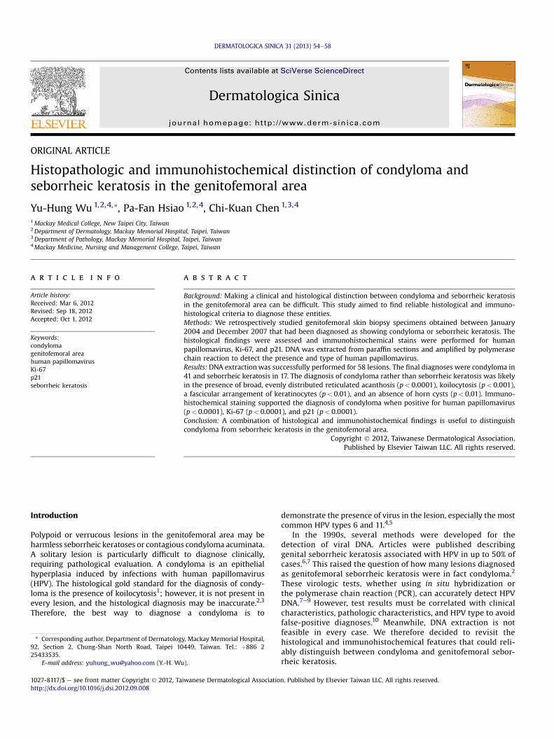
Figure 1 Important diagnostic features of condyloma. (A) Evenly distributed broad reticulgranular cell layer. (C) Fascicular arrangement of the keratinocytes (H&E stain, B, C 200�)
HPV staining. The final diagnoses thus included 41 condylomas and17 seborrheic keratoses.
Comparison between two groups
Clinical comparisonThe average age of patients with condyloma (31 years) was youngerthan those with seborrheic keratosis (54 years). Condylomas weremore common in males (32) than females (9), whereas the genderdistribution was closer to equality in seborrheic keratosis (7 malesand 10 females). Lesions occurring on the scrotum, penis, vulva, andperineum were more likely to be condyloma, although seborrheickeratosis could be present in any of those areas (Table 2).
Histopathologic findings in two groupsCondylomas and seborrheic keratosis may both have a reticulararchitecture. The rete ridges were significantly more likely to bebroad, with a similar width in condyloma (Figure 1A) compared toa variable size in seborrheic keratosis (p< 0.01) (Table 3). Koilocy-tosis was present in 68% (28/41) of condylomas (Figure 1B), but canoccasionally be observed in seborrheic keratosis (3/17, 18%;p< 0.01). Keratinocytes in condylomas were often arranged intightly interwoven fascicles (Figure 1C), a finding rarely present inseborrheic keratosis (p¼ 0.01). Horn cysts were frequently seenin seborrheic keratosis but are not common in condylomas(p< 0.01). The presence of a cauliflower shape, pigmentation,papillomatosis, spongiosis, and dermal inflammation did not differsignificantly between the two disorders.
Immunohistochemical stains in two groupsImmunohistochemical staining (HPV, Ki-67, and p21) resultsdiffered significantly between condyloma and seborrheic keratosis(all p< 0.01; Table 3). The HPV stain was specific but not sensitive.Only 20% of condylomas (8/41) had a strong positive result (þþor þþþ), and most of those eight lesions could be easily diagnosed
ated acanthosis and cauliflower architecture (H&E stain, 40�). (B) Koilocytosis of the(D) Coexistence of both koilocytosis and a fascicular arrangement of keratinocytes.

Figure 2 Positive immunohistochemical staining in the diagnosis of condyloma. Human papillomavirus stain: (A) strongly positive (þþþ); (B) moderately positive (þþ); (C) weaklypositive (þ). Ki-67 stain: (D) strongly positive (þþþ); (E) moderately positive (þþ); (F) weakly positive (þ). p21 stain: (G) strongly positive (þþþ); (H) moderately positive (þþ); (I)weakly positive (þ). (AeC¼ 400�; DeII¼ 200�).
Y.-H. Wu et al. / Dermatologica Sinica 31 (2013) 54e58 57
histopathologically. Conversely, Ki-67 and p21 were not specific butwere very sensitive for the diagnosis of condyloma, with more than80% of condylomas positive for at least one of the stains. Nearly half(46%, 19/41 for Ki-67; 49%, 21/41 for p21) of the specimens hada strongly positive result (þþ or þþþ; Figure 2).
Sensitivity, specificity, predictive value, and likelihood ratio in twogroupsSeven featuresdkoilocytosis, broad reticulated acanthosis,a fascicular arrangement of keratinocytes, horn cysts, HPV-
Table 4 Diagnostic characteristics of various features in evaluating condyloma in the ge
Koilocytosis Broad reticulatedacanthosis
Fasciculararrangement
Sensitivity, % 68 98 46Specificity, % 82 59 94PPV, % 90 85 95NPV, % 52 91 42LRþ 3.9 2.4 7.9LR� 0.4 0.04 0.6
LRþ¼ positive likelihood ratio; LRe¼ negative likelihood ratio; NPV¼ negative predictia Divided by zero.
positive staining, Ki67-positive staining, and p21 pos-itivityddiffered significantly between the two types of lesion(Table 3). The characteristics of the various diagnostic criteria areshown in Table 4). Based on likelihood ratios, the most helpfulfindings to rule in the diagnosis of condyloma (positive likelihoodratio � 3) were the presence of koilocytosis, a fasciculararrangement, and positive staining for HPV, Ki-67, and p21. Themost helpful findings to rule out condyloma (negative likelihoodratio � 0.2) were an absence of broad reticulated acanthosis andnegative Ki-67 and p21 staining.
nitofemoral area.
Horn cysts HPV-positivestaining
Ki-67-positivestaining
p21-positivestaining
29 63 88 8329 100 71 9450 100 88 9715 53 71 700.4 a 3.0 14.12.4 0.4 0.2 0.2
ve value; PPV¼ positive predictive value.

Y.-H. Wu et al. / Dermatologica Sinica 31 (2013) 54e5858
Discussion
This study examined the reliability of old criteria and provided newinformation on the distinction between condyloma and seborrheickeratosis in the genitofemoral area. In low-power fields, condylomaoften had broad and evenly distributed reticulated acanthosis,a finding that was more sensitive and useful than cauliflower orpapillomatous architecture. In higher magnification, about two-thirds of condylomas had diagnostic koilocytosis. However, thereare two pitfalls which clinicians should be aware of when inter-preting the histopathologic findings. First, koilocytes can be occa-sionally seen in seborrheic keratosis; second, horn cysts, a verycommon finding usually indicating seborrheic keratosis, mayoccasionally appear in a condyloma.
A very helpful sign that has not previously been reported is thefascicular arrangement of keratinocytes, found in half the condy-lomas in our series but infrequently in seborrheic keratosis. In theauthors’ personal experience, this pattern may only appear occa-sionally in nevus sebaceus. One possible explanation for thispeculiar arrangement in condylomas is that the disorder involvessquamoid keratinocyte proliferation. Seborrheic keratosis, on theother hand, involves the proliferation of both squamous andbasoloid cells, with the latter often predominating. Therefore,a fascicular arrangement of keratinocytes would not be common inseborrheic keratosis.
For lesions that cannot easily be diagnosed using histopatho-logical features, immunohistochemical stains for Ki-67 and p21were helpful. The most frequently used immunohistochemicalstains in the diagnosis of HPV-associated genital intraepithelialneoplasia are those for Ki-67 and p16.9,15,16 Condylomas have beendemonstrated to be a proliferative keratinocytic lesion with Ki-67expression9,17; however, Ki-67 staining was normally present inthe basal cell layer of all specimens, and interpretation requiredskill to distinguish normal from abnormal staining. p16 is a cell-cycle regulatory protein overexpressed in cell nuclei infected byhigh-risk HPV.15,16 It has been found that p16 expression is nothelpful with vulvar lesions associated with low-risk HPV infection,including condylomas.9
In recent research, another cell-cycle control protein, p21, wasnoted to be produced in cells infected with low-risk HPV types.11
Our study demonstrated a similar result. p21 is a cyclin-dependent kinase inhibitor that usually results in G1-phase cell-cycle arrest.11,18 One would expect p21 to be not expressed in theproliferation of HPV-infected keratinocytes. In contrast, increasedp21 expression has been found in the suprabasal cells of condy-lomas,11,18 and this was confirmed in our study. HPV-infectedkeratinocytes expressing p21 can still proliferate, as shown by theco-expression of p21 and Ki-67 studies,18 might be attributed tohost cell reaction. The findings are very useful in the diagnosis ofcondyloma because cells in normal epithelium do not showa concurrent expression of both positive and negative regulatoryproteins. Moreover, p21 nuclear staining was present in the upperepidermis without basal cell positivity, which is easier to readcompared to Ki-67 staining.
The primary limitation of this investigation was the smallnumber of specimens available for study. In addition, bias waspossible between different observers who made the diagnosis ofthe lesions. A greater variety of lesions, including resolvingcondylomas for example, should be examined to confirm theresults. In particular, a variety of proliferative epidermal lesionsshould be evaluated to see if the fascicular arrangement of
keratinocytes is a valid histological marker for HPV infection. Theexpression of cell-cycle proteins in various subtypes of HPV infec-tion also needs to be more fully understood.
In conclusion, broad reticulated acanthosis and a fasciculararrangement of keratinocytes are helpful findings at scanningmagnification that raise the possibility of condyloma. If thesepatterns are present, high-power fields should be carefullyexamined for koilocytosis. If the diagnosis is still in question,staining with ki-67 and p21 may provide indirect evidence of HPVinfection.
Acknowledgments
We thank Mr Po-Tsang Chen and Mr Schu-Rern Chern (Depart-ment of Medical Research, Mackay Memorial Hospital) andDr Chih-Ping Chen (Department of Gynecology, Mackay MemorialHospital) for their help with HPV DNA extraction and sequencing.This work was supported by grants from the Mackay MemorialHospital MMH-E-9730.
References
1. Kimura S, Masuda M. A comparative immunoperoxidase and histopathologicstudy of condylomata acuminata. J Cutan Pathol 1985;12:142e6.
2. Li J, Ackerman AB. “Seborrheic keratoses” that contain human papillomavirusare condylomata acuminata. Am J Dermatopathol 1994;16:398e405.
3. Zhao YK, Lin YX, Luo RY, et al. Human papillomavirus (HPV) infection inseborrheic keratosis. Am J Dermatopathol 1989;11:209e12.
4. Wang H, Qiao YL. Human papillomavirus type-distribution in condylomataacuminata of mainland China: a meta-analysis. Int J STD AIDS 2008;19:680e4.
5. D’Ambrogio A, Yerly S, Sahli R, et al. Human papilloma virus type and recur-rence rate after surgical clearance of anal condylomata acuminata. Sex TransmDis 2009;36:536e40.
6. Leonardi CL, Zhu WY, Kinsey WH, Penneys NS. Seborrheic keratoses from thegenital region may contain human papillomavirus DNA. Arch Dermatol1991;127:1203e6.
7. Zhu WY, Leonardi C, Penneys NS. Detection of human papillomavirus DNA inseborrheic keratosis by polymerase chain reaction. J Dermatol Sci 1992;4:166e71.
8. Brown DR, Bryan JT, Cramer H, Katz BP, Handy V, Fife KH. Detection of multiplehuman papillomavirus types in condylomata acuminata from immunosup-pressed patients. J Infect Dis 1994;170:759e65.
9. Bai H, Cviko A, Granter S, Yuan L, Betensky RA, Crum CP. Immunophenotypicand viral (human papillomavirus) correlates of vulvar seborrheic keratosis.Hum Pathol 2003;34:559e64.
10. Forslund O, Lindelof B, Hradil E, et al. High prevalence of cutaneous humanpapillomavirus DNA on the top of skin tumors but not in “Stripped” biopsiesfrom the same tumors. J Invest Dermatol 2004;123:388e94.
11. Lyman RC, Wilson ML, Herrington CS. Cell-cycle control protein expression isdisrupted in anogenital condylomata infected with low-risk human papillo-mavirus types. J Low Genit Tract Dis 2008;12:224e31.
12. Lorincz AT, Quinn AP, Goldsborough MD, Schmidt BJ, Temple GF. Cloning andpartial DNA sequencing of two new human papillomavirus types associatedwith condylomas and low-grade cervical neoplasia. J Virol 1989;63:2829e34.
13. Degener AM, Laino L, Pierangeli A, Accappaticcio G, Innocenzi D, Pala S. Humanpapillomavirus-32-positive extragenital Bowenoid papulosis (BP) in a HIVpatient with typical genital BP localization. Sex Transm Dis 2004;31:619e22.
14. Cheng YP, Chen CW, Sheen YS, Tsai TF. Genotype distribution of humanpapillomavirus in anogenital warts of male patients in Taiwan. Dermatol Sinica2012;30:85e9.
15. Bean SM, Eltoum I, Horton DK, Whitlow L, Chhieng DC. Immunohistochemicalexpression of p16 and Ki-67 correlates with degree of anal intraepithelialneoplasia. Am J Surg Pathol 2007;31:555e61.
16. Walts AE, Lechago J, Bose S. P16 and Ki67 immunostaining is a useful adjunct inthe assessment of biopsies for HPV-associated anal intraepithelial neoplasia.Am J Surg Pathol 2006;30:795e801.
17. Pirog EC, Chen YT, Isacson C. MIB-1 immunostaining is a beneficial adjunct testfor accurate diagnosis of vulvar condyloma acuminatum. Am J Surg Pathol2000;24:1393e9.
18. Zehbe I, Ratsch A, Alunni-Fabbroni M, et al. Overriding of cyclin-dependentkinase inhibitors by high and low risk human papillomavirus types: evidencefor an in vivo role in cervical lesions. Oncogene 1999;18:2201e11.