histopathological effects of aspergillus clavatus ... · histopathological effects of aspergillus...
TRANSCRIPT
f u n g a l b i o l o g y 1 2 0 ( 2 0 1 6 ) 4 8 9e4 9 9
journa l homepage : www.e lsev ier . com/ loca te / funb io
Histopathological effects of Aspergillus clavatus(Ascomycota: Trichocomaceae) on larvae of thesouthern house mosquito, Culex quinquefasciatus(Diptera: Culicidae)
Thomas BAWINa,*, Fawrou SEYEa,b, Slimane BOUKRAAa,Jean-Yves ZIMMERa, Fara Nantenaina RAHARIMALALAa,c,Mady NDIAYEb, Philippe COMPEREd, Frank DELVIGNEe, Fr�ed�eric FRANCISa
aFunctional and Evolutionary Entomology, Gembloux Agro-Bio Tech, University of Liege, Passage des D�eport�es 2,
B-5030 Gembloux, BelgiumbLaboratory of Reproductive Biology, Department of Animal Biology, Faculty of Science and Technology, University
Cheikh Anta Diop, B-5005 Dakar Fann, SenegalcMedical Entomology, Pasteur Institute, Ambatofotsikely, 101, Antananarivo, MadagascardFunctional and Evolutionary Morphology, Department of Biology, Ecology and Evolution, Faculty of Sciences,
University of Liege, All�ee de la Chimie 3, B-4000 Li�ege, BelgiumeMicrobial Processes and Interactions, Gembloux Agro-Bio Tech, University of Liege, Passage des D�eport�es 2, B-5030
Gembloux, Belgium
a r t i c l e i n f o
Article history:
Received 22 December 2015
Accepted 4 January 2016
Available online 14 January 2016
Corresponding Editor:
N.P. Money
Keywords:
Aspergillosis
Bioassay
Electron microscopy
Entomopathogenic fungi
Histopathology
Microbial control
* Corresponding author. Tel.: þ32 81 62 22 87E-mail addresses: entomologie.gemblo
[email protected] (S. Boukraa),(F. N. Raharimalala), [email protected]@ulg.ac.be (F. Francis).http://dx.doi.org/10.1016/j.funbio.2016.01.0021878-6146/ª 2016 The British Mycological So
a b s t r a c t
Aspergillus clavatus (Ascomycota: Trichocomaceae) was previously found to be an opportu-
nistic pathogen of mosquitoes (Diptera: Culicidae). In the present study, the mechanism
leading to its insecticidal activity was investigated regarding histological damages on Culex
quinquefasciatus larvae exposed to A. clavatus spores. Multiple concentration assays using
spore suspensions (0.5e2.5 � 108 spores ml�1) revealed 17.0e74.3 % correctedmortalities af-
ter 48hexposure.Heat-deactivatedspores induceda lowermortality compared tononheated
spores suggesting that insecticidal effects are actively exerted. Spore-treated and untreated
larvae were prepared for lightmicroscopy as well as for scanning and transmission electron
microscopy. Spores failed to adhere to the external body surface (except themouth parts) of
these aquatic immature stages but progressively filled the digestive tract where theirmetab-
olismseemed toactivate. Inparallel, the internal tissuesof the larvae, i.e. themidgutwall, the
skeletalmuscles, and the cuticle-secreting epidermis,were progressively destroyedbetween
8and24hof exposure. Theseobservations suggest that toxins secretedbyactive germinating
spores of A. clavatus in the digestive tract altered the larval tissues, leading to their necrosis
; fax: þ32 81 62 23 [email protected], [email protected] (T. Bawin), [email protected] (F. Seye),[email protected] (J. -Y. Zimmer), [email protected] (M. Ndiaye), [email protected] (P. Compere), [email protected] (F. Delvigne),
ciety. Published by Elsevier Ltd. All rights reserved.
490 T. Bawin et al.
and causing larval death. Fungal proliferation and sporulation then occurred during a sapro-
phytic phase. A. clavatus enzymes or toxins responsible for these pathogenic effects need to
be identified in further studies before any use of this fungus in mosquito control.
ª 2016 The British Mycological Society. Published by Elsevier Ltd. All rights reserved.
Introduction Aspergillus clavatus Desmazi�eres (Ascomycota: Trichoco-
Many mosquito (Diptera: Culicidae) species are vectors re-
sponsible for the transmission of infectious diseases of medi-
cal and veterinary importance including filariasis, malaria,
and arboviruses (Goddard 2008; Mullen & Durden 2009;
Medlock et al. 2012). Risk for human infection considerably in-
creased during the last decades due to climate changes and in-
creasing global trade (Reiter 2001; Medlock et al. 2012; Boukraa
et al. 2013). Integrated pest management is now promoted due
to harmful side effects of the chemical insecticides classically
used for mosquito control and insect resistance development
(Nauen 2007; Rattner 2009; Rivero et al. 2010). Entomopatho-
genic microorganisms including fungi are increasingly stud-
ied in a biological control context regarding their ability to
infect and kill insect hosts with more or less selectivity
(Shah & Pell 2003; Becker et al. 2010; Bawin et al. 2015). In this
context, soil-borne generalist fungal pathogens including
Beauveria (Ascomycota: Cordycipitaceae) and Metarhizium
(Ascomycota: Clavicipitaceae) isolates have been extensively
studied due to their simple life-cycle and thereby easy produc-
tion of stable aerial sporeswhich are the infectious propagules
(Scholte et al. 2004; Kanzok & Jacobs-Lorena 2006; Seye et al.
2013). Despite a possible production of hazardous metabolites
to vertebrates (Gugnani 2003; Br€ase et al. 2009), Aspergillus
(Ascomycota: Trichocomaceae) species are now studied due
to the opportunistic insect pathogenic behaviour of some iso-
lates (de Moraes et al. 2001; Pereira et al. 2009; Maketon et al.
2014), their ubiquity in the environment (Gugnani 2003), and
their potential for biotechnological applications (Powell et al.
1994).
Infection of terrestrial insects as adult mosquitoes by such
fungi proceeds by several steps following a consistent pattern
(Shah & Pell 2003; Charnley & Collins 2007; Khachatourians &
Qazi 2008), starting with spore adhesion to the host cuticle,
followed by germination and penetration using enzymatic
and mechanical forces. Colonization of the insect haemocoel,
commonly reliedwith hyphal proliferationwithin host tissues
and enzymatic activities, is responsible for lethal histopatho-
logical damages. The virulence of insect pathogenic fungi is
thus related to the secretion of proteolytic, lipolytic, and chiti-
nolytic enzymes as well as secondary metabolites (Hajek & St
Leger 1994; Khachatourians & Qazi 2008). Fungal eruption
through the integument of the insect finally leads to the
dissemination of newly produced aerial spores. By contrast,
the mechanisms by which these pathogens affect aquatic
mosquito larval instars appear to be inconsistent and often
unclear. Many invasion routes have been reported, including
penetration of the cuticle as described above, or entry via
the respiratory siphon or alimentary canal (Lacey et al. 1988;
Miranpuri & Khachatourians 1991; Butt et al. 2013).
maceae) was previously investigated as an effective opportu-
nistic mosquito pathogen (Seye & Ndiaye 2008; Seye et al.
2009, 2014a).However, theprecisemechanism leading to larval
death remains elusive. In the present study, histological dam-
ages on larvae of the southern house mosquito, Culex quinque-
fasciatus Say, during exposition to A. clavatus spores are
described through light and electronmicroscopy observations.
Materials and methods
Fungal strain
A. clavatus was isolated from the locust cricket Oedaleus sene-
galensisKrauss (Orthoptera: Acrididae) in the Botanical Garden
of the Faculty of Sciences, Cheikh Anta Diop University,
Dakar, Senegal (accession number MUCL 55275, Mycoth�eque
de l’Universit�e Catholique de Louvain, Belgian Coordinated
Collections of Microorganisms, Belgium) and maintained on
Potato Dextrose Agar (PDA) medium as pure culture. This
strain was previously shown to have an insecticidal activity
against mosquitoes (Seye et al. 2009).
To obtain large amounts of spores, the funguswas grown in
250 ml Erlenmeyer flasks on a wheat bran-based solid-state
substrate (5 gwheat branper flask and 20mlnutritive solution:
peptone 1 %, yeast extract 1 %, chloramphenicol 0.005 %).
Wheat bran and nutritive solution were separately sterilized
at 121 �C for 20 min before being mixed. Each flask was inocu-
latedwitha spore suspension (1ml, 5�106 sporesml�1) and in-
cubated 7 d at 30 �C. Fungal masses produced on the media
were then washed with a 0.05 % Tween 80 solution in distilled
water (150 ml) on a rotary shaker (150 rpm) for 2 h. Fungal fila-
ments and wheat bran residues were discarded by filtration
throughadouble layer of sterilemuslin, and the resulting solu-
tion was centrifuged (3000 g, 5 min) to remove the newly pro-
duced spores as pellet. Spores were resuspended in a 0.05 %
Tween 80 solution and the concentration was assessed using
a haemocytometer (Thoma, Assistent, Sondheim/Rh€on, Ger-
many) before application onmosquito larvae.
Mosquito larvae
C. quinquefasciatus adults (S-lab strain, native from Riverside,
California) were reared in 50� 50� 50 cmnet cages (BugDorm,
MegaView Science, Taichung, Taiwan) and fed with 10 % (w/v)
sucrose solutions in water. Females were exposed three times
a week to glass membrane feeders (manufactured by CNAP-
MAD, Antananarivo, Madagascar) maintained at 37 �C by cir-
culating water, to allow blood meal through stretched
Parafilm sheets. Plastic cups filled of water were placed in
Histopathological effects of A. clavatus on mosquito larvae 491
the cages as oviposition sites. Egg rafts were daily collected
and maintained in 25 (length) � 15 (width) cm containers
with 5 (depth) cm distilled water. After hatching, larvae were
fed every 2 d with a powder made with 2:1 crushed fish food
(TetraMin, Tetra, Blacksburg, USA) and natural brewer’s yeast
(Biover, Brugge, Belgium). Water was renewed every week.
Rearing conditions were 25 � 2 �C temperature, 70 � 5 % rela-
tive humidity, and 16:8 h (light:dark) photoperiod.
Bioassays
Conventional toxicity tests were first conducted in aqueous
suspensions according to the World Health Organization
protocol (2005) to assess lethal concentrations of A. clavatus
spores required to kill 50 % (LC50) and 90 % (LC90) of the larvae.
Batches of 20 third-instar individuals were exposed to a range
of final concentrations of 0.5 � 108, 1.0 � 108, 1.5 � 108,
2.0 � 108, and 2.5 � 108 spores ml�1 in separated bottles
(50 ml) for 48 h. Control larvae were maintained in 0.05 %
Tween 80 solution. Treatments (i.e. a control batch of larvae
associated to a set of the tested concentrations) were repli-
cated four times at different time intervals with spores
produced independently. Laboratory conditions were similar
to rearing conditions. Mortality was daily recorded and
corrected using the Abbott’s formula (Abbott 1925). Corrected
mortality proportions were linearized using logit transforma-
tion (Dagnelie 1970): logit(P) ¼ ln(P/1 � P). A simple linear
regression was used for modelling the relationship between
logit-transformed mortality and logarithm-transformed
values of fungal concentrations as explanatory variable:
logit(P) ¼ slope � ln(concentration) þ intercept. The relation-
ship between larval mortality and spore concentrations was
assessed considering Snedecor-F distribution and p-value.
The lethal activity of the funguswas then investigated over
time. Toxicity tests were carried out as described above using
a concentration of 1.5 � 108 spores ml�1 (that is related to
LC50s). Mortality was recorded after 6, 12, 18, 24, 30, 36, and
48 h exposure. The lethal time required to kill 50 % of the
larvae (LT50) was calculated using KaplaneMeier analysis,
and log-rank test (providing chi-square and p-value) was
carried out to check for significant differences in survival
between spore-treated and control larvae.
Heat-treated spores were used to determine whether spore
germination is required to impact mosquito larvae. Spores in
aqueous suspension were killed by autoclaving for 20 min at
121 �C. Spore viability was assessed using PDA plates over
an incubation time of 72 h at 30 �C. Toxicity tests were con-
ducted as described above using a 2.5 � 108 spores ml�1 con-
centration. Differences in survival between individuals
exposed to heat-treated and nonheated spores, and control
larvae were analysed using KaplaneMeier analysis with pair-
wise comparison over log-rank test.
Dead larvae treated with either 2.5 � 108 intact or heat-
deactivated sporesml�1 (n ¼ 10 per treatment) were randomly
sampled, rinsed thrice with distilled water, and incubated on
awet paper in Petri dishes (30 �C) in order to observe the emer-
gence of fungal aerial filaments outside the body.
All statistical analyses were performed using Minitab v.17
software (Minitab, Coventry, UK). For all tests, the significant
threshold was p < 0.050.
Light and electron microscopy
Fifty third-instar larvae (125 ml) were exposed to a concentra-
tion of 1.5 � 108 spores ml�1 according to the protocol
described above. Control larvae were maintained in 0.05 %
Tween 80 solution. In both treatments, five larvae were taken
after 8, 12, 24, and 48 h exposure for light microscopy (LM) and
transmission electronmicroscopy (TEM). Three additional lar-
vae were sampled after 8 and 24 h for scanning electron mi-
croscopy (SEM).
Spore-treated and control larvae were cut behind the head
and in front of the respiratory siphon to facilitate the perme-
ation of chemicals. Body segments were fixed for 12 h in
a 2.5 % glutaraldehyde solution buffered with 0.1 M sodium
cacodylate at pH 7.3. They were then rinsed and stored for
few days in 0.2 M cacodylate buffer, before postfixation in
1% osmium tetroxide (1 h), and three rinses (3� 10min) in dis-
tilled water. Dehydration was performed through a graded
ethanol series of increasing concentrations: 30 %, 50 %, 70 %,
90 % (1 � 10 min), and 100 % (3 � 20 min).
For LM and TEM, body segments were embedded in epoxy
resin (SPI-PON 812, SPI-CHEM, SPI supplies, Leuven, Belgium)
with propylene oxide as intermediate solvent for impregna-
tion (2 � 30 min in pure solvent, 2 h 30 min in solvent/resin
mixture, and overnight in pure resin). Embedding was
performed in flat silicone molds to facilitate sample orienta-
tion for sectioning, then placed in a stove (60 �C, 72 h) to allow
polymerization. Semithin (1e2 mm) and ultrathin (60e80 nm)
sections were performed between the second and fourth ab-
dominal segment using a diamond knife on a Reichert-Jung
Ultracut E (Reichert-Jung, Vienna, Austria) ultramicrotome.
Semithin sections were stained with 1 % toluidine blue (pH
9) before observation for general histology and orientation of
further ultrathin sections, using an Olympus Provis Ax70
(Olympus, Tokyo, Japan) light microscope equipped with an
Olympus DP50-CU (Olympus) digital camera. Ultrathin sec-
tions were contrasted with uranyl acetate and lead citrate
according to the conventional method; then observed in
a Jeol JEM 100-SX (JEOL, Tokyo, Japan) transmission electron
microscope under 80 kV accelerating voltage.
For SEM, dehydrated body segments were critical-point
dried with CO2 and mounted on glass slides with double-
side carbon tape. All were then sputter-coated with Pt
(20 nm) in a Blazers SCD 030 unit (Oerlikon Balzers Coating,
Balzers, Liechtenstein) before observation in a Jeol JSM-840A
(JEOL, Tokyo, Japan) scanning electron microscope under
20 kV accelerating voltage.
Results
Bioassays
Control mortalities ranged from 0.0 (24 h) to 2.9 % (48 h). Cor-
rected mortalities increased with the spore concentration
(0.5e2.5 � 108 spores ml�1), ranging from 12.5 to 67.5 % at 24 h
and from 17.0 to 74.3 % at 48 h. Corresponding lethal concentra-
tions (LC50 and LC90) were respectively 1.4 � 108 and
4.9 � 108 spores ml�1 (slope ¼ 1.8 � 0.3; intercept ¼ �33.5 � 4.9;
492 T. Bawin et al.
F(1,18) ¼ 45.8; p < 0.001), and 1.2 � 108 and 4.3 � 108 spores ml�1
(slope¼ 1.7� 0.2; intercept¼�31.4� 4.1; F(1,18)¼ 58.1; p< 0.001).
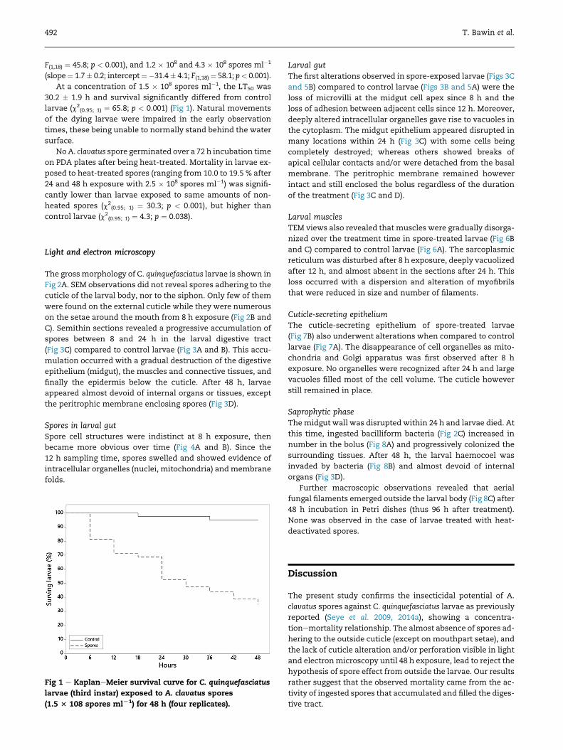
At a concentration of 1.5 � 108 spores ml�1, the LT50 was
30.2 � 1.9 h and survival significantly differed from control
larvae (c2(0.95; 1) ¼ 65.8; p < 0.001) (Fig 1). Natural movements
of the dying larvae were impaired in the early observation
times, these being unable to normally stand behind the water
surface.
NoA. clavatus spore germinated over a 72 h incubation time
on PDA plates after being heat-treated. Mortality in larvae ex-
posed to heat-treated spores (ranging from 10.0 to 19.5 % after
24 and 48 h exposure with 2.5 � 108 spores ml�1) was signifi-
cantly lower than larvae exposed to same amounts of non-
heated spores (c2(0.95; 1) ¼ 30.3; p < 0.001), but higher than
control larvae (c2(0.95; 1) ¼ 4.3; p ¼ 0.038).
Light and electron microscopy
The grossmorphology of C. quinquefasciatus larvae is shown in
Fig 2A. SEM observations did not reveal spores adhering to the
cuticle of the larval body, nor to the siphon. Only few of them
were found on the external cuticle while they were numerous
on the setae around the mouth from 8 h exposure (Fig 2B and
C). Semithin sections revealed a progressive accumulation of
spores between 8 and 24 h in the larval digestive tract
(Fig 3C) compared to control larvae (Fig 3A and B). This accu-
mulation occurred with a gradual destruction of the digestive
epithelium (midgut), the muscles and connective tissues, and
finally the epidermis below the cuticle. After 48 h, larvae
appeared almost devoid of internal organs or tissues, except
the peritrophic membrane enclosing spores (Fig 3D).
Spores in larval gutSpore cell structures were indistinct at 8 h exposure, then
became more obvious over time (Fig 4A and B). Since the
12 h sampling time, spores swelled and showed evidence of
intracellular organelles (nuclei, mitochondria) andmembrane
folds.
Fig 1 e KaplaneMeier survival curve for C. quinquefasciatus
larvae (third instar) exposed to A. clavatus spores
(1.5 3 108 spores mlL1) for 48 h (four replicates).
Larval gutThe first alterations observed in spore-exposed larvae (Figs 3C
and 5B) compared to control larvae (Figs 3B and 5A) were the
loss of microvilli at the midgut cell apex since 8 h and the
loss of adhesion between adjacent cells since 12 h. Moreover,
deeply altered intracellular organelles gave rise to vacuoles in
the cytoplasm. The midgut epithelium appeared disrupted in
many locations within 24 h (Fig 3C) with some cells being
completely destroyed; whereas others showed breaks of
apical cellular contacts and/or were detached from the basal
membrane. The peritrophic membrane remained however
intact and still enclosed the bolus regardless of the duration
of the treatment (Fig 3C and D).
Larval musclesTEM views also revealed that muscles were gradually disorga-
nized over the treatment time in spore-treated larvae (Fig 6B
and C) compared to control larvae (Fig 6A). The sarcoplasmic
reticulumwas disturbed after 8 h exposure, deeply vacuolized
after 12 h, and almost absent in the sections after 24 h. This
loss occurred with a dispersion and alteration of myofibrils
that were reduced in size and number of filaments.
Cuticle-secreting epitheliumThe cuticle-secreting epithelium of spore-treated larvae
(Fig 7B) also underwent alterations when compared to control
larvae (Fig 7A). The disappearance of cell organelles as mito-
chondria and Golgi apparatus was first observed after 8 h
exposure. No organelles were recognized after 24 h and large
vacuoles filled most of the cell volume. The cuticle however
still remained in place.
Saprophytic phaseThemidgut wall was disrupted within 24 h and larvae died. At
this time, ingested bacilliform bacteria (Fig 2C) increased in
number in the bolus (Fig 8A) and progressively colonized the
surrounding tissues. After 48 h, the larval haemocoel was
invaded by bacteria (Fig 8B) and almost devoid of internal
organs (Fig 3D).
Further macroscopic observations revealed that aerial
fungal filaments emerged outside the larval body (Fig 8C) after
48 h incubation in Petri dishes (thus 96 h after treatment).
None was observed in the case of larvae treated with heat-
deactivated spores.
Discussion
The present study confirms the insecticidal potential of A.
clavatus spores against C. quinquefasciatus larvae as previously
reported (Seye et al. 2009, 2014a), showing a concentra-
tionemortality relationship. The almost absence of spores ad-
hering to the outside cuticle (except on mouthpart setae), and
the lack of cuticle alteration and/or perforation visible in light
and electronmicroscopy until 48 h exposure, lead to reject the
hypothesis of spore effect from outside the larvae. Our results
rather suggest that the observed mortality came from the ac-
tivity of ingested spores that accumulated and filled the diges-
tive tract.
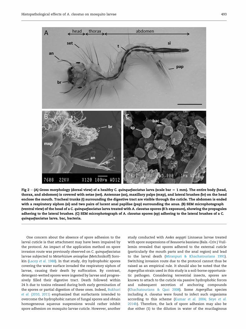
Fig 2 e (A) Gross morphology (dorsal view) of a healthy C. quinquefasciatus larva (scale bar [ 1 mm). The entire body (head,
thorax, and abdomen) is covered with setae (set). Antennae (an), maxillary palps (mxp), and lateral brushes (br) on the head
enclose the mouth. Tracheal trunks (t) surrounding the digestive tract are visible through the cuticle. The abdomen is ended
with a respiratory siphon (si) and two pairs of lucent anal papillae (pap) surrounding the anus. (B) SEM microphotograph
(ventral view) of the head of a C. quinquefasciatus larva treated with A. clavatus spores (8 h exposure), showing the propagules
adhering to the lateral brushes. (C) SEM microphotograph of A. clavatus spores (sp) adhering to the lateral brushes of a C.
quinquefasciatus larva. bac, bacteria.
Histopathological effects of A. clavatus on mosquito larvae 493
One concern about the absence of spore adhesion to the
larval cuticle is that attachment may have been impaired by
the protocol. An impact of the application method on spore
invasion route was previously observed on C. quinquefasciatus
larvae subjected to Metarhizium anisopliae (Metchnikoff) Soro-
kin (Lacey et al. 1988). In that study, dry hydrophobic spores
covering the water surface invaded the respiratory siphon of
larvae, causing their death by suffocation. By contrast,
detergent-wetted spores were ingested by larvae and progres-
sively filled their digestive tract. Death followed within
24 h due to toxins released during both early germination of
the spores or partial digestion of these ones. Indeed, Bukhari
et al. (2010, 2011) emphasized that surfactants intended to
overcome the hydrophobic nature of fungal spores and obtain
homogeneous aqueous suspensions would rather inhibit
spore adhesion on mosquito larvae cuticle. However, another
study conducted with Aedes aegypti Linnaeus larvae treated
with spore suspensions of Beauveria bassiana (Bals.-Criv.) Vuil-
lemin revealed that spores adhered to the external cuticle
(particularly the mouth parts and the anal region) and lead
to the larval death (Miranpuri & Khachatourians 1991).
Switching invasion route due to the protocol cannot thus be
raised as an empirical rule. It should also be noted that the
Aspergillus strain used in this study is a soil-borne opportunis-
tic pathogen. Considering terrestrial insects, spores are
known to attach to the cuticle via passive hydrophobic forces
and subsequent secretion of anchoring compounds
(Khachatourians & Qazi 2008). Some Aspergillus species
including A. clavatus were found to infect such organisms
according to this scheme (Kumar et al. 2004; Seye et al.
2014b). Therefore, the lack of spore adhesion may also be
due either (1) to the dilution in water of the mucilaginous
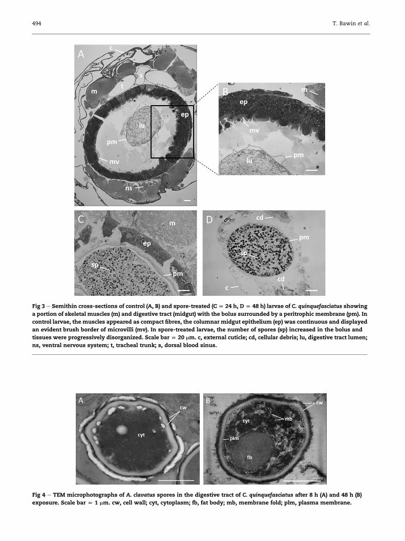
Fig 3 e Semithin cross-sections of control (A, B) and spore-treated (C [ 24 h, D [ 48 h) larvae of C. quinquefasciatus showing
a portion of skeletal muscles (m) and digestive tract (midgut) with the bolus surrounded by a peritrophic membrane (pm). In
control larvae, the muscles appeared as compact fibres, the columnar midgut epithelium (ep) was continuous and displayed
an evident brush border of microvilli (mv). In spore-treated larvae, the number of spores (sp) increased in the bolus and
tissues were progressively disorganized. Scale bar [ 20 mm. c, external cuticle; cd, cellular debris; lu, digestive tract lumen;
ns, ventral nervous system; t, tracheal trunk; s, dorsal blood sinus.
Fig 4 e TEM microphotographs of A. clavatus spores in the digestive tract of C. quinquefasciatus after 8 h (A) and 48 h (B)
exposure. Scale bar [ 1 mm. cw, cell wall; cyt, cytoplasm; fb, fat body; mb, membrane fold; plm, plasma membrane.
494 T. Bawin et al.
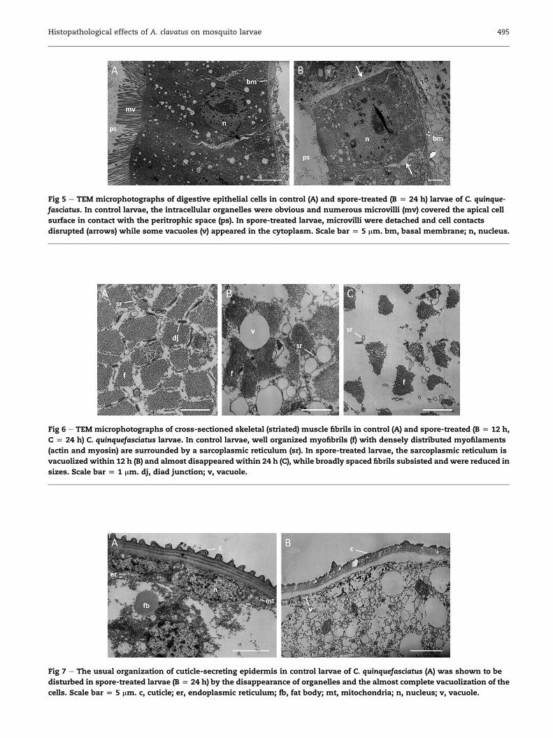
Fig 5 e TEM microphotographs of digestive epithelial cells in control (A) and spore-treated (B [ 24 h) larvae of C. quinque-
fasciatus. In control larvae, the intracellular organelles were obvious and numerous microvilli (mv) covered the apical cell
surface in contact with the peritrophic space (ps). In spore-treated larvae, microvilli were detached and cell contacts
disrupted (arrows) while some vacuoles (v) appeared in the cytoplasm. Scale bar [ 5 mm. bm, basal membrane; n, nucleus.
Fig 6 e TEM microphotographs of cross-sectioned skeletal (striated) muscle fibrils in control (A) and spore-treated (B [ 12 h,
C [ 24 h) C. quinquefasciatus larvae. In control larvae, well organized myofibrils (f) with densely distributed myofilaments
(actin and myosin) are surrounded by a sarcoplasmic reticulum (sr). In spore-treated larvae, the sarcoplasmic reticulum is
vacuolized within 12 h (B) and almost disappeared within 24 h (C), while broadly spaced fibrils subsisted and were reduced in
sizes. Scale bar [ 1 mm. dj, diad junction; v, vacuole.
Fig 7 e The usual organization of cuticle-secreting epidermis in control larvae of C. quinquefasciatus (A) was shown to be
disturbed in spore-treated larvae (B [ 24 h) by the disappearance of organelles and the almost complete vacuolization of the
cells. Scale bar [ 5 mm. c, cuticle; er, endoplasmic reticulum; fb, fat body; mt, mitochondria; n, nucleus; v, vacuole.
Histopathological effects of A. clavatus on mosquito larvae 495

Fig 8 e (A) Ingested bacilliform bacteria (bac), associated with spores (sp) in the bolus that is surrounded by the peritrophic
membrane (pm), increased in number since 24 h after treatment (scale bar [ 1 mm). (B) Some invaded the larval haemocoel
and were found nearby cellular debris (cd) (scale bar [ 1 mm). (C) C. quinquefasciatus larva incubated in Petri dish 48 h after
death, displaying A. clavatus aerial filaments that emerged outside the body (scale bar [ 1 mm). he, head; ch, conidial head;
si, siphon.
496 T. Bawin et al.
compounds produced by the fungus to attach the cuticle (Butt
et al. 2013), or (2) to the physico-chemical cues of the cuticle
with some compounds protecting the insect against potential
microbial pathogens by altering hydrophobicity (Lord &
Howard 2004) or being fungistatic (Koidsumi 1957; Sosa-
Gomez et al., 1997; Urbanek et al. 2012; Golebiowski et al.
2015). It was also hypothesized that the gut of aquatic
insects may be the preferred site for fungal spore develop-
ment due to physiologically favourable features (Miranpuri &
Khachatourians 1991), suggesting stress from the water envi-
ronment. Under these assumptions, spore adhesion previ-
ously reported on mosquito larvae with A. clavatus by Seye
et al. (2009) appears to be aminor epiphenomenon responsible
for a limited part of the observedmortality compared to inges-
tion. By contrast,mosquitocidal aquatic pathogenswith better
spore water affinity such as Lagenidium giganteum Couch
(Oomycota: Pythiaceae) (Kerwin 2007) or Tolypocladium cylin-
drosporum Gams (Ascomycota: Ophiocordycipitaceae) (Soares
1982) were found to adhere and penetrate the larval cuticle
as reported with terrestrial insects. Whether naturally
mosquito-associated Aspergillus isolates (de Moraes et al.
2001; Pereira et al. 2009; Mohanty & Prakash 2010) are more
adapted to aquatic environments or simply ingested by larvae
as described above before being isolated remains to be
investigated.
Whether ingested spores mechanically blocked the mouth
parts while others attached inside the digestive tract (Federici
1981) or not (Butt et al. 2013) has still to be elucidated. Anyway,
they remained enclosed in the peritrophic membrane that
was apparently not affected. This suggests that damages to
larval tissues were caused at distance by an early diffusion
of toxins or enzymes. On the one hand, dead larvae were
reported after exposition to heat-deactivated spores, and
one might expect that at least partial digestion of spores
and/or passive diffusion of toxins occurred being responsible
for a few part of the mortality. However, living spores were
Histopathological effects of A. clavatus on mosquito larvae 497
required to fully bring about larval mortality. As revealed by
TEM observations, damages to larval tissues may indeed be
related to the appearance of ultrastructural features of spore
metabolic activity. Considering previous reports, Lacey et al.
(1988) observed the early germination steps of M. anisopliae
spores in the digestive tract of dying larvae suggesting that
death arose due to secreted toxins. Miranpuri &
Khachatourians (1991) have established the kinetics of B.
bassiana spore development in the digestive tract ofAe. aegypti
larvae. An ingested spore (stage I) swells twice its size in
24 h (stage II). A germ tube (stage III) then emerges up to
48 h later, followed by linear (stage IV) and branched (stage
V) mycelial growth. These observations are consistent with
the results presented here with respect to the first two stages.
Extended observation times would allow to show the emer-
gence of a germ tube, as incubated dead larvae displayed
aerial fungal filaments. On the other hand, the ultrastructural
damages observed in larval tissues are characteristic of cell ly-
sis and tissue necrosis. Especially, the serious damages affect-
ing the epithelial midgut cells are similar to that caused by
Bacillus thuringiensis var. israelensis Berliner endotoxins against
Simulium pertinax Kollar (Cavados et al. 2004) and Aedes albopic-
tus Skuse larvae (Silva et al. 2008), as well as aqueous plant
extracts against Anopheles gambiae Giles (Koua et al. 1998)
and C. quinquefasciatus (Almehmadi, 2011), where loss of
microvilli, vacuolization of digestive cells and rupture of the
epithelium, and rejection of cytoplasmic material in the peri-
trophic space were also described. Taking together, these
observations (spore activity and characteristic tissue necrosis)
are consistent with the hypothesis where spores actively
released during germination toxic compounds that are
responsible for cellular damages. The degradation of the larval
tissues may then have been reinforced by ingested sapro-
phytic bacteria that benefited from the knockdown of the
immune system and invaded the general cavity in the last
steps.
A. clavatus is a commonly encountered fungus in the envi-
ronment known to produce a large number of enzymes and
mycotoxins (Gugnani 2003; Br€ase et al. 2009). Many studies
showed that A. clavatus germination was accompanied by
a secretion of toxins such as clavatol (Bergel et al. 1944), ascla-
diol (Suzuki et al. 1971), tryptoquivalone and tryptoquivaline
(Clardy et al. 1975), glyantrypine (Penn et al. 1992), cytochalasin
E (Lopez-Diaz & Flannigan 1997), kojic acid and xanthocilin
(Pitt 2000), and patulin (Sabater-Vilar et al. 2004). Some of these
compounds acting synergistically are responsible for human
diseases and animal poisoning (Flannigan & Pearce 1994;
Lopez-Diaz & Flannigan 1997). Whether the observed tissue
disorders are due to such nonselective compounds
remain to be determined as these features depend on the fun-
gal isolate. By example, patulin production was detected in
three of eight tested A. clavatus strains (Varga et al. 2003),
with amounts being strain dependent (Lopez-Diaz &
Flannigan 1997). However, the fact that the chitinous peritro-
phicmembrane aswell as the external cuticle remained intact
until at least 48 h exposure suggests that early germinating
spores did not secrete adapted chitinolytic nor proteolytic
enzymes, contrary to that was reported with other insect
pathogenic fungi (Schrank & Vainstein 2010). Because heat-
denatured spores induced some larval mortality, heat-stable
compounds such as mycotoxins may be at least in part re-
sponsible for the pathogenic effects. Patulin, whichwas found
to slowly decompose at 90 �Cwhile over 20 % of the compound
only disappeared in 30 min at 120 �C (Kubacki 1986), can be
one of such expected toxins. Also, cytochalasin E produced
by A. clavatus was reported to induce an inhibition of glucose
transport and actin polymerization (Brenner & Korn 1980).
This could explain why natural movements started to stop
down in the early observation times. At this time, some larvae
were nomore able to breathe air on thewater surface probably
causing their asphyxia.
Conclusions
Our results suggest that toxic compounds caused damages to
larval tissues through ingestion and accumulation of spores in
the digestive tract, which would therefore appear as the pri-
mary invasion route. Living spores are required to fully bring
about larval mortality, suggesting that these compounds are
actively secreted. Their nature and properties are thus of great
concern before using A. clavatus spores in environmental con-
ditions due to a likely lack of specificity andmaybe side effects
on nontargeted organisms. Further studies dealing with the
purification of A. clavatus toxins are required in order to char-
acterize the compounds responsible for the lethal effects.
Acknowledgements
We thank the Islamic Development Bank (IDB) for the finan-
cial support of a postdoctoral scholarship to Fawrou Seye. Nic-
ole Decloux (Functional Morphology and Evolutionary,
University of Liege) is acknowledged for her technical assis-
tance in the ultrastructural study and the Center of Applied
Technology in Microscopy (CATm, University of Liege) for pro-
viding access to electron microscopy equipment.
r e f e r e n c e s
Abbott WS, 1925. A method of computing effectiveness of insec-ticides. Journal of Economic Entomology 18: 265e267.
Almehmadi RM, 2011. Larvicidal, histopathological and ultra-structure studies of Matricharia chamomella extracts againstthe Rift Valley Fever mosquito Culex quinquefasciatus (Culici-dae: Diptera). Journal of Entomology 8: 63e72.
Bawin T, Seye F, Boukraa S, Zimmer J-Y, Delvigne F, Francis F,2015. La lutte contre les moustiques (Diptera: Culicidae):diversit�e des approches et application du controle biologique.The Canadian Entomologist 147: 476e500. http://dx.doi.org/10.4039/tce.2014.56.
Becker N, Petric D, Zgomba M, Dahl C, Boase C, Lane J, Kaiser A,2010. Mosquitoes and Their Control, 2nd edn. Springer-Verlag,Heidelberg.
Bergel F, Morrison AL, Moss AR, Rinderknecht H, 1944. An anti-bacterial substance from Aspergillus clavatus. Journal of theChemical Society, 415e421.
Boukraa S, Raharimalala FN, Zimmer J-Y, Schaffner F, Bawin T,Haubruge E, Francis F, 2013. Reintroduction of the invasivemosquito species Aedes albopictus in Belgium in July 2013.Parasite 20: 54. http://dx.doi.org/10.1051/parasite/2013054.

498 T. Bawin et al.
Br€ase S, Encinas A, Keck J, Nising CF, 2009. Chemistry and biologyof mycotoxins and related fungal metabolites. ChemicalReviews 109: 3903e3990.
Brenner SL, Korn ED, 1980. The effect of cytochalasins on actinpolymerization and actin ATPase provide insights into themechanism of polymerization. The Journal of Biological Chem-istry 255: 841e844.
Bukhari T, Middelman A, Koenraadt CJM, Takken W, Knols BGJ,2010. Factors affecting fungus-induced larval mortality inAnopheles gambiae and Anopheles stephensi. Malaria Journal 9: 22.http://dx.doi.org/10.1186/1475-2875-9-22.
Bukhari T, Takken W, Koenraadt JMC, 2011. Development ofMetarhizium anisopliae and Beauveria bassiana formulations forcontrol of malaria mosquito larvae. Parasites & Vectors 4: 23.http://dx.doi.org/10.1186/1756-3305-4-23.
Butt TM, Greenfield BPJ, Greig C, Maffeis TGG, Taylor JWD,Piasecka J, Dudley E, Abdulla A, Dubovskiy IM, Garrido-Jurado I, Quesada-Moraga E, Penny MW, Eastwood DC, 2013.Metarhizium anisopliae pathogenesis of mosquito larvae: a ver-dict of accidental death. PLOS ONE 8: e81686. http://dx.doi.org/10.1371/journal.pone.0081686.
Cavados CFG, Majerowicz S, Chaves JQ, Ara�ujo-Coutinho CJPC,Rabinovitch L, 2004. Histopathological and ultrastructural ef-fects of d-endotoxins of Bacillus thuringiensis serovar israelensisin the midgut of Simulium pertinax larvae (Diptera, Simuliidae).Memorias do Instituto Oswaldo Cruz 99: 493e498.
Charnley AK, Collins SA, 2007. Entomopathogenic fungi and theirrole in pest control. In: Kubicek CP, Druzhinina IS (eds), TheMycota: environmental and microbial relationships. Springer-Ver-lag, Heidelberg, pp. 159e187.
Clardy J, Springer JP, B€uchi G, Matsuo K, Wrightman R, 1975.Tryptoquivaline and tryptoquivalone, two new tremorgenicmetabolites of Aspergillus clavatus. Journal of the AmericanChemical Society 97: 663e665.
Dagnelie P, 1970. Th�eorie et m�ethodes statistiques: applicationsagronomiques. Les m�ethodes d’inf�erence statistique, vol. 2, Ducu-lot, Gembloux.
de Moraes AM, da Costa GL, Barcellos MZ, de Oliveira RL, deOliveira PC, 2001. The entomopathogenic potential of Asper-gillus spp. in mosquitoes vectors of tropical diseases. Journal ofBasic Microbiology 41: 45e49.
Federici BA, 1981. Mosquito control by the Fungi Culicinomyces,Lagenidium and Coelomomyces. In: Burges HD (ed.), MicrobialControl of Pests and Plant Diseases 1970e1980. Academic Press,London, pp. 555e572.
Flannigan B, Pearce AR, 1994. Aspergillus spoilage: spoilage ofcereals and cereal products by the hazardous species Asper-gillus clavatus. In: Powell KA, Renwick A, Peberdy JF (eds), TheGenus Aspergillus: from taxonomy and genetics to industrialapplication. Plenum Press, NY, pp. 115e127.
Goddard J, 2008. Mosquito-borne diseases. In: Goddard J (ed.),Infectious Diseases and Arthropods. Humana Press, Totowa,pp. 31e79.
Golebiowski M, Cerkowniaka M, Urbanek A, Dawgul M,Kamysz W, Bogus MI, Stepnowski P, 2015. Identification andantifungal activity of novel organic compounds found incuticular and internal lipids of medically important flies.Microbiological Research 170: 213e222.
Gugnani HC, 2003. Ecology and taxonomy of pathogenic asper-gilli. Frontiers in Bioscience 8: s346es357.
Hajek AE, St Leger RJ, 1994. Interactions between fungal pathogensand insect hosts. Annual Review of Entomology 39: 293e322.
Kanzok SM, Jacobs-Lorena M, 2006. Entomopathogenic fungi asbiological insecticides to control malaria. Trends in Parasitology22: 49e51. http://dx.doi.org/10.1016/j.pt.2005.12.008.
Kerwin JL, 2007. Oomycetes: Lagenidium giganteum. Journal of theAmerican Mosquito Control Association 23: 50e57.
Khachatourians GG, Qazi SS, 2008. Entomopathogenic fungi:biochemistry and molecular biology. In: Brakhage AA,Zipfel PF (eds), The Mycota VI, Human and Animal Relationships.Springer-Verlag, Heidelberg, pp. 33e61.
Koidsumi K, 1957. Antifungal action of cuticular lipids in insects.Journal of Insect Physiology 1: 40e51.
Koua HK, Han SH, d’Almeida M-A, 1998. Histopathologied’Anopheles gambiae s.l. Giles, 1902 (Diptera, Culicidae) soumis�a l’activit�e larvicide de l’extrait aqueux de Persea americanaMiller, 1768 (Lauraceae). Bulletin de la Soci�et�e de Pathologie Exo-tique 91: 252e256.
Kubacki SJ, 1986. The analysis and occurrence of patulin in applejuice. In: Steyn PS, Vleggaar (eds), Mycotoxins and Phytotoxins.Elsevier Science, Amsterdam, pp. 293e304.
Kumar V, Singh GP, Babu AM, 2004. Surface ultrastructural stud-ies on the germination, penetration and conidial developmentof Aspergillus flavus Link:Fries infecting silkworm, Bombyx moriLinn. Mycopathologia 157: 127e135.
Lacey CM, Lacey LA, Roberts DR, 1988. Route of invasion andhistopathology of Metarhizium anisopliae in Culex quinquefas-ciatus. Journal of Invertebrate Pathology 52: 108e118.
Lopez-Diaz TM, Flannigan B, 1997. Production of patulin and cy-tochalasin E by Aspergillus clavatus during malting of barleyand wheat. International Journal of Food Microbiology 35:129e136.
Lord JC, Howard RW, 2004. A proposed role for the cuticular fattyamides of Liposcelis bostrychophila (Psocoptera: Liposcelidae) inpreventing adhesion of entomopathogenic fungi with dry-conidia. Mycopathologia 158: 211e217.
Maketon M, Amnuaykanjanasin A, Kaysorngup A, 2014. A rapidknockdown effect of Penicillium citrinum for control of themosquito Culex quinquefasciatus in Thailand. World Journal ofMicrobiology and Biotechnology 30: 727e736.
Medlock JM, Hansford KM, Schaffner F, Versteirt V, Hendrickx G,Zeller H, Van Bortel W, 2012. A review of the invasive mos-quitoes in Europe: ecology, public health risks, and controloptions. Vector-Borne and Zoonotic Diseases 12: 435e447.
Miranpuri GS, Khachatourians GG, 1991. Infection sites of theentomopathogenic fungus Beauveria bassiana in the larvae ofthe mosquito Aedes aegypti. Entomologia Experimentalis et Ap-plicata 59: 19e27.
Mohanty SS, Prakash S, 2010. Comparative efficacy and patho-genicity of keratinophilic soil fungi against Culex quinquefas-ciatus larvae. Indian Journal of Microbiology 50: 299e302.
Mullen G, Durden L, 2009. Medical and Veterinary Entomology, 2ndedn. Elsevier Academic Press, Amsterdam.
Nauen R, 2007. Insecticide resistance in disease vectors of publichealth importance. Pest Management Science 63: 628e633.
Penn J, Mantle PG, Bilton JN, Sheppard RN, 1992. Glyantrypine,a novel anthranilic acid-containing metabolite of Aspergillusclavatus. Journal of the Chemical Society, Perkin Transactions 1:1495e1496.
Pereira A da S, Sarquis MI de M, Ferreira-Keppler RL, Hamada N,Alencar YB, 2009. Filamentous fungi associated with mosquitolarvae (Diptera: Culicidae) in municipalities of the BrazilianAmazon. Neotropical Entomology 38: 352e359.
Pitt JI, 2000. Toxigenic fungi and mycotoxins. British Medical Bul-letin 56: 184e192.
Powell K, Renwick A, Pebordy J, 1994. The Genus Aspergillus: fromtaxonomy and genetics to industrial applications. Springer US,Boston. http://doi.org/10.1007/978-1-4899-0981-7.
Rattner BA, 2009. History of wildlife toxicology. Ecotoxicology 18:773e783.
Reiter P, 2001. Climate change and mosquito-borne disease. En-vironmental Health Perspectives 109: 141e161.
Rivero A, V�ezilier J, Weill M, Read AF, Gandon S, 2010. Insecticidecontrol of vector-borne diseases: when is insecticide
Histopathological effects of A. clavatus on mosquito larvae 499
resistance a problem? PLOS Pathogens 6: e1001000. http://dx.doi.org/10.1371/journal.ppat.1001000.
Sabater-Vilar M, Maas RFM, De Bosschere H, Ducatelle R,Johanna FG, 2004. Patulin produced by an Aspergillus clavatusisolated from feed containingmalting residues associated witha lethal neurotoxicosis in cattle. Mycopathologia 158: 419e426.
Scholte EJ, Knols BGJ, Samson RA, Takken W, 2004. Entomopa-thogenic fungi for mosquito control: a review. Journal of InsectScience 4: 19.
Schrank A, Vainstein MH, 2010. Metarhizium anisopliae enzymesand toxins. Toxicon 56: 1267e1274.
Seye F, Ndiaye M, 2008. Compatibilit�e entre Aspergillus clavatus(Hyphomycetes) et l’huile de neem (Azadirachta indica) contrele moustique vecteur de filarioses Culex quinquefasciatus (Say,1823) (Diptera: Culicidae). Bacteriologia, Virusologia, Parazitolo-gia, Epidemiologia 53: 43e48.
Seye F, Faye O, Ndiaye M, Njie E, Afoutou JM, 2009. Pathogenicityof the fungus, Aspergillus clavatus, isolated from the locust,Oedaleus senegalensis, against larvae of the mosquitoes Aedesaegypti, Anopheles gambiae and Culex quinquefasciatus. Journal ofInsect Science 9: 53.
Seye F, Ndione RD, Tour�e M, Ndiaye M, Boukraa S, Bawin T,Zimmer J-Y, Francis F, 2013. Laboratory and semi-field envi-ronment tests for the control efficacy of Metarhizium anisopliaeformulated with neem oil (suneem) against Anopheles gambiaes.l. adult emergence. Academia Journal of Biotechnology 1: 46e52.
Seye F, Bawin T, Boukraa S, Zimmer J-Y, Ndiaye M, Delvigne F,Francis F, 2014a. Pathogenicity of Aspergillus clavatus in a fun-gal biofilm bioreactor toward Culex quinquefasciatus (Diptera:Culicidae). Journal of Pesticide Science 39: 127e132.
Seye F, Bawin T, Boukraa S, Zimmer J-Y, Ndiaye M, Delvigne F,Francis F, 2014b. Effect of entomopathogenic Aspergillusstrains against the pea aphid, Acyrthosiphon pisum (Hemiptera:Aphididae). Applied Entomology and Zoology 49: 453e458.
Shah PA, Pell JK, 2003. Entomopathogenic fungi as biologicalcontrol agents. Applied Microbiology and Biotechnology 61:413e423.
Silva VC, Pinheiro NL, Scherer PO, Falc~ao SS, Ribeiro VR,Mendes RM, Chagas R, Cardozo-De-Almeida M, Dos Santos-Mallet JR, 2008. Histology and ultrastructure of Aedesalbopictus larval midgut infected with Bacillus thuringiensisvar. israelensis. Microscopy Research and Technique 71:663e668.
Soares Jr GG, 1982. Pathogenesis of infection by the hyphomy-cetous fungus, Tolypocladium cylindrosporum in Aedes sierren-sis and Culex tarsalis [Dip.: Culicidae]. Entomophaga 27:283e300.
Sosa-Gomez DR, Boucias DG, Nation JL, 1997. Attachment ofMetarhizium anisopliae to the southern green stink bug Ne-zara viridula cuticle and fungistatic effect of cuticular lipidsand aldehydes. Journal of Invertebrate Pathology 69: 31e39.
Suzuki T, Takeda M, Tanabe H, 1971. A new mycotoxin producedby Aspergillus clavatus. Chemical and Pharmaceutical Bulletin 19:1786e1788.
Urbanek A, Szadziewski R, Stepnowski P, Boros-Majewska J,Gabriel I, Dawgul M, Kamysz W, Sosnowska D, Go1ebiowski M,2012. Composition and antimicrobial activity of fatty acidsdetected in the hygroscopic secretion collected from the se-cretory setae of larvae of the biting midge Forcipomyia nigra(Diptera: Ceratopogonidae). Journal of Insect Physiology 58:1265e1276.
Varga J, Rigo K, Molnar J, Toth B, Szencz S, Teren J, Kozakiewicz Z,2003. Mycotoxin production and evolutionary relationshipsamong species of Aspergillus section Clavati. Antonie van Leeu-wenhoek 83: 191e200.
World Health Organization, 2005. Guidelines for Laboratory and FieldTesting of Mosquito Larvicides. World Health Organization, Ge-neva, Document WHO/CDS/WHOPES/GCDPP/13.