histophilus somni-induced thrombotic meningoencephalitis ... · pesq. et. bras. :-, abril 1...
TRANSCRIPT

Pesq. Vet. Bras. 35(4):329-336, abril 2015DOI: 10.1590/S0100-736X2015000400003
329
RESUMO.- [Meningoencefalite trombótica-induzida por Histophilus somni em bovinos da região norte do Para-ná.] Meningoencefalite trombótica (Thrombotic meningo-
Histophilus somni-induced thrombotic meningoencephalitis in cattle from northern Paraná, Brazil1
Selwyn A. Headley2*, Ana Paula F.R.L. Bracarense2, Victor H.S. Oliveira3, Gustavo R. Queiroz4, Werner Okano5, Alice F. Alfieri3, Karina K.M.C. Flaiban6, Júlio A.N. Lisbôa4
and Amauri A. Alfieri3
ABSTRACT.- Headley S.A., Bracarense A.P.F.R.L., Oliveira V.H.S., Queiroz G.R., Okano W., Alfieri A.F., Flaiban K.K.M.C., Lisbôa J.A.N. & Alfieri A.A. 2015. Histophilus somni-induced throm-botic meningoencephalitis in cattle from northern Paraná, Brazil. Pesquisa Veterinária Brasileira 35(4):329-336. Laboratório de Patologia Animal, Departamento de Medicina Vete-rinária Preventiva, Universidade Estadual de Londrina, Rodovia Celso Garcia Cid, PR-445 Km 380, Cx. Postal 10.011, Londrina, PR 860571-970, Brazil. E-mail: [email protected]
Thrombotic meningoencephalitis (TME) is a fatal neurological disease of cattle, predo-minantly from North America, that is caused by Histophilus somni with sporadic descrip-tions from other countries. This manuscript describes the occurrence of spontaneous TME in cattle from northern Paraná, Brazil. Most cattle had acute neurological manifestations characteristic of brain dysfunction. Hematological and cerebrospinal fluid analyses were not suggestive of bacterial infections of the brain. Histopathology revealed meningoencephalitis with vasculitis and thrombosis of small vessels that contained discrete neutrophilic and/or lymphocytic infiltrates admixed with fibrin at the brainstem, cerebral cortex, and trigeminal nerve ganglion of all animals. All tissues from the central nervous system used during this study were previously characterized as negative for rabies virus by the direct immunoflu-orescence assay. PCR and RT-PCR assays investigated the participation of infectious agents associated with bovine neurological disease by targeting specific genes of H. somni, Listeria monocytogenes, bovine herpesvirus -1 and -5, bovine viral diarrhea virus, and ovine herpes-virus-2. PCR and subsequent sequencing resulted in partial fragments of the 16S rRNA gene of H. somni from brain sections of all animals with histopathological diagnosis of TME; all other PCR/RT-PCR assays were negative. These findings confirmed the participation of H. somni in the neuropathological disease observed in these animals, extend the geographical distribution of this disease, and support previous findings of H. somni from Brazil.INDEX TERMS: Histophilus somni, thrombotic meningoencephalitis, bovine disease, neuropathology, central nervous system, molecular diagnostics, histophilosis.
1 Received on October 3, 2014.Accepted for publication on February 10, 2015.
2 Laboratório de Patologia Animal, Departamento de Medicina Veteri-nária Preventiva, Universidade Estadual de Londrina (UEL), Londrina, PR, 860571-970, Brasil. *Corresponding author: [email protected]
3 Laboratório de Biologia Molecular, Departamento de Medicina Veteri-nária Preventiva, UEL, Londrina, PR 860571-970.
4 Clínica de Grande Animais, Departamento de Clínicas Veterinárias, UEL, Londrina, PR, 860571-970.
5 Laboratório de Patologia Animal, Escola de Medicina Veterinária, Uni-versidade Norte do Paraná, Arapongas, PR, 86702-670, Brasil.
6 Laboratório de Patologia Clínica, Departamento de Medicina Veteriná-ria Preventiva, UEL, Londrina, PR 860571-970.
encephalitis- TME) é uma doença neurológica fatal de bovi-nos ocasionada por Histophilus somni. A infecção tem sido descrita predominantemente na América do Norte e de for-ma esporádica em outros países. O objetivo deste estudo é relatar a ocorrência de TME em bovinos da região norte do estado do Paraná, Brasil. A maioria dos animais apresenta-ram sinais clínicos neurológicos característicos de disfun-ção cerebral aguda. Análises hematológicas e do fluido ce-rebrospinal não foram sugestivas de infecção bacteriana do cérebro. A histopatologia revelou meningoencefalite com vasculite e trombose de pequenos vasos com discreto infil-trado neutrofílico e/ou linfocítico mesclada com fibrina no tronco e córtex cerebral e no gânglio do nervo trigêmio de todos os animais. As amostras de sistema nervoso central incluídas nesse estudo foram previamente caracterizadas

Pesq. Vet. Bras. 35(4):329-336, abril 2015
330 Selwyn A. Headley et al.
como negativas para raiva por meio de técnica de imuno-fluorescência direta. A participação de agentes infecciosos associados à doença neurológica em bovinos foi avaliada por técnicas moleculares como PCR e RT-PCR para amplifi-cação parcial de genes de H. somni, Listeria monocytogenes, herpesvírus bovino 1 e 5, vírus da diarreia viral bovina e herpesvírus ovino 2. As seções do cérebro de todos os ani-mais com diagnóstico histopatológico de TME foram positi-vas em PCR para a detecção do gene 16S rRNA de H. somni. O sequenciamento dos produtos amplificados confirmou a presença de DNA de H. somni nos fragmentos de cérebro avaliados. As reações de PCR/RT-PCR para todos os outros micro-organismos avaliados resultaram negativas. Os re-sultados desse estudo confirmaram a participação do H. somni nos episódios de doença neurológica observada nos animais avaliados, amplia a distribuição geográfica da TME e ratifica estudos prévios realizados no Brasil que demons-traram a presença de H. somni em outras formas de mani-festação clínica das infecções por essa bactéria.TERMOS DE INDEXAÇÃO: Meningoencefalite trombótica, Histo-philus somni, doença de bovinos, neuropatologia, sistema nervoso central, diagnóstico molecular, histofilose.
INTRODUCTIONHistophilosis is a multisystemic disease entity of rumi-nants caused by the Gram-negative bacterium Histophilus somni (formerly known as Haemophilus somnus) which produces several clinical syndromes including thrombo-tic meningoencephalitis (TME), bronchopneumonia, pleu-ritis, polysynovitis, septicemia, myocarditis, otitis media, infertility, abortion, and mastitis (Inzana & Corbeil 2004, Pérez et al. 2010). Histophilosis has been a major problem of cattle in North America (Van Dreumel et al. 1970, Ma-cDonald et al. 1973, George 2009) and Australia (Lancaster et al. 1984, Hick et al. 2012) for decades, resulting in seve-re economic loss to the affected livestock industry (Saun-ders et al. 1980). Additionally, there are sporadic reports of histophilosis from countries such as the UK (Wessels & Wessels 2005), Argentina (Descarga et al. 2002), and Japan (Tanaka et al. 2005). Recently, we have described histophi-losis in cattle from different geographical regions of Bra-zil due to the characterization of systemic disease in five animals (Headley et al. 2013b), and reproductive manifes-tations have been confirmed in bovine fetuses (Headley et al. 2015). Further, we believe that histophilosis might be a threat to the beef cattle feedlot industry of Brazil and South America (Headley et al. 2014a), while H. somni is conside-red as an emerging disease pathogen of beef cattle feedlots in Argentina (Descarga et al. 2002).
TME is a fulminating neurological disease of cattle that occurs after septicemia induced by H. somni (George 2009), which produces fatality rates in affected herds and affects predominantly feedlot cattle relative to dairy or pastured animals (Fecteau & George 2004). Affected cattle can de-monstrate lateral recumbency, profound depression, a sle-epy appearance (MacDonald et al. 1973; Fecteau & George 2004), head tilt, nystagmus, strabismus, blindness, coma, and convulsions (MacDonald et al. 1973, George 2009).
Gross neurological lesions are characteristic and visible in most untreated cases, and consist of 1 – 30 mm diameter hemorrhagic areas randomly distributed throughout the brain (Van Dreumel et al. 1970, Maxie & Youssef 2007, Za-chary 2012). Histologically, TME is characterized by vas-culitis and thrombosis, which predominantly affects small arteries, resulting in tissue necrosis with a marked neutro-philic response (Inzana & Corbeil 2004, Maxie & Youssef 2007, George 2009); thrombi frequently contains intrale-sional bacterial colonies (Maxie & Youssef 2007, Zachary 2012). This paper presents the clinical, hematological, ce-rebrospinal fluid, and neuropathological findings observed in cases of H. somni-induced thrombotic meningoencepha-litis from northern Paraná.
MATERIALS AND METHODSAnimals, clinical history, and necropsy. All affected cattle
originated from several breeds, and between 1.5-6 years of age; the signalment and principal clinical manifestations of the affec-ted cattle are given in Table 1. Further, these animals were part of an ongoing study that is investigating the cause of neurological disease in cattle from the state of Paraná. Most properties (n=4) were small subsistence farms, while herd no 5 raised cattle for fattening. Cattle at all herds are routinely vaccinated against Foot and Mouth Disease and clostridiosis, maintained predominantly on green pastures, and supplemented with commercially prepa-red mineral salts; water was given ad libitum.
The first two cases occurred in late July 2013, with the other three cases occurring during August, September, and December 2013. All animals were depressed, but most (n=4) were in perma-nent recumbency, a few were blind (n=2), and with convulsions (n=2); only one animal was febrile (no 1). The course of clinical manifestations until spontaneous death or euthanasia was acute in most cases (n=4), with variation of 2 to 8 days, but animal no 2 was reportedly ill during 22 days. This animal was hospitalized due to a history of progressive emaciation and muscular weak-ness, and was maintained on supportive therapy. Additionally, most animals (n=4) were euthanized in extremis, with owners consent, due to the severity of clinical manifestations; animal no 4 died spontaneously. Blood samples and cerebrospinal fluid (CSF) of all animals were collected before death for laboratory analyses.
Routine necropsies were performed soon after death; selec-ted tissue fragments (brain, liver, lungs, kidneys, spleen, intesti-ne, myocardium, and lymph nodes) from each animal necropsied were fixed by immersion in 10% buffered formalin solution and routinely processed for histopathological evaluation. Neurologi-cal tissue fragments of all animals were obtained from five pre--defined anatomical locations of the brain: brainstem, cerebrum, cerebellum, thalamus, and the trigeminal nerve ganglion and the carotid rete miriabile (TNG-CRM) complex. Duplicate sections of the organs mentioned above were collected freshly during ne-cropsy, and maintained at -80°C until processed for molecular diagnostics. Additionally, to avoid cross contamination, necropsy equipment used for sample collection were cleaned and then im-mersed in a mixture of antiseptic and disinfectant solution betwe-en each collected sample.
This study adhered to the guidelines for the usage of animals/samples in studies as required by CONCEA (approved by CEUA/UEL protocol # 32340.2012.04).
Characterization of rabies virus. The direct immunofluor-escence assay was performed for the identification of Lyssavirus from brain samples of all animals submitted to the Official Dia-
Pesq. Vet. Bras. 35(4):329-336, abril 2015
331Histophilus somni-induced thrombotic meningoencephalitis in cattle from northern Paraná, Brazil
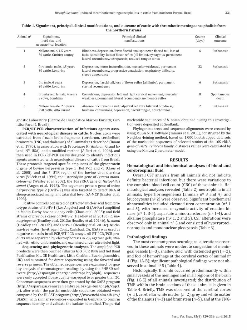
Table 1. Signalment, principal clinical manifestations, and outcome of cattle with thrombotic meningoencephalitis from the northern Paraná
Animal nº Signalment, Principal clinical Course Clinical herd size, and manifestations (days) outcome geographical location
1 Nellore, male, 1.5 years Blindness, depression, fever, flaccid anal sphincter, flaccid tail, loss of 6 Euthanasia 50 cattle, Curiúva county facial sensibility, loss of flexor reflex (all limbs), nystagmus, permanent lateral recumbency, tetraparesis, reduced tongue tonus
2 Girolando, male, 1.5 years Depression, motor incoordination, muscular weakness, permanent 22 Euthanasia 30 cattle, Londrina lateral recumbency, progressive emaciation, respiratory difficulty, sleepy appearance
3 Gir, male, 6 years Depression, flaccid tail, loss of flexor reflex (all limbs), permanent 6 Euthanasia 20 cattle, Londrina sternal recumbency
4 Crossbreed, female, 4 years Convulsions, depression left and right cervical movement, muscular 8 Spontaneous 3 cows, Londrina weakness, permanent lateral recumbency, no menace reflex death
5 Nellore, female, 2.5 years Absence of cutaneous and palpebral reflexes, bilateral blindness, 2 Euthanasia 250 cattle, Alto Paraná bruxism, convulsions, depression, flaccid tongue, opisthotonos
gnostic Laboratory (Centro de Diagnóstico Marcos Enrietti; Cur-itiba, Paraná, Brazil).
PCR/RT-PCR characterization of infectious agents asso-ciated with neurological disease in cattle. Nucleic acids were extracted from frozen brain fragments (cerebrum, cerebellum, brainstem, TNG, and thalamus) of all animals as described (Boom et al. 1990), in association with Proteinase K (Ambion, Grand Is-land, NY, USA), and a modified method (Alfieri et al. 2006), and then used in PCR/RT-PCR assays designed to identify infectious agents associated with neurological disease of cattle from Brazil. These protocols targeted specific amplicons of the glycoprotein C gene of bovine herpesvirus type 1 (BoHV-1) and -5 (Claus et al. 2005), and the 5’-UTR region of the bovine viral diarrhea virus (Vilček et al. 1994), the listeriolysin gene of Listeria mono-cytogenes (Wesley et al. 2002), the 16s rRNA gene of Histophilus somni (Angen et al. 1998). The tegument protein gene of ovine herpesvirus type 2 (OvHV-2) was also targeted to detect DNA of sheep-associated malignant catarrhal fever, SA-MCF (Baxter et al. 1993).
Positive controls consisted of extracted nucleic acid from pro-totype strains of BoHV-1 (Los Angeles) and -5 (AA-Par) amplified in Madin-Darby bovine kidney cells (Claus et al. 2005); and field strains of previous cases of OvHv-2 (Headley et al. 2013c), L. mo-nocytogenes (Headley et al. 2013a; Headley et al. 2014b), H. somni (Headley et al. 2013b), and OvHV-2 (Headley et al. 2013c). Nucle-ase-free water (Invitrogen Corp., Carlsbad, CA, USA) was used as negative controls in all PCR/RT-PCR assays. All RT-PCR/PCR pro-ducts were separated by electrophoresis in 2% agarose gels, stai-ned with ethidium bromide, and examined under ultraviolet light.
Sequencing and phylogenetic analyses. The amplified PCR products were then purified (illustra GFX PCR DNA and Gel Band Purification Kit, GE Healthcare, Little Chalfont, Buckinghamshire, UK) and submitted for direct sequencing using the forward and reverse primers. The obtained sequences were examined for qua-lity analysis of chromatogram readings by using the PHRED sof-tware (http://asparagin.cenargen.embrapa.br/phph); sequences were only accepted if base quality was equal to or greater than 20. Consensus sequences were then generated by the CAP3 program (http://asparagin.cenargen.embrapa.br/cgi-bin/phph/cap3.pl), after which the partial nucleotide sequences obtained were compared by the BLAST program (http://www.ncbi.nlm.nih.gov/BLAST) with similar sequences deposited in GenBank to confirm sequence identity and validate the isolates identified. The partial
nucleotide sequences of H. somni obtained during this investiga-tion were deposited at GenBank.
Phylogenetic trees and sequence alignments were created by using MEGA 6.01 software (Tamura et al. 2011), constructed by the Neighbor-Joining method, based on 1,000 bootstrapped data sets of the nucleotide sequences of selected strains of the 16S rRNA gene of Pasteurellaceae family; distances values were calculated by using the Kimura 2 parameter model.
RESULTSHematological and biochemical analyses of blood and cerebrospinal fluid
Overall CSF analysis from all animals did not indicate definite bacterial infections, but there were variations to the complete blood cell count (CBC) of these animals. He-matological analyses revealed (Table 2) neutrophilia in all animals; hyperfibrinogenemia (animals nº 3 and 4), and leucocytosis (nº 2) were observed. Significant biochemical abnormalities included elevated urea concentration (nº 1 and 5), with increased enzymatic activity of creatine ki-nase (nº 1, 3-5), aspartate aminotransferase (nº 1-4), and alkaline phosphatase (nº 1, 2 and 5). CSF alterations were observed only in animal nº 5 and consisted of hyperprotei-norraquia and mononuclear pleocytosis (Table 3).
Pathological findingsThe most constant gross neurological alterations obser-
ved in these animals were moderate congestion of menin-geal vessels (n=3), shallow sulci with distended gyri (n=2), and foci of hemorrhage at the cerebral cortex of animal no 4 (Fig. 1A-B); significant pathological findings were not ob-served in animal no 5 (Table 4).
Histologically, thrombi occurred predominantly within small vessels of the meninges and in all regions of the brain (Fig. 1C-E) of all animals investigated; the distribution of TME within the brain sections of these animals is given in Table 4. Briefly, TME was observed at the cerebral cortex (n=5), cerebellar white matter (n=2), grey and white matter of the thalamus (n=3) and brainstem (n=5), and at the TNG-

Pesq. Vet. Bras. 35(4):329-336, abril 2015
332 Selwyn A. Headley et al.
-CRM complex (n=5). Thrombi consisted of discrete intra-vascular accumulations of neutrophils and/or lymphocytes admixed with fibrin and moderate to marked proliferation of the affected vascular endothelium; intralesional bacteria were not frequently observed and there was no associated perivascular inflammatory exudate. Additionally, varying degrees of cortical edema, gliosis, and hemorrhage were observed in the brain sections evaluated. Other significant non-neurological pathological alterations included throm-bi within small vessels of the lung (Fig.1F), myocardium (no 2 and 5), liver and intestine (no 5).
PCR, sequencing, and phylogenetic analysisThe H. somni PCR successfully amplified bacterial DNA
from the brain of all animals with histopathological dia-
gnosis of TME (Table 4). Bacterial DNA was amplified from multiple sections of the brain from all animals, except no 2, who had H. somni DNA identified only from the trigeminal nerve ganglion. In summary, H. somni DNA was amplified from the TNG-CRM complex (n=5), thalamus (n=4), the brainstem and cerebral cortex (n=3), and cerebellum (n=2) of affected animals. All other PCR/RT-PCR assays provided negative results and all animals were negative for rabies virus by the immunofluorescence assay.
Direct sequencing revealed the desired 408 base pairs of the partial nucleotide sequence of the H. somni 16 rRNA gene from the TNG-CRM complex (animals no 1, 2, and 4), cerebral cortex (no 3), and the brainstem (no 5). Initial BLAST analyses revealed that all nucleotide sequences (GenBank Accession no KM374589, animal no 1; KM374590, no 2; KM374591, no 3; KM374592, no 4; and KM374593, no 5) derived from this study demonstrated 99-100% identity with similar sequences deposited in GenBank. Further, the generated phylogenetic tree revealed that all isolates of H. somni formed a large cluster, which was distinct from other members of the Pasteurellaceae family (Fig. 2).
DISCUSSIONThe histopathological findings observed within several brain sections of these animals are consistent with those described in H. somni-induced TME of ruminants (Fecteau & George 2004; Maxie & Youssef 2007; George 2009), and similar to the brain lesions previously described in cases of systemic (Headley et al. 2013b) and reproductive (Headley et al. 2015) histophilosis from Brazil. Additionally, the amp-lification and consequent sequencing of H. somni from these
Table 3. Cerebrospinal fluid analyses of cattle with thrombotic meningoencephalitis
Parameters Reference Animals values 1 2 3 4 5
Color Unco- Unco- Unco- Unco- Unco- Unco- lored lored lored lored lored lored Aspect Clear Clear Clear Clear Clear Protein (mg/dL) 40 39.3 21.6 19.9 17.7 133.7 Density >1,010 1,004 1,008 1,006 1,006 1,004 Total nucleated 10 2 0 0 2 14 cell count (mm3) Segmented 0 0 0 0 28 neutrophils (%) Band 0 0 0 0 0 neutrophils (%) Mononuclear 0 0 0 0 72 cells (%)
Table 2. Hematological and biochemical findings of cattle with thrombotic meningoencephalitis
Laboratory Parameters Reference Animals nº values 1 2 3 4 5
Hemogram Packed Cell Volume (%) 27 – 48 42.8 36.8 44.1 42.1 38.0 Red Blood cells (x10⁶/µL) 6 – 11.6 8.51 8.20 7.88 7.6 7.97 Hemoglobin (g/dL) 8.50 – 16.5 11.2 11.1 11.1 10.7 12.6 Total plasma protein (g/dL) 6.6 – 7.8 7.8 7.0 8.0 9.8 7.4 Fibrinogen (mg/dL) 300 - 700 400 400 1,000 800 600 Leucocytes (x10³/ µL) 6,200 – 12,200 10,100 14,200 8,800 7,600 7,000 Lymphocytes (x10³/ µL) 3,300 – 8,000 4,141 5,112 1,496 1,672 2,030 Eosinophils (x10³/ µL) 0 – 2,400 0 0 0 0 0 Monocytes (x10³/ µL) 200 - 700 0 284 0 0 0 Bastonetes (x10³/ µL) 0 - 200 0 0 88 304 0 Segmented neutrophils (x10³/µL) 1,300 – 3,400 5,959 8,804 7,216 5,624 4,970 MCV (fL) 30.5 – 55.5 50.3 44.9 56.0 55.5 47.6 MCHC (%) 28 – 38 26.1 30.1 35.1 25.4 34.0 MCH (pg) 9.7 – 19 13.1 13.5 14.0 14.0 15.8 Serum biochemistry Albumin (g/dL) 2.7 – 4.3 2.3 1.5 2.6 2.3 2.6 Creatinine (mg/dL) 1 – 2.7 2.7 0.60 1.5 1.2 2.3 Urea (mg/dL) 5.8 – 35.9 111 15 35 39 70 Creatine kinase, CK (UI/L) 35 – 280 952 136 4,117 4,164 5,676 Aspartate aminotransferase, AST (UI/L) 32 – 71 207 88 962 399 ND Gamma-glutamyl transferase GGT (UI/L) 3.7 – 31.3 34 45 23 41 30 Alkaline phosphatase, ALP (UI/L) 33 – 100 256 160 ND ND 198 Total bilirubin (mg/dL) 0.32 – 0.74 0.47 0.55 0.54 0.22 0.47 Conjugated bilirubin (mg/dL) 0.01 – 0.34 0.35 0.35 0.12 0.05 0.06 Unconjugated bilirubin (mg/dL) 0.16 – 0.67 0.12 0.20 0.42 0.17 0.41
ND = not determined.

Pesq. Vet. Bras. 35(4):329-336, abril 2015
333Histophilus somni-induced thrombotic meningoencephalitis in cattle from northern Paraná, Brazil
Fig.1. Gross and histopathological features of Histophilus somni induced thrombotic meningoencephalitis in cattle from northern Paraná. (A) Congestion of meningeal vessels and hemorrhage with distended gyri and shallow sulci. (B) Edema of the sectioned surface of the cerebrum. There are thrombi (*) at the meninges (C-D), carotid rete miriabile (E), and the lungs (F). (B) Scale in centimeters. (C-F) Hematoxylin and eosin stain. (C and F) Bar = 20µm. (D-E) 100µm.
Table 4. Principal gross findings with the distribution of thrombotic meningoencephalitis and Histophilus somni DNA in brain sections of cattle
Animal nº Gross findings Thrombotic meningoencephalitis Histophilus somni DNA Brain- Cere- Cere- TNG- Thala- Brain- Cere- Cere- TNG- Thala- stem bellum brum CRM mus stem bellum brum CRM mus
1 Congestion of meningeal vessels; + - + + - + + + + + myocardial and endocardial petechial, hemorrhage, ulcerativeabomastits 2 Cerebrum; shallow sulci with distended gyri; + + + + + - - - + - congestion of meningeal vessels, hydropericardium, hyperemia of intestinal mucosa 3 Congestion of meningeal vessels, pulmonary + - + + + + - + + + emphysema, and atelectasis, ulcerative abomastits 4 Ascites, cerebrum: shallow sulci with distended + - + + - - + - + + gyri, cranioventral consolidation of lungs, enlarged liver, hydrothorax, renal infarction, tongue and oesophagus: ulcerations 5 Significant pathological alterations were + + + + + + - + + + not observed
TNG-CRM = Trigeminal nerve ganglion-carotid rete miriabile complex, + present, - absent.

Pesq. Vet. Bras. 35(4):329-336, abril 2015
334 Selwyn A. Headley et al.
areas, suggest that this bacterium was associated with the neuropathological lesions; the amplification of H. somni DNA and sequencing were previously used to identify this pathogen (Angen et al. 1998, Headley et al. 2013b, 2015). Moreover, the negative PCRs and RT-PCRs results with the absence of other histopathological disease patterns sug-gest that other common pathogens associated with neur-ological disease of cattle were not involved in the develop-ment of the clinicopathological manifestations observed during this investigation. Of extreme importance was the negative PCR assay for OvHV-2, since this is a common cause of neurological disease of cattle from Brazil (Head-ley et al. 2012, 2013c). Additionally, in the cases from this study, lesions at the carotid rete miriabile were thrombotic and not the characteristic fibrinoid necrosis frequently dia-gnosed in SA-MCF (Headley et al. 2012, 2013c).
The clinical manifestations, the acute incubation period culminating in spontaneous death or euthanasia, and the reduced number of animals affected at each herd during this study are frequent clinical and epidemiological find-ings associated with TME (MacDonald et al. 1973, Fecteau & George 2004, George 2009). During this investigation, bacterial isolation was not attempted in any instance since diagnoses of TME were only achieved after histopatholo-gical evaluation. It is worthy to mention that the hemato-logical and more specifically, the CSF findings were not in-dicative of classical bacterial infections of the brain (Scott 2004, Stokol et al. 2009). Although the CBC in cases of TME is nonspecific, the CSF findings normally demonstrate al-terations indicative of bacterial disease with an increase in the number of nucleated cells, elevated protein concen-trations (Van Dreumel et al. 1970, Fecteau & George 2004, Scott 2004), xanthochromia, and hemorrhage (Scott 2004). Alterations to CSF levels were only observed in animal no5 but were indicative of hyperproteinorraquia and lympho-cytic pleocytosis, without xanthochromia and hemorrhage. Alternatively, a retrospective study that classified the CSF relative to neurological disease in 101 cattle, observed marked variation in the three cases of TME described, with pleocytosis being neutrophilic, lymphocytic, and histiocytic (Stokol et al. 2009). Interestingly, the biochemical altera-tions observed in most animals are due to prolonged re-cumbency. However, the results of the CSF presented in this study are important for the ante-mortem diagnosis and the understanding of TME, since there are few documentations of the alterations associated with CSF in spontaneous cases of neurological histophilosis and the classical represent-ation of CSF alterations in TME is not fully known (Scott 2004).
The distribution of TME was predominant at the brain-stem, cerebral cortex, and the TNG-CRM complex of all animals, while these lesions were not consistent at the cerebellum and thalamus. However, H. somni DNA was recovered more frequently from the TGN-CRM complex, followed by the thalamus (n=4), cerebral cortex, brain-stem (n=3), and cerebellum (n=2). This might suggest that there is no definite correlation between the demonstra-tion of TME in a particular anatomical region of the brain and the amplification of H. somni DNA from these areas. Nevertheless, histopathological lesions of TME seem to have predilection for the cerebral cortex (MacDonald et al. 1973, Maxie & Youssef 2007, Zachary 2012) and thal-amus (Maxie & Youssef 2007), but can be randomly dis-tributed throughout any part of the brain (MacDonald et al. 1973). These findings are in agreement with the distri-bution of TME observed during this investigation, and sug-gest that fragments of multiple anatomical regions of the brain must be collected to obtain an accurate histopatho-logical diagnosis of TME, particularly in non-conventional areas of this disease. Additionally, since histopathological confirmation and the amplification of H. somni DNA was constant from the TGN-CRM complex of all animals, this neuroanatomical region, of the brain, particularly the ca-rotid rete miriabile, should always be investigated when TME is suspected.
Fig.2. The phylogenetic relationship of selected strains of Histo-philus somni based on the 16S rRNA gene of the Pasteurel-laceae family. The isolates from this study are highlighted (ar-rows); the GenBank accession numbers and country of origin of the sequences used are given. E. coli was used as the out--group.

Pesq. Vet. Bras. 35(4):329-336, abril 2015
335Histophilus somni-induced thrombotic meningoencephalitis in cattle from northern Paraná, Brazil
The identification of H. somni DNA from these cases with typical histopathological findings of TME represent the first successfully characterization of this important neuro-logical disease of cattle from Brazil, and definitely extends the geographical distribution of this disease and pathogen. This is because histophilosis was previously described by our group in cattle from southern Brazil (Headley et al. 2013b), while the bacterium has been associated with steers that demonstrated pulmonary distress at a feedlot (Headley et al. 2014a), and in sheep with endometritis (Rizzo et al. 2012). In addition, H. somni has been linked to abortions in cattle from different geographical regions of Brazil (Headley et al. 2015). Further, H. somni DNA was identified from the bronquialveolar lavage of cattle with and without pulmonary discomfort (Oliveira et al., manu-script in preparation), and in a lamb with omphalitis (Head-ley et al., manuscript in preparation). Interestingly, vaccin-ation of cattle against H. somni is not a routine practice in beef and dairy cattle herds of Brazil, so additional cases of histophilosis are likely to occur nationwide. Consequently, this disease might be a possible threat to the local cattle industry (Headley et al. 2014a), resulting in serious eco-nomic losses as have been documented in North America (Saunders et al. 1980). However, since the distribution of H. somni in cattle herds of Brazil is unknown, seroepidemi-ological studies are being implemented to determine the prevalence of this pathogen in beef and dairy herds.
CONCLUSIONSHistophilus somni DNA was identified from brain frag-
ments of cattle with neuropathological manifestations and histopathological lesions that are consistent with those described in TME of cattle from North America.
Further, sequencing of the H. somni 16S rRNA gene from affected brain fragments of all animals confirmed the par-ticipation of this pathogen in the lesions herein described.
These results contribute to the documentation of this important emerging infectious disease of cattle from Bra-zil and South America, complements previous findings of histophilosis by our group (Headley et al. 2013b, 2014a, 2015), and produces novel CSF findings associated with spontaneous cases of TME.
Acknowledgements.- Selwyn A. Headley, Ana Paula F.R.L. Bracarense, Alice F. Alfieri, Júlio A.N. Lisbôa, and Amauri A. Alfieri are recipients of the National Council for Scientific and Technological Development (CNPq, Brazil) fellowships. This study was partially funded by a CNPq grant (Pro-posal #448797/2014-3) and a joint CNPq/Ministry of Agriculture (MAPA) grant (Protocols #578645/2008-4 and #478254/2012-1).
The authors contributed for publication of the article as follows:SAH and AAA: conceived and designed the study;SAH, APFRB and WO: were involved in the pathological evaluations and interpretations;SAH, VHSO, AFA and AAA: participated in the molecular analyses and interpretations;GRQ and JANL: did the clinical and neurological examinations and vi-sited the farms;KKMC and JANL: were responsible for the clinical laboratiory inves-tigation;All authors participated in the drafting of the manuscript, read, and approved the final version.
REFERENCESAlfieri A.A., Parazzi M.E., Takiuchi E., Medici K.C. & Alfieri A.F. 2006. Fre-
quency of group A rotavirus in diarrhoeic calves in Brazilian cattle herds, 1998-2002. Trop. Anim. Health Prod. 38:521-526.
Angen O., Ahrens P. & Tegtmeier C. 1998. Development of a PCR test for identification of Haemophilus somnus in pure and mixed cultures. Vet. Microbiol. 63:39-48.
Baxter S.I., Pow I., Bridgen A. & Reid H.W. 1993. PCR detection of the sheep-associated agent of malignant catarrhal fever. Archs Virol. 132: 145-159.
Boom R., Sol C.J., Salimans M.M., Jansen C.L., Wertheim-Van Dillen P.M. & Van der Noordaa J. 1990. Rapid and simple method for purification of nucleic acids. J. Clin. Microbiol. 28:495-503.
Claus M.P., Alfieri A.F., Folgueras-Flatschart A.V., Wosiacki S.R., Medici K.C. & Alfieri A.A. 2005. Rapid detection and differentiation of bovine herpesvirus 1 and 5 glycoprotein C gene in clinical specimens by multi-plex-PCR. J. Virol. Methods 128:183-188.
Descarga C.O., Piscitelli H.G., Zielinski G.C. & Cipolla A.L. 2002. Throm-boembolic meningoencephalitis due to Haemophilus somnus in feedlot cattle in Argentina. Vet. Rec. 150:817.
Fecteau G. & George L.W. 2004. Bacterial meningitis and encephalitis in ruminants. Vet. Clin. North. Am., Food. Anim. Pract. 20:363-377.
George L.W. 2009. Thrombotic meningoencephalitis (Histophilus somni [Hae-mophilus somuns] infection; sleeper calves), p.1048-1050. In: Smith B.P. (Ed.), Large Animal Internal Medicine. Mosby/Elsevier, St Louis, Missouri.
Headley S.A., Sousa I.K.F., Minervino A.H.H., Barros I.O., Barrêto Júnior R.A., Alfieri A.F., Ortolani E.L. & Alfieri A.A. 2012. Molecular confirmation of ovine herpesvirus 2-induced malignant catarrhal fever lesions in cattle from Rio Grande do Norte, Brazil. Pesq. Vet. Bras. 32:1213-1218.
Headley S.A., Bodnar L., Fritzen J.T., Bronkhorst D.E., Alfieri A.F., Okano W. & Alfieri A.A. 2013a. Histopathological and molecular characterization of encephalitic listeriosis in small ruminants from northern Parana, Brazil. Braz. J. Microbiol. 44:889-896.
Headley S.A., Oliveira V.H., Figueira G.F., Bronkhorst D.E., Alfieri A.F., Okano W. & Alfieri A.A. 2013b. Histophilus somni-induced infections in cattle from southern Brazil. Trop. Anim. Health Prod. 45:1579-1588.
Headley S.A., Queiroz G.R., Fritzen J.T.T., Lisboa J.A.N., Alfieri A.F., Oliveira R.A.M., Bracarense A.P.F.R.L. & Alfieri A.A. 2013c. Ovine herpesvirus type 2-induced malignant catarrhal fever in a heifer. Semina, Ciênc. Agrarias 34:3895-3900.
Headley S.A., Alfieri A.F., Oliveira V.H.S., Beuttemmüller E.A. & Alfieri A.A. 2014a. Histophilus somni is a potential threat to beef cattle feedlots from Brazil. Vet. Rec. 175:249.
Headley S.A., Fritzen J.T.T., Queiroz G.R., Oliveira R.A.M., Alfieri A.F., Santis G.W.D., Lisbôa J.A.N. & Alfieri A.A. 2014b. Molecular characterization of encephalitic bovine listeriosis from southern Brazil. Trop. Anim. Health Prod. 46:19-25.
Headley S.A., Voltarelli D., Oliveira V.H.S., Bronkhorst D.E., Alfieri A.F., Negri L.C., Okano W. & Alfieri A.A. 2015. Association of Histophilus somni with spontaneous abortions in dairy cattle herds from Brazil. Trop. Anim. Health Prod. 47:403-413.
Hick P.M., Read A.J., Lugton I., Busfield F., Dawood K.E., Gabor L., Hornitzky M. & Kirkland P.D. 2012. Coronavirus infection in intensively managed cattle with respiratory disease. Aust. Vet. J. 90:381-386.
Inzana T.J. & Corbeil L. 2004. Haemophilus, p.243-258. In: Gyles C.L., Prescott J.F., Songer G. & Thoen C.O. (Eds), Pathogenesis of Bacterial In-fections in Animals. Blackwell Publishing, Ames, Iowa.
Lancaster M.J., McGillivery D.J., Patterson R.M. & Irwin S. 1984. Pneumonia associated with Haemophilus somnus in a calf. Aust. Vet. J. 61:269.
MacDonald D.W., Christian R.G. & Chalmers G.A. 1973. Infectious throm-boembolic meningoencephalitis: literature review and occurrence in Alberta, 1969-71. Can. Vet. J. 14:57-61.
Maxie M.G. & Youssef S. 2007. Nervous system, p.408-411. In: Maxie M.G. (Ed.), Jubb, Kennedy, and Palmer’s Pathology of Domestic Animals, Vol.1. Saunders/Elsevier, Philadelphia.

Pesq. Vet. Bras. 35(4):329-336, abril 2015
336 Selwyn A. Headley et al.
Pérez D.S., Pérez F.A. & Bretschneider G. 2010. Histophilus somni: patho-genecity in cattle an update. An. Vet. Murcia 26:5-21.
Rizzo H., Gregory L., Carvalho A.F. & Pinheiro E.S. 2012. Histophilus somni isolation on sheep with endometritis first case report on Brazil. Revta Bras. Reprod. Anim. 36:136-138.
Saunders J.R., Thiessen W.A. & Janzen E.D. 1980. Haemophilus somnus in-fections I. A ten year (1969-1978) retrospective study of losses in cattle herds in Western Canada. Can. Vet. J. 21:119-123.
Scott P.R. 2004. Diagnostic techniques and clinicopathologic findings in ruminant neurologic disease. Vet. Clin. North Am., Food Anim. Pract. 20: 215-230.
Stokol T., Divers T.J., Arrigan J.W. & McDonough S.P. 2009. Cerebrospinal fluid findings in cattle with central nervous system disorders: a retro-spective study of 102 cases (1990-2008). Vet. Clin. Pathol. 38:103-112.
Tamura K., Peterson D., Peterson N., Stecher G., Nei M. & Kumar S. 2011. MEGA5: molecular evolutionary genetics analysis using maximum like-lihood, evolutionary distance, and maximum parsimony methods. Mol. Biol. Evol. 28:2731-2739.
Tanaka A., Hoshinoo K., Hoshino T. & Tagawa Y. 2005. Differentiation between bovine and ovine strains of Histophilus somni based on the se-quences of 16S rDNA and rpoB gene. J. Vet. Med. Sci. 67:255-262.
Van Dreumel A.A., Curtis R.A. & Ruhnke H.L. 1970. Infectious thromboem-bolic meningoencephalitis in Ontario feedlot cattle. Can. Vet. J. 11:125-130.
Vilček Š., Herring A.J., Herring J.A., Nettleton P.F., Lowings J.P. & Paton D.J. 1994. Pestiviruses isolated from pigs, cattle and sheep can be allocated into at least three genogroups using polymerase chain reaction and re-striction endonuclease analysis. Archs Virol. 136:309-323.
Wesley I.V., Larson D.J., Harmon K.M., Luchansky J.B. & Schwartz A.R. 2002. A case report of sporadic ovine listerial menigoencephalitis in Iowa with an overview of livestock and human cases. J. Vet. Diagn. Invest. 14:314-321.
Wessels J. & Wessels M.E. 2005. Histophilus somni myocarditis in a beef rearing calf in the United Kingdom. Vet. Rec. 157:420-421.
Zachary J.F. 2012. Nervous system, p.771-870. In: Zachary J.F. & McGavin M.D. (Eds), Pathological Basis of Veterinary Disease. Elsevier/Mosby, St Louis, Missouri.1322p.