historical development of the cell theory (p. 6-11)
TRANSCRIPT

Biology 2201 Unit 1: Cells and Energy for Life Page 1 of 25
Historical Development of the Cell Theory (p. 6-11)
Biology
- The study of life (living things).
- Living things are composed of units called cells.
Unicellular organism
- A one-celled organism.
o Ex: paramecium, bacteria, amoeba
Multicellular organism
- Organisms with more than one cell.
o Ex: cat, dog, worm, human
The Cell Theory
Contains four Statements: (First stated in 1858)
1. All living organisms are composed of one or more cells.
2. Cells are the basic unit of structure and function in all organisms.
3. All cells are derived from pre-existing cells.
4. In a multicellular organism, the activity of the entire organism depends on the total
activity of its independent cells.
In the Beginning…
Spontaneous generation
- Idea that living things come from non-living sources
o Ex: maggots appear on meat if left out too long
o Ex: After it rains — frogs
o Ex: Insects and plants seem to come out of mud in ponds.
- In 1870 abiogenesis was used to describe spontaneous generation
Aristotle (334 BC)
- Put forward the idea of spontaneous generation.
- He classified all organisms as plants or animals.

Biology 2201 Unit 1: Cells and Energy for Life Page 2 of 25
Francesco Redi (l668)
- Challenged the idea of spontaneous generation.
- Helped proposed the idea known as biogenesis – Life comes from Life.
o Did the first controlled experiment.
o He hypothesized that if maggots come from fly eggs, then maggots will appear
only in open jars where flies can deposit eggs on meat. When testing, he placed
some meat samples in covered jars and some in uncovered jars. He found that
maggots appeared only in uncovered containers. He tested many times and
obtained the same results even with different meats.
John Needham (1748)
- Performed an experiment similar to Redi's.
- He boiled a meat broth (to kill microbes); sealed one container (not airtight --- sterile)
and left another open.
- The result was microbes were present.
- This supported spontaneous generation.
Lorenzo Spallanzani (1776)
- Repeated Needham’s experiment.
o He boiled the containers for one hour; then, sealed the
flasks tightly.
o No microbes were present.
o The microbes appeared hours after the seals were
broken.
o He believed that microorganisms were carried in air
and multiplies when they had a food supply.

Biology 2201 Unit 1: Cells and Energy for Life Page 3 of 25
Louis Pasteur (1861)
- Repeated Spallanzani’s work.
o He used S - shaped necked flasks (heat flask and bend into an S-shaped curve)
which allowed air and microbes in.
o When he boiled the solution in the base of the flask this created steam which
condensed and formed water droplets which trapped microbes in the neck of
the flask. The broth remained clear. He broke the necks of the flasks. The broth
turned cloudy. Flasks were tipped and the microbes mixed with the broth. The
broth turned cloudy. Some of his flasks (on display) are still sterile today.
Early Microscopes and Cells
Leeuwenhoek (1675)
- Invents the simple (single lens) microscope.
o Considered the Father of the Microscope
- Using his microscope, he sees microorganisms.
Robert Hooke (1665)
- Studied slices of cork and saw hollow sacs he called “Cells”.

Biology 2201 Unit 1: Cells and Energy for Life Page 4 of 25
Contributions to the Development of the Cell Theory
Schleiden (1838)
- Cells were present in plant tissue.
Schwann (1839)
- Cells were present in animal tissue.
This suggested that all organisms were composed of one or more cells.
Robert Brown (1831)
- Discovered the center of the cell. He called it the nucleus.
Virchow (1858)
- Observed dividing cells and concluded that cells can arise only from other cells.
Braun (1845)
- “the cell is the basic unit of life”
Conclusion:
Crossword puzzle
Assignment: Create a Timeline
QUIZ – Scientists
Biology 2201 Unit 1: Cells and Energy for Life Page 5 of 25
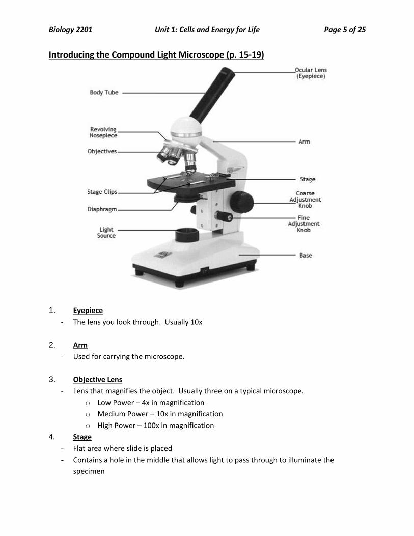
Introducing the Compound Light Microscope (p. 15-19)
1. Eyepiece
- The lens you look through. Usually 10x
2. Arm
- Used for carrying the microscope.
3. Objective Lens
- Lens that magnifies the object. Usually three on a typical microscope.
o Low Power – 4x in magnification
o Medium Power – 10x in magnification
o High Power – 100x in magnification
4. Stage
- Flat area where slide is placed
- Contains a hole in the middle that allows light to pass through to illuminate the
specimen

Biology 2201 Unit 1: Cells and Energy for Life Page 6 of 25
5. Diaphragm
- Flat disc with holes in it just below the stage
- Controls the amount og light reaching the object
6. Light source
- Mirror or electrical light that illuminates the object being viewed
7. Base
- Bottom of the microscope that provides support for the other parts.
8. Coarse adjustment (focus) knob
- Knob which brings the object into focus quickly
- Usually used only on low power.
9. Fine adjustment (focus) knob
- Knob used for fine focussing of objects.
- Used on medium and high power.
Using the Microscope
1. Magnification: The ratio of image size to actual specimen size.
a. Calculating Magnification – Use the formula below.
Magnification = objective lens X eyepiece
Ex: Magnification = 40x (high power) X 10x (eyepiece) = 400x total magnification.
Note: maximum magnification of a light microscope is usually 2000X
2. Resolving Power: The ability of a microscope to distinguish between two objects that
are close together.
Note: Maximum resolving power of Light microscope = 0.2 µm or
200nm
Maximum resolving power of Electron Microscope = 0.2 nm

Biology 2201 Unit 1: Cells and Energy for Life Page 7 of 25
Remember:
Any light microscope with a resolving power LESS than 0.2 µm
you will not see the objects as distinct and separate.
They will appear as one object.
3. Field of View: The area you see as you look through the eyepiece. The area is round.
Determining the Size of the Field of View
a. Low Power Measure using a mm ruler.
b. Medium and High Power use the formula below
FOV for med or high = FOV on low X Mag. low
Mag. med or high
Ex: FOV(med) = 3.3 mm X 40x
100x
FOV(med) = 3.3 mm x 0.4
FOV(med) = 1.32mm
4. Specimen Size: The actual size of a specimen. Found by doing a calculation.
Specimen size = Field of View
# specimens fitting across FOV
Practice:

Biology 2201 Unit 1: Cells and Energy for Life Page 8 of 25
5. Making a biological drawing
RULES: 1. ________________________________________________________________________
2. ________________________________________________________________________
3. ________________________________________________________________________
4. ________________________________________________________________________
5. ________________________________________________________________________
6. ________________________________________________________________________
7. ________________________________________________________________________
8. ________________________________________________________________________
9. ________________________________________________________________________
10. ________________________________________________________________________

Biology 2201 Unit 1: Cells and Energy for Life Page 9 of 25
Lab #1: Caring For and Using a Microscope
Follow your Lab Write-up Guidelines with these additional instructions. Answer your 3 pre-lab questions first. There is no need for a HYPOTHESIS.
We will do the lab in a different order (be sure to note this in your procedure)
- Part A: Becoming familiar with a compound microscope (done in a previous class)
- Part F: Preparing a wet mount of onion skin cells
o Additional assessment will be for preparing a wet mount
- Part B: Calculating Field of View (misnamed in the text)
o Magnification
o Field of view
o Specimen size
- Part D: Making scale drawings.
- Part E: Observing depth of field
Part C: Omitted
Refer to MHR Biology for your MATERIALS and PROCEDURE.
Your RESULTS section will include the following:
Calculation of the magnification for all three objective lenses.
Calculation of the field of view for all three objective lenses.
Calculation of your specimen size (in m).
A scale drawing of your specimen (see p. 742)
Answer the post-lab questions #s 1, 2, 3, 5, 6, 7 and 8.
In a CONCLUSION:
Answer the question in the problem
summarize your findings (how you found your specimen size and what it was)
include any sources of error
ways that your lab could be improved

Biology 2201 Unit 1: Cells and Energy for Life Page 10 of 25
Practical Assessment: Preparing a Wet Mount
1) Obtain a microscope employing good handling techniques and set it up at your work
station
2) Prepare your wet mount using the following technique
Add one drop of water to a slide
Carefully use a razor blade and/ or forceps to obtain a sample of onion
epithelium
Place the onion on the slide into the water drop
Apply the cover slip using the 45o angle technique demonstrated in the previous
laboratory exercise
A. B. C.
Apply a single drop of stain in the center, on one edge of the cover slip
Using paper-towel or tissue paper, draw the stain into the wet mount by placing
it on the opposite edge of the cover slip, as previously explained
A. B.
View specimen under low power on the light microscope.
3) Clean up your work station and return your microscope to the storage cabinet.

Biology 2201 Unit 1: Cells and Energy for Life Page 11 of 25
Advances in Microscope Technology
Light Microscope
- Uses light as a source of illumination for objects.
o Simple Light Microscope -- Microscope composed of only 1 lens.
o Compound Light Microscope – Microscope composed of 2 or more lenses.
Electron Microscope
- Uses Electrons as a source of illumination for objects.
o TEM – Transmission Electron Microscope
Shoots a beam of electrons through specimen.
Produces a 2D micrograph (picture) of a specimen.
o SEM – Scanning Electron Microscope
Shoots a beam of electrons at the surface of a specimen.
Produces a 3D micrograph.
Comparison of the light microscope with an electron microscope
Type of
Microscope
Source of
Illumination
Magnification Resolution Specimen
Preparation
Light visible light up to 2000 x about 0.2 um
(200 nm)
usually killed,
fixed and stained
Electron electrons TEM : typically
10 000 x to
500 000 x
SEM : typically
1 000 x to
10 000 x
TEM : about
0.2 nm SEM :
about 1 to 10
nm
usually killed,
dried and fixed
fixed, cleaned,
and coated with
metal
Microscope Comparison Chart

Biology 2201 Unit 1: Cells and Energy for Life Page 12 of 25
More About Cells
Cell
- Basic Structural Unit of Life.
Prokaryotic cells
- Cells that DO NOT contain a true membrane-bound nucleus;
- DNA is concentrated in an area called the nucleoid.
1.
2.
3.
- Ex: ALL bacterial cells
Eukaryotic cells
- Cells that DO contain a true membrane – bound nucleus.
- The nucleus is an enclosed region that separates the DNA from the rest of the cell
contents.
- Eukaryotes contain a number of specialized structures called organelles.
- There are TWO types of Eukaryotic Cells
o Animal Cells
o Plant Cells
Organelles
- Specialized structures within a Eukaryotic cell that carries out a specific function.
- Organelles work together to keep cell functioning.
Organelle name
Description of structure Description of function
Plant or
Animal or
BOTH?
nucleus
round; occupies center
of cell, largest
control center of the cell
stores genetic info on DNA
mitochondria “peanut-shaped”
Has 2 (double
membranes
cellular respiration
“powerhouse of cell”

Biology 2201 Unit 1: Cells and Energy for Life Page 13 of 25
ribosome
little balls found on ER protein synthesis with
mRNA
vacuole
round vesicle (large in
plants, small in animals)
storage of food, wastes,
water etc.
chloroplast
“cucumber-shaped”
Contains discs called
Thylakoids.
Thylakoids are in stacks
called Grana
photosynthesis
lysosome round sac – contains
digestive enzymes
Digestion
Recycling materials
nucleolus An area of chromatin
(uncoiled DNA) that
produces ribosomes
Golgi apparatus stacked discs – like
pancakes
vesicle formation, packages
products for shipment to
other cells
Endoplasmic
reticulum - 2 types
Smooth – No
ribosomes
Rough – ribosomes
Folded long, thin
membranes;
thick structure surrounding
transport of materials
cell wall
Made of chitin and
cellulose structural support
cytoskeleton
thin, long structures
2 types:
Microtubules- Hollow
Microfilaments-Solid
skeletal support
cell membrane thin phospholipid bilayer
separates the inside from
the outside
controls entry/ exit of
materials

Biology 2201 Unit 1: Cells and Energy for Life Page 14 of 25
Cytoplasm
Fluid of cell
Gel-like Site of many reactions.
Vesicle
Cilia
Flagella
Complete above chart with colored diagrams and types of cells where found
(Plant/Animal/BOTH?)
Label the plant and animal cell diagrams
QUIZ - Organelles

Biology 2201 Unit 1: Cells and Energy for Life Page 15 of 25
Cellular Processes (p. 50-64)
Cell Membrane
- controls what enters and leaves the cell to maintain homeostasis.
- transports raw materials into the cell
- transports created materials and waste out of the cell
- Prevents unwanted material from entering the cell.
- Prevents escape of materials from cell needed for cell functions.
Homeostasis
- The process of maintaining a stable internal environment.
Fluid Mosaic Model
- A model that describes the cell membrane as being a double layer (bilayer) composed of
a phospholipid backbone with proteins embedded throughout. The layer is flexible and
is able to move.
Phosphoplipid Bilayer
- Composed of a Head and Tail section.
Head: Hydrophilic or “water loving” (points to the outside of the cell)
Tail: Hydrophobic or “water hating” (points to the inside of the cell).
Activity: Build-A-Membrane Model

Biology 2201 Unit 1: Cells and Energy for Life Page 16 of 25
Cellular Transport
The cell carries out transport in one of two ways – Passive or Active Transport.
1. Passive Transport: Transport of materials without the use of energy.
There are three types of Passive Transport.
a) Simple Diffusion
b) Facilitated Diffusion
c) Osmosis – a type of special diffusion
a) Simple Diffusion
The movement of particles from an area of high concentration to an area of low concentration
along a concentration gradient. Movement occurs until particles are scattered evenly.
High Concentration:
An area having a large amount of particles.
Low Concentration:
An area having a small amount of particles.
Concentration Gradient: The difference between an area with many particles and one
with few particles.
Q. What drives Diffusion?
A. Molecules and particles are in constant random motion which creates a concentration
gradient and as such causes particles to move.

Biology 2201 Unit 1: Cells and Energy for Life Page 17 of 25
Note:
Particles such as Oxygen(O2), Carbon Dioxide(CO2) and Alcohols
cross the cell membrane by simple diffusion.
These particles are small enough to pass right through the membrane.
Factors Affecting the Rate of Diffusion
1. Temperature --- the higher the temperature the greater the rate of diffusion.
2. Size of Molecule --- the smaller the molecule the faster diffusion occurs.
3. Size of Concentration Gradient – the greater the difference between areas with high
concentration and areas with low concentration, the faster diffusion occurs.
b) Facilitated Diffusion
Similar to diffusion except, the particles trying to cross the cell membrane are too large, so they
are assisted/facilitated by special proteins in the cell membrane.
Two types of Proteins in the cell membrane
1. Carrier Protein Facilitated Diffusion – Diffusion of NONCHARGED molecules across
the cell membrane. Ex: Glucose
2. Channel Protein Facilitated Diffusion - Diffusion of CHARGED particles such as ions
across the cell membrane. Ex: sodium, Na+, K+
Biology 2201 Unit 1: Cells and Energy for Life Page 18 of 25
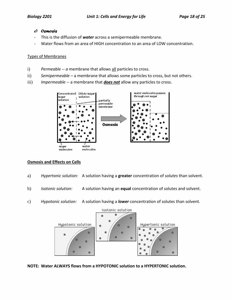
c) Osmosis
- This is the diffusion of water across a semipermeable membrane.
- Water flows from an area of HIGH concentration to an area of LOW concentration.
Types of Membranes
i) Permeable -- a membrane that allows all particles to cross.
ii) Semipermeable – a membrane that allows some particles to cross, but not others.
iii) Impermeable -- a membrane that does not allow any particles to cross.
Osmosis and Effects on Cells
a) Hypertonic solution: A solution having a greater concentration of solutes than solvent.
b) Isotonic solution: A solution having an equal concentration of solutes and solvent.
c) Hypotonic solution: A solution having a lower concentration of solutes than solvent.
NOTE: Water ALWAYS flows from a HYPOTONIC solution to a HYPERTONIC solution.

Biology 2201 Unit 1: Cells and Energy for Life Page 19 of 25
The Effect of Osmosis on Animal and Plant Cells
Questions to consider?
- Why would plants be negatively affected by too much fertilizer?
- Why are vegetables in grocery stores sprayed with water?
- Why does IV fluid have to be isotonic?
Core Lab Activity: A Cell Membrane Model
Extracellular Fluid:
- Fluid located immediately outside a cell that surrounds and bathes the cell.
- Sometimes called interstitial fluid
Intercellular fluid
- Fluid located inside the cell membrane

Biology 2201 Unit 1: Cells and Energy for Life Page 20 of 25
2. Active Transport: Transport of materials with the use of Energy (ATP)
ATP -- Adenosine Tri-Phosphate.
It is the energy molecule of the cell.
The movement of materials AGAINST the concentration gradient.
Active transport requires the cell to use energy (ATP).
For example:
- Plants pumping nutrients in from the soil
- Intestinal cells pumping nutrients from the gut to the blood stream
- Kidneys pumping glucose and amino acids from the urine back into the blood
- Sodium potassium pump in nerve cells
Sodium – Potassium Pump
What’s missing from this picture?
Biology 2201 Unit 1: Cells and Energy for Life Page 21 of 25
Bulk Transport:
- Transport of large materials through the cell membrane.
- These particles are too large to cross the cell membrane by normal means.
- The cell has to use energy to accomplish this.
Basically:
- The membrane folds in on itself to create vesicles (vacuoles) that contain the materials.
Two types of Bulk Transport
1. Exocytosis:
- This is the movement of large particles
OUT of the cell.
2. Endocytosis:
- This is the movement of large particles
INTO the cell.
There are two types of Endocytosis.
A. Phagocytosis: Referred to as “Cell eating”. The cell ingests a piece of solid material such
as a Red Blood Cell or a bacterium.
B. Pinocytosis: Referred to as “Cell Drinking”. The cell ingests a small amount of “extra
cellular fluid” from the outside of the cell.
Questions p. 61 # 1, 3-11, 13, 14, 16, 17

Biology 2201 Unit 1: Cells and Energy for Life Page 22 of 25
Why Are Cells So Small?
Cells are microscopic. The reason cells are microscopic has to do with two critical factors.
a) Surface area of the cell membrane
b) Volume of cytoplasm within a cell.
The cell membrane is responsible for controlling what enters and leaves the cell.
The volume of a cell is a measure of the cytoplasm of the cell. The cytoplasm contains all the
organelles in a cell and is the site of many reactions.
As a cell’s volume grows, the cell membrane has to increase in size in order to:
- bring in enough nutrients
AND
- get rid of wastes
If the cell membrane is not able to increase enough to account for the increasing
volume, the cell will not be able to survive. That is the cell membrane gets too
small to meet the cell’s needs.
A useful ratio called the Surface Area to Volume Ratio is a good measure of the efficiency of
the interaction between the cell membrane and the volume of the cytoplasm.
A ratio of 1:1 indicates a balance between the volume of the cytoplasm and the cell
membrane’s surface area.
The larger the ratio the more efficient the cell is at exchanging materials between the cell
membrane and the cytoplasmic volume.
In other words, the cell membrane is large enough in surface area to bring in enough materials
to supply the volume of the cell while at the same time getting rid of enough wastes.
Biology 2201 Unit 1: Cells and Energy for Life Page 23 of 25
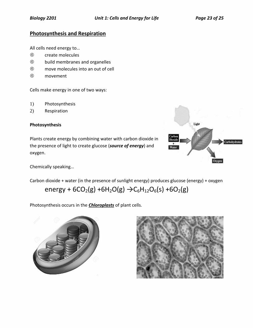
Photosynthesis and Respiration
All cells need energy to…
create molecules
build membranes and organelles
move molecules into an out of cell
movement
Cells make energy in one of two ways:
1) Photosynthesis
2) Respiration
Photosynthesis
Plants create energy by combining water with carbon dioxide in
the presence of light to create glucose (source of energy) and
oxygen.
Chemically speaking…
Carbon dioxide + water (in the presence of sunlight energy) produces glucose (energy) + oxygen
energy + 6CO2(g) +6H2O(g) →C6H12O6(s) +6O2(g)
Photosynthesis occurs in the Chloroplasts of plant cells.

Biology 2201 Unit 1: Cells and Energy for Life Page 24 of 25
Cellular Respiration
Cells create energy by combining glucose with oxygen to produce water and carbon dioxide.
Chemically speaking…
C6H12O6(s) +6O2(g) →6H2O(g) +6CO2(g) + energy
Respiration occurs within the mitochondrion of a cell.
Two types of Respiration
1. Aerobic Respiration:
This is respiration that occurs WITH oxygen. This type of respiration
releases water, oxygen and MORE energy
2. Anaerobic Respiration:
This is respiration that occurs WITHOUT oxygen. There are two types
of anaerobic respiration.
- Fermentation – The breakdown of vegetables and fruits by bacteria. Releases alcohol,
carbon dioxide and LESS energy.
- Lactic Acid Fermentation – Occurs in muscle cells. Results in a build of lactic acid that
makes muscles sore (a stitch). Releases lactic acid, carbon dioxide and LESS energy.

Biology 2201 Unit 1: Cells and Energy for Life Page 25 of 25
Respiration and Photosynthesis are OPPOSITE, but COMPLEMENTARY processes.
This means that what is created in one is used as a reactant in another.
One process depends upon the other to run.
Consider the importance of photosynthesis and cellular respiration on a global scale:
- Remember what we learned in Science 1206
- How have humans impacted the carbon cycle?
- What could this mean for primary industries of agriculture, forestry and fisheries?
- What are we doing to reduce our impact?