hiv-1 tat recruits hdm2 e3 ligase to target irf-1 for...
TRANSCRIPT

HIV-1 Tat Recruits HDM2 E3 Ligase To Target IRF-1 forUbiquitination and Proteasomal Degradation
Anna Lisa Remoli,a Giulia Marsili,a Edvige Perrotti,a Chiara Acchioni,a Marco Sgarbanti,a Alessandra Borsetti,b John Hiscott,c
Angela Battistinia
Department of Infectious, Parasitic and Immune-Mediated Diseases, Istituto Superiore di Sanità, Rome, Italya; National AIDS Center, Istituto Superiore di Sanità, Rome,Italyb; Istituto Pasteur-Fondazione Cenci Bolognetti, Rome, Italyc
ABSTRACT In addition to its ability to regulate HIV-1 promoter activation, the viral transactivator Tat also functions as a deter-minant of pathogenesis and disease progression by directly and indirectly modulating the host anti-HIV response, largelythrough the capacity of Tat to interact with and modulate the activities of multiple host proteins. We previously demonstratedthat Tat modulated both viral and host transcriptional machinery by interacting with the cellular transcription factor interferonregulatory factor 1 (IRF-1). In the present study, we investigated the mechanistic basis and functional significance of Tat�IRF-1interaction and demonstrate that Tat dramatically decreased IRF-1 protein stability. To accomplish this, Tat exploited the cellu-lar HDM2 (human double minute 2 protein) ubiquitin ligase to accelerate IRF-1 proteasome-mediated degradation, resulting ina quenching of IRF-1 transcriptional activity during HIV-1 infection. These data identify IRF-1 as a new target of Tat-inducedmodulation of the cellular protein machinery and reveal a new strategy developed by HIV-1 to evade host immune responses.
IMPORTANCE Current therapies have dramatically reduced morbidity and mortality associated with HIV infection and haveconverted infection from a fatal pathology to a chronic disease that is manageable via antiretroviral therapy. Nevertheless, HIV-1infection remains a challenge, and the identification of useful cellular targets for therapeutic intervention remains a major goal.The cellular transcription factor IRF-1 impacts various physiological functions, including the immune response to viral infec-tion. In this study, we have identified a unique mechanism by which HIV-1 evades IRF-1-mediated host immune responses andshow that the viral protein Tat accelerates IRF-1 proteasome-mediated degradation and inactivates IRF-1 function. Restorationof IRF-1 functionality may thus be regarded as a potential strategy to reinstate both a direct antiviral response and a morebroadly acting immune regulatory circuit.
Received 18 August 2016 Accepted 13 September 2016 Published 18 October 2016
Citation Remoli AL, Marsili G, Perrotti E, Acchioni C, Sgarbanti M, Borsetti A, Hiscott J, Battistini A. 2016. HIV-1 Tat recruits HDM2 E3 ligase to target IRF-1 for ubiquitination andproteasomal degradation. mBio 7(5):e01528-16. doi:10.1128/mBio.01528-16.
Invited Editor Eric A. Cohen, Institut de Recherches Cliniques de Montréal Editor Vinayaka R. Prasad, Albert Einstein College of Medicine
Copyright © 2016 Remoli et al. This is an open-access article distributed under the terms of the Creative Commons Attribution 4.0 International license.
Address correspondence to Angela Battistini, [email protected].
The complex pathogenesis of HIV-1 infection is determined inpart by interactions between viral regulatory proteins and cel-
lular factors that are responsible for both viral gene expression indifferent tissues and virus-induced physiological changes. TheHIV-1 transactivator Tat is essential for efficient transcription ofthe integrated provirus and for efficient HIV-1 replication (1, 2).By specifically binding to the transactivation-responsive elementregion in the viral promoter, Tat enhances both transcription andtranscriptional elongation (3). Independent of its ability to regu-late HIV-1 transcription, Tat also contributes to viral persistenceand dissemination by exerting a variety of other activities thatdirectly or indirectly modulate the host antiviral immune re-sponse, including deregulation of cytokine expression (4), inhibi-tion of dendritic cell maturation (5), suppression of antigen (Ag)-induced lymphocyte activation (6, 7), as well as activation of cellproliferation and increase of cell survival (8, 9). Tat protein is alsoreleased from acutely infected cells into the extracellular environ-ment and taken up by neighboring noninfected cells, where sim-ilarly, it increases virus infectivity and modulates cellular func-tions (4, 10). Many of these functions depend on the ability of Tat
to interact with host regulatory proteins and interfere with theexpression of multiple cellular functions (11, 12).
Among the numerous Tat-interacting proteins, we previouslydemonstrated that Tat interacted with interferon regulatory factor1 (IRF-1), the founding member of a family of nine transcriptionalregulators that impacts various physiological functions, includingthe immune response to viral infection, oncogenesis, and devel-opment of an immune system (13–16). Although originally iden-tified as a regulator of type I IFN gene expression, IRF-1 is notconsidered essential for IFN gene expression, except in cell-specific contexts (17–19). However, as an interferon (IFN)-regulated gene, IRF-1 is involved in IFN-induced antiviral immu-nity through the regulation of selected antiviral genes thatcooperatively promote an effective antiviral program against abroad spectrum of viruses (20–22). By inducing a rapid IFN-independent expression of antiviral factors (18), IRF-1 thus pro-vides a rapid antiviral defense upstream of the IRF3-activated IFNaxis, that is particularly relevant for those pathogens, includingHIV-1, that evade innate immunity by disrupting the inductionand function of IFN. In addition to its antiviral activity, IRF-1 also
RESEARCH ARTICLE
crossmark
September/October 2016 Volume 7 Issue 5 e01528-16 ® mbio.asm.org 1
on August 20, 2018 by guest
http://mbio.asm
.org/D
ownloaded from

impacts other aspects of immune regulation, including adaptiveimmunity and inflammation (23). IRF-1 is predominantly regu-lated at the transcriptional level (24, 25), but posttranslationalmodifications also play a significant, nonredundant role in theregulation of its activity (26–31). Like many other transcriptionfactors, IRF-1 is a short-lived protein that is rapidly degraded viathe ubiquitin-proteasome pathway (32, 33). The ubiquitinationand degradation signals reside in the C-terminal portion of IRF-1(32), and the degradation rate can be regulated in response tocellular conditions and specific stress (34, 35).
In the context of HIV-1 infection, IRF-1 can act both as aninducer of viral gene expression and as an antiviral factor, depend-ing on the physical interactions between Tat and IRF-1 in HIV-1-infected cells. In particular, during the early phase of infection,IRF-1 is induced by HIV-1 and, in combination with NF-�B, ac-tivates proviral transcription irrespective of the presence of Tat.Later, when discrete amounts of Tat are produced and IRF-1 be-comes dispensable for long terminal repeat (LTR) activity, inter-action with Tat sequesters IRF-1, resulting in the quenching of itstranscriptional activity on target genes (36–39).
In the present study, we have examined the mechanistic basisof IRF-1 expression modulation by viral Tat and now demonstratethat Tat targets IRF-1 for ubiquitin-mediated, K48-dependentproteasome degradation. We also identify human double minute2 protein (HDM2) as the IRF-1-specific ligase utilized by Tat todecrease IRF-1 stability. HDM2 (also known as mouse doubleminute 2 [Mdm2] in mice) is an E3 ubiquitin ligase that ubiquiti-nates the tumor suppressor p53 and is required for proteasome-dependent degradation and nuclear export of p53 (40, 41). HDM2also targets other viral and cellular substrates (40), includingmembers of the IRF family (42, 43), although the physiologicalsignificance of HDM2-IRF interactions has not been fully ad-dressed. Our observations identify an additional mechanism bywhich HIV-1 may suppress the antiviral immune response andcontribute to immune dysfunctions that favor viral replicationand disease progression.
RESULTSTat affects IRF-1 protein stability. In previous studies, we re-ported that HIV-1 Tat physically interacted in vitro and in vivowith IRF-1 (37–39), and we also observed that coexpression of Tatand IRF-1 cause a reproducible decrease in IRF-1 accumulation.Because IRF-1 expression is primarily regulated at the transcrip-tional level, the effect of Tat on IRF-1 transcription was initiallyevaluated. Analysis of the transcriptional activity of a 3,500-bpfragment of the IRF-1 promoter linked to the luciferase reportergene indicated that the basal and tumor necrosis factor alpha(TNF-�)-stimulated IRF-1 promoter activity was not affected byincreasing amounts of Tat expression compared with cells ex-pressing an empty vector (Fig. 1A). Similarly, no variation inIRF-1 mRNA levels was observed in the presence of increasing Tat(Fig. 1B). Therefore, to determine whether Tat could modulateIRF-1 protein stability, IRF-1 was coexpressed together with Flag-tagged Tat in the presence of the protein synthesis inhibitor cyclo-heximide (CHX). In the presence of Tat, a significant accelerationof IRF-1 decay was detected at 24 h posttransfection (Fig. 1C, lanes5 and 6 versus lanes 2 and 3). Densitometric quantification ofprotein levels indicated that in the absence of Tat expression, thehalf-life of IRF-1 was 50 min, whereas in the presence of Tat, IRF-1protein half-life was reduced to ~30 min. Since it is known that
IRF-1 is degraded through the ubiquitin/proteasome pathway, wewondered whether Tat could stimulate IRF-1 polyubiquitinationand proteasome degradation. IRF-1 and six-histidine-taggedubiquitin (His6-Ub) were coexpressed in the presence or absenceof Flag-tagged Tat. IRF-1 ubiquitination was then determined bycapturing His6-Ub in cell extracts with nickel beads (nickel-nitrilotriacetic acid [Ni-NTA]), followed by Western blot analysisof the purified ubiquitin conjugates with IRF-1-specific antibod-ies. Monoubiquitinated IRF-1 was detected in the His-Ub-expressing cells, while IRF-1 polyubiquitination was easily de-tected in Tat-containing extracts (Fig. 1D). To assess theproteasome-mediated IRF-1 degradation, IRF-1 expression wasanalyzed in cells expressing IRF-1 alone or in combination withFlag-tagged Tat in the presence or absence of the proteasome in-hibitor MG132. MG132 clearly blocked the ability of Tat to accel-erate IRF-1 turnover (Fig. 1E, lane 3 versus lane 4), suggesting thatTat may indeed increase the proteasome-mediated degradation ofIRF-1.
HDM2 E3 ligase mediates Tat-induced IRF-1 turnover. Insearching for the E3 ubiquitin ligase that mediates Tat-inducedpolyubiquitination/degradation of IRF-1, HDM2 was selected forfurther investigation, given previous studies that established a linkbetween HDM2 and both Tat (44) and IRF-1 (42). HDM2 wasreported to interact with Tat as an E3 ligase that increases Tat-mediated transactivation of the LTR upon K63 ubiquitination inTat-expressing cells (44). Similarly, HDM2 was shown to bind andubiquitinate IRF-1 (42). We therefore initially examined whetherHDM2 affected IRF-1 stability. In the presence of CHX and in-creasing amounts of green fluorescent protein (GFP)-taggedHDM2, HDM2 per se decreased IRF-1 levels in a dose-dependentmanner (Fig. 2A, lanes 4 and 5 and graph). Then, to assess whetherthe Tat-mediated decrease in IRF-1 stability involved HDM2,IRF-1 was coexpressed together with Flag-tagged Tat using smallamounts of HDM2 that did not per se affect IRF-1 stability. In thepresence of Tat, IRF-1 degradation was dramatically acceleratedby HDM2 coexpression compared to cells not expressing HDM2(Fig. 2B, lanes 5 and 6 versus lanes 2 and 3). Densitometric quan-tification of protein levels indicated that in the absence of HDM2expression, the half-life of IRF-1 was ~30 min, whereas in thepresence of HDM2, the half-life of IRF-1 protein was reduced to~18 min.
The specificity of the effect of Tat on IRF-1 stability was thenevaluated in expression studies using a mutation in cysteine 22(TatC22G) of Tat that reduces both interaction with HDM2 andTat ubiquitination (44). Compared with the effect of wild-typeTat, the TatC22G mutant was unable to accelerate HDM2-mediated IRF-1 degradation (Fig. 2C, lane 2 versus lane 4).
In support of the interrelationship between Tat, IRF-1, andHDM2, coimmunoprecipitation experiments with Flag-taggedIRF-1, GFP-tagged HDM2, and Tat indicated a physical associa-tion of the three proteins (Fig. 2D). Collectively, these results in-dicate that Tat accelerates IRF-1 turnover upon recruitment of theHDM2 E3 ligase.
Tat increases HDM2-induced IRF-1 K48 polyubiquitination.The nickel capture assay was next used to evaluate the effect of Taton HDM2-dependent IRF-1 ubiquitination; expression of eitherTat or HDM2 stimulated the accumulation of the ubiquitinatedIRF-1 (Fig. 3A, lanes 2 and 3), and the coexpression of Tat togetherwith HDM2 greatly increased the accumulation of the polyubiq-uitinated forms of IRF-1 (Fig. 3A, lane 4). The low level of IRF-1
Remoli et al.
2 ® mbio.asm.org September/October 2016 Volume 7 Issue 5 e01528-16
on August 20, 2018 by guest
http://mbio.asm
.org/D
ownloaded from

ubiquitination present in the control extract (Fig. 3A, lane 1) islikely mediated by endogenous E3-ligase activity. Since ubiquiti-nation is not limited to proteasomal degradation, we next deter-mined whether degradation of IRF1 by Tat and HDM2 involvedthe formation of K48-linked polyubiquitination chains that act asa bona fide signal for targeting substrates to proteasomal degrada-
tion. Using Flag-tagged IRF1 with GFP-tagged HDM2 in the pres-ence or absence of Tat, immunoprecipitation was performed us-ing an anti-Flag antibody-conjugated resin, and IRF-1-linkedpolyubiquitination chains were detected using antibodies specificfor K48- or K63-linked ubiquitin, respectively. Both Tat andHDM2 individually induced K48-linked IRF-1 ubiquitination
FIG 1 Tat affects IRF-1 stability. (A) HEK293 cells were transfected with a 3,500-bp fragment of the IRF-1 promoter linked to the luciferase reporter gene aloneor in combination with increasing amounts of Tat-expressing vector. After 24 h, cells were treated for 4 h with 10 ng/ml of TNF-� (�), where indicated, and thenprocessed for luciferase activity. Data shown are the means plus standard errors of the means (SEM) (error bars) from three separate experiments calculated afternormalization with the Renilla activity. The values for untreated cells were set at 1. (B) HEK293 cells were transfected with increasing amounts of Tat-expressingvector. At 24 h after transfection, cells were harvested, and IRF-1 RNA levels were assessed using real-time RT-PCR. The levels were normalized to GAPDHmRNA abundance. The means plus SEM of three independent experiments are shown as relative expression units. The values for untreated cells were set at 1. (C)HEK293 cells were transfected with IRF-1 expression vector in the presence (�) or absence (�) of Flag-Tat expression vector. At 24 h posttransfection, the cellswere treated with CHX for the indicated time. IRF-1 and Tat proteins were detected with anti-IRF-1 (�-IRF-1) and anti-Flag (�-Flag) antibodies, respectively.Data plotted in the graph represent the means � SEM from three different assays of IRF-1 protein bands quantified from Western blots and normalized to actinprotein levels as the loading control and presented as percentage values relative to those without CHX treatment set at 100%. (D) HEK293 cells were cotransfectedwith expression vectors for His6-Ub and IRF-1 in the presence or absence of Flag-Tat-expressing vector. His-Ub-conjugated proteins were captured bynickel-agarose beads, eluted, and analyzed by Western blotting with anti-IRF-1 antibody. Western blotting of cell lysates shows the expression of ectopicallyexpressed proteins. (E) HEK293 cells were cotransfected with expression vectors for IRF-1 and Flag-Tat and then treated with MG132 for 2 h where indicated.IRF-1 and Tat expression was detected by Western blotting. Data plotted in the graph represent the means plus SEM from three different assays of IRF-1 proteinbands quantified from Western blots and normalized to actin protein levels as the loading control. Results are presented as percentage values relative to basalIRF-1 expression set at 100. Blots are representative of at least three independent experiments with similar results.
Tat Promotes HDM2-Mediated Degradation of IRF-1
September/October 2016 Volume 7 Issue 5 e01528-16 ® mbio.asm.org 3
on August 20, 2018 by guest
http://mbio.asm
.org/D
ownloaded from

FIG 2 HDM2 E3 ligase mediates Tat-induced IRF-1 turnover. (A) HEK293 cells were transfected with expression vectors encoding IRF-1 alone or incombination with increasing amounts of GFP-tagged HDM2 (GFP-HDM2), as indicated. The cells were treated with CHX for the indicated time, and cell lysateswere then subjected to immunoblotting. Data plotted in the graph represent the means � SEM from three different assays of IRF-1 protein bands quantified from
(Continued)
Remoli et al.
4 ® mbio.asm.org September/October 2016 Volume 7 Issue 5 e01528-16
on August 20, 2018 by guest
http://mbio.asm
.org/D
ownloaded from
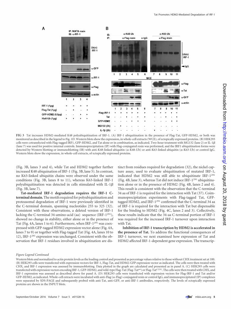
(Fig. 3B, lanes 3 and 4), while Tat and HDM2 together furtherincreased K48 ubiquination of IRF-1 (Fig. 3B, lane 5). In contrast,no K63-linked ubiquitin chains were observed under the sameconditions (Fig. 3B, lanes 8 to 11), whereas K63-linked IRF-1polyubiquitination was detected in cells stimulated with IL-1�(Fig. 3B, lane 7).
Tat-mediated IRF-1 degradation requires the IRF-1 C-terminal domain. The motifs required for polyubiquitination andproteasomal degradation of IRF-1 were previously identified inthe C-terminal domain, spanning nucleotides 255 to 325 (32).Consistent with these observations, a deleted version of IRF-1lacking the C-terminal 34-amino-acid (aa) sequence (IRF-1291),showed no change in stability, either alone or in the presence ofTat (Fig. 4A, lanes 1 to 6). Furthermore, when IRF-1291 was coex-pressed with GFP-tagged HDM2 expression vector alone (Fig. 4A,lanes 7 to 9) or together with Flag-tagged Tat (Fig. 4A, lanes 10 to12), IRF-1291 expression was unchanged. Consistent with the ob-servation that IRF-1 residues involved in ubiquitination are dis-
tinct from residues required for degradation (32), the nickel cap-ture assay, used to evaluate ubiquitination of mutated IRF-1,indicated that HDM2 was still able to ubiquitinate IRF-1291
(Fig. 4B, lane 3), whereas Tat did not induce IRF-1291 ubiquitina-tion alone or in the presence of HDM2 (Fig. 4B, lanes 2 and 4).This result is consistent with the observation that the C-terminal34 aa of IRF-1 is required for the interaction with Tat (37). Coim-munoprecipitation experiments with Flag-tagged Tat, GFP-tagged HDM2, and IRF-1291 confirmed that the C-terminal 34 aaof IRF-1 is required for the interaction with Tat but dispensablefor the binding to HDM2 (Fig. 4C, lanes 2 and 3). Collectively,these results indicate that the 34-aa C-terminal portion of IRF-1was required for the increased IRF-1 turnover upon interactionwith Tat.
Inhibition of IRF-1 transcription by HDM2 is accelerated inthe presence of Tat. To address the functional consequences ofIRF-1 turnover, we next examined how expression of Tat andHDM2 affected IRF-1-dependent gene expression. The transcrip-
Figure Legend Continued
Western blots and normalized to actin protein levels as the loading control and presented as percentage values relative to those without CHX treatment set at 100.(B) HEK293 cells were transfected with expression vectors for IRF-1, Flag-Tat, and HDM2-GFP expression vector as indicated. The cells were then treated withCHX, and IRF-1 expression was assessed by immunoblotting. Data plotted in the graph are calculated and presented as in panel A. (C) HEK293 cells weretransfected with expression vectors encoding IRF-1, GFP-HDM2, and wild-type Flag-Tat (Flag-Tatwt) or Flag-TatC22G. The cells were then treated with CHX, andIRF-1 expression was assessed as described above for panel A. (D) HEK293 cells were transfected with expression vectors for Flag-IRF-1 and Tat and/orGFP-HDM2, as indicated. Whole-cell extracts were incubated with anti-Flag (�-Flag)-conjugated resin or control IgG, and immunoprecipitated (IP) complexeswere separated by SDS-PAGE and subsequently probed with anti-Tat, anti-GFP, or anti-IRF-1 antibodies, respectively. The levels of ectopically expressedproteins are shown in the INPUT blots.
FIG 3 Tat increases HDM2-mediated K48 polyubiquitination of IRF-1. (A) IRF-1 ubiquitination in the presence of Flag-Tat, GFP-HDM2, or both wasmonitored as described in the legend to Fig. 1D. Western blots show the expression, in whole-cell extracts (WCE), of ectopically expressed proteins. (B) HEK293cells were cotransfected with Flag-tagged IRF1, GFP-HDM2, and Tat alone or in combination, as indicated. Two-hour treatment with MG132 (lane 2) or IL-1�(lane 7) was used for positive internal controls. Immunoprecipitation (IP) with Flag-conjugated resin was performed, and the IRF1 ubiquitination forms weredetected by Western blotting or immunoblotting (IB) with anti-K48-linked ubiquitin (�-K48-Ub) or anti-K63-linked ubiquitin (�-K63-Ub) or control IgG.Western blots show the expression, in whole-cell extracts, of ectopically expressed proteins.
Tat Promotes HDM2-Mediated Degradation of IRF-1
September/October 2016 Volume 7 Issue 5 e01528-16 ® mbio.asm.org 5
on August 20, 2018 by guest
http://mbio.asm
.org/D
ownloaded from

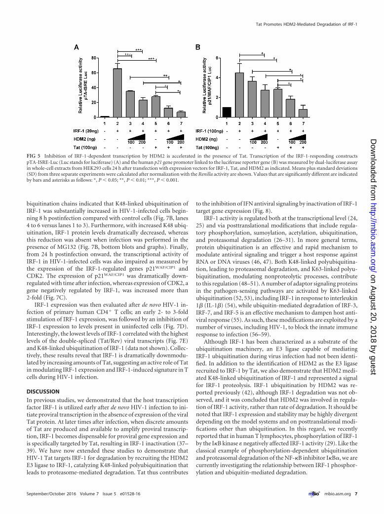
tional activity of IRF-1-responsive luciferase reporter constructspISRE-TA (bearing five copies of the consensus IRF-E motif) andthe IRF-1-responsive p21WAF/CIP1 gene promoter was evaluated inHEK293 cells transiently cotransfected with IRF-1 and increasingamounts of HDM2 in the presence or absence of Tat (Fig. 5A andB). Expression of the IRF-1-responsive promoters was reduced~50 percent by Tat expression, and increasing amounts of HDM2also decreased IRF-1 promoter expression by 20% to 70%; in thepresence of both Tat and HDM2, IRF-1 driven promoter activitywas essentially abolished and returned to basal levels. In contrast,IRF-1291 was not affected by Tat alone or in combination withHDM2 (data not shown).
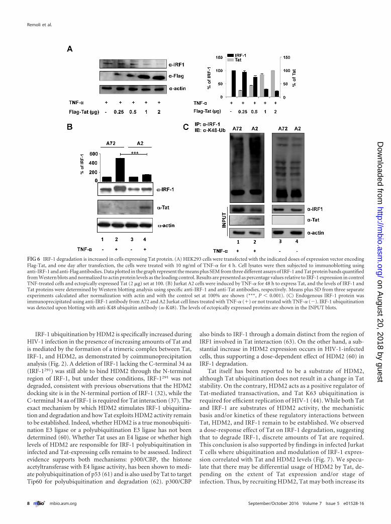
IRF-1 degradation is increased in Jurkat cells inducibly ex-pressing Tat protein. To evaluate whether Tat also decreased ac-cumulation of endogenous IRF-1, we transfected a Tat-expressingconstruct in cells where expression of endogenous IRF-1 was stim-ulated by TNF-�� Increasing amounts of Tat were indeed able toaffect IRF-1 expression in a dose-response manner (Fig. 6A andgraph). Moreover, the Tat-mediated IRF-1 proteolysis was alsoevaluated in a more physiologically relevant cell model, i.e., in aJurkat T cell clone (termed A2) that inducibly expresses Tat fol-
lowing TNF-� treatment (45). The content of IRF-1 in controlA72 and in Tat-expressing A2 cells was thus evaluated, and aspreviously reported, TNF-� treatment stimulated IRF-1 expres-sion in A72 control cells (Fig. 6B, lane 2 and graph). Conversely, inTat-expressing A2 cells, IRF-1 did not accumulate (Fig. 6B, lane 4and graph). Importantly, in A2 cells, Tat expression followingTNF-� treatment resulted in K48-linked ubiquitination of IRF-1(Fig. 6C, lane 2) compared to A72 cells that do not express Tat(Fig. 6C, lane 1). As expected, similar basal levels of IRF-1 ubiq-uitination were observed in the two cell lines in the absence ofTNF-� treatment (Fig. 6C, lanes 3 and 4).
IRF-1 is downregulated and K48 polyubiquitinated in HIV-1-infected Jurkat T cells and during HIV-1 de novo infection ofhuman primary CD4� T cells. To assess the biological relevanceof the above findings, the turnover of IRF-1 was also evaluated inthe context of HIV-1 infection. In HIV-infected Jurkat T cellsbeginning 24 h postinfection, IRF-1 expression was substantiallydecreased (Fig. 7A, lanes 4 to 6 versus lanes 1 to 3), while HDM2expression increased during the course of infection (Fig. 7A). Theturnover of IRF-1 expression mirrored the increase in Tat/Revtranscripts (Fig. 7A, top panel). Detection of IRF-1-linked polyu-
FIG 4 Tat-mediated IRF-1 degradation requires the IRF-1 C-terminal domain. (A) HEK293 cells were cotransfected with expression vectors for an IRF-1mutant with the 34-aa COOH terminus deleted (IRF1291), Flag-Tat, and GFP-HDM2, alone or in combination. One day after transfection, the cells were treatedwith CHX for the indicated time points, and expression of IRF1291, Tat, and HDM2 was detected by Western blotting using specific antibodies, as indicated. (B)IRF1291 ubiquitination in the presence of Flag-Tat, GFP-HDM2, or both, was monitored as described in the legend to Fig. 3A. Western blots show the expressionof ectopically expressed proteins in whole-cell extracts. (C) HEK293 cells were cotransfected with the indicated expression vectors, immunoprecipitated withanti-IRF-1 antibodies, and Tat and HDM2 were detected by Western blotting using the indicated antibodies. INPUT shows the level of ectopically expressedproteins.
Remoli et al.
6 ® mbio.asm.org September/October 2016 Volume 7 Issue 5 e01528-16
on August 20, 2018 by guest
http://mbio.asm
.org/D
ownloaded from
biquitination chains indicated that K48-linked ubiquitination ofIRF-1 was substantially increased in HIV-1-infected cells begin-ning 8 h postinfection compared with control cells (Fig. 7B, lanes4 to 6 versus lanes 1 to 3). Furthermore, with increased K48 ubiq-uitination, IRF-1 protein levels dramatically decreased, whereasthis reduction was absent when infection was performed in thepresence of MG132 (Fig. 7B, bottom blots and graphs). Finally,from 24 h postinfection onward, the transcriptional activity ofIRF-1 in HIV-1-infected cells was also impaired as measured bythe expression of the IRF-1-regulated genes p21WAF/CIP1 andCDK2. The expression of p21WAF/CIP1 was dramatically down-regulated with time after infection, whereas expression of CDK2, agene negatively regulated by IRF-1, was increased more than2-fold (Fig. 7C).
IRF-1 expression was then evaluated after de novo HIV-1 in-fection of primary human CD4� T cells; an early 2- to 3-foldstimulation of IRF-1 expression, was followed by an inhibition ofIRF-1 expression to levels present in uninfected cells (Fig. 7D).Interestingly, the lowest levels of IRF-1 correlated with the highestlevels of the double-spliced (Tat/Rev) viral transcripts (Fig. 7E)and K48-linked ubiquitination of IRF-1 (data not shown). Collec-tively, these results reveal that IRF-1 is dramatically downmodu-lated by increasing amounts of Tat, suggesting an active role of Tatin modulating IRF-1 expression and IRF-1-induced signature in Tcells during HIV-1 infection.
DISCUSSION
In previous studies, we demonstrated that the host transcriptionfactor IRF-1 is utilized early after de novo HIV-1 infection to ini-tiate proviral transcription in the absence of expression of the viralTat protein. At later times after infection, when discrete amountsof Tat are produced and available to amplify proviral transcrip-tion, IRF-1 becomes dispensable for proviral gene expression andis specifically targeted by Tat, resulting in IRF-1 inactivation (37–39). We have now extended these studies to demonstrate thatHIV-1 Tat targets IRF-1 for degradation by recruiting the HDM2E3 ligase to IRF-1, catalyzing K48-linked polyubiquitination thatleads to proteasome-mediated degradation. Tat thus contributes
to the inhibition of IFN antiviral signaling by inactivation of IRF-1target gene expression (Fig. 8).
IRF-1 activity is regulated both at the transcriptional level (24,25) and via posttranslational modifications that include regula-tory phosphorylation, sumoylation, acetylation, ubiquitination,and proteasomal degradation (26–31). In more general terms,protein ubiquitination is an effective and rapid mechanism tomodulate antiviral signaling and trigger a host response againstRNA or DNA viruses (46, 47). Both K48-linked polyubiquitina-tion, leading to proteasomal degradation, and K63-linked polyu-biquitination, modulating nonproteolytic processes, contributeto this regulation (48–51). A number of adaptor signaling proteinsin the pathogen-sensing pathways are activated by K63-linkedubiquitination (52, 53), including IRF-1 in response to interleukin1� (IL-1�) (54), while ubiquitin-mediated degradation of IRF-3,IRF-7, and IRF-5 is an effective mechanism to dampen host anti-viral response (55). As such, these modifications are exploited by anumber of viruses, including HIV-1, to block the innate immuneresponse to infection (56–59).
Although IRF-1 has been characterized as a substrate of theubiquitination machinery, an E3 ligase capable of mediatingIRF-1 ubiquitination during virus infection had not been identi-fied. In addition to the identification of HDM2 as the E3 ligaserecruited to IRF-1 by Tat, we also demonstrate that HDM2 medi-ated K48-linked ubiquitination of IRF-1 and represented a signalfor IRF-1 proteolysis. IRF-1 ubiquitination by HDM2 was re-ported previously (42), although IRF-1 degradation was not ob-served, and it was concluded that HDM2 was involved in regula-tion of IRF-1 activity, rather than rate of degradation. It should benoted that IRF-1 expression and stability may be highly divergentdepending on the model systems and on posttranslational modi-fications other than ubiquitination. In this regard, we recentlyreported that in human T lymphocytes, phosphorylation of IRF-1by the I�B kinase � negatively affected IRF-1 activity (29). Like theclassical example of phosphorylation-dependent ubiquitinationand proteasomal degradation of the NF-�B inhibitor I�B�, we arecurrently investigating the relationship between IRF-1 phosphor-ylation and ubiquitin-mediated degradation.
FIG 5 Inhibition of IRF-1-dependent transcription by HDM2 is accelerated in the presence of Tat. Transcription of the IRF-1-responding constructspTA-ISRE-Luc (Luc stands for luciferase) (A) and the human p21 gene promoter linked to the luciferase reporter gene (B) was measured by dual-luciferase assayin whole-cell extracts from HEK293 cells 24 h after transfection with expression vectors for IRF-1, Tat, and HDM2 as indicated. Means plus standard deviations(SD) from three separate experiments were calculated after normalization with the Renilla activity are shown. Values that are significantly different are indicatedby bars and asterisks as follows: *, P � 0.05; **, P � 0.01; ***, P � 0.001.
Tat Promotes HDM2-Mediated Degradation of IRF-1
September/October 2016 Volume 7 Issue 5 e01528-16 ® mbio.asm.org 7
on August 20, 2018 by guest
http://mbio.asm
.org/D
ownloaded from
IRF-1 ubiquitination by HDM2 is specifically increased duringHIV-1 infection in the presence of increasing amounts of Tat andis mediated by the formation of a trimeric complex between Tat,IRF-1, and HDM2, as demonstrated by coimmunoprecipitationanalysis (Fig. 2). A deletion of IRF-1 lacking the C-terminal 34 aa(IRF-1291) was still able to bind HDM2 through the N-terminalregion of IRF-1, but under these conditions, IRF-1291 was notdegraded, consistent with previous observations that the HDM2docking site is in the N-terminal portion of IRF-1 (32), while theC-terminal 34 aa of IRF-1 is required for Tat interaction (37). Theexact mechanism by which HDM2 stimulates IRF-1 ubiquitina-tion and degradation and how Tat exploits HDM2 activity remainto be established. Indeed, whether HDM2 is a true monoubiquiti-nation E3 ligase or a polyubiquitination E3 ligase has not beendetermined (60). Whether Tat uses an E4 ligase or whether highlevels of HDM2 are responsible for IRF-1 polyubiquitination ininfected and Tat-expressing cells remains to be assessed. Indirectevidence supports both mechanisms: p300/CBP, the histoneacetyltransferase with E4 ligase activity, has been shown to medi-ate polyubiquitination of p53 (61) and is also used by Tat to targetTip60 for polyubiquitination and degradation (62). p300/CBP
also binds to IRF-1 through a domain distinct from the region ofIRF1 involved in Tat interaction (63). On the other hand, a sub-stantial increase in HDM2 expression occurs in HIV-1-infectedcells, thus supporting a dose-dependent effect of HDM2 (60) inIRF-1 degradation.
Tat itself has been reported to be a substrate of HDM2,although Tat ubiquitination does not result in a change in Tatstability. On the contrary, HDM2 acts as a positive regulator ofTat-mediated transactivation, and Tat K63 ubiquitination isrequired for efficient replication of HIV-1 (44). While both Tatand IRF-1 are substrates of HDM2 activity, the mechanisticbasis and/or kinetics of these regulatory interactions betweenTat, HDM2, and IRF-1 remain to be established. We observeda dose-response effect of Tat on IRF-1 degradation, suggestingthat to degrade IRF-1, discrete amounts of Tat are required.This conclusion is also supported by findings in infected JurkatT cells where ubiquitination and modulation of IRF-1 expres-sion correlated with Tat and HDM2 levels (Fig. 7). We specu-late that there may be differential usage of HDM2 by Tat, de-pending on the extent of Tat expression and/or stage ofinfection. Thus, by recruiting HDM2, Tat may both increase its
FIG 6 IRF-1 degradation is increased in cells expressing Tat protein. (A) HEK293 cells were transfected with the indicated doses of expression vector encodingFlag-Tat, and one day after transfection, the cells were treated with 10 ng/ml of TNF-� for 4 h. Cell lysates were then subjected to immunoblotting usinganti-IRF-1 and anti-Flag antibodies. Data plotted in the graph represent the means plus SEM from three different assays of IRF-1 and Tat protein bands quantifiedfrom Western blots and normalized to actin protein levels as the loading control. Results are presented as percentage values relative to IRF-1 expression in controlTNF-treated cells and ectopically expressed Tat (2 �g) set at 100. (B) Jurkat A2 cells were induced by TNF-� for 48 h to express Tat, and the levels of IRF-1 andTat proteins were determined by Western blotting analysis using specific anti-IRF-1 and anti-Tat antibodies, respectively. Means plus SD from three separateexperiments calculated after normalization with actin and with the control set at 100% are shown (***, P � 0.001). (C) Endogenous IRF-1 protein wasimmunoprecipitated using anti-IRF-1 antibody from A72 and A2 Jurkat cell lines treated with TNF-� (�) or not treated with TNF-� (�). IRF-1 ubiquitinationwas detected upon blotting with anti-K48 ubiquitin antibody (�-K48). The levels of ectopically expressed proteins are shown in the INPUT blots.
Remoli et al.
8 ® mbio.asm.org September/October 2016 Volume 7 Issue 5 e01528-16
on August 20, 2018 by guest
http://mbio.asm
.org/D
ownloaded from
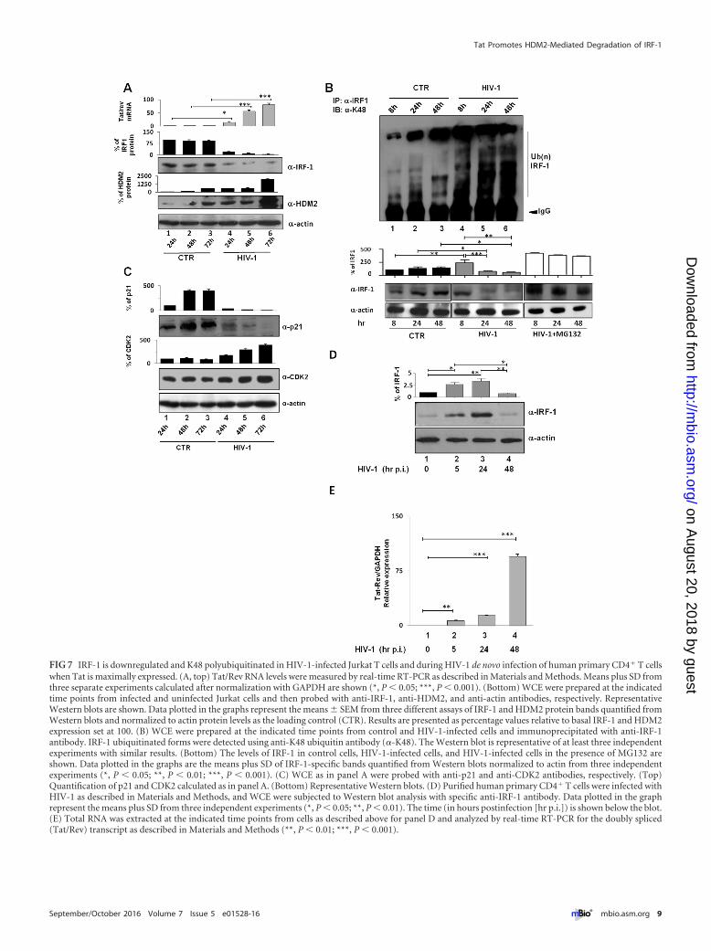
FIG 7 IRF-1 is downregulated and K48 polyubiquitinated in HIV-1-infected Jurkat T cells and during HIV-1 de novo infection of human primary CD4� T cellswhen Tat is maximally expressed. (A, top) Tat/Rev RNA levels were measured by real-time RT-PCR as described in Materials and Methods. Means plus SD fromthree separate experiments calculated after normalization with GAPDH are shown (*, P � 0.05; ***, P � 0.001). (Bottom) WCE were prepared at the indicatedtime points from infected and uninfected Jurkat cells and then probed with anti-IRF-1, anti-HDM2, and anti-actin antibodies, respectively. RepresentativeWestern blots are shown. Data plotted in the graphs represent the means � SEM from three different assays of IRF-1 and HDM2 protein bands quantified fromWestern blots and normalized to actin protein levels as the loading control (CTR). Results are presented as percentage values relative to basal IRF-1 and HDM2expression set at 100. (B) WCE were prepared at the indicated time points from control and HIV-1-infected cells and immunoprecipitated with anti-IRF-1antibody. IRF-1 ubiquitinated forms were detected using anti-K48 ubiquitin antibody (�-K48). The Western blot is representative of at least three independentexperiments with similar results. (Bottom) The levels of IRF-1 in control cells, HIV-1-infected cells, and HIV-1-infected cells in the presence of MG132 areshown. Data plotted in the graphs are the means plus SD of IRF-1-specific bands quantified from Western blots normalized to actin from three independentexperiments (*, P � 0.05; **, P � 0.01; ***, P � 0.001). (C) WCE as in panel A were probed with anti-p21 and anti-CDK2 antibodies, respectively. (Top)Quantification of p21 and CDK2 calculated as in panel A. (Bottom) Representative Western blots. (D) Purified human primary CD4� T cells were infected withHIV-1 as described in Materials and Methods, and WCE were subjected to Western blot analysis with specific anti-IRF-1 antibody. Data plotted in the graphrepresent the means plus SD from three independent experiments (*, P � 0.05; **, P � 0.01). The time (in hours postinfection [hr p.i.]) is shown below the blot.(E) Total RNA was extracted at the indicated time points from cells as described above for panel D and analyzed by real-time RT-PCR for the doubly spliced(Tat/Rev) transcript as described in Materials and Methods (**, P � 0.01; ***, P � 0.001).
Tat Promotes HDM2-Mediated Degradation of IRF-1
September/October 2016 Volume 7 Issue 5 e01528-16 ® mbio.asm.org 9
on August 20, 2018 by guest
http://mbio.asm
.org/D
ownloaded from

activity on the HIV-1 LTR and inactivate a transcriptional pro-tein involved in the host antiviral response (Fig. 8).
The interplay between IRF-1, Tat, and HDM2 may provide aselective advantage to HIV-1 replication. Consistently, we haveobserved a substantial increase in HIV-1 replication, measured byp24 accumulation, when IRF-1 expression is constitutivelyknocked out in T cells (data not shown). This is not surprisingsince IRF-1 represents a network hub in the regulation of the hostantiviral, immunomodulatory, and growth modulatory func-tions. More specifically, the antiviral activities of IRF-1 have beenreemphasized by interferon-stimulated gene(ISG) expressionscreening studies that identified IRF-1 as a potent antiviral effectorthat inhibited a broad range of viruses, including HIV (20, 22).Consistent with these observations, many ISGs are directly acti-vated by IRF-1 after viral infection (18, 64).
IRF-1 also exerts a number of functions beyond its antimicro-bial effects. By targeting IRF-1, Tat may regulate cell growth byinhibiting p21WAF/CIP1 and CDK2. In this respect, the loss of theG1/S checkpoint associated with the loss of p21WAF/CIP1 gene ex-pression in HIV-infected cells provides a selective advantage forHIV-1 by allowing viral transcription and replication (65). Thus,the targeting of IRF-1 by HIV-1 Tat again illustrates that a singleviral protein can modulate a number of cellular pathways, thuscontributing to a replicative advantage for the virus.
MATERIALS AND METHODSPlasmids, transient transfection, and reporter gene assay. CMVBL,CMVBL IRF-1, and mutant CMVBL IRF1291 and CMV-Tat expression
vectors have been described previously (37, 66). Flag-tagged IRF1 (Flag-IRF1) was obtained by PCR from CMVBL IRF1 and inserted intopCMV2-Flag (CMV stands for cytomegalovirus) (Clontech Laboratories,Inc.) expression vector using HindIII and XbaI restriction enzymes. Flag-tagged Tat (Flag-Tat) was obtained by de novo gene synthesis Gene-Scriptand then cloned in pCMV2-Flag (Clontech) using BamHI and EcoR1;pTatC22G was generated by site-directed mutagenesis (QuikChange; Strat-agene, Cedar Creek, TX) using Flag-Tat as the substrate according to themanufacturer’s protocol. Mutated clones were fully sequenced after iden-tification. pEGFP-C2-Hdm2 (EGFP stands for enhanced green fluores-cent protein), p21/WAF/CIP1 luciferase reporter gene and pCDNA3.1Ub-His(6x) were generous gifts of G. D’Orazi and T. Haas. IRF-1-responding luciferase reporter constructs pISRE-TA was from Clontech.The constructs for p3500 (encoding the entire IRF-1 promoter from�3400 bp to �168 bp) cloned upstream of the luciferase reporter genewas a generous gift of Richard Pine.
Transient transfections were performed using JetPei reagent (PolyplusTransfection SA, Illkirch, France) or the calcium phosphate transfectionsystem (Life Technologies, Invitrogen Corporation, Carlsbad, CA) ac-cording to the manufacturer’s protocol. The amounts of transfected DNAwere normalized by using an empty vector.
Reagents from Promega (Promega Corporation, Madison, WI) wereused to assay extracts for dual-luciferase activity in a Lumat LB9501 lumi-nometer (E&G Berthold, Bad Wildbad, Germany).
Cell culture and reagents. J-Lat Tat-GFP cells (clone A2/A72) fromEric Verdin (45) was obtained through the NIH AIDS Reagent Program,Division of AIDS, NIAID, NIH. Jurkat, Jurkat clone A2/A72, and HEK293cells were grown in RPMI 1640 medium and Dulbecco’s modified Eagle’smedium (DMEM) (Bio-Whittaker, Cambrex Bio Science, Verviers, Bel-gium), containing 10% fetal calf serum (FCS) and antibiotics. Human
FIG 8 Schematic representation of the dual effect of Tat on IRF-1 activity in the course of HIV-1 infection. In early phases of HIV-1 replication, IRF-1 istranscriptionally stimulated by viral infection, and it is recruited by small amounts of Tat on the viral promoter to drive, with NF-�B, initial transcription of theintegrated provirus. Later, when discrete amounts of Tat are produced and IRF1 activity on LTR is dispensable for the virus to replicate, Tat nullifies the functionof IRF-1, accelerating its proteasome-mediated degradation upon recruitment of the HDM2 E3 ligase, thus quenching its activity on target gene promoters.
Remoli et al.
10 ® mbio.asm.org September/October 2016 Volume 7 Issue 5 e01528-16
on August 20, 2018 by guest
http://mbio.asm
.org/D
ownloaded from

peripheral blood mononuclear cells (PBMCs) from healthy donors wereisolated by Ficoll-Hypaque gradient centrifugation, and the CD4� T cellpopulation was purified by negative selection using magnetic beads(Miltenyi Biotech GmbH, Bergisch Gladbach, Germany), as previouslydescribed (29). Recovered cells were �96% CD4�, as determined byfluorescence-activated cell sorter (FACS) analysis. The cells were culturedin RPMI 1640 medium (Bio-Whittaker, Cambrex Bio Science) containing20% FCS and antibiotics and activated with anti-CD3 monoclonal anti-bodies (MAbs) (R&D Systems, Minneapolis, MN). MG132 (Sigma) wasused at 50 �M, cycloheximide (CHX) (Sigma) was used at 25 �g/ml, andtumor necrosis factor alpha (TNF-�) was used at 10 ng/ml.
HIV stock preparation and infection. Replication-competent dual-tropic virus was generated by calcium phosphate-mediated transienttransfection of HEK293 cells with the pHXB2R molecular clone. Virus-containing supernatant was filtered, frozen in aliquots at �70°C, andtitrated on TZM-bl cells. Jurkat and primary CD4� T cells were inocu-lated with HIV-1/HXB2 at a multiplicity of infection of 0.05 50% tissueculture infective dose (TCID50) per cell, as previously described (37).
Quantitative real-time reverse transcription-PCR. Total RNA ex-tracted using the RNeasy total RNA extraction kit (Qiagen) was treatedwith RNase-free DNase (Qiagen) and then reverse transcribed with HighCapacity cDNA reverse transcription kit (Applied Biosystems) accordingto the manufacturer’s instructions. cDNA was subjected to quantitativereal-time PCR on ABI 7000 sequence detection system (PE Applied Bio-systems, Warrington, United Kingdom) by using SYBR green PCR mastermix (Applied Biosystems). Primers used for quantitative real-time reversetranscription-PCR (qRT-PCR) were IRF-1 forward primer 5=-AGCTCAGCTGTGCGAGTGTA-3= and reverse primer 5=-CATGACTTCCTCTTGGCCTT-3= and Tat/Rev forward primer 5=-CTTAGGCATCTCCTATGGCAGGAA-3= and reverse primer 5=-GGATCTGTCTCTGTCTCTCTCTCCACC-3=. Transcript levels were normalized to glyceraldehyde-3-phosphate dehydrogenase (GAPDH) (forward, 5=-GGGTGTGAACCATGAGAAG-3=; reverse, 5=-GCTAAGCAGTTGGTGGTGC-3=) as aninternal control and expressed as fold increase according to the �CT
methods (means � standard deviations).Coimmunoprecipitation, Western blot analysis, and protein quan-
tifications. Total protein extracts (whole-cell extracts [WCE]) were pre-pared and subjected to Western blot analysis or immunoprecipitation, aspreviously described (39). Briefly, for coimmunoprecipitation, 300 �g ofWCE was incubated with 1 �g of polyclonal anti-IRF-1 antibody (sc-13041; Santa Cruz Biotechnology Inc., Santa Cruz, CA) overnight at 4°C,and then Ultralink immobilized protein A/G-Sepharose (Pierce Biotech-nology, Rockford, IL) was added for 2 h at room temperature. Alterna-tively, anti-FlagM2 antibody cross-linked resin (Sigma) was added to ly-sate and processed according to the manufacturer’s instructions. Afterextensive washing, immunoprecipitates were eluted by boiling the beadsfor 5 min in 2� sodium dodecyl sulfate (SDS) sample buffer and thensubjected to Western blot analysis. IRF-1, IRF-1 deleted form (IRF-1291),HDM2, GFP-tagged, and Flag-tagged proteins were detected by anti-IRF1(sc-497; Santa Cruz Biotechnology), anti-IRF1 (sc-13041; Santa Cruz Bio-technology), anti-HDM2 (oncogene Ab2 clone 2A10), anti-Flag M2(Sigma), and anti-GFP (Santa Cruz Biotechnology) primary antibodies,respectively. Polyclonal antibody against Tat was a generous gift of B.Ensoli. Anti-UbK48 (Apu2; Millipore), anti-UbK63 (HWA4C4; eBiosci-ences), anti-p21 (Santa Cruz Biotechnology), anti-CDK2 (clone AN4.3;Millipore), and anti-actin antibody (Santa Cruz Biotechnology). Second-ary antibodies were from Calbiochem (San Diego, CA). The levels ofIRF-1 protein relative to the levels of endogenous actin protein werequantified on Western blots using a Fluor-S Multi-Imager (BioRad) sys-tem and Quantity One Fluor S software.
Nickel capture assay. HEK293 cells were plated in 10-cm dishes andcotransfected with expression plasmids encoding ubiquitin-His(6�),Flag-Tat, and full-length or mutant IRF-1 (IRF-1 or IRF-1291). The cellswere harvested 24 h after transfection, and 20% were lysed and used fordirect Western blot analysis as previously described (39). The remaining
cells were lysed in 6 ml of highly denaturing buffer A (6 M guanidium-HCl, 10 mM Tris-HCl [pH 8], 100 mM Na2HPO4/NaH2PO4 [pH 8.0],5 mM imidazole, and 10 mM �-mercaptoethanol). The lysates were son-icated to reduce the viscosity. His-Ub-conjugated proteins were purifiedby nickel chromatography upon incubation with 70 �l of nickel-NTA-agarose beads (Qiagen) overnight at 4°C. The beads were then washedonce in buffer B (8 M urea, 100 mM Na2HPO4/NaH2PO4 [pH 8.0],10 mM Tris-HCl [pH 8], and 10 mM �-mercaptoethanol), twice in bufferC (8 M urea, 100 mM Na2HPO4/NaH2PO4 [pH 6.3], 10 mM Tris-HCl[pH 6.3], 10 mM �-mercaptoethanol, and 0.2% Triton X-100), once inbuffer C plus Triton 0.1%. His-Ub-conjugated proteins were then elutedwith 50 �l of buffer D (0.15 M Tris-HCl [pH 6.7], 30% glycerol, 0.72 M�-mercaptoethanol, 5% SDS supplemented with 200 mM imidazole)while being stirred for 20 min at room temperature. Sample buffer wasadded, and the supernatants were subjected to SDS-PAGE and Westernblot analysis.
Statistical analysis. Significant differences between experimentalpoints measured by qRT-PCR and luciferase assays were assessed by usingthe Student-Newman-Keuls posttest following significant (P � 0.05, P �0.01, and P � 0.001) repeated-measure analysis of variance (ANOVA).
ACKNOWLEDGMENTS
We thank Roberto Orsatti for technical assistance.This work was supported in part by ISS intramural research grant
524/2013-2015 to A. Battistini and in part by a grant from the NationalInstitutes of Health (AI108861) to J. Hiscott.
We declare that we have no conflict of interest.
FUNDING INFORMATIONThis work, including the efforts of John Hiscott, was funded by NationalInstitutes of Health (AI108861). This work, including the efforts of AngelaBattistini, was funded by Istituto Superiore di Sanità (ISS) (524/2013-2015).
REFERENCES1. Fisher AG, Feinberg MB, Josephs SF, Harper ME, Marselle LM, Reyes
G, Gonda MA, Aldovini A, Debouk C, Gallo RC, Wong-Staal F. 1986.The trans-activator gene of HTLV-III is essential for virus replication.Nature 320:367–371. http://dx.doi.org/10.1038/320367a0.
2. Dayton AI, Sodroski JG, Rosen CA, Goh WC, Haseltine WA. 1986. Thetrans-activator gene of the human T cell lymphotropic virus type III isrequired for replication. Cell 44:941–947. http://dx.doi.org/10.1016/0092-8674(86)90017-6.
3. Dingwall C, Ernberg I, Gait MJ, Green SM, Heaphy S, Karn J, Lowe AD,Singh M, Skinner MA, Valerio R. 1989. Human immunodeficiency virus1 tat protein binds trans-activation-responsive region (TAR) RNA invitro. Proc Natl Acad Sci U S A 86:6925– 6929. http://dx.doi.org/10.1073/pnas.86.18.6925.
4. Ensoli B, Buonaguro L, Barillari G, Fiorelli V, Gendelman R, MorganRA, Wingfield P, Gallo RC. 1993. Release, uptake, and effects of extra-cellular human immunodeficiency virus type 1 Tat protein on cell growthand viral transactivation. J Virol 67:277–287.
5. Fanales-Belasio E, Moretti S, Fiorelli V, Tripiciano A, Pavone CossutMR, Scoglio A, Collacchi B, Nappi F, Macchia I, Bellino S, FrancavillaV, Caputo A, Barillari G, Magnani M, Laguardia ME, Cafaro A, Titti F,Monini P, Ensoli F, Ensoli B. 2009. HIV-1 Tat addresses dendritic cells toinduce a predominant Th1-type adaptive immune response that appearsprevalent in the asymptomatic stage of infection. J Immunol 182:2888 –2897. http://dx.doi.org/10.4049/jimmunol.0711406.
6. Subramanyam M, Gutheil WG, Bachovchin WW, Huber BT. 1993.Mechanism of HIV-1 Tat induced inhibition of antigen-specific T cellresponsiveness. J Immunol 150:2544 –2553.
7. Viscidi RP, Mayur K, Lederman HM, Frankel AD. 1989. Inhibition ofantigen-induced lymphocyte proliferation by Tat protein from HIV-1.Science 246:1606 –1608. http://dx.doi.org/10.1126/science.2556795.
8. Chauhan A, Turchan J, Pocernich C, Bruce-Keller A, Roth S, Butter-field DA, Major EO, Nath A. 2003. Intracellular human immunodefi-ciency virus Tat expression in astrocytes promotes astrocyte survival but
Tat Promotes HDM2-Mediated Degradation of IRF-1
September/October 2016 Volume 7 Issue 5 e01528-16 ® mbio.asm.org 11
on August 20, 2018 by guest
http://mbio.asm
.org/D
ownloaded from

induces potent neurotoxicity at distant sites via axonal transport. J BiolChem 278:13512–13519. http://dx.doi.org/10.1074/jbc.M209381200.
9. Chipitsyna G, Slonina D, Siddiqui K, Peruzzi F, Skorski T, Reiss K,Sawaya BE, Khalili K, Amini S. 2004. HIV-1 Tat increases cell survival inresponse to cisplatin by stimulating Rad51 gene expression. Oncogene23:2664 –2671. http://dx.doi.org/10.1038/sj.onc.1207417.
10. Huigen MC, Kamp W, Nottet HS. 2004. Multiple effects of HIV-1 trans-activator protein on the pathogenesis of HIV-1 infection. Eur J Clin Invest34:57– 66. http://dx.doi.org/10.1111/j.1365-2362.2004.01282.x.
11. Jones KA. 1997. Taking a new TAK on tat transactivation. Genes Dev11:2593–2599. http://dx.doi.org/10.1101/gad.11.20.2593.
12. Debaisieux S, Rayne F, Yezid H, Beaumelle B. 2012. The ins and outsof HIV-1 Tat. Traffic 13:355–363. http://dx.doi.org/10.1111/j.1600-0854.2011.01286.x.
13. Tamura T, Yanai H, Savitsky D, Taniguchi T. 2008. The IRF familytranscription factors in immunity and oncogenesis. Annu Rev Immunol2 6 : 5 3 5 – 5 8 4 . h t t p : / / d x . d o i . o r g / 1 0 . 1 1 4 6 / a n n u r e v . i m m u n o l.26.021607.090400.
14. Battistini A. 2009. Interferon regulatory factors in hematopoietic cell dif-ferentiation and immune regulation. J Interferon Cytokine Res 29:765–780. http://dx.doi.org/10.1089/jir.2009.0030.
15. Savitsky D, Tamura T, Yanai H, Taniguchi T. 2010. Regulation ofimmunity and oncogenesis by the IRF transcription factor family. CancerImmunol Immunother 59:489 –510. http://dx.doi.org/10.1007/s00262-009-0804-6.
16. Taniguchi T, Ogasawara K, Takaoka A, Tanaka N. 2001. IRF family oftranscription factors as regulators of host defense. Annu Rev Immunol19:623– 655. http://dx.doi.org/10.1146/annurev.immunol.19.1.623.
17. Negishi H, Fujita Y, Yanai H, Sakaguchi S, Ouyang X, Shinohara M,Takayanagi H, Ohba Y, Taniguchi T, Honda K. 2006. Evidence forlicensing of IFN-gamma-induced IFN regulatory factor 1 transcriptionfactor by MyD88 in Toll-like receptor-dependent gene induction pro-gram. Proc Natl Acad Sci U S A 103:15136 –15141. http://dx.doi.org/10.1073/pnas.0607181103.
18. Dixit E, Boulant S, Zhang Y, Lee AS, Odendall C, Shum B, Hacohen N,Chen ZJ, Whelan SP, Fransen M, Nibert ML, Superti-Furga G, KaganJC. 2010. Peroxisomes are signaling platforms for antiviral innate immu-nity. Cell 141:668 – 681. http://dx.doi.org/10.1016/j.cell.2010.04.018.
19. Hoshino K, Sasaki I, Sugiyama T, Yano T, Yamazaki C, Yasui T,Kikutani H, Kaisho T. 2010. Critical role of IkappaB kinase alpha inTLR7/9-induced type I IFN production by conventional dendritic cells. JImmunol 184:3341–3345. http://dx.doi.org/10.4049/jimmunol.0901648.
20. Schoggins JW, Wilson SJ, Panis M, Murphy MY, Jones CT, Bieniasz P,Rice CM. 2011. A diverse range of gene products are effectors of the typeI interferon antiviral response. Nature 472:481– 485. http://dx.doi.org/10.1038/nature09907.
21. Odendall C, Dixit E, Stavru F, Bierne H, Franz KM, Durbin AF,Boulant S, Gehrke L, Cossart P, Kagan JC. 2014. Diverse intracellularpathogens activate type III interferon expression from peroxisomes. NatImmunol 15:717–726. http://dx.doi.org/10.1038/ni.2915.
22. Schoggins JW, Rice CM. 2011. Interferon-stimulated genes and theirantiviral effector functions. Curr Opin Virol 1:519 –525. http://dx.doi.org/10.1016/j.coviro.2011.10.008.
23. Dou L, Liang HF, Geller DA, Chen YF, Chen XP. 2014. The regulationrole of interferon regulatory factor-1 gene and clinical relevance. HumI m m u n o l 7 5 : 1 1 1 0 – 1 1 1 4 . h t t p : / / d x . d o i . o r g / 1 0 . 1 0 1 6 / j.humimm.2014.09.015.
24. Fujita T, Reis LF, Watanabe N, Kimura Y, Taniguchi T, Vilcek J. 1989.Induction of the transcription factor IRF-1 and interferon-beta mRNAsby cytokines and activators of second-messenger pathways. Proc NatlAcad Sci U S A 86:9936 –9940. http://dx.doi.org/10.1073/pnas.86.24.9936.
25. Sims SH, Cha Y, Romine MF, Gao PQ, Gottlieb K, Deisseroth AB. 1993.A novel interferon-inducible domain: structural and functional analysis ofthe human interferon regulatory factor 1 gene promoter. Mol Cell Biol13:690 –702. http://dx.doi.org/10.1128/MCB.13.1.690.
26. Watanabe N, Sakakibara J, Hovanessian AG, Taniguchi T, Fujita T.1991. Activation of IFN-beta element by IRF-1 requires a posttranslationalevent in addition to IRF-1 synthesis. Nucleic Acids Res 19:4421– 4428.http://dx.doi.org/10.1093/nar/19.16.4421.
27. Park J, Kim K, Lee EJ, Seo YJ, Lim SN, Park K, Rho SB, Lee SH, Lee JH.2007. Elevated level of SUMOylated IRF-1 in tumor cells interferes withIRF-1-mediated apoptosis. Proc Natl Acad Sci U S A 104:17028 –17033.http://dx.doi.org/10.1073/pnas.0609852104.
28. Lin R, Hiscott J. 1999. A role for casein kinase II phosphorylation in theregulation of IRF-1 transcriptional activity. Mol Cell Biochem 191:169 –180. http://dx.doi.org/10.1023/A:1006850009017.
29. Sgarbanti M, Marsili G, Remoli AL, Stellacci E, Mai A, Rotili D, PerrottiE, Acchioni C, Orsatti R, Iraci N, Ferrari M, Borsetti A, Hiscott J,Battistini A. 2014. IkappaB kinase epsilon targets interferon regulatoryfactor 1 in activated T lymphocytes. Mol Cell Biol 34:1054 –1065. http://dx.doi.org/10.1128/MCB.01161-13.
30. Nakagawa K, Yokosawa H. 2002. PIAS3 induces SUMO-1 modificationand transcriptional repression of IRF-1. FEBS Lett 530:204 –208. http://dx.doi.org/10.1016/S0014-5793(02)03486-5.
31. Marsili G, Remoli AL, Sgarbanti M, Battistini A. 2004. Role of acetylasesand deacetylase inhibitors in IRF-1-mediated HIV-1 long terminal repeattranscription. Ann N Y Acad Sci 1030:636 – 643. http://dx.doi.org/10.1196/annals.1329.074.
32. Pion E, Narayan V, Eckert M, Ball KL. 2009. Role of the IRF-1 enhancerdomain in signalling polyubiquitination and degradation. Cell Signal 21:1479 –1487. http://dx.doi.org/10.1016/j.cellsig.2009.05.004.
33. Nakagawa K, Yokosawa H. 2000. Degradation of transcription factorIRF-1 by the ubiquitin-proteasome pathway. The C-terminal region gov-erns the protein stability. Eur J Biochem 267:1680 –1686. http://dx.doi.org/10.1046/j.1432-1327.2000.01163.x.
34. Pamment J, Ramsay E, Kelleher M, Dornan D, Ball KL. 2002. Regula-tion of the IRF-1 tumour modifier during the response to genotoxic stressinvolves an ATM-dependent signalling pathway. Oncogene 21:7776 –7785. http://dx.doi.org/10.1038/sj.onc.1205981.
35. Narayan V, Pion E, Landré V, Müller P, Ball KL. 2011. Docking-dependent ubiquitination of the interferon regulatory factor-1 tumorsuppressor protein by the ubiquitin ligase CHIP. J Biol Chem 286:607– 619. http://dx.doi.org/10.1074/jbc.M110.153122.
36. Battistini A, Marsili G, Sgarbanti M, Ensoli B, Hiscott J. 2002. IRFregulation of HIV-1 long terminal repeat activity. J Interferon CytokineRes 22:27–37. http://dx.doi.org/10.1089/107999002753452638.
37. Sgarbanti M, Borsetti A, Moscufo N, Bellocchi MC, Ridolfi B, Nappi F,Marsili G, Marziali G, Coccia EM, Ensoli B, Battistini A. 2002. Modu-lation of human immunodeficiency virus 1 replication by interferon reg-ulatory factors. J Exp Med 195:1359 –1370. http://dx.doi.org/10.1084/jem.20010753.
38. Sgarbanti M, Remoli AL, Marsili G, Ridolfi B, Borsetti A, Perrotti E,Orsatti R, Ilari R, Sernicola L, Stellacci E, Ensoli B, Battistini A. 2008.IRF-1 is required for full NF-kappaB transcriptional activity at the humanimmunodeficiency virus type 1 long terminal repeat enhancer. J Virol82:3632–3641. http://dx.doi.org/10.1128/JVI.00599-07.
39. Remoli AL, Marsili G, Perrotti E, Gallerani E, Ilari R, Nappi F, CafaroA, Ensoli B, Gavioli R, Battistini A. 2006. Intracellular HIV-1 Tat proteinrepresses constitutive LMP2 transcription increasing proteasome activityby interfering with the binding of IRF-1 to STAT1. Biochem J 396:371–380. http://dx.doi.org/10.1042/BJ20051570.
40. Marine JC, Lozano G. 2010. Mdm2-mediated ubiquitylation: p53 andbeyond. Cell Death Differ 17:93–102. http://dx.doi.org/10.1038/cdd.2009.68.
41. Brooks CL, Gu W. 2006. p53 ubiquitination: Mdm2 and beyond. Mol Cell21:307–315. http://dx.doi.org/10.1016/j.molcel.2006.01.020.
42. Landré V, Pion E, Narayan V, Xirodimas DP, Ball KL. 2013. DNA-binding regulates site-specific ubiquitination of IRF-1. Biochem J 449:707–717. http://dx.doi.org/10.1042/BJ20121076.
43. Pettersson S, Kelleher M, Pion E, Wallace M, Ball KL. 2009. Role ofMdm2 acid domain interactions in recognition and ubiquitination of thetranscription factor IRF-2. Biochem J 418:575–585. http://dx.doi.org/10.1042/BJ20082087.
44. Brès V, Kiernan RE, Linares LK, Chable-Bessia C, Plechakova O,Tréand C, Emiliani S, Peloponese JM, Jeang KT, Coux O, Scheffner M,Benkirane M. 2003. A non-proteolytic role for ubiquitin in Tat-mediatedtransactivation of the HIV-1 promoter. Nat Cell Biol 5:754 –761. http://dx.doi.org/10.1038/ncb1023.
45. Jordan A, Bisgrove D, Verdin E. 2003. HIV reproducibly establishes alatent infection after acute infection of T cells in vitro. EMBO J 22:1868 –1877. http://dx.doi.org/10.1093/emboj/cdg188.
46. Liu X, Wang Q, Pan Y, Wang C. 2015. Sensing and responding tocytosolic viruses invasions: an orchestra of kaleidoscopic ubiquitinations.Cytokine Growth Factor Rev 26:379 –387. http://dx.doi.org/10.1016/j.cytogfr.2015.03.001.
47. Davis ME, Gack MU. 2015. Ubiquitination in the antiviral immune re-
Remoli et al.
12 ® mbio.asm.org September/October 2016 Volume 7 Issue 5 e01528-16
on August 20, 2018 by guest
http://mbio.asm
.org/D
ownloaded from
sponse. Virology 479-480:52– 65. http://dx.doi.org/10.1016/j.virol.2015.02.033.
48. Shi HX, Liu X, Wang Q, Tang PP, Liu XY, Shan YF, Wang C. 2011.Mitochondrial ubiquitin ligase MARCH5 promotes TLR7 signaling byattenuating TANK action. PLoS Pathog 7:e1002057. http://dx.doi.org/10.1371/journal.ppat.1002057.
49. Lauwers E, Jacob C, André B. 2009. K63-linked ubiquitin chains as aspecific signal for protein sorting into the multivesicular body pathway. JCell Biol 185:493–502. http://dx.doi.org/10.1083/jcb.200810114.
50. Chen ZJ, Sun LJ. 2009. Nonproteolytic functions of ubiquitin in cellsignaling. Mol Cell 33:275–286. http://dx.doi.org/10.1016/j.molcel.2009.01.014.
51. Komander D, Rape M. 2012. The ubiquitin code. Annu Rev Biochem81:203–229. http://dx.doi.org/10.1146/annurev-biochem-060310-170328.
52. Kawai T, Sato S, Ishii KJ, Coban C, Hemmi H, Yamamoto M, Terai K,Matsuda M, Inoue J, Uematsu S, Takeuchi O, Akira S. 2004. Interferon-alpha induction through Toll-like receptors involves a direct interactionof IRF7 with MyD88 and TRAF6. Nat Immunol 5:1061–1068. http://dx.doi.org/10.1038/ni1118.
53. Huye LE, Ning S, Kelliher M, Pagano JS. 2007. Interferon regulatoryfactor 7 is activated by a viral oncoprotein through RIP-dependent ubiq-uitination. Mol Cell Biol 27:2910 –2918. http://dx.doi.org/10.1128/MCB.02256-06.
54. Harikumar KB, Yester JW, Surace MJ, Oyeniran C, Price MM, HuangWC, Hait NC, Allegood JC, Yamada A, Kong X, Lazear HM, BhardwajR, Takabe K, Diamond MS, Luo C, Milstien S, Spiegel S, Kordula T.2014. K63-linked polyubiquitination of transcription factor IRF1 is essen-tial for IL-1-induced production of chemokines CXCL10 and CCL5. NatImmunol 15:231–238. http://dx.doi.org/10.1038/ni.2810.
55. Yu Y, Hayward GS. 2010. The ubiquitin E3 ligase RAUL negatively reg-ulates type I interferon through ubiquitination of the transcription factorsIRF7 and IRF3. Immunity 33:863– 877. http://dx.doi.org/10.1016/j.immuni.2010.11.027.
56. Gustin JK, Moses AV, Früh K, Douglas JL. 2011. Viral takeover of thehost ubiquitin system. Front Microbiol 2:161. http://dx.doi.org/10.3389/fmicb.2011.00161.
57. Viswanathan K, Früh K, DeFilippis V. 2010. Viral hijacking of the hostubiquitin system to evade interferon responses. Curr Opin Microbiol 13:517–523. http://dx.doi.org/10.1016/j.mib.2010.05.012.
58. Coccia EM, Battistini A. 2015. Early IFN type I response: learning frommicrobial evasion strategies. Semin Immunol 27:85–101. http://dx.doi.org/10.1016/j.smim.2015.03.005.
59. Remoli AL, Marsili G, Sgarbanti M, Perrotti E, Fragale A, Orsatti R,Battistini A. 2011. HIV-1 targeting of IFN regulatory factors. Future Virol6:1397–1405. http://dx.doi.org/10.2217/fvl.11.125.
60. Li M, Brooks CL, Wu-Baer F, Chen D, Baer R, Gu W. 2003. Mono-versus polyubiquitination: differential control of p53 fate by Mdm2. Sci-ence 302:1972–1975. http://dx.doi.org/10.1126/science.1091362.
61. Grossman SR, Deato ME, Brignone C, Chan HM, Kung AL, Tagami H,Nakatani Y, Livingston DM. 2003. Polyubiquitination of p53 by a ubiq-uitin ligase activity of p300. Science 300:342–344. http://dx.doi.org/10.1126/science.1080386.
62. Col E, Caron C, Chable-Bessia C, Legube G, Gazzeri S, Komatsu Y,Yoshida M, Benkirane M, Trouche D, Khochbin S. 2005. HIV-1 Tattargets Tip60 to impair the apoptotic cell response to genotoxic stresses.EMBO J 24:2634 –2645. http://dx.doi.org/10.1038/sj.emboj.7600734.
63. Dornan D, Eckert M, Wallace M, Shimizu H, Ramsay E, Hupp TR, BallKL. 2004. Interferon regulatory factor 1 binding to p300 stimulates DNA-dependent acetylation of p53. Mol Cell Biol 24:10083–10098. http://dx.doi.org/10.1128/MCB.24.22.10083-10098.2004.
64. Stirnweiss A, Ksienzyk A, Klages K, Rand U, Grashoff M, Hauser H,Kröger A. 2010. IFN regulatory factor-1 bypasses IFN-mediated antiviraleffects through viperin gene induction. J Immunol 184:5179 –5185. http://dx.doi.org/10.4049/jimmunol.0902264.
65. Clark E, Santiago F, Deng L, Chong S, de la Fuente C, Wang L, Fu P,Stein D, Denny T, Lanka V, Mozafari F, Okamoto T, Kashanchi F. 2000.Loss of G(1)/S checkpoint in human immunodeficiency virus type1-infected cells is associated with a lack of cyclin-dependent kinase inhib-itor p21/Waf1. J Virol 74:5040 –5052. http://dx.doi.org/10.1128/JVI.74.11.5040-5052.2000.
66. Lin R, Mustafa A, Nguyen H, Gewert D, Hiscott J. 1994. Mutationalanalysis of interferon (IFN) regulatory factors 1 and 2. Effects on theinduction of IFN-beta gene expression. J Biol Chem 269:17542–17549.
Tat Promotes HDM2-Mediated Degradation of IRF-1
September/October 2016 Volume 7 Issue 5 e01528-16 ® mbio.asm.org 13
on August 20, 2018 by guest
http://mbio.asm
.org/D
ownloaded from