hiv protease inhibitors activate the unfolded protein...
TRANSCRIPT

MOL (12898)
1
Title Page
HIV Protease Inhibitors Activate the Unfolded Protein
Response in Macrophages: Implication for Atherosclerosis
and Cardiovascular Disease
Huiping Zhou, William M. Pandak, Jr., Vijay Lyall, Ramesh Natarajan,
Phillip B. Hylemon
Department of Microbiology & Immunology (H.Z, P.B.H), Department of Internal
Medicine/Gastroenterology and McGuire Veterans Affairs Medical Center (W.M.P),
Department of Physiology (V.L), Department of Internal Medicine/Division of Pulmonary and
Critical Care Medicine (R.N)
Virginia Commonwealth University
Richmond, Virginia, 23298
Molecular Pharmacology Fast Forward. Published on June 23, 2005 as doi:10.1124/mol.105.012898
Copyright 2005 by the American Society for Pharmacology and Experimental Therapeutics.
This article has not been copyedited and formatted. The final version may differ from this version.Molecular Pharmacology Fast Forward. Published on June 23, 2005 as DOI: 10.1124/mol.105.012898
at ASPE
T Journals on July 2, 2018
molpharm
.aspetjournals.orgD
ownloaded from

MOL (12898)
2
Running Title Page
a). Running Title:
HIV protease inhibitor and unfolded protein response
b). Correspondence to:
Phillip B. Hylemon, Ph.D.
Department of Microbiology & Immunology
Virginia Commonwealth University
P.O. Box 980678
Richmond, VA 23298-0678
Tel: 804-828-2331
Fax: 804-828-0676
Email: [email protected]
c). Number of text pages (double spaced), 18
Number of figures, 11
Number of references, 37
Word count-Abstract, 189
Word count-Introduction, 464
Word count-Discussion, 1193
d). List of non-standard abbreviations:
ATF, activating transcription factor
CHOP, C/EBP homologous protein
ERSE, ER stress response element
GADD, growth arrest and DNA-damage inducible
HAART, highly active anti-retroviral treatment
IRE1, inositol-requiring enzyme 1
PERK, doubled-stranded RNA-activated protein kinase-like ER kinase
SREBP, sterol regulatory element binding protein
UPR, unfolded protein response
XBP-1, X-box-binding protein 1
This article has not been copyedited and formatted. The final version may differ from this version.Molecular Pharmacology Fast Forward. Published on June 23, 2005 as DOI: 10.1124/mol.105.012898
at ASPE
T Journals on July 2, 2018
molpharm
.aspetjournals.orgD
ownloaded from

MOL (12898)
3
Abstract
HIV protease inhibitors have been successfully used in highly active anti-retrovial
therapy for HIV-1 infection. Unfortunately, treatment of HIV infected patients with HIV
protease inhibitors is associated with a number of clinically significant metabolic abnormalities
and an increased risk of premature atherosclerosis and myocardial infarction. However, the
cellular/ molecular mechanisms of the HIV protease inhibitor-induced lipid dysregulation and
atherosclerosis remain elusive. Macrophages are the most prominent cell type present in
atherosclerotic lesions and play essential roles in both early lesion development and late lesion
complications. In this study, we demonstrate that three different HIV protease inhibitors
(ritonavir, indinavir and atazanavir) induce endoplasmic reticulum stress and activate the
unfolded protein response in mouse macrophages. Furthermore, at therapeutic concentrations (5-
15 µM), these HIV protease inhibitors were found to increase the levels of transcriptionally
active sterol regulatory element binding proteins (SREBPs), decrease endogenous cholesterol
esterification, cause the accumulation of free cholesterol in intracellular membranes, deplete
endoplasmic reticulum calcium stores, activate caspase-12, and increase apoptosis in
macrophages. These findings provide possible cellular mechanisms by which HIV protease
inhibitors promote atherosclerosis and cardiovascular disease in HIV-1 infected patients treated
with HIV protease inhibitors.
This article has not been copyedited and formatted. The final version may differ from this version.Molecular Pharmacology Fast Forward. Published on June 23, 2005 as DOI: 10.1124/mol.105.012898
at ASPE
T Journals on July 2, 2018
molpharm
.aspetjournals.orgD
ownloaded from

MOL (12898)
4
HIV protease inhibitors (PIs) have been successfully used in highly active anti-retroviral
therapy (HAART) for HIV-1 infection, and is the most effective treatment currently available.
Incorporation of PIs in HAART has significantly reduced the morbidity and mortality, and
prolonged the lifespan of patients with HIV infection. Unfortunately, the benefits of HIV PIs are
compromised by a number of clinically significant adverse side effects (Hui, 2003). Most
patients on HAART develop hyperlipidemia, lipodystrophy, and insulin resistance (Beregszaszi
et al., 2003; Bongiovanni et al., 2004; Carr et al., 1998;Koster et al., 2003). Moreover, HAART
significantly increases the risk of premature atherosclerosis and myocardial infarction (Holmberg
et al., 2004). Recent studies suggest that PIs disrupt cellular lipid homeostasis by increasing the
levels of transcriptionally active sterol regulatory element-binding proteins (SREBPs) (Williams
et al., 2004; Riddle et al., 2001), endoplasmic reticulum (ER) membrane-bound transcription
factors that when proteolytically activated increase the expression of dozens of genes involved in
lipid metabolism (Horton et al., 2002). However, the cellular/molecular mechanisms underlying
the HIV PI-associated metabolic abnormalities remain elusive and maybe multifactorial.
The unfolded protein response (UPR) is an intracellular signaling pathway, which utilizes
unique regulatory mechanisms to cope with the accumulation of unfolded or misfolded proteins
in the ER, and plays an important role in regulating cell growth, differentiation, and apoptosis
(Zhang and Kaufman, 2004; Oyadomari and Mori, 2004). Several UPR components have been
identified in mammalian cells, which include three transducers: ER transmembrane
kinase/endoribonuclease IRE1, PKR-like ER kinase (PERK) and activating transcription factor 6
(ATF-6), and one master regulator, an ER chaperone protein (BiP/GRP78) (Zhang and Kaufman,
2004). Under various physiological and pathological conditions, the protein folding in the ER is
This article has not been copyedited and formatted. The final version may differ from this version.Molecular Pharmacology Fast Forward. Published on June 23, 2005 as DOI: 10.1124/mol.105.012898
at ASPE
T Journals on July 2, 2018
molpharm
.aspetjournals.orgD
ownloaded from

MOL (12898)
5
impaired which causes ER stress. The UPR may be activated by many ER stress inducers
including: accumulation of misfolded proteins, inhibition of N-linked glycosylation, depletion of
ER calcium stores, glucose deprivation, redox status, and accumulation of free cholesterol in the
ER (Feng et al., 2003; Rutkowski and Kaufman, 2004). Although an elicited response aids the
cell in surviving stress, prolonged activation of the UPR ultimately leads to programmed cell
death.
Macrophages are the most prominent cell type present in atherosclerotic lesions and play
essential roles in all phases of atherosclerosis (Tabas, 2004). It has been recently demonstrated
that accumulation of excess free cholesterol in the ER can activate the UPR and induce apoptosis
in macrophages (Feng et al., 2003). In the present study, we examined the effects of different
HIV PIs on lipid metabolism and the UPR activation in macrophages. The results show that HIV
PIs increased levels of transcriptionally active SREBPs, induced accumulation of intracellular
free cholesterol, decreased ER calcium stores, activated the UPR and significantly increased
apoptosis in macrophages. These results provide novel insights into the cellular mechanisms
whereby HIV PIs induce lipid dysregulation and accelerate atherosclerosis.
This article has not been copyedited and formatted. The final version may differ from this version.Molecular Pharmacology Fast Forward. Published on June 23, 2005 as DOI: 10.1124/mol.105.012898
at ASPE
T Journals on July 2, 2018
molpharm
.aspetjournals.orgD
ownloaded from
MOL (12898)
6
Materials and Methods
Materials. Mouse J774A.1 macrophage cells were purchased from American Type Culture
(ATCC) (Manassas, VA). Cell culture reagents and NuPAGE Novex Bis-Tris and Tris-acetate
Gels were obtained from Invitrogen (Carlsbad, CA). Fetal Bovine Serum was from Atalanata
Biologicals (Norcross, GA) and was heat-inactivated for 30 mins at 65°C. Lipoprotein deficient
bovine Calf Serum was from Biomedical Technologies Inc. (Stoughton, MA). Antibodies
against CHOP, ATF-4, XBP-1, Lamin B, SREBP-1 and SREBP-2 and Horseradish peroxidase
(HRP)-conjugated donkey anti-goat IgG were from Santa Cruz Biotechnology (Santa Cruz, CA).
Bio-Rad protein assay reagent, HRP-conjugated goat anti-rabbit IgG and Precision Plus Protein
Kaleidoscope Standards were from Bio-Red (Hercules, CA). BD ApoAlert Annexin V kit was
from BD Biosciences (Palo Alto, CA). Anti-fade mounting solution and Fura-2 AM were from
Molecular Probes (Eugene, OR). SIL 1 B gel plates were from J.T. Baker (Phillipsburg, NJ).
[3H]oleate and [14C]cholesteryl oleate were from PerkElmer Life Sciences, Inc (Boston, MA).
Ritonavir, indinavir and atazanavir were generous gifts from Abbott laboratories (Abbott Park,
IL), Merck&Co., Inc (Whitehouse Station, NJ) and Bristol-Meyers-Squibb (New Brunswick,
NJ). Biomax MS films were obtained from Eastman Kodak Company (Rochester, NY).
Concanavalin A was purchased from Biomeda (Foster City, CA). Caspase-12 Fluorometric
Assay Kit was from BioVision (Mountain View, CA). Free Cholesterol C and Cholesterol E
assay kits were from Wako (Richmond, VA). RNAqueous total RNA isolation kit was from
Ambion (Austin, TX). High-Capacity cDNA Archive Kit and gene expression kits for mouse
ATP-binding cassette (ABC) A1, ABCG1, CD36 and mouse LDL receptor (LDLR) were from
Applied Biosystems (Foster City, CA). Modified human lipoprotein acetylated LDL (Ac-LDL)
This article has not been copyedited and formatted. The final version may differ from this version.Molecular Pharmacology Fast Forward. Published on June 23, 2005 as DOI: 10.1124/mol.105.012898
at ASPE
T Journals on July 2, 2018
molpharm
.aspetjournals.orgD
ownloaded from

MOL (12898)
7
was from Intracel (Frederick, MD). All chemical reagents including filipin III and Oil Red O
were from Sigma (St Louis, MO). C57BL/6 male mice were from Harlan Sprague Dawley, Inc.
Cell Culture and HIV PIs Treatment. Mouse J774A.1 macrophages were maintained in
DMEM supplemented with 10% FBS, 100 U/ml penicillin, and 100 µg/ml streptomycin at 37°C
with 5% CO2. Cells from passages six to nine were used in these studies. Ritonavir and
atazanavir were dissolved in ethanol. Indinavir was dissolved in H2O. HIV PIs were directly
added to culture medium (final concentration 5 to 50 µM) and incubated for 0.5 to 24 h.
Isolation of Mouse Peritoneal Macrophages. Adult male C57BL/6 mice were injected
intraperitoneally with 0.5 ml of phosphate-buffered saline (PBS) containing 40 µg concanavalin
A. The macrophages were harvested 72 h after injection by peritoneal lavage. The harvested
cells were cultured in DMEM containing 10% fetal bovine serum (FBS) and 20% L-cell-
conditioned medium (Koster et al., 2003; Stanley et al., 1976). The medium was replaced every
24 h until the macrophages were confluent.
Filipin Staining of Free Cholesterol. Mouse J774A.1 macrophages were plated on 22 x 22 mm
glass cover slips in 6-well plates. The medium was replaced after 24 h and cells were treated
with control vehicle, or HIV PIs (5 to 50 µM) or thapsigargin (100 nM) for 24h. The cells were
fixed with 3.7% formaldehyde in PBS for 10 min and permabilized with 0.1% Triton-100 in PBS
for 3 min at 4°C. After washing with PBS, the cells were stained with filipin (50 µg/ml)
dissolved in PBS containing 0.5% BSA for 30 min at 37°C followed by extensive washing with
PBS. The coverslips were mounted on to glass slides using anti-fade mounting solution. The
This article has not been copyedited and formatted. The final version may differ from this version.Molecular Pharmacology Fast Forward. Published on June 23, 2005 as DOI: 10.1124/mol.105.012898
at ASPE
T Journals on July 2, 2018
molpharm
.aspetjournals.orgD
ownloaded from
MOL (12898)
8
filipin-free cholesterol complexes were visualized by fluorescence microscopy using Olympus
epifluorescence microscope with excitation at 380/40 nm and emission at 485/35 nm.
Measurement of Intracellular Free Cholesterol and Cholesterol Ester. Mouse J774A.1
macrophages were plated on 100-mm plates. The medium was replaced after 24 h and cells were
treated with control vehicle, or HIV PIs (5 to 25 µM) or thapsigargin (100 nM) for 24 h. The
cells were collected and washed with PBS. The intracellular total and free cholesterol were
measured by using Wako Cholesterol E and Free Cholesterol assay kits (Wako Chemicals USA).
The amount of cholesterol ester was calculated by subtracting free cholesterol from total
cholesterol.
Western Blot Analysis. The nuclear extract was isolated from cells as previously described
(Williams et al., 2004). The protein concentration was determined using Bio-Rad protein assay
reagent. The nuclear extract (15 µg protein) were resolved on 10% Bis-Tris or 7% Tris-Acetate
NuPAGE Novex gels and transferred to Nitrocellulose membranes. Immunoblots were blocked
overnight at 4 °C with 5% non-fat milk in TBS buffer and incubated with antibodies to CHOP,
XBP-1, ATF-4, SREBP-1, SREBP-2, or lamin B. Immunoreactive bands were detected using
horseradish peroxidase–conjugated secondary antibody and the Western Lightning
Chemiluminescence reagent plus. The density of immunoblot was analyzed using Image J
computer software (NIH).
Cholesterol Esterification Assay. Mouse J774A.1 macrophages were plated in DMEM
containing 10% FBS for 24 h. Media were replaced with DMEM containing 10% lipoprotein
deficient bovine Calf Serum. Cells were treated with ritonavir (15 or 25 µM) or control vehicle
This article has not been copyedited and formatted. The final version may differ from this version.Molecular Pharmacology Fast Forward. Published on June 23, 2005 as DOI: 10.1124/mol.105.012898
at ASPE
T Journals on July 2, 2018
molpharm
.aspetjournals.orgD
ownloaded from

MOL (12898)
9
for 24 h in the presence (20 µg/ml) or absence of LDL and pulsed with [3H]oleate (2 Ci/ml) for
2 h. The lipids were extracted with hexane:isopropyl alcohol (3:2, v/v) as described previously
(Williams et al., 2004). A recovery standard (30 µg cholesteryl oleate, 30 µg triolein 0.0005 µCi
[14C]cholesteryl oleate was added. The extracted samples were dried under a nitrogen gas
atmosphere. The lipids were separated by TLC using heptane:ethylether:acetic acid (90:30:1,
v/v/v) and visualized by iodine. The [3H]cholesteryl oleate was quantified by liquid scintillation
spectrometry.
Analysis of Apoptosis by Annexin V and Propidium Iodine Staining. Mouse J774A.1
macrophages were treated with various concentrations of ritonavir for 24 h and stained with
Annexin V and propidium iodine using BD ApoAlert Annexin V kit according to the protocol
recommended by the manufacturer. Annexin V/propidium iodine-stained cells were visualized
under confocal fluorescence microscope with a 40 x oil immersion objective using a dual filter
set for FITC and rhodamine. Cells stained with Annexin V and propidium iodine were further
analyzed by two-color flow cytometry to quantify the apoptotic cells. Annexin V and propidium
iodine emissions were detected in the FL1 and FL2 channels of a CYTINICS FC 500 Flow
Cytometer. At least 10,000 cells were analyzed in each experiment.
Measurement of Caspase-12 Activity. Mouse J774A.1 macrophages were treated with various
concentrations of ritonavir or thapsigargin (100 nM) for 24 h. The total cell lysate was prepared.
The caspase-12 activity was measured by using Caspase-12 Fluorometric Assay Kit according to
the manufacture instruction (BioVison).
Assay of Endoplasmic Reticulum Calcium Pools. Mouse J774A.1 macrophages were grown
This article has not been copyedited and formatted. The final version may differ from this version.Molecular Pharmacology Fast Forward. Published on June 23, 2005 as DOI: 10.1124/mol.105.012898
at ASPE
T Journals on July 2, 2018
molpharm
.aspetjournals.orgD
ownloaded from

MOL (12898)
10
on 22 x 40 mm coverslips and treated with vehicle or ritonavir for 24 h. The cells were loaded
with 4 µM Fura-2 AM and 0.3% Plurinic F-127 in HBSS at 37°C for 60 min and incubated in
HBSS for additional 30 min, then mounted on the stage of a Zeiss Axioskop 2 plus upright
fluorescence microscope equipped with a 40 x objective. After washing with HBSS without
Ca2+ and Mg2+ for three times, the cells were stimulated with 100 nM thapsigargin. Fluorescence
images (510-nm emission after alternate 340- and 380-nm excitation) before and after addition of
thapsigargin were collected at 15 s intervals through a cooled CCD camera (Imago, TILL
Photonics, Applied Scientific Instrumentation, Eugene, OR, USA) which is attached to an image
intensifier (VS4-1845 Videoscope, Washington, DC, USA), an epifluorescent light source (TILL
Photonics Polychrome IV), a 515 nm dichroic beam splitter, and a 535 nm emission filter (20 nm
band pass; Omega Optical). The 340:380 ratios of individual cells in these images were analysed
using TILLvisION v3.1 imaging software.
RNA Isolation and Real-time Quantitative PCR. Total cellular RNA was isolated from
mouse J774A.1 macrophages after treatment with ritonavir (15 µM), thapsigargin (100 nM) or
vehicle control for 24 h, using Ambion RNAqueous kit. Total RNA (10 µg) was used for first-
strand cDNA synthesis using High-Capacity cDNA Archive Kit. The mRNA levels of ABCA1,
ABCG1, CD36 and LDLR were quantified using the specific gene expression assay kits for
mouse ABCA1, ABCG1, CD36 and LDLR on an ABI PRISM7700 Sequence Detection System.
The mRNA values for each gene were normalized to internal control β-actin mRNA. The ratio
of normalized mean value for each treatment groups to vehicle control was calculated.
Oil Red O Staining. Mouse J774A.1 macrophages were plated on 22 x 22 mm glass cover slips
in 6-well plates. The medium was replaced after 24 h. Cells were loaded with Ac-LDL (50
This article has not been copyedited and formatted. The final version may differ from this version.Molecular Pharmacology Fast Forward. Published on June 23, 2005 as DOI: 10.1124/mol.105.012898
at ASPE
T Journals on July 2, 2018
molpharm
.aspetjournals.orgD
ownloaded from
MOL (12898)
11
µg/ml) or vehicle control for 24 h, then treated with ritonavir (15 µM), thapsigargin (100 nM) or
vehicle control for 24 h. Cells were fixed with 3.7% formaldehyde in PBS for 30 min followed
by washing twice with PBS. The cells were stained with 0.2% Oil Red O in 60% 2-propanol for
10 min and washed with PBS for three times. The images were taken by Olympus microscope
equipped with image recorder under 40 x lenses.
Statistical Methods. Student's t test was employed to analyze the differences between sets of
data. Statistics were performed using GraphPad Pro (GraphPad, San Diego, CA).
This article has not been copyedited and formatted. The final version may differ from this version.Molecular Pharmacology Fast Forward. Published on June 23, 2005 as DOI: 10.1124/mol.105.012898
at ASPE
T Journals on July 2, 2018
molpharm
.aspetjournals.orgD
ownloaded from

MOL (12898)
12
Results
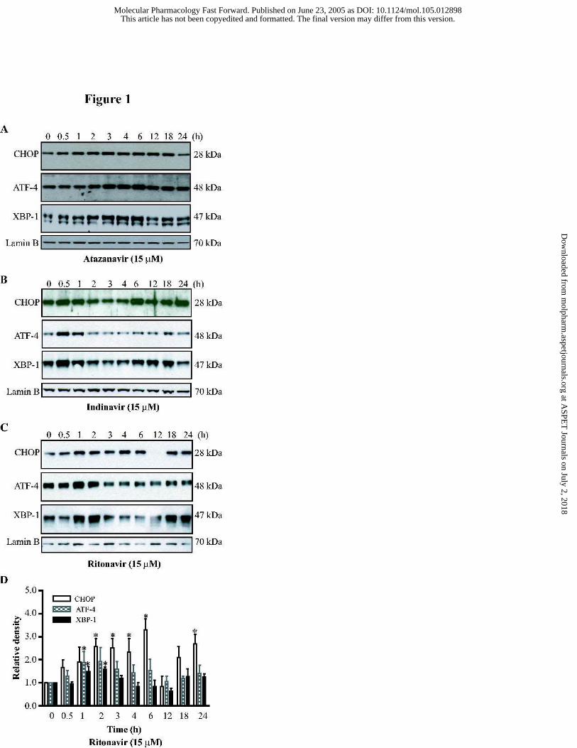
HIV PIs Activate the UPR and Induce Apoptosis in Macrophages. We initially examined the
effects of HIV PIs on activation of the UPR in cultured mouse J774A.1 macrophages. Induction
of the downstream transcription factors, X-box-binding protein 1 (XBP-1), activating
transcription factor 4 (ATF-4) and C/EBP-homologous protein (CHOP), are markers for
activation of the UPR. Treatment of mouse macrophages with therapeutically relevant
concentration (5 to 15 µM) of PIs significantly increased the expression of CHOP, ATF-4 and
XBP-1 (Fig.1). The expression levels of CHOP, ATF-4 and XBP-1 induced by atazanavir all
peaked at 6 h (Fig. 1A). The indinavir-induced activation of CHOP, ATF-4 and XBP-1 peaked
at 6 h, 0.5 h and 0.5h, respectively (Fig. 1B). The expression levels of CHOP, ATF-4 and XBP-
1 induced by ritonavir peaked at 6 h, 1 h and 2 h, respectively (Fig. 1C and 1D). As shown in
Fig. 2, these HIV PIs induced activation of these transcription factors was concentration-
dependent. At 15 µM, ritonavir increased the expression of CHOP, ATF-4 and XBP-1 by 66%,
53% and 170%, respectively, after 3 h treatment.
To further confirm the HIV PI-induced activation of the UPR was not a phenomenon of
cultured macrophages, we isolated primary mouse peritoneal macrophages. Treatment (3 h) of
these cells with HIV PIs also markedly activated the UPR. The expression of CHOP was
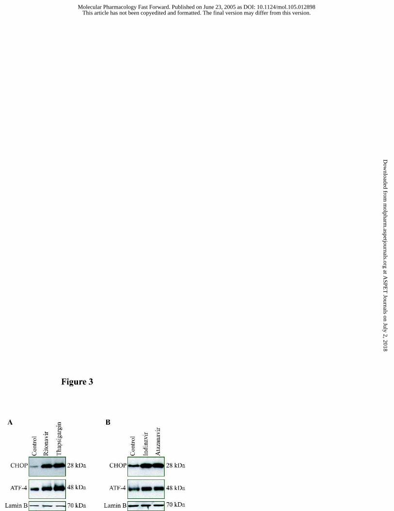
increased by 240%, 144% and 140% after treatment with ritonavir, indinavir and atazanavir (15
µM), respectively, (Fig. 3).
CHOP is one of the highest inducible gene during ER stress and is involved in ER stress-
induced apoptosis. When mouse J774A.1 macrophages were treated with ritonavir (5 to 50 µM)
This article has not been copyedited and formatted. The final version may differ from this version.Molecular Pharmacology Fast Forward. Published on June 23, 2005 as DOI: 10.1124/mol.105.012898
at ASPE
T Journals on July 2, 2018
molpharm
.aspetjournals.orgD
ownloaded from

MOL (12898)
13
for 24 h, morphological changes characteristic of apoptosis were observed (Fig. 4A). To
quantify the apoptotic or necrotic cells induced by ritonavir, mouse macrophages were treated
with drug for 24 h, then stained with Annexin V and propidium iodine, and analyzed by flow
cytometry. As shown in Fig. 4B, treatment with ritonavir resulted in a concentration-dependent
increase in apoptotic cells. The percentages of apoptotic cells were increased by 17% and 30%,
respectively, after treatment with 5 µM and 15 µM of ritonavir for 24 h. Indinavir and
atazanavir also induced apoptosis in macrophages (data not shown).
Caspases are cysteine proteases involved in programmed cell death. More than a dozen
different caspases have been identified to date. It has been demonstrated that caspase-12 is
predominantly associated with the ER, and is believed to mediate an ER specific apoptotic
pathway (Nakagawa et al., 2000). Treatment of mouse macrophages with ritonavir (5 to 25 µM)
significantly activated caspase-12. At the concentration of 15 µM, the caspase-12 activity was
increased by 1.9 fold (Fig. 4C).
Depletion of ER Calcium Stores by HIV PIs. The ER is a principal site of the synthesis for
protein, sterols, cholesterol, and other lipids. Maintenance of ER calcium homeostasis is
essential for many cellular functions. Perturbations of ER calcium homeostasis is expected to
induce the ER stress. Thapsigaragin (a sacroplasmic/ER calcium-ATPase inhibitor) depletes the
ER calcium stores and activates the UPR in many different cells (Yamaguchi and Wang, 2004).
To investigate the possible mechanisms of the HIV PI induced ER stress and activation of the
UPR, we assessed the effect of ritonavir on ER calcium stores in mouse J774A.1 macrophages
with the fluorescent calcium indicator fura-2/AM. After treatment with different concentrations
of ritonavir for 24 h, cells were loaded with fura-2/AM and switched to calcium-free medium.
This article has not been copyedited and formatted. The final version may differ from this version.Molecular Pharmacology Fast Forward. Published on June 23, 2005 as DOI: 10.1124/mol.105.012898
at ASPE
T Journals on July 2, 2018
molpharm
.aspetjournals.orgD
ownloaded from

MOL (12898)
14
Thapsigargin (100 nM) induced ER calcium release was recorded by fluorescence microscopy.
Treatment of mouse J774A.1 macrophage with ritonavir decreased the ER calcium content in a
dose dependent manner (Fig. 5). Cells treated with 25 µM of ritonavir remarkably reduced the
response to thapsigargin, indicating that ER calcium stores were depleted. Atazanavir and
indinavir had similar effects on ER calcium stores (data not shown).
Effects of HIV PIs on SREBPs in Macrophages. SREBPs play critical roles in lipid
homeostasis and directly activate the expression of dozens of genes involved in lipid metabolism.
SREBP-1 is primarily involved in regulating fatty acid and triglyceride biosynthesis; whereas,
SREBP-2 is involved in sterol biosynthesis and metabolism. It has been reported that HIV PIs
cause severe hyperlipidemia and lipodystrophy in some patients (Fontas et al., 2004; Hui, 2003;
The Data Collection on Adverse Events of Anti-HIV Drugs (DAD) Study Group, 2003). Our
previous studies showed that indinavir increased the levels of activated SREBP-1 and SREBP-2
in rat hepatocytes (Williams et al., 2004). To examine the effect of HIV PIs on the expression of
SREBPs in mouse J774A.1 macrophages, cells were treated with HIV PIs (ritonavir, indinavir
and atazanavir) at therapeutic concentration (15 µM) for various times. The levels of
transcriptionally active mature SREBP-1 and SREBP-2 were significantly increased as shown in
Fig. 6. Treatment of these cells with ritonavir (15 µM) for 6 h, increased the levels of mature
SREBP-1 and SREBP-2 by 273% and 82% (p<0.05), respectively. This HIV PI also increased
the level of the precursor form of SREBP-1 (data not shown).
Effects of Ritonavir on Free Cholesterol Accumulation and Esterification in Macrophages.
Maintenance of the cellular cholesterol homeostasis is crucial to various cellular functions. To
further define whether HIV PI-induced activation of SREBPs is associated with a corresponding
increase in intracellular free cholesterol, mouse macrophages were treated with different
This article has not been copyedited and formatted. The final version may differ from this version.Molecular Pharmacology Fast Forward. Published on June 23, 2005 as DOI: 10.1124/mol.105.012898
at ASPE
T Journals on July 2, 2018
molpharm
.aspetjournals.orgD
ownloaded from

MOL (12898)
15
concentrations of ritonavir (5 to 50 µM) for 24 h and the free cholesterol was detected with
filipin staining. As shown in Fig. 7A, ritonavir modestly induced the accumulation of free
cholesterol in intracellular membranes of mouse macrophages. Treatment with 15 to 25 µM
ritonavir increased intracellular free cholesterol content (Fig. 7B). However, treatment with
thapsigargin (100 nM) for 24 h did not cause the accumulation of free cholesterol in macrophage
(Fig. 7A and 7B). These results indicate that activation of UPR is not the direct cause of the
HIV PI-induced free cholesterol accumulation in mouse macrophages. Surprisingly, even
though intracellular cholesterol levels increased, there was a significant decrease in endogenous
cholesterol esterification (Fig.7C). In contrast, exogenous cholesterol esterification was not
affected (Fig. 7D). The amount of total esterified cholesterol was not affected by ritonavir (Fig.
7B).
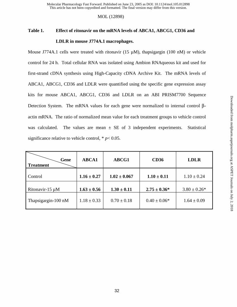
Effects of Ritonavir on the Expression of ABCA1, ABCG1, CD36 and LDLR in
Macrophages. To determine whether HIV PIs regulate the genes related to cholesterol uptake
and efflux, such as LDLR, CD36, ABCA1, and ABCG1, we utilized quantitative PCR to
measure the mRNA level of these receptors and lipid transporters in control and ritonavir treated
cells. Mouse J774A.1 cells were treated with ritonavir (15 µM), thapsigargin (100 nM), or
control vehicle for 24 h, and total RNA was isolated. The mRNA levels of ABCA1, ABCG1,
LDLR and CD 36 were quantified using specific gene expression assay kits. The results (Table
1) show that ritonavir had no effect on mRNA levels of ABCA1 and ABCG1, but significantly
increased CD36 and LDLR mRNA levels. However, thapsigargin, a positive control of ER
stress inducer, significantly decreased CD36 mRNA level and had little effects on ABCA1,
ABCG1 and LDLR mRNA levels.
This article has not been copyedited and formatted. The final version may differ from this version.Molecular Pharmacology Fast Forward. Published on June 23, 2005 as DOI: 10.1124/mol.105.012898
at ASPE
T Journals on July 2, 2018
molpharm
.aspetjournals.orgD
ownloaded from
MOL (12898)
16
Ritonavir Activates the UPR and Induce Apoptosis in Foam Cells. Lipid-laden
macrophages (foam cells) are found in all stages of atherosclerosis. Unstable human
atherosclerotic plaques constitute numerous foam cells. Apoptosis of foam cells are extremely
detrimental. In order to further examine whether HIV PIs also can induce ER stress, activate the
UPR and increase apoptosis in foam cells, mouse J774A.1 cells were loaded with Ac-LDL (50
µg/ml) or vehicle control for 24 h, then treated with ritonavir (15 µM), thapsigargin (100 nM) or
vehicle for 24 h. The intracellular lipids were stained with oil red O; the expression of CHOP,
ATF-4 and XBP-1 were detected by western blot; the apoptotic cells were detected by Annexin
V-FITC/propidium iodine staining with confocal fluorescence microscope. As shown in Fig.
8A&B, ritonavir not only increased the lipid accumulation in Ac-LDL loaded cells, but also in
normal macrophages, suggesting that ritonavir may promote the foam cell formation. In Ac-
LDL induced foam cells, ritonavir modestly increased the accumulation of the intracellular free
cholesterol, but significantly increased cholesterol ester accumulation (Fig. 8C). Treatment of
foam cells with ritonavir (0 to 50 µM) induced dose-dependent apoptosis. As shown in Fig. 9,
Ac-LDL induced foam cells were more sensitive to ritonavir compare to normal cholesterol
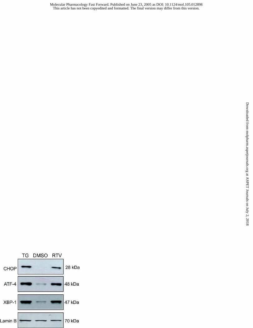
loaded macrophages. Similarly, ritonavir activated the UPR in foam cells (Fig. 10).
This article has not been copyedited and formatted. The final version may differ from this version.Molecular Pharmacology Fast Forward. Published on June 23, 2005 as DOI: 10.1124/mol.105.012898
at ASPE
T Journals on July 2, 2018
molpharm
.aspetjournals.orgD
ownloaded from

MOL (12898)
17
Discussion
Clinical studies report that HIV PIs increase serum lipids (Hui, 2003), promote
atherosclerosis (Sklar and Masur, 2003) and increase the risk of myocardial infarction (The Data
Collection on Adverse Events of Anti-HIV Drugs (DAD) Study Group, 2003) in patients with
HIV infection. Studies using rodent macrophages (Dressman et al., 2003) and hepatocytes
(Riddle et al., 2001;Liang et al., 2001) have shown that HIV PIs dysregulate cellular lipid
metabolism. However, the exact mechanism(s) altering lipid metabolism is unclear and maybe
multi-factorial. Our current studies show that HIV PIs (ritonavir, indinavir and atazanavir)
activate the UPR induce apoptosis both in normal cholesterol loaded macrophages and Ac-LDL
loaded macrophages. Surprisingly, HIV PIs markedly increased the mature forms of nuclear
SREBP-1 and SREBP-2 (Fig. 6) while increasing cholesterol in intracellular membranes (Fig. 7).
HIV-PIs induced disruption of lipid homeostasis and enhanced programmed cell death occurred
in macrophages at concentrations (5-15 µM) that are within serum levels reported for HIV
infected patients taking these medications (Flexner, 1998).
In mammalian cells, the ER is involved in the proper folding and posttranslational
modification of proteins. The ER also maintains very low membrane cholesterol content (Feng
et al., 2003) and is very sensitive to alterations in cellular homeostatic mechanisms. A number
of cellular stress conditions can alter the normal rate of folding and processing of newly
synthesized proteins (Pahl, 1999). The ER has evolved cell signaling pathways to respond to the
accumulation of unfolded or misfolded proteins, collectively referred to as the UPR [reviewed in
(Zhang and Kaufman, 2004)]. Activation of the UPR allows mammalian cells to increase protein
This article has not been copyedited and formatted. The final version may differ from this version.Molecular Pharmacology Fast Forward. Published on June 23, 2005 as DOI: 10.1124/mol.105.012898
at ASPE
T Journals on July 2, 2018
molpharm
.aspetjournals.orgD
ownloaded from
MOL (12898)
18
folding and degradation pathways, and differentially inhibits most protein synthesis. However,
prolonged activation of the UPR can lead to apoptosis.
Macrophages are the major cell type found in atherosclerotic lesions, and inappropriate
and/or excessive apoptosis of these cells is believed to play key roles both in the initiation and in
the progression of atherosclerosis. Compounds that activate the UPR in vivo appear to accelerate
cardiovascular disease. For example, patients with hyperhomocysteinemia accumulate high
serum levels of homocysteine due to genetic defects in genes involved in homocysteine
metabolism (Outinen et al., 1999). Hyperhomocysteinemia is an independent risk factor for
atherosclerosis and cardiovascular disease. Homocysteine has previuosly been shown to be an
activator of the UPR in hepatocytes and vascular endothelial cells (Werstuck et al., 2001).
Activation of the UPR by homocysteine is associated with dysregulation of lipid metabolism,
increased apoptosis, and accelerated atherosclerosis. We hypothesize that HIV PIs accelerate
atherosclerosis and cardiovascular disease as a result of their ability to disrupt normal lipid
metabolism and to activate the UPR.
It is currently unclear how HIV protease inhibitors activate the UPR as a number of
cellular stress signals can activate this system such as depletion of ER calcium stores, increased
cholesterol in ER membranes, deprivation of glucose (Oyadomari and Mori, 2004) or inhibition
of proteasome activity (Parker et al., 2005). Recent studies have shown that overloading of free
cholesterol in the ER causes depletion of ER calcium stores (by inhibiting the sarcoplasmic-
endoplasmic reticulum calcium ATPase-2b)(Li et al., 2004), activates the UPR, and induces
apoptosis in macrophages (Feng et al., 2003). In the present study, we discovered that HIV PIs
This article has not been copyedited and formatted. The final version may differ from this version.Molecular Pharmacology Fast Forward. Published on June 23, 2005 as DOI: 10.1124/mol.105.012898
at ASPE
T Journals on July 2, 2018
molpharm
.aspetjournals.orgD
ownloaded from
MOL (12898)
19
induced accumulation of free cholesterol and lipid in intracellular membranes of mouse
macrophages and depleted ER calcium stores (as shown in Fig. 5, Fig. 7A&B and Fig. 8).
However, there was no direct effect of HIV PIs on ER calcium release (data not shown).
Therefore, one possible mechanism by which HIV PIs activate the UPR in macrophages is the
accumulation of intracellular free cholesterol followed by the depletion of ER calcium stores.
Macrophages have acquired multiple mechanisms to prevent the accumulation of excess
free cholesterol, including an increase in cholesterol esterification, induction of free cholesterol
efflux, downregulation of cholesterol biosynthesis (Zhang and Kaufman, 2003). HIV PIs may
promote foam cell formation and atherosclerotic by modulation of CD36 (Dressman et al., 2003)
and the LDLR (Tran et al., 2003), and by increasing cholesterol biosynthesis. In the present
study, we demonstrated that ritonavir significantly up-regulates CD36 and LDLR mRNA
expression, but has no effect on ABCA1 and ABCG1 mRNA levels. It has been demonstrated
that over loading of free cholesterol in macrophages decreases ABCA1-mediated cholesterol
efflux by accelerating the degradation of ABCA1 protein (Feng and Tabas, 2002). Thus, HIV
PIs induced accumulation of intracellular free cholesterol and lipid may indirectly impair the
normal function of ABCA1 protein. It has also been shown that disruption of intracellular
cholesterol transport may adversely affect cholesterol efflux and cholesterol esterification
(Tabas, 2002).
It has been reported that depletion of glucose causes the accumulation of unfolded
proteins, induces ER stress and activates the UPR (Pahl, 1999). It has been demonstrated that
indinavir selectively and reversibly inhibits the glucose transporter isoform GluT4 at therapeutic
concentrations (Koster et al., 2003; Murata et al., 2002) and impairs SREBP-1 intranuclear
This article has not been copyedited and formatted. The final version may differ from this version.Molecular Pharmacology Fast Forward. Published on June 23, 2005 as DOI: 10.1124/mol.105.012898
at ASPE
T Journals on July 2, 2018
molpharm
.aspetjournals.orgD
ownloaded from

MOL (12898)
20
localization (Caron et al., 2001). These observations suggest another possible mechanism by
which HIV PIs might induce ER stress and activate the UPR. However, whether HIV PIs disrupt
glucose homeostasis in macrophages needs further investigation.
Most recent studies done by Parker et al suggest that inhibition of proteasome activity
and differential glucose transport by HIV PIs are proximal events eliciting the UPR which can
regulate lipogenic pathways in hepatocytes or adipocytes (Parker et al., 2005). Several studies
have shown that ritonavir is a reversible inhibitor of proteasome and inhibits the chymotrypsin-
like activity of the proteasome while enhancing the trypsin-like activity at high concentration
(50-100 µM) (Lagathu et al., 2004; Laurent et al., 2004). However, a block in proteasomal
housekeeping functions in intact cells by ritonavir requires 50 to 100 µM of the drug (Andre et
al., 1998). In the present studies, the UPR is activated by ritonavir at concentrations (5 to 15
µM) where essential functions of the proteasome are not yet blocked. In vitro studies have also
shown that different HIV PIs differ in their effects on proteasome activity. Indinavir and
nelfinavir have no effect on proteasome activity even at high concentration (100 µM) (Andre et
al., 1998). However, in our studies we observed that indinavir induced the UPR activation and
disrupted lipid metabolism at clinically relevant concentration (15 µM) in macrophages.
Nelfinavir is also able to induce the UPR activation at even lower concentration (1 to 10 µM)
(unpublished data). Therefore, the HIV PIs induced activation of the UPR may not be due to the
dysfunction of the proteasome in macrophages.
In summary, the current study provides novel insights into how HIV PIs induce ER
stress, activating the UPR, and inducing apoptosis in macrophages (Fig. 11). The effects of HIV
This article has not been copyedited and formatted. The final version may differ from this version.Molecular Pharmacology Fast Forward. Published on June 23, 2005 as DOI: 10.1124/mol.105.012898
at ASPE
T Journals on July 2, 2018
molpharm
.aspetjournals.orgD
ownloaded from
MOL (12898)
21
PIs on accumulation of intracellular free cholesterol and lipids, and depletion of ER calcium
stores represent a potential mechanism by which HIV PIs induce atherosclerosis and
cardiovascular disease in HIV patients undergoing HARRT. Different HIV PIs have different
effects in different cell types. We cannot rule out the involvement of other target proteins in the
HIV PI-induced activation of the UPR and apoptosis in macrophages. A better understanding of
the cellular and molecular mechanisms of HIV PIs induced metabolic abnormality may provide
useful information for the development of new drugs and therapeutic strategies.
Acknowledgements. We would like to thank the following companies for providing us with
the following compounds used in this research: Abbott Laboratories (Ritonavir); Merck & Co.,
Inc. (Indinavir); and Bristol-Meyers-Squibb (Atazanavir).
This article has not been copyedited and formatted. The final version may differ from this version.Molecular Pharmacology Fast Forward. Published on June 23, 2005 as DOI: 10.1124/mol.105.012898
at ASPE
T Journals on July 2, 2018
molpharm
.aspetjournals.orgD
ownloaded from
MOL (12898)
22
References
Andre P, Groettrup M, Klenerman P, de G R, Booth B L, Jr., Cerundolo V, Bonneville M, Jotereau F, Zinkernagel R M and Lotteau V (1998) An Inhibitor of HIV-1 Protease Modulates Proteasome Activity, Antigen Presentation, and T Cell Responses. Proc Natl Acad Sci U S A95:13120-13124.
Beregszaszi M, Jaquet D, Levine M, Ortega-Rodriguez E, Baltakse V, Polak M and Levy-Marchal C (2003) Severe Insulin Resistance Contrasting With Mild Anthropometric Changes in the Adipose Tissue of HIV-Infected Children With Lipohypertrophy. Int J Obes Relat Metab Disord 27:25-30.
Bongiovanni M, Bini T, Chiesa E, Cicconi P, Adorni F and Monforte d A (2004) Lopinavir/Ritonavir Vs. Indinavir/Ritonavir in Antiretroviral Naive HIV-Infected Patients: Immunovirological Outcome and Side Effects. Antiviral Res 62:53-56.
Caron M, Auclair M, Vigouroux C, Glorian M, Forest C and Capeau J (2001) The HIV Protease Inhibitor Indinavir Impairs Sterol Regulatory Element-Binding Protein-1 Intranuclear Localization, Inhibits Preadipocyte Differentiation, and Induces Insulin Resistance. Diabetes50:1378-1388.
Carr A, Samaras K, Burton S, Law M, Freund J, Chisholm D J and Cooper D A (1998) A Syndrome of Peripheral Lipodystrophy, Hyperlipidaemia and Insulin Resistance in Patients Receiving HIV Protease Inhibitors. AIDS 12:F51-F58.
Dressman J, Kincer J, Matveev S V, Guo L, Greenberg R N, Guerin T, Meade D, Li X A, Zhu W, Uittenbogaard A, Wilson M E and Smart E J (2003) HIV Protease Inhibitors Promote Atherosclerotic Lesion Formation Independent of Dyslipidemia by Increasing CD36-Dependent Cholesteryl Ester Accumulation in Macrophages. J Clin Invest 111:389-397.
Feng B and Tabas I (2002) ABCA1-Mediated Cholesterol Efflux Is Defective in Free Cholesterol-Loaded Macrophages. Mechanism Involves Enhanced ABCA1 Degradation in a Process Requiring Full NPC1 Activity. J Biol Chem 277:43271-43280.
Feng B, Yao P M, Li Y, Devlin C M, Zhang D, Harding H P, Sweeney M, Rong J X, Kuriakose G, Fisher E A, Marks A R, Ron D and Tabas I (2003) The Endoplasmic Reticulum Is the Site of Cholesterol-Induced Cytotoxicity in Macrophages. Nat Cell Biol 5:781-792.
Flexner C (1998) HIV-Protease Inhibitors. N Engl J Med 338:1281-1292.
Fontas E, van L F, Sabin C A, Friis-Moller N, Rickenbach M, d'Arminio M A, Kirk O, Dupon M, Morfeldt L, Mateu S, Petoumenos K, El-Sadr W, de W S, Lundgren J D, Pradier C and Reiss P (2004) Lipid Profiles in HIV-Infected Patients Receiving Combination Antiretroviral Therapy: Are Different Antiretroviral Drugs Associated With Different Lipid Profiles? J Infect Dis 189:1056-1074.
This article has not been copyedited and formatted. The final version may differ from this version.Molecular Pharmacology Fast Forward. Published on June 23, 2005 as DOI: 10.1124/mol.105.012898
at ASPE
T Journals on July 2, 2018
molpharm
.aspetjournals.orgD
ownloaded from
MOL (12898)
23
Holmberg SD, Moorman A C, Greenberg A E, Friis-Moller N, Sabin C, Lundgren J D and the DAD Steering Committee (2004) Trends in Rates of Myocardial Infarction Among Patients With HIV. N Engl J Med 350:730-732.
Horton JD, Goldstein J L and Brown M S (2002) SREBPs: Activators of the Complete Program of Cholesterol and Fatty Acid Synthesis in the Liver. J Clin Invest 109:1125-1131.
Hui DY (2003) Effects of HIV Protease Inhibitor Therapy on Lipid Metabolism. Prog Lipid Res 42:81-92.
Koster JC, Remedi M S, Qiu H, Nichols C G and Hruz P W (2003) HIV Protease Inhibitors Acutely Impair Glucose-Stimulated Insulin Release. Diabetes 52:1695-1700.
Lagathu C, Bastard J P, Auclair M, Maachi M, Kornprobst M, Capeau J and Caron M (2004) Antiretroviral Drugs With Adverse Effects on Adipocyte Lipid Metabolism and Survival Alter the Expression and Secretion of Proinflammatory Cytokines and Adiponectin in Vitro. Antivir Ther 9:911-920.
Laurent N, de B S, Guillamo J S, Christov C, Zini R, Jouault H, Andre P, Lotteau V and Peschanski M (2004) Effects of the Proteasome Inhibitor Ritonavir on Glioma Growth in Vitro and in Vivo. Mol Cancer Ther 3:129-136.
Li Y, Ge M, Ciani L, Kuriakose G, Westover E J, Dura M, Covey D F, Freed J H, Maxfield F R, Lytton J and Tabas I (2004) Enrichment of Endoplasmic Reticulum With Cholesterol Inhibits Sarcoplasmic-Endoplasmic Reticulum Calcium ATPase-2b Activity in Parallel With Increased Order of Membrane Lipids: Implications for Depletion of Endoplasmic Reticulum Calcium Stores and Apoptosis in Cholesterol-Loaded Macrophages. J Biol Chem 279:37030-37039.
Liang JS, Distler O, Cooper D A, Jamil H, Deckelbaum R J, Ginsberg H N and Sturley S L (2001) HIV Protease Inhibitors Protect Apolipoprotein B From Degradation by the Proteasome: a Potential Mechanism for Protease Inhibitor-Induced Hyperlipidemia. Nat Med7:1327-1331.
Murata H, Hruz P W and Mueckler M (2002) Indinavir Inhibits the Glucose Transporter Isoform Glut4 at Physiologic Concentrations. AIDS 16:859-863.
Nakagawa T, Zhu H, Morishima N, Li E, Xu J, Yankner B A and Yuan J (2000) Caspase-12 Mediates Endoplasmic-Reticulum-Specific Apoptosis and Cytotoxicity by Amyloid-Beta. Nature 403:98-103.
Outinen PA, Sood S K, Pfeifer S I, Pamidi S, Podor T J, Li J, Weitz J I and Austin R C (1999) Homocysteine-Induced Endoplasmic Reticulum Stress and Growth Arrest Leads to Specific Changes in Gene Expression in Human Vascular Endothelial Cells. Blood 94:959-967.
Oyadomari S and Mori M (2004) Roles of CHOP/GADD153 in Endoplasmic Reticulum Stress. Cell Death Differ 11:381-389.
This article has not been copyedited and formatted. The final version may differ from this version.Molecular Pharmacology Fast Forward. Published on June 23, 2005 as DOI: 10.1124/mol.105.012898
at ASPE
T Journals on July 2, 2018
molpharm
.aspetjournals.orgD
ownloaded from
MOL (12898)
24
Pahl HL (1999) Signal Transduction From the Endoplasmic Reticulum to the Cell Nucleus. Physiol Rev 79:683-701.
Parker RA, Flint O P, Mulvey R, Elosua C, Wang F, Fenderson W, Wang S, Yang W P and Noor M A (2005) Endoplasmic Reticulum Stress Links Dyslipidemia to Inhibition of Proteasome Activity and Glucose Transport by HIV Protease Inhibitors. Mol Pharmacol.
Riddle TM, Kuhel D G, Woollett L A, Fichtenbaum C J and Hui D Y (2001) HIV Protease Inhibitor Induces Fatty Acid and Sterol Biosynthesis in Liver and Adipose Tissues Due to the Accumulation of Activated Sterol Regulatory Element-Binding Proteins in the Nucleus. J Biol Chem 276:37514-37519.
Rutkowski DT and Kaufman R J (2004) A Trip to the ER: Coping With Stress. Trends Cell Biol 14:20-28.
Sklar P and Masur H (2003) HIV Infection and Cardiovascular Disease -- Is There Really a Link? N Engl J Med 349:2065-2067.
Stanley ER, Cifone M, Heard P M and Defendi V (1976) Factors Regulating Macrophage Production and Growth: Identity of Colony-Stimulating Factor and Macrophage Growth Factor. J Exp Med 143:631-647.
Tabas I (2004) Apoptosis and Plaque Destabilization in Atherosclerosis: the Role of Macrophage Apoptosis Induced by Cholesterol. Cell Death Differ 11 Suppl 1:S12-S16.
Tabas I (2002) Consequences of Cellular Cholesterol Accumulation: Basic Concepts and Physiological Implications. J Clin Invest 110:905-911.
The Data Collection on Adverse Events of Anti-HIV Drugs (DAD) Study Group (2003) Combination Antiretroviral Therapy and the Risk of Myocardial Infarction. N Engl J Med349:1993-2003.
Tran H, Robinson S, Mikhailenko I and Strickland D K (2003) Modulation of the LDL Receptor and LRP Levels by HIV Protease Inhibitors. J Lipid Res 44:1859-1869.
Werstuck GH, Lentz S R, Dayal S, Hossain G S, Sood S K, Shi Y Y, Zhou J, Maeda N, Krisans S K, Malinow M R and Austin R C (2001) Homocysteine-Induced Endoplasmic Reticulum Stress Causes Dysregulation of the Cholesterol and Triglyceride Biosynthetic Pathways. J Clin Invest 107:1263-1273.
Williams K, Rao Y P, Natarajan R, Pandak W M and Hylemon P B (2004) Indinavir Alters Sterol and Fatty Acid Homeostatic Mechanisms in Primary Rat Hepatocytes by Increasing Levels of Activated Sterol Regulatory Element-Binding Proteins and Decreasing Cholesterol 7alpha-Hydroxylase MRNA Levels. Biochem Pharmacol 67:255-267.
Yamaguchi H and Wang H G (2004) CHOP Is Involved in Endoplasmic Reticulum Stress-Induced Apoptosis by Enhancing DR5 Expression in Human Carcinoma Cells. J Biol Chem279:45495-45502.
This article has not been copyedited and formatted. The final version may differ from this version.Molecular Pharmacology Fast Forward. Published on June 23, 2005 as DOI: 10.1124/mol.105.012898
at ASPE
T Journals on July 2, 2018
molpharm
.aspetjournals.orgD
ownloaded from

MOL (12898)
25
Zhang K and Kaufman R J (2004) Signaling the Unfolded Protein Response From the Endoplasmic Reticulum. J Biol Chem 279:25935-25938.
Zhang K and Kaufman R J (2003) Unfolding the Toxicity of Cholesterol. Nat Cell Biol 5:769-770.
This article has not been copyedited and formatted. The final version may differ from this version.Molecular Pharmacology Fast Forward. Published on June 23, 2005 as DOI: 10.1124/mol.105.012898
at ASPE
T Journals on July 2, 2018
molpharm
.aspetjournals.orgD
ownloaded from
MOL (12898)
26
Footnotes
This work is supported by grants from the National Institutes of Health (R01 AI057189 and
P01 DK38030).
This article has not been copyedited and formatted. The final version may differ from this version.Molecular Pharmacology Fast Forward. Published on June 23, 2005 as DOI: 10.1124/mol.105.012898
at ASPE
T Journals on July 2, 2018
molpharm
.aspetjournals.orgD
ownloaded from
MOL (12898)
27
Figure Legends
Figure 1. Activation of the UPR by HIV PIs. Representative immunoblots against
CHOP, ATF-4, XBP-1 and lamin B from the nuclear extracts of mouse J774A.1 macrophages
treated with HIV PIs (15 µM) for 0, 0.5, 1, 2, 3, 4, 6, 12, 18 and 24 h. Lamin B was used as a
loading control. (A) Atazanavir; (B) Indinavir; (C) Ritonavir; (D) Relative density of the
immunoblots against CHOP, ATF-4 and XBP-1 activated by ritonavir. The density of the
immunoreactive bands was analyzed using Image J software and normalized to lamin B
control. Values are mean ± SE of 3 independent experiments. Statistical significance relative
to vehicle control, * p < 0.05.
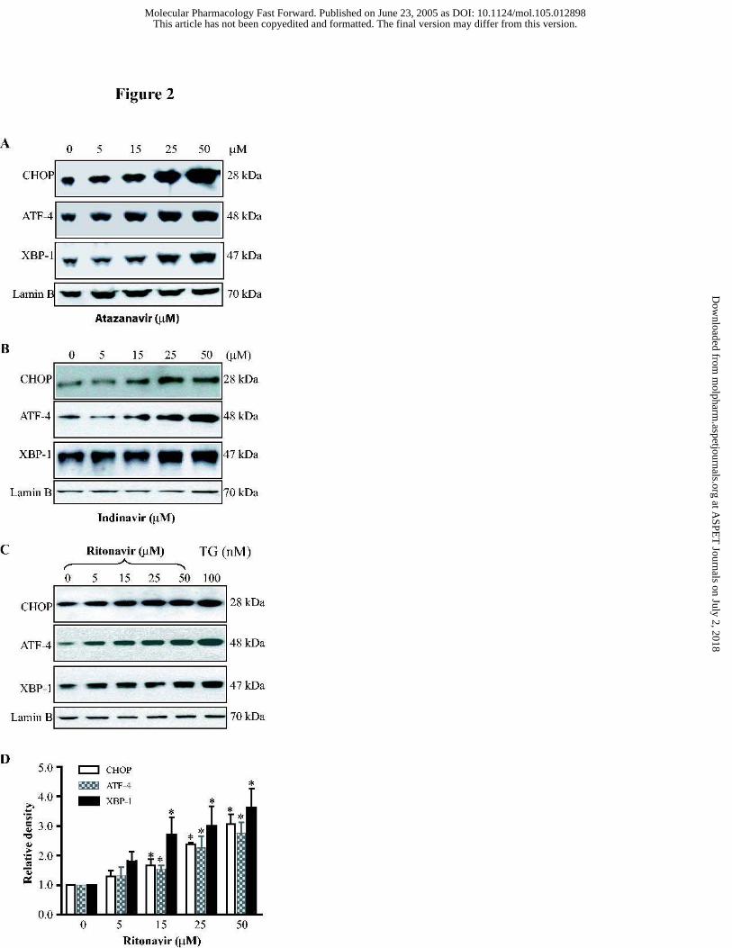
Figure 2. Concentration dependent activation of the UPR by HIV PIs in
macrophages. Representative immunoblots against CHOP, ATF-4, XBP-1 and lamin B from
the nuclear extracts of mouse J774A.1 macrophages treated with the different concentrations of
(A) Atazanavir; (B) Indinavir; (C) Ritonavir (0 to 50 µM) or thapsigargin (100 nM) for 3 h.
Thapsigargin, a known inducer of CHOP, ATF-4, and the UPR, is shown for control purposes.
Lamin B was used as a loading control. (D) Relative density of the immunoblots against
CHOP, ATF-4 and XBP-1 activated by ritonavir. The density of the immunoreactive bands
was analyzed using Image J software and normalized to lamin B control. Values are mean ±
SE of 3 independent experiments. Statistical significance relative to vehicle control, * p< 0.05.
Figure 3. HIV PIs activate the UPR in primary mouse peritoneal macrophages. (A)
Ritonavir induced activation of the UPR. Representative immunoblots against CHOP, ATF-4
This article has not been copyedited and formatted. The final version may differ from this version.Molecular Pharmacology Fast Forward. Published on June 23, 2005 as DOI: 10.1124/mol.105.012898
at ASPE
T Journals on July 2, 2018
molpharm
.aspetjournals.orgD
ownloaded from
MOL (12898)
28
and lamin B from the nuclear extracts of mouse peritoneal macrophages treated with ritonavir
(15 µM) or thapsigargin (100 nM) for 3 h. Thapsigargin, a known inducer of CHOP, ATF-4,
and the UPR, is shown for control purposes. Lamin B was used as a loading control. (B)
Indinavir and atazanavir induced activation of the UPR. Representative immunoblots against
CHOP, ATF-4 and lamin B from the nuclear extracts of mouse peritoneal macrophages treated
with indinavir (15 µM) or atazanavir (15 µM) for 3 h. Lamin B was used as a loading control.
Figure 4. Ritonavir induces apoptosis and activates caspase-12 in mouse J774A.1
macrophages. Cells were treated with vehicle control or various concentrations of ritonavir (5
to 50 µM) for 24 h, then stained with Annexin V-FITC/propidium iodine. (A) Images of
Annexin V/propidium iodine-stained cells were visualized under confocal fluorescence
microscope with a dual filter set for FITC and rhodamine. (B) The percentages of apoptotic
cells were analyzed by flow cytometry. The results are the mean ± SE for three independent
experiments. Statistical significance relative to vehicle control, * p< 0.05. (C) Activity of
caspase-12 in whole cell lysates of mouse J774A.1 macrophages treated with ritonavir (0 to 25
µM) or thapsigargin (100 nM) for 24 h was measured using Caspase-12 Fluorometric Assay
Kit according to the manufacture instruction. Statistical significance relative to vehicle
control, * p< 0.05.
Figure 5. Depletion endoplasmic reticulum calcium stores by ritonavir. (A)
Assessment of endoplasmic reticulum calcium stores in mouse J774A.1 macrophages treated
for 24 h with different concentrations of ritonavir (0, 5, 15 and 25 µM). Representative
tracings of the Fura-2 fluorescence ratio of 340:380 nm in an individual macrophage for each
This article has not been copyedited and formatted. The final version may differ from this version.Molecular Pharmacology Fast Forward. Published on June 23, 2005 as DOI: 10.1124/mol.105.012898
at ASPE
T Journals on July 2, 2018
molpharm
.aspetjournals.orgD
ownloaded from

MOL (12898)
29
treatment group before and after addition of 100 nM thapsigargin are shown. (B) Relative
calcium content was calculated by total area under the curve for each treatment group and
expressed as percent of the vehicle control. Statistical significance relative to vehicle control,
* p< 0.05.
Figure 6. Time course of the HIV PIs induced activation of SREBP-1 and SREBP-2.
Representative immunoblots against SREBP-1, SREBP-2 and lamin B from the nuclear
extracts of mouse J774A.1 macrophages treated with HIV PIs (15 µM) for 0, 0.5, 1, 2, 3, 4, 6,
12, 18 and 24 h. Lamin B was used as a loading control. (A) Atazanavir; (B) Indinavir; (C)
Ritonavir; (D) Relative density of the immunoblots against SREBP-1 and SREBP-2 activated
by ritonavir. The density of the immunoreactive bands was analyzed using Image J software
and normalized to lamin B control. Values are mean ± SE of 3 independent experiments.
Statistical significance relative to vehicle control, * p< 0.05.
Figure 7. Effects of ritonavir on the accumulation of free cholesterol in mouse
J774A.1 macrophages and cholesterol esterification. (A) Cells were treated with various
concentrations of ritonavir (0 to 50 µM) or thapsigargin (100 nM) for 24 h and stained with
Filipin for free cholesterol as described under Methods. Fluroscence images and merged with
phase-contrast images are shown. (B) Cells were treated with various concentrations of
ritonavir (0 to 25 µM) or thapsigargin (100 nM) for 24 h. The intracellular total and free
cholesterol contents were measured using Wako Cholesterol E and Free Cholesterol assay kits.
Values are mean ± SE of 3 independent experiments. Statistical significance relative to vehicle
control, * p< 0.05. (C) Endogenous cholesterol esterification. Cells were treated with ritonavir
This article has not been copyedited and formatted. The final version may differ from this version.Molecular Pharmacology Fast Forward. Published on June 23, 2005 as DOI: 10.1124/mol.105.012898
at ASPE
T Journals on July 2, 2018
molpharm
.aspetjournals.orgD
ownloaded from

MOL (12898)
30
(15 and 25 µM) for 24 h in LDL deficient medium and labeled with [3H] oleate. The
[3H]cholesteryl oleate was analyzed and quantified as described under Methods. Data is
expressed as a percentage of control vehicle and values are mean ± SE of 3 independent
experiments. Statistical significance relative to vehicle control, * p< 0.05. (D) Exogenous
cholesterol esterification. Cells were treated with ritonavir (15 and 25 µM) for 24 h in LDL
containing medium and labeled with [3H] oleate. The [3H]cholesteryl oleate was analyzed and
quantified as described under Methods. Data is expressed as a percentage of control vehicle
and values are mean ± SE of 3 independent experiments. Statistical significance relative to
vehicle control, * p< 0.05.
Figure 8. Effects of Ritonavir on intracellular cholesterol and lipid content in
macrophages . Normal mouse J774A.1 macrophages (A) or Ac-LDL loaded (50 µg/ml for 24
h) macrophages (B) were treated with (a) vehicle control, (b) ritonavir (15 µM) or (c)
thapsigargin (100 nM) for 24 h. The intracellular lipids were stained with 0.2% oil red O as
described under Methods. The images were taken by Olympus microscope equipped with
image recorder. (C) Ac-LDL loaded macrophages were treated with vehicle control (DMSO),
ritonavir (RTV, 15 µM) or thapsigargin (TG, 100 nM) for 24 h. The intracellular cellular total
and free cholesterol contents were measured using Wako Cholesterol E and Free Cholesterol
assay kits. Values are mean ± SE of 3 independent experiments. Statistical significance
relative to vehicle control, * p< 0.05.
Figure 9. Ritonvair induces apoptosis of Ac-LDL loaded cells (foam cells). Mouse
macrophages were loaded with Ac-LDL (50 µg/ml) for 24 h, then treated with different
This article has not been copyedited and formatted. The final version may differ from this version.Molecular Pharmacology Fast Forward. Published on June 23, 2005 as DOI: 10.1124/mol.105.012898
at ASPE
T Journals on July 2, 2018
molpharm
.aspetjournals.orgD
ownloaded from
MOL (12898)
31
concentrations of ritonavir (RTV, 0 to 50 µM) or thapsigargin (TG, 100 nM) for 24 h.
Apoptotic cells were stained with Annexin V-FITC/propidium iodine and visualized with a
dual filter set for FITC and rhodamine. (a): propidium iodine staining; (b): phase contrast
image; (c): annexin V-FITC staining; (d): FITC and propidium iodine merged images.
Fig. 10. Activation of the UPR by ritonavir in Ac-LDL loaded macrophages.
Representative immunoblots against CHOP, ATF-4, XBP-1 and lamin B from the nuclear
extracts of mouse macrophages loaded with Ac-LDL (50 µg/ml) and treated with ritonavir
(RTV, 15 µM) for 24 h. Lamin B was used as a loading control. Thapsigagin (TG, 100 nM)
was used as a positive control. DMSO was the vehicle control.
Figure 11. Proposed model of HIV PIs induced UPR signaling pathways in
macrophages. HIV PIs induce the ER stress and activate the UPR in macrophages.
Activation of ATF-4, XBP-1 and ATF-6 results in transcriptional induction of the CHOP gene.
HIV PIs also increase cytosolic calcium and activate caspase-12. Both CHOP and caspase-12
mediate ER stress-induced apoptosis. UPR: unfolded protein response; ER: endoplasmic
reticulum; IRE1: inositol-requiring enzyme, a transmembrane protein kinase/
endoribonuclease; PERK: doubled-stranded RNA-activated protein kinase-like ER kinase;
ATF-6: activating transcription factor 6; ATF-4: activating transcription factor 4; XBP-1: X-
box-binding protein; CHOP: C/EBP-homologous protein, also called GADD153 (growth arrest
and DNA damage-inducing gene); ERSE: ER stress response element.
This article has not been copyedited and formatted. The final version may differ from this version.Molecular Pharmacology Fast Forward. Published on June 23, 2005 as DOI: 10.1124/mol.105.012898
at ASPE
T Journals on July 2, 2018
molpharm
.aspetjournals.orgD
ownloaded from
MOL (12898)
32
Table 1. Effect of ritonavir on the mRNA levels of ABCA1, ABCG1, CD36 and
LDLR in mouse J774A.1 macrophages.
Mouse J774A.1 cells were treated with ritonavir (15 µM), thapsigargin (100 nM) or vehicle
control for 24 h. Total cellular RNA was isolated using Ambion RNAqueous kit and used for
first-strand cDNA synthesis using High-Capacity cDNA Archive Kit. The mRNA levels of
ABCA1, ABCG1, CD36 and LDLR were quantified using the specific gene expression assay
kits for mouse ABCA1, ABCG1, CD36 and LDLR on an ABI PRISM7700 Sequence
Detection System. The mRNA values for each gene were normalized to internal control β-
actin mRNA. The ratio of normalized mean value for each treatment groups to vehicle control
was calculated. The values are mean ± SE of 3 independent experiments. Statistical
significance relative to vehicle control, * p< 0.05.
Gene Treatment
ABCA1 ABCG1 CD36 LDLR
Control 1.16 ± 0.27 1.02 ± 0.067 1.10 ± 0.11 1.10 ± 0.24
Ritonavir-15 µM 1.63 ± 0.56 1.30 ± 0.11 2.75 ± 0.36* 3.80 ± 0.26*
Thapsigargin-100 nM 1.18 ± 0.33 0.70 ± 0.18 0.40 ± 0.06* 1.64 ± 0.09
This article has not been copyedited and formatted. The final version may differ from this version.Molecular Pharmacology Fast Forward. Published on June 23, 2005 as DOI: 10.1124/mol.105.012898
at ASPE
T Journals on July 2, 2018
molpharm
.aspetjournals.orgD
ownloaded from
This article has not been copyedited and formatted. The final version may differ from this version.Molecular Pharmacology Fast Forward. Published on June 23, 2005 as DOI: 10.1124/mol.105.012898
at ASPE
T Journals on July 2, 2018
molpharm
.aspetjournals.orgD
ownloaded from
This article has not been copyedited and formatted. The final version may differ from this version.Molecular Pharmacology Fast Forward. Published on June 23, 2005 as DOI: 10.1124/mol.105.012898
at ASPE
T Journals on July 2, 2018
molpharm
.aspetjournals.orgD
ownloaded from
This article has not been copyedited and formatted. The final version may differ from this version.Molecular Pharmacology Fast Forward. Published on June 23, 2005 as DOI: 10.1124/mol.105.012898
at ASPE
T Journals on July 2, 2018
molpharm
.aspetjournals.orgD
ownloaded from

This article has not been copyedited and formatted. The final version may differ from this version.Molecular Pharmacology Fast Forward. Published on June 23, 2005 as DOI: 10.1124/mol.105.012898
at ASPE
T Journals on July 2, 2018
molpharm
.aspetjournals.orgD
ownloaded from

This article has not been copyedited and formatted. The final version may differ from this version.Molecular Pharmacology Fast Forward. Published on June 23, 2005 as DOI: 10.1124/mol.105.012898
at ASPE
T Journals on July 2, 2018
molpharm
.aspetjournals.orgD
ownloaded from

This article has not been copyedited and formatted. The final version may differ from this version.Molecular Pharmacology Fast Forward. Published on June 23, 2005 as DOI: 10.1124/mol.105.012898
at ASPE
T Journals on July 2, 2018
molpharm
.aspetjournals.orgD
ownloaded from

This article has not been copyedited and formatted. The final version may differ from this version.Molecular Pharmacology Fast Forward. Published on June 23, 2005 as DOI: 10.1124/mol.105.012898
at ASPE
T Journals on July 2, 2018
molpharm
.aspetjournals.orgD
ownloaded from

This article has not been copyedited and formatted. The final version may differ from this version.Molecular Pharmacology Fast Forward. Published on June 23, 2005 as DOI: 10.1124/mol.105.012898
at ASPE
T Journals on July 2, 2018
molpharm
.aspetjournals.orgD
ownloaded from

This article has not been copyedited and formatted. The final version may differ from this version.Molecular Pharmacology Fast Forward. Published on June 23, 2005 as DOI: 10.1124/mol.105.012898
at ASPE
T Journals on July 2, 2018
molpharm
.aspetjournals.orgD
ownloaded from

This article has not been copyedited and formatted. The final version may differ from this version.Molecular Pharmacology Fast Forward. Published on June 23, 2005 as DOI: 10.1124/mol.105.012898
at ASPE
T Journals on July 2, 2018
molpharm
.aspetjournals.orgD
ownloaded from
This article has not been copyedited and formatted. The final version may differ from this version.Molecular Pharmacology Fast Forward. Published on June 23, 2005 as DOI: 10.1124/mol.105.012898
at ASPE
T Journals on July 2, 2018
molpharm
.aspetjournals.orgD
ownloaded from
This article has not been copyedited and formatted. The final version may differ from this version.Molecular Pharmacology Fast Forward. Published on June 23, 2005 as DOI: 10.1124/mol.105.012898
at ASPE
T Journals on July 2, 2018
molpharm
.aspetjournals.orgD
ownloaded from