hma1 and hma6 are essential components of metal homeostasis in
TRANSCRIPT

HMA1 and HMA6 are essential components of metal homeostasis in
Arabidopsis thaliana
by
Ana María Avalos
A Thesis
Submitted to the Faculty of the
WORCESTER POLYTECHNIC INSTITUTE
in partial fulfillment of the requirements for the
Degree of Master of Science
In Biochemistry
April 2004
APPROVED:
Dr. José Argüello, Major Advisor
Dr. James Dittami, Head of Department

TABLE OF CONTENTS
TABLE OF CONTENTS ............................................................................................................. ii
ABSTRACT.................................................................................................................................. iii
ACKNOWLEDGMENTS ........................................................................................................... iv
Heavy metal homeostasis in plants .......................................................................................... 1
Chelators and chaperones ........................................................................................................ 1
Transporters .............................................................................................................................. 3
P1B-ATPases............................................................................................................................... 5
Metal specificities of P1B-ATPases ........................................................................................... 8
P1B-ATPases in Arabidopsis................................................................................................... 10
MATERIALS AND METHODS ............................................................................................... 13
RESULTS .................................................................................................................................... 22
HMA1 and HMA6: Predicted topology and metal specificity ............................................ 22
Cloning of HMA1 and HMA6 in pBADTOPO .................................................................... 24
Cloning of HMA1 and HMA6 in pYES2/CT........................................................................ 26
Transcript levels in plant organs and in seedlings upon metal stress ................................ 29
Screening for homozygous plants for T-DNA insertions..................................................... 32
Characterization of hma1-1 mutant ...................................................................................... 35
DISCUSSION .............................................................................................................................. 41
Cloning of HMA1 and HMA6 genes ..................................................................................... 41
Expression of HMA1 and HMA6 in organs and upon metal stress ................................... 42
Characterization of hma1-1 insertion mutant ...................................................................... 43
BIBLIOGRAPHY ....................................................................................................................... 45
ii

ABSTRACT
Metal homeostasis in plants is regulated by diverse mechanisms that act together to
maintain optimal metal ion concentrations inside the cell. P1B-ATPases are heavy metal transport
ATPases that are likely to be related to these processes. The sequencing of the genome of
Arabidopsis thaliana revealed the presence of eight putative P1B-ATPases, HMA1-8.
The main goal in this work is to characterize of the role of P1B-ATPases in plant metal
homeostasis. Toward this goal, the P1B-ATPases HMA1 and HMA6 from Arabidopsis thaliana
were cloned from leaves and sequenced. Results from RT-PCR experiments show ubiquitous
expression in planta of this two ATPases, except for HMA1 that does not express in roots. Upon
Cu2+ exposure during growth, expression of HMA6 increases in seedlings. HMA1 expression
increases when seedlings are grown in high Cu2+ and Co2+ media, and decreases when grown in
high concentrations of Zn2+ and Ni2+. hma1-1 plants have smaller size and less chlorophyll
content than WT plants. Growth is affected in hma1-1 seedlings when grown in Zn2+, Mn2+, Fe2+,
Co2+ and Cu2+ deficient media, or when these metals are in excess. Moreover, hma1-1 plants
show an increase in Zn2+, Mn2+ and Fe2+ content in whole plants compared to WT plants. Mutant
plants also show increased levels of HMA3 and HMA4 transcripts (Zn2+/Cd2+/Pb2+ P1B-
ATPases), upregulation of metallothioneins 1a and 2b, downregulation of metallothionein 1c,
and a decrease in the phytochellatin synthases 1 and 2 transcripts, compared to WT plants.
Homozygous for mutation in HMA6 seems to be lethal, given that none was recovered after
screening. These results indicate HMA1 and HMA6 as essential components of plant metal
homeostasis in Arabidopsis thaliana.
iii

ACKNOWLEDGMENTS
First and foremost, I would like to thank my advisor, Dr. José Argüello for his constant
attention and concern so we could receive, as Graduate Students, the best formation. I would also
like to thank him for the help received during my first weeks in the US.
Thanks also to Professors Craig Fairchild and Kristin Wobbe, and Prof. Elsbeth Walker
from UMass Amherst, for their help and advice during research.
To Dr. James Dittami, Head of Department, for his advice and for being always
accessible when I needed help.
To my current and alumni labmates and friends, Atin, Geetha, Majo, Elif, Ying, Fred,
Seba, Kasia, Ye, Serdar, a huge THANKS!, for making such a great environment to work, and
for their constant support.
To the people who made my settling in this country and this University a fun and
extremely social experience: Pascal, Fede, Fabien, Seba.
To my friends, for the fun times out of the lab, and for being there to listen and to support
me: Geetha, Majo, Fede, Chummu.
To my parents, sister and brothers, for being an inspiration for strength and tenacity, and
for supporting me no matter what in this adventure.
To James, for the great times we enjoyed together and for being a key support during
these last six months.
To the many people I met during this years and made my experience in this country
diverse and exciting.
To all of you, THANKS!
Ana.
iv

INTRODUCTION
Heavy metal homeostasis in plants
The mineral nutrition of higher plants is of fundamental importance to agriculture and
human health. Amongst the minerals required by plants, heavy metal ions such as Cu2+, Zn2+,
Mn2+, Fe2+, Ni2+ and Co2+, are essential micronutrients, while other heavy metals like Cd2+, Pb2+
or Hg2+ are nonessential and highly toxic for the cell (Clemens, 2001). For example, Cu2+ is a
vital component for electron-transfer reactions mediated by proteins such as superoxide
dismutase, cytochrome c oxidase and plastocyanin, while Zn2+ serves as a cofactor for many
enzymes. Inside cells, metal concentration is tightly regulated, and typically metals remain
bound to metal-containing molecules to avoid the detrimental effects that they could cause if
present in a free form; formation of hazardous free radicals that could damage DNA or plasma
membrane structure, for example (Hall, 2002; Marschner, 1995; Outten & O'Halloran, 2001; Rae
et al., 1999; Williams et al., 2000). Little is known about the mechanisms that govern metal ion
concentrations in plants. However, the identities of some components have been determined,
unveiling a complex set of peptides, proteins, transporters and organic molecules important for
metal homeostasis and regulation in plants.
Chelators and chaperones
Chelators contribute to metal detoxification by buffering cytosolic metal concentrations.
In plants, metal chelators include phytochelatins, metallothioneins, organic and amino acids.
Phytochelatins (PC) are small, enzymatically synthesized peptides that present the
general structure (γ-Glu-Cys)n-Gly (n=2-11) (Cobbett, 2000). PCs are rapidly induced in vivo by
1

a wide range of heavy metal ions, although it only has been shown that Cd2+ positively regulates
PC synthase (Ortiz et al., 1995).
Metallothioneins (MTs) are ubiquitous, small proteins, which bind metal ions by metal-
thiolate clusters (Hamer, 1986). MTs, unlike PCs, are gene-encoded proteins, and are involved in
copper detoxification, cytosolic zinc buffering and scavenging of metals during leaf senescence
(Garcia-Hernandez et al., 1998; Rauser, 1999; Robinson et al., 1996). Depending on the plant
species, the effect of metals on the expression of MTs varies. In Arabidopsis, the transcription of
MTs is enhanced by various metals (Murphy & Taiz, 1995).
Carboxylic and amino acids represent potential ligands for metals due to the reactivity of
metals with S, N and O. To date, these chelators have been linked to metal translocation through
the plant vascular system, although they could also interact with metals inside the cell. Citrate,
for example, has been hypothesized to be a Cd2+ ligand, to form complexes with Ni2+ in Ni-
hyperaccumulating plants, and to contribute to Zn2+ accumulation and tolerance. Nicotianamine
is a non-proteinaceous amino acid, which chelates Fe2+ and other divalent cations as well as Fe3+
(Clemens, 2001).
Chaperones are peptides that deliver metal ions to metal-requiring proteins. Most of the
current knowledge on these proteins has been obtained by studying copper chaperones and their
target proteins inside the cell. In yeast, the copper chaperone ATX1 interacts and delivers copper
to the Cu+-ATPase CCC2 (Lin et al., 1997). The ATPase CCC2 is located in a post-Golgi
compartment (Pufahl et al., 1997), and upon interaction with ATX1, copper is transported by
CCC2 inside the lumen of a post-Golgi vesicle. Copper is then inserted into copper requiring
proteins as they make their way to their specific intracellular targets (Lin et al., 1997). In
humans, a homologue of ATX1, the copper chaperone HAH1, was cloned and shown to interact
2

with the Menkes disease protein (MNK) (Hamza et al., 1999). MNK is a Cu+-ATPase, and
mutations in this protein produce copper deficiency symptoms in humans (Lutsenko & Petris,
2002). CCH, a copper chaperone from Arabidopsis that presented homology to ATX1 has been
cloned. It was observed that CCH is upregulated in senescent leaves (Himelblau et al., 1998).
The presence of four putative Cu+ transporting ATPases in Arabidopsis suggest that in plant cells
a Cu+ trafficking network analogous to the ones described in yeast and humans might exist.
Transporters
A number of transporters have been reported to have a role in metal transport through the
plasma membrane, translocation through vesicular membranes and tonoplast or insertion into
chloroplast and mitochondria. These transporters belong to the ZIP, NRAMP and CDF protein
families, and to the heavy metal transport ATPases subfamily, also known as P1B-ATPases. This
subfamily of proteins uses the energy of ATP to transport metals against their concentration
gradients.
The ZIP (ZRT, IRT-like Protein) family of transporters has been shown to be responsible
for the transport of Fe2+ and Zn2+ ions. Arabidopsis sequence indicates the presence of 15
putative ZIP members (Maser et al., 2001). IRT1 and IRT2 are ZIP members thought to transport
Fe2+, and their transcription is induced in roots after iron starvation (Eide et al., 1996;
Korshunova et al., 1999). Additional studies showed that IRT1 also transports Mn2+, Zn2+ and
Cd2+ (Cohen et al., 1998; Korshunova et al., 1999; Vert et al., 2001). Arabidopsis ZIP
transporters 1-3 confer Zn2+ uptake activity when expressed in yeast cells and their transcripts
are induced under Zn2+ starvation conditions (Grotz et al., 1998). A role for ZIP members in Zn2+
accumulation in hyperaccumulating species has also been proposed (Pence et al., 2000).
3

NRAMP (Natural Resistance-Associated Macrophage Protein) is a family of proteins that
is also involved in the transport of heavy metal ions. The sequence of Arabidopsis genome
revealed six genes encoding proteins with high homology to NRAMP genes. AtNRAMP1, 3 and
4 complement a yeast strain deficient in Mn2+ and Fe2+ uptake, and their expression increase its
sensitivity to Cd2+ and its content inside yeast cells (Thomine et al., 2000). Moreover,
overexpression of NRAMP1 in plants decreases iron sensitivity, thereby suggesting a role in
intracellular sequestering (Curie et al., 2000).
The CDF (Cation Diffusion Facilitator) protein family has also been involved in heavy
metal transport. Members of this family present six putative transmembrane domains plus a
signature sequence in the N terminus (Paulsen & Saier, 1997). ZAT1 is an Arabidopsis CDF that
transports Zn2+ (Bloss et al., 2002; van der Zaal et al., 1999). A search of the complete
Arabidopsis genome revealed the existence of eight genes encoding for CDF family members,
although their specific role in metal transport is still to be elucidated (Maser et al., 2001).
Other proteins are related to metal homeostasis but appear not belong to any of the
mentioned families. The Arabidopsis COPT1 transporter shows significant homology with a
yeast copper uptake transporter, CTR1. Expression of COPT1 in yeast restores growth of ∆ctr1-3
mutant strain, with a concomitant increase of sensitivity to high copper concentrations
(Kampfenkel et al., 1995). The ABC-ATPases are involved in the efflux of many substrates
using ATP as energy donor. In plants, ABC-type proteins are involved in the transport of plant
toxins, Cd2+ (Li et al., 2002), Fe2+ (Rogers & Guerinot, 2002) and phytochelatin-Cd2+ complexes
(Ortiz et al., 1995).
4

P1B-ATPases
Heavy metal ATPases have been implicated in the transport of heavy metals across cell
membranes (Axelsen & Palmgren, 1998; Lutsenko & Kaplan, 1995), typically by catalyzing
metal export from cytoplasm to either extracellular space or intracellular compartments
(Argüello, 2003). Heavy metal ATPases belong to the P-type family of ATPases, which are
primary transporters that couple hydrolysis of ATP to ion translocation. P-type ATPases
transport cations against their concentration gradients, and transport can be in one direction (one
cation is being transported, e.g. Ca2+-ATPase, H+-ATPases) or in both directions (interchange of
cations, e.g Na+/K+ ATPase, H+/K+ ATPase). Most P-type ATPases have a single subunit with
eight to twelve transmembrane segments, N- and C-termini exposed to the cytoplasm, and a large
central cytoplasmic domain including phosphorylation and ATP binding sites (Axelsen &
Palmgren, 2001; Lutsenko & Kaplan, 1995). P-type ATPases form a phosphorylated
intermediate during their catalytic cycle, and are inhibited by vanadate. The P-type ATPase
family was subdivided into subfamilies based on sequence and functional similarities. Subfamily
1B comprises heavy metal ATPases, like the Cu+-ATPases from Wilson and Menkes diseases,
2C/D includes the highly characterized Na+/K+ ATPase in animals, subfamily 3A contains the
H+-ATPases in fungi and plants and subfamily 2A/B comprises the Ca2+-ATPases (Axelsen &
Palmgren, 1998). During their catalytic cycle, all P-type ATPases adopt two conformations, E1
and E2, upon cation binding. Figure 1 shows the catalytic cycle for a typical P1B-ATPase, which
is analogous to all P-type ATPases that drive the outward movement of cations.
P1B-ATPases contain 6 to 8 transmembrane fragments, as shown by hydrophobicity
analysis (Fig 2). The topology of CadA from H. pylori (Melchers et al., 1996) and CadA from S.
aureus (Tsai et al., 2002) has been experimentally determined, showing the presence of eight
5

transmembrane fragments. P1B-ATPases also share the common feature of a conserved CPC,
CPH, CPS, SPC or TPC motif in the transmembrane fragment six, which could be involved in
metal ion binding and transport (Argüello, 2003). Experimental evidence indicates the Cys
present in those domains in essential for enzyme function. Mutation in the Cys from CPC from
CopA of A. fulgidus produces a loss of ATPase activity (Mandal & Argüello, 2003), and similar
results were observed when Cys were mutated in CPC sequences of C. elegans Cu-ATPase
(Yoshimizu et al., 1998), in E. hirae CopB (Bissig et al., 2001) and E. coli CopA (Fan, 2002).
Figure 1: Catalytic cycle of a model P1B-ATPase. Binding of ATP produces a conformational change from E2 to
E1, which allow binding of the metal ion from the cytoplasmic side. This is followed by phosphorylation of a
conserved aspartate residue in the protein, and the release of the metal ion on the other side (either extracellular or
intracellular compartments) provokes a conformational change to E2, with subsequent dephosphorylation.
Many of these enzymes also present a cytoplasmic metal binding domain in the N
terminus (Fig 2), that consists of a conserved CXXC sequence or a His stretch (Argüello, 2003).
The two human Cu+-ATPases, the Wilson and Menkes disease proteins, contain six CXXC
sequences in the N terminus, and copper binds with a stoichiometry of one copper atom per
metal-binding repeat (Lutsenko et al., 1997). Other examples are CadA from Listeria
monocytogenes, which transports Cd2+ and contains one CXXC repeat (Mitra & Sharma, 2001)
6

and ZntA from E. coli also contains one CXXC motif (Rensing et al., 1997). Additionally,
proteins with His rich motifs include E. hirae CopB (Odermatt et al., 1993) and CopB from A.
fulgidus (Mana-Capelli et al., 2003).
Figure 2: Scheme of a typical P1B-ATPase. These ATPases present eight transmembrane fragments (H1 to H8),
with a conserved CPC, CPH or CPS in H6. The metal binding domain consists of a consensus sequence CXXC or a
His repeat stretch; phosphorylation of the aspartate in the conserved sequence DKTGT occurs during catalytic cycle.
Originally, the N terminal metal binding domain was thought to participate in metal
binding and transport. The role of the metal binding domain was further explored, and it was
shown it is not essential for enzymatic activity; either removal of this domain or point mutations
in the Cys of the CXXC consensus sequence did not suppress completely protein activity.
Therefore, this domain was proposed to be regulatory (Mana-Capelli et al., 2003; Mandal &
Argüello, 2003; Mitra & Sharma, 2001; Voskoboinik et al., 2001; Voskoboinik et al., 1999).
7
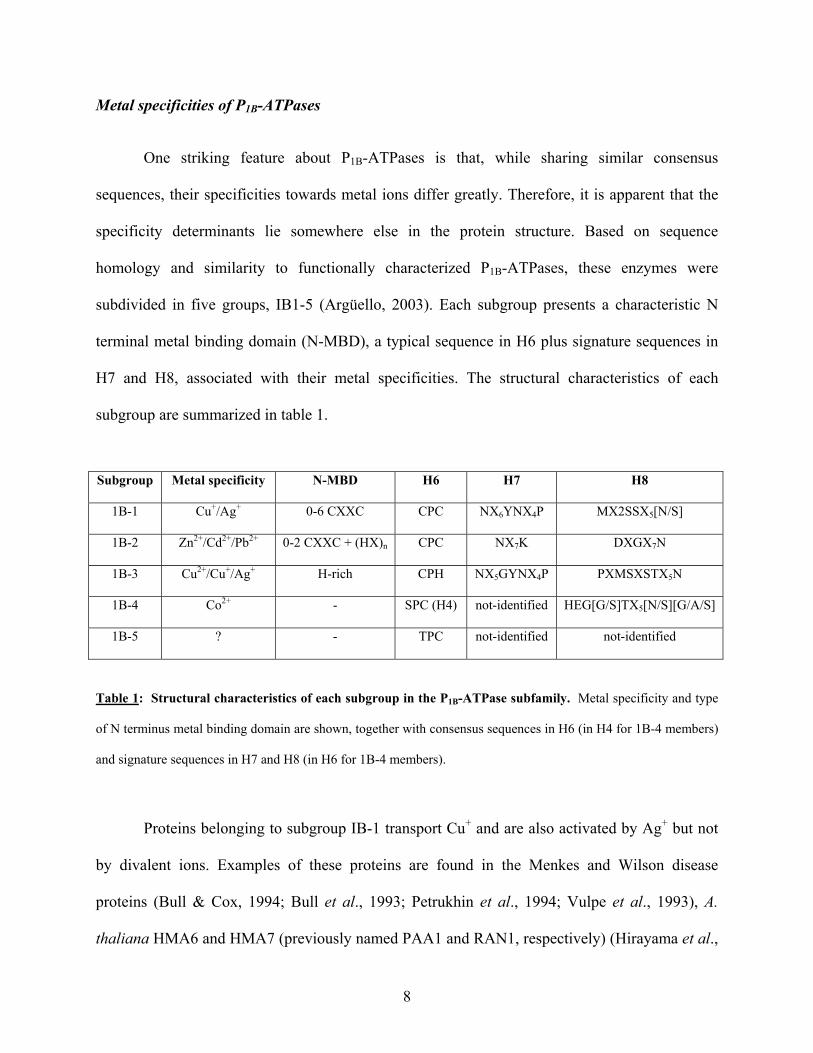
Metal specificities of P1B-ATPases
One striking feature about P1B-ATPases is that, while sharing similar consensus
sequences, their specificities towards metal ions differ greatly. Therefore, it is apparent that the
specificity determinants lie somewhere else in the protein structure. Based on sequence
homology and similarity to functionally characterized P1B-ATPases, these enzymes were
subdivided in five groups, IB1-5 (Argüello, 2003). Each subgroup presents a characteristic N
terminal metal binding domain (N-MBD), a typical sequence in H6 plus signature sequences in
H7 and H8, associated with their metal specificities. The structural characteristics of each
subgroup are summarized in table 1.
Subgroup Metal specificity N-MBD H6 H7 H8
1B-1 Cu+/Ag+ 0-6 CXXC CPC NX6YNX4P MX2SSX5[N/S]
1B-2 Zn2+/Cd2+/Pb2+ 0-2 CXXC + (HX)n CPC NX7K DXGX7N
1B-3 Cu2+/Cu+/Ag+ H-rich CPH NX5GYNX4P PXMSXSTX5N
1B-4 Co2+ - SPC (H4) not-identified HEG[G/S]TX5[N/S][G/A/S]
1B-5 ? - TPC not-identified not-identified
Table 1: Structural characteristics of each subgroup in the P1B-ATPase subfamily. Metal specificity and type
of N terminus metal binding domain are shown, together with consensus sequences in H6 (in H4 for 1B-4 members)
and signature sequences in H7 and H8 (in H6 for 1B-4 members).
Proteins belonging to subgroup IB-1 transport Cu+ and are also activated by Ag+ but not
by divalent ions. Examples of these proteins are found in the Menkes and Wilson disease
proteins (Bull & Cox, 1994; Bull et al., 1993; Petrukhin et al., 1994; Vulpe et al., 1993), A.
thaliana HMA6 and HMA7 (previously named PAA1 and RAN1, respectively) (Hirayama et al.,
8

1999; Shikanai et al., 2003; Tabata et al., 1997; Woeste & Kieber, 2000), E. coli CopA (Rensing
2000), the yeast Cu+-ATPase CCC2 (Pufahl et al., 1997) and A. fulgidus CopA (Mandal et al.,
2002). Subgroup 1B-2 includes Zn2+/Cd2+/Pb2+ transporters. Members of this group are found in
archaea, prokaryotes and plants, being the latter the only eukaryotes were these proteins are
found. Proteins belonging to this subgroup include E. coli ZntA (Rensing et al., 1997; Sharma et
al., 2000), H. pylori CadA and HMA2, the first Zn2+ATPase functionally characterized in
eukaryotic organisms (Eren & Argüello, submitted for publication). Proteins belonging to
subgroup 1B-3 include only prokaryotic members. These proteins transport Cu2+, Cu+ and Ag+,
the former at a higher rate than the latter (Mana-Capelli et al., 2003). Examples of these proteins
can be found in CopB from A. fulgidus (Mana-Capelli et al., 2003) and CopB from E. hirae
(Odermatt et al., 1993).
Subgroup 1B-4 contains P1B-ATPases with a total of six transmembrane fragments
compared to the eight that characterizes this subfamily of proteins. Metal specificity was
determined experimentally for only one member of this subgroup, CoaT from Synechocystis
PCC6803. Disruption of CoaT gene leads to Co2+ sensitivity and accumulation, therefore
indicating a role in Co2+ transport (Rutherford et al., 1999). In this subgroup, the consensus
sequence usually found in H6, is present in H4, the functional equivalent of H6, and it is SPC,
with no signature sequence identified in H5. Another protein belonging to this group is HMA1
from A. thaliana, one of the targets of this study. Subgroups 1B-5 and 1B-6 include the
remainder of the proteins that could not be included in any of the mentioned groups. Their
specificity determinants are still to be found (Argüello, 2003).
9

P1B-ATPases in Arabidopsis
Arabidopsis thaliana contains eight P1B-ATPases, named HMA1-8, and together with
Oryza sativa, comprises the highest number of P1B-ATPases found in a single organism
(Argüello, 2003; Axelsen & Palmgren, 2001; Baxter et al., 2003). Phylogenetic analysis of their
sequences, show that HMA2-4 are closely related, HMA5-8 cluster together and HMA1 is more
distantly related to the other two groups (Axelsen & Palmgren, 2001; Baxter et al., 2003;
Cobbett et al., 2003; Williams et al., 2000). Based on Argüello’s classification in functional
subgroups, HMA1 belongs to the 1B-4 subgroup, HMA2-4 to the 1B-2 and HMA5-8 to the 1B-1
subgroup. Moreover, among eukaryotic organisms, only plants contain members of subgroups
1B-2 and 1B-4, as humans and yeast present P1B-ATPases only belonging to subgroup 1B-1
(Argüello, 2003).
The first P1B-ATPase reported in higher plants was HMA6 from Arabidopsis thaliana
(Tabata et al., 1997). No variation in HMA6 expression was observed when Arabidopsis
thaliana plants were grown in different CuSO4 concentrations. Furthermore, no functional data
supporting a role in metal transport was provided. Recently, mutant plants in HMA6 gene that
presented a high chlorophyll fluorescence phenotype were isolated (Shikanai et al., 2003). These
mutants were defective in photosynthetic electron transport, exhibited less concentrations of
copper-bound plastocyanin, a chloroplastic protein, and lower activity of other chloroplastic
proteins compared to wild type plants. Mutant plants also contained lower Cu2+ content in
chloroplasts, and the mutant phenotype was recovered upon Cu2+ treatment. This experimental
evidence suggests that HMA6 pumps copper into the chloroplast. Functional evidence has also
been provided for the role of HMA7 in Cu+ transport. The HMA7 gene was identified when
plants were screened for constitutive ethylene response in the presence of a potent ethylene
10

antagonist, TCO. The expression of HMA7 gene complemented growth of yeast mutant for the
CCC2 Cu+-ATPase, therefore indicating a role in Cu+ transport. Based in this evidence, it was
proposed that HMA7 might act delivering Cu+ in the trans Golgi for the assembly of functional
ethylene receptors (Hirayama et al., 1999). In a later work, the essential role of HMA7 for plant
metabolism was shown as mutant plants exhibited a rosette-lethal phenotype when grown in soil,
and smaller and unfertile plants when grown in Gamborg’s media (Woeste & Kieber, 2000).
HMA4 was the first protein from subgroup 1B-2 to be reported (Mills et al., 2003).
Expression of HMA4 complemented growth of ∆zntA E.coli in high Zn2+ media. HMA4 also
confers resistance to Cd2+ when expressed in the mutant Saccaromyces cerevisiae strain ∆ycf1.
This evidence suggests that HMA4 transports Zn2+/Cd2+, consistent with its placement in
subgroup 1B-2. HMA4 transcript is also found in all plant organs, except for siliques, and it
increases upon Zn2+, Cd2+ and Ni2+ treatment and decreases upon Mn2+ treatment (Mills et al.,
2003; Orofino & Argüello, unpublished results). A recent publication provides evidence that
HMA3 might be involved in Cd2+ and Pb2+ transport to intracellular compartments, as its
expression complemented strain ∆ycf1 when grown in high Cd2+ or Pb2+ containing-media
(Gravot et al., 2004). Biochemical evidence of Zn2+ and Cd2+ transport has been provided for
HMA2 (Eren & Argüello, submitted for publication). HMA2 transports Zn2+ and Cd2+ with high
affinity, and other metals to a lesser extent. As a typical P-type ATPase, HMA2 forms a stable
phosphorylated intermediate and it is inhibited by vanadate. Metal transport experiments
evidence that HMA2 drives the outward movement of metals from the cytoplasm to the
extracellular milieu. HMA2 transcript is present in all plant organs and does not change upon
metal exposure. Under normal grown conditions, mutant plants for HMA2 gene present higher
Zn2+ concentration, and further Zn2+ accumulation if grown in Zn2+ containing media. When
11

exposed to high Cd2+ concentrations, mutant plants accumulate this metal to a higher extent than
wild type plants, further supporting a role in Zn2+ and Cd2+ transport (Eren & Argüello,
submitted for publication).
Functional evidence for HMA1, HMA5 and HMA8 metal transport has not yet been
provided. Results obtained in our laboratory indicate that HMA5 transcript is found only in roots
and is upregulated in high Cu2+ media; HMA8 transcript in found in all plant organs, and also
upregulated in high Cu2+ media (Eren, Orofino & Argüello, unpublished results).
The high number of P1B-ATPases found in Arabidopsis, and the distinct expression
patterns found in members belonging to the same functional subgroup, indicates different
transport processes in different compartments of the plant body. Where these proteins are
expressed in the whole plant provides information on the roles they may play in metal
homeostasis. Changes in expression upon metal stress or deficiency may indicate if they are also
related to metal tolerance mechanisms. Moreover, the effect of P1B-ATPase gene mutation in
plant metabolism might provide insight on how essential these genes are for integral plant
physiology. In this work, two P1B-ATPases from Arabidopsis thaliana, HMA1 and HMA6
were chosen for study. The goals of this work were to clone and sequence these genes for
functional studies, to determine their mRNA-transcript levels in different plant organs and in
seedlings grown at different metal concentrations, and to determine the effect of T-DNA
insertions in these genes in plant growth and development.
12

MATERIALS AND METHODS
Plant material. Seeds of Arabidopsis thaliana ecotype Columbia 0 (Col0) were grown in soil or
in growth media at 23ºC with a photoperiod 14 hours light:10 hours dark. For metal tolerance
experiments, seeds from Arabidopsis thaliana ecotype Col0 were surface-sterilized by
submerging them in 70 % ethanol for 2 min, 10 min in 20 % bleach, 0.2 % SDS and washed ten
times with sterile water. Sterile seeds were placed in plates containing Murashige-Skoog (MS)
Salt Mixture media with vitamins (Invitrogen Co, Carlsbad, CA) with 1% agar plus addition of
different metal concentrations, stratified at 4°C for two days and grown at the same photoperiod
and temperature stated before. For seedling growth determination, the metals and final
concentrations tested were 0, 0.1, 0.25 and 0.5 mM of CuSO4, ZnSO4, CdCl2, NiSO4, CoCl2,
FeSO4, MnCl2 and AgNO3. For root length measurements, excess metal media was prepared by
mixing MS media with each metal to final concentrations of 0.1 mM CuSO4, 0.25 mM ZnSO4,
0.25 mM CdCl2, 0.25 mM NiSO4, 0.25 mM CoCl2, 0.5 mM MnCl2 or 0.1 mM AgNO3. Metal
deficient media was prepared by mixing all MS media components except for the metal salt for
which deficiency was tested.
Cloning in pBADTOPO vector and expression in bacteria. DNA sequences for HMA1 and
HMA6 genes were obtained through GenBank (http://www.ncbi.nlm.nih.gov/). Leaves from 4
week-old plants were harvested, frozen in liquid N2 and kept at -80ºC for RNA extraction.
Approximately 100 mg of frozen leaf tissue from 4 week-old Col0 plants was grinded in liquid
N2, and total RNA was extracted using the RNeasy Plant mini kit (QIAGEN Inc, Valencia, CA)
following the manufacturer’s specifications. RNA integrity was analyzed in formaldehyde
agarose-gels (Sambrook et al., 1989). Single stranded cDNA was synthesized using Superscript
13

II-H- Reverse Transcriptase (Invitrogen Co, Carlsbad, CA) following the manufacturer’s
protocol. 2 µg of total RNA was used per 20 µl of RT reaction final volume.
For PCR amplification of coding sequence of HMA1 gene, primers used were 5HMA1
and 3HMA1; for HMA6 gene, primers 5PAA1 and 3PAA103 were used (Table 2). For HMA1
gene, a stop codon was included given that HMA1 contains a six His residues in tandem at its N
terminal. The positive control for the polymerase function consisted in two primers specific for
the flanking sequence of a clone of known size (~200pb). The PCR program consisted in 95° 2
min, 15 cycles of 95° 15 sec, 60° 30 sec, 68° 8 min, and then 25 cycles of 95° 15 sec, 60° 30 sec,
68° 8 min plus 20 seconds per cycle. Expand Polymerase (Roche) was used, which exhibits good
proofreading activity and, like Taq Polymerase, adds an A overhang in the 3’ end. PCR
fragments were analyzed in 1% agarose gels containing 0.5 µg/ml Ethidium bromide (Sambrook
et al., 1989). Amplified fragments were cloned in pBAD-TOPO vector (Invitrogen Co, Carlsbad,
CA) following manufacturer’s protocol.
The DNA sequence of HMA1 and HMA6 was confirmed by automated DNA sequence
analysis (Davis, Keck and Macrogen facilities) with the primers listed on Table 2.
For expression in bacteria, E. coli strains used were TOP10, TOP10 CP, BL21
DE3(pLys)S, BL21 DE3(pLys)SStar and BL21 AI (Invitrogen). All protein concentration
determinations were performed using the Bradford reagent (Bradford, 1976). An aliquot of an
overnight culture of transformed cells was inoculated in 25 ml 2xYT media plus addition of 100
µg/ml Ampicillin, or plus 100 µg/ml Ampicillin and 34 µg/ml Chloramphenicol for BL21 cells.
These cultures were grown at 37°C or 22°C until O.D.600 of 0.6 (exponential phase of growth), at
which arabinose was added to a final concentration of 0.002 % to induce expression. After three
hours, 1 ml aliquots of culture were taken; cells were spun and lysed by sonication.
14

HMA1 gene 5’ PRIMERS
Primer name
Position in ORF
Position in gene
Comments Use in this work
5HMA1 4-32 4-32 - Cloning-Sequence-T-DNA 5HMA102 334-357 334-357 - Sequence 5HMA103 1330-1365 2264-2299 D A (NheI site)a Sequence 5HMA104 - 3691-3715 Intron localized T-DNA screening Y5HMA1 1-20 1-20 SacI + AATAb Cloning in pYES
3’ PRIMERS 3HMA1 2431-2460 4338-4366 Stop codon c Cloning-Sequence
3HMA102 2251-2271 4069-4092 - Sequence 3HMA103 1337-1364 2264-2299 D A (NheI site)a Sequence 3HMA104 228-252 228-252 - T-DNA screening 3HMA105 - 3973-3998 - T-DNA screening
Y3HMA102 2439-2447 4345-4363 NotI sited Cloning in pYES HMA6 gene 5’ PRIMERS
Primer name
Position in ORF
Position in gene
Comments Use in this work
5PAA1 1-30 1-30 - Cloning-Sequencing 51PAA102 473-496 559-582 - Sequencing 51PAA103 1645-1651 4295-4318 Last 16bp introne Sequencing 5PAA104 - 1332-1361 Intron localized T-DNA screening 5PAA105 1-3 -57-3 5’UTR T-DNA screening 5PAA106 - -557-(-536) 5’UTR T-DNA screening Y5PAA1 1-20 1-20 KpnI site +AATAb Cloning in pYES
3’ PRIMERS 3PAA1 2822-2850 6829-6857 Stop codonc Sequencing
3PAA102 2264-2286 5597-5619 - Sequencing 3PAA103 2818-2847 6825-6854 Plus 15bp EK sitef Cloning 3PAA104 2489-2511 6088-6110 - Sequencing 3PAA105 486-511 555-579 CGGC AGGA
(NaeI)a Sequencing
3PAA106 1668-1695 3721-3748 CPC APA (NotI)a Sequencing 3PAA107 - 1521-1549 Intron localized T-DNA screening 3PAA108 334-354 334-354 - T-DNA screening Y31PAA1 2829-2847 6840-6854 NotI sited Cloning in pYES
Table 2 legend on next page
15
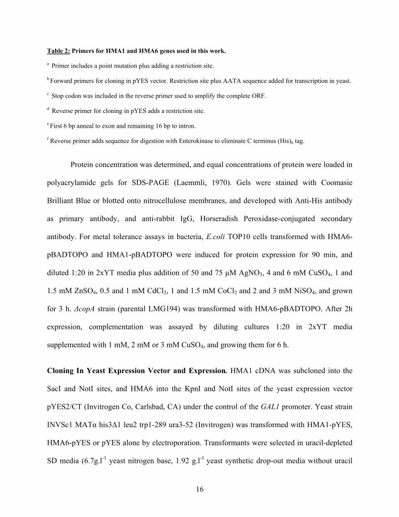
Table 2: Primers for HMA1 and HMA6 genes used in this work.
a Primer includes a point mutation plus adding a restriction site.
b Forward primers for cloning in pYES vector. Restriction site plus AATA sequence added for transcription in yeast.
c Stop codon was included in the reverse primer used to amplify the complete ORF.
d Reverse primer for cloning in pYES adds a restriction site.
e First 6 bp anneal to exon and remaining 16 bp to intron.
f Reverse primer adds sequence for digestion with Enterokinase to eliminate C terminus (His)6 tag.
Protein concentration was determined, and equal concentrations of protein were loaded in
polyacrylamide gels for SDS-PAGE (Laemmli, 1970). Gels were stained with Coomasie
Brilliant Blue or blotted onto nitrocellulose membranes, and developed with Anti-His antibody
as primary antibody, and anti-rabbit IgG, Horseradish Peroxidase-conjugated secondary
antibody. For metal tolerance assays in bacteria, E.coli TOP10 cells transformed with HMA6-
pBADTOPO and HMA1-pBADTOPO were induced for protein expression for 90 min, and
diluted 1:20 in 2xYT media plus addition of 50 and 75 µM AgNO3, 4 and 6 mM CuSO4, 1 and
1.5 mM ZnSO4, 0.5 and 1 mM CdCl2, 1 and 1.5 mM CoCl2 and 2 and 3 mM NiSO4, and grown
for 3 h. ∆copA strain (parental LMG194) was transformed with HMA6-pBADTOPO. After 2h
expression, complementation was assayed by diluting cultures 1:20 in 2xYT media
supplemented with 1 mM, 2 mM or 3 mM CuSO4, and growing them for 6 h.
Cloning In Yeast Expression Vector and Expression. HMA1 cDNA was subcloned into the
SacI and NotI sites, and HMA6 into the KpnI and NotI sites of the yeast expression vector
pYES2/CT (Invitrogen Co, Carlsbad, CA) under the control of the GAL1 promoter. Yeast strain
INVSc1 MATα his3∆1 leu2 trp1-289 ura3-52 (Invitrogen) was transformed with HMA1-pYES,
HMA6-pYES or pYES alone by electroporation. Transformants were selected in uracil-depleted
SD media (6.7g.l-1 yeast nitrogen base, 1.92 g.l-1 yeast synthetic drop-out media without uracil
16

(Sigma)) supplemented with 20 g.l-1 glucose. To induce HMA1 or HMA6 expression, cells were
diluted to O.D.600 0.6 in the same media containing 20 g.l-1 galactose and grown for 8 h. For
complementation assays, different yeast strains were transformed with the same constructs. Two
∆ccc2 mutant strains were transformed with HMA6-pYES and pYES vectors, parental strain
2809 (MATα his3-200 leu2 trp1-101 ura-52 ade5) (kindly provided by Dr. Andrew Dancis,
University of Pennsylvania) and BY4743 (MAT a/α his3∆1/ his3∆1 leu2∆0/leu2∆0
lys2∆0/LYS2 MET15/met15∆0 ura3∆0/ ura3∆0) (Invitrogen). ∆fet3 in the background BY4743
(Openbiosystems) was also transformed with HMA6-pYES and pYES vectors. ∆ycf1
(Invitrogen) and ∆cot1 (Openbiosystems) in the BY4743 background were transformed with
HMA1-pYES or pYES vectors.
Complementation assays in yeast. One fresh yeast colony was grown overnight in uracil
depleted SD media with 20 g.l-1 glucose and diluted 1:10 the next day. Yeast cells were grown
until O.D.600 of 0.6, diluted in the same media but supplemented with 20 g.l-1 galactose and
grown for 4 h to allow protein expression. At that time, 1 ml of a culture with O.D.600 0.8 was
spun down, and pellet resupended in 100 µl SD media. 1:10 serial dilutions were made and 5 µl
were spotted in SD media supplemented with 20 g.l-1 galactose and 10 g.l-1 raffinose (2809
strain) or 20 g.l-1 galactose (BY4743 strain) plus 1% agar. For growth of the mutant strains in the
BY4743 background, 200 µg/ml of Geneticin was added to the media. For complementation of
∆ccc2 in the 2809 background, 60 µM Bathophenanthroline disulfonic acid (BSDS) and 60 µM
Bathocuproine disulfonate (BCS) were added to the solid media, for ∆ccc2 strain BY4743, 30
µM BSDS or 300 µM BCS were added to the solid media. Complementation of ∆fet3 strain was
tested in media plus addition of 0.5 mM to 4 mM CuSO4. ∆ycf1 and ∆cot1 (BY4743) were
transformed with HMA1-pYES but no expression was detected up to 24 h post induction.
17

E. coli and yeast membrane preparation. Membrane fractions of E. coli cells were prepared
according to Mandal et al (2002) with some variations. Briefly, cells were washed with 25 mM
Tris-HCl, 100 mM KCl buffer pH 7, weighed and stored at -80°C. The cells were resuspended in
Buffer A (25 mM Tris-HCl pH 7.5, 100 mM sucrose) plus 1 mM phenylmethylsulfonyl fluoride
(PMSF). The extracts were lysed using French Press 3 times at 20000 psi and incubated with 2
mM MgCl2 and 0.02 mg/ml of DNAse I at 4°C 30 min. The cell lysate was diluted 1:1 in Buffer
A plus 1 mM PMSF and spun at 27,000xg 30 min. The supernatant (cleared lysate) was
centrifuged at 100,000 x g 1 h and the pellet (membrane fraction) was washed 1 h in buffer A
plus 1 mM PMSF, resuspended in buffer A plus 1 mM PMSF, total protein concentration was
determined by Bradford method and approximately 10 mg protein aliquots were stored at -20°C
in 20 % glycerol.
Total membranes of yeast cells were prepared as described by Voskoboinik et al (2001)
with some modifications. Briefly, cells were suspended in 25 mM Tris HCl (pH 7.4), 250 mM
sucrose, 10 mM ascorbate, 1 mM PMSF, 1 µg.ml-1 leupeptin and 1 µg.ml-1 aprotinin. Cells were
disrupted in a Beads Beater (BioSpec, Bartlesville, OK) 4 x 30 sec homogeneization with 30 sec
intervals plus 1 min interval after each cycle. The homogenate was centrifuged at 10,000 x g for
20 min. The supernatant was centrifuged at 110,000 x g for 1 h and the pellet was resuspent in
the same buffer described before but with 0.2 mM ascorbate instead.
ATPase activity assays. All phosphate determinations for assessing ATPase activity were
performed as described by Mandal et al. (2002). The ATPase assay mix contained 50 mM Tris-
HCl pH 7, 3 mM MgCl2 and 3 mM ATP in a final volume of 250 µl, with enzyme, metal and
cofactor additions. All reactions were incubated at 37°C for 20 min. The reaction was stopped by
placing tubes in ice. 750 µl of the color reagent (3 volumes of 0.045 % Malachite green and 1
18

volume of 4.2 % Ammonium Molybdate in 4 N HCl) was added, vortexed and the colorimetric
reaction was stopped by addition of 100 µl of 34 % Sodium Citrate. The absorbance at 660 nm
was recorded.
Phosphorylation assays. Phosphorylation by ATP was assayed with 100 µg yeast membrane
protein in buffer containing 50 mM Tris HCl pH 7.5, 1 mM MgCl2, 50 mM NaCl, 5 µM [γ-32P]
ATP, 20% Dimethyl sulphoxide (DMSO) and addition of 10 and 100 µM AgNO3 or 10 and 100
µM CuSO4 plus 2.5 mM Dithiothreitol (DTT) in a 100 µl final volume. 5 mM EDTA was added
to controls. The reactions were initiated by addition of [γ-32P] ATP. After 60 sec incubation,
phosphorylation was stopped by addition of five volumes of stopping solution (ice cold 10%
trichloroacetic acid, 1 mM Na2HPO4). Samples were centrifuged 14,000 x g 10 min, pellets were
washed with five volumes of stopping solution and centrifuged 14,000 x g 10 min. Pellets were
resuspended in acidic loading buffer (5 mM Tris-PO4, pH 5.8, 6.7 M urea, 0.4 M DTT, 5% SDS
and 0.014% bromophenol blue) and resolved by SDS-PAGE in 8% acidic gels (Sarkadi et al.,
1986).
Transcript level determination by RT-PCR. RNA from seedlings growing in MS media plus
0.1 mM CuSO4, 0.25mM ZnSO4, 0.25 mM CdCl2, 0.25mM NiSO4, 0.25 mM CoCl2, 0.5mM
MnCl2 and 0.1mM AgNO3, and from roots, leaves, flowers and stems of 6 week-old plants was
extracted using the RNeasy Plant Midi kit (QIAGEN Inc, Valencia, CA) following the
manufacturer’s specifications. RT reaction was done following manufacturer’s protocols and
PCR cycle consisted in 95° 2 min, x number of cycles of 95° 15 sec, 55° 30 sec, 72° 3 min, and a
final elongation of 3 min at 72°C. Amplification of EF1α transcription factor was used as
control. For amplification of EF1α from seedlings and organs, x equaled 25. For HMA1
19

amplification in seedlings, 30 cycles were used, and 25 cycles for organs. For HMA6
amplification in seedlings and organs, 30 cycles were used. For determination of HMA1
transcript in hma1-1 mutant, and transcript levels of proteins involved in metal homeostasis,
RNA was extracted from leaves of 6 weeks-old WT and hma1-1 plants. The PCR program was
the same than before, and the number of cycles for HMA3 and HMA4 genes was 30; for EF1α,
MT1a, MT1b, PCS1 and PCS2 genes, 25 cycles; and for MT2a and MT2b genes, 20 cycles.
PCR screening T-DNA Insertion Plants. Seeds of Col0 ecotype and T-DNA insertion mutant
lines (Alonso et al., 2003) were grown in soil after two-day stratification at 4°C. The T-DNA
insertion lines screened were, for HMA1 gene, SALK_088042 and 043265; for HMA6 gene,
SALK_072581 and 109629 (Table 3). One leaf from each of 2 week-old plants was harvested
and genomic DNA preparation was made according to Edwards et al. (1991). Briefly, the lid of
an eppendorf tube was used to pinch out a leaf disc, the tissue was macerated with disposable
grinders (Scienceware, NJ) 15 sec and 400 µl of extraction buffer was added (200 mM Tris HCl
pH 7.5, 250 mM NaCl, 25 mM EDTA and 0.5 % SDS). The extract was centrifuged 13,000 x g 1
min in a microfuge, and 300 µl of the supernatant are added to 300 µl of room temperature
isopropanol, left 2 min at room temperature and then spun at 13,000 x g for 5 min. The pellet
was air-dried and resuspended in 50 µl 1x TE buffer. 2 µl of this preparation are used for PCR
screening. The primers used for the screening of each line are shown in Table 3. The program
used for PCR screening was 94°C 2 min, 30 cycles of 94°C 15 sec, 60°C 30 sec, and 72°C 2 min.
The same program was used to amplify a 600 bp fragment from the T-DNA sequence, used as a
control (with primers 5LBb1 and 3LBb2).
20

Gene SALK insertion line WT pair Insertion pair HMA1 088042 HMA1-3HMA104 5HMA1-3LBb1 HMA1 043265 5HMA104-3HMA105 3LBb1-3HMA105a HMA6 072581 5PAA104-3PAA107 5PAA104-3LBb1 HMA6 109629 5PAA105-3PAA107 3LBb1-3PAA107a HMA6 109629 5PAA106-3PAA108 3LBb1-3PAA108a
Table 3: T-DNA insertion lines and primer pairs used for screening of mutants. WT pair amplifies only if the
insertion is not in the expected site; insertion pair amplifies if T-DNA is present at the expected position.
a Primer 3LBb1 was used as the forward primer since the T-DNA insertion was in the opposite direction
Chlorophyll and metal content determinations. Chlorophyll from WT and hma1-1 plants was
extracted according to Wu et al (2002) with some modifications. 0.1 to 0.2 gr of leaves were
frozen in liquid N2 and pulverized. 2 ml of ice-cold 80 % acetone was added per 0.15 g of leaves
and left for 1 h on ice. Samples were centrifuged for 15 min at 5000 x g. A663 (Chlorophyll A)
and A645 (Chlorophyll B) was measured and total Chlorophyll was calculated using the equation:
µg Chl/ml = 20.2 x A663 + 8.02 x A645
For metal content measurements, approximately 700 mg of leaf samples were digested in 7 ml of
4.5 N HNO3 at 80˚C for 4 h, and left overnight at room temperature. The next day, 0.5 ml of
30% H2O2 were added, samples were filtered in Wathman #1 filters, and diluted with water to 15
ml. Fe2+, Zn2+, Cd2+ and Mn2+ contents were analyzed by atomic absorption spectroscopy (Perkin
Elmer AAnalyst 300). For metal content determination per dry weight, approximately 3 g of
whole plant tissue was dried at 80ºC for 3 days, ashed at 480ºC for 16 h and digested with HNO3
as described above.
21

RESULTS
HMA1 and HMA6: Predicted topology and metal specificity
The sequence of HMA1 and HMA6 indicates they are membrane bound proteins.
Therefore, topological maps were constructed to localize which segments are inside the
membrane, and which fragments are oriented to cytoplasm or extracellular milieu. The prediction
of the membrane spanning regions was made using the TMHMM 2.0 prediction program
(http://www.cbs.dtu.dk/services/TMHMM/) and topogical maps of HMA1 and HMA6 were
constructed using the TOPO2 program (http://www.sacs.ucsf.edu/TOPO-run/wtopo.pl). HMA1
contains six transmembrane fragments (H1 to H6), while HMA6 contains eight (H1 to H8).
HMA1 differs from the typical eight transmembrane fragments array found in most P1B-
ATPases. Topologically, HMA1 can be viewed as any other P1B-ATPase with the exception of
lacking the first two transmembrane fragments. Therefore, while HMA6 presents a CPC
sequence in H6, HMA1 contains an SPC in H4, the functional equivalent of H6 (Fig 3). HMA1
presents a His stretch in the N terminus that could be involved in heavy metal binding, while
HMA6 presents one CGGC sequence in the cytoplasmic N terminus. Both of them present a
phosphorylation site, DKTGT, and the HP consensus sequence in the large cytoplasmic loop.
HMA1 contains a mitochondria-targeting sequence in the N terminus, while HMA6 has a
chloroplast signal peptide, thus indicating a possible localization to mitochondria and
chloroplast, respectively.
Analysis of HMA1 and HMA6 sequences shows that they belong to subgroups 1B-4 and
1B-1, respectively (Argüello, 2003). Proteins that are apparently involved in Co2+ transport share
the signature sequence H-E-G-[G/S]-T-X5-[N/S]-[G/A/S] in H6. Cu+ and Ag+ transporters
contain the sequence N-X6-Y-N-X4-I-P-X-A in H7 and M-X2-S-S-X5-N in H8.
22

A
B
Figure 3: Topological maps of HMA1 and HMA6. For the two proteins, the phosphorylation site DKTGT in the
large cytoplasmic loop is shown in yellow and the N-terminal binding domain in blue. In (A), map of HMA1,
showing the six transmembrane fragments, a His stretch at the cytoplasmic N terminus (blue) and the SPC sequence
in the H4 (red). In (B), sequence map of HMA6, showing one CGGC metal binding domain at the N terminus (blue)
and the CPC sequence at the H6 (in red). In both, an arrow is indicating the transit peptide cleavage site, at amino
acid 17 for HMA1 and amino acid 14 for HMA6, respectively.
23

HMA1 contains a signature sequence HEGGTLLVCLNS in H6, as it is shown in Figure
4, and as CoaT from Synechocystis PCC6803. HMA6 contains a NLWWAFGYNIVGIPAA in
H7 and MGVSSLGVMTN in H8, as the well characterized Cu+-ATPases of Wilson and Menkes
diseases (Bull & Cox, 1994; Bull et al., 1993; Petrukhin et al., 1994; Vulpe et al., 1993).
K Q I VT V L A
SK Q
N F S L A V I C L L I C A N F L Q A M E L P F G V I G H E G S T I L V I L N G L R 590 B.subtilisN I A A T F I A V L V L W D L F G Q L P L P L G V V G H E G S T V L V A L N G M R 598 M.tuberculosis
R Q N L V F A M G A M A V L V L S G L F F E L P L P V A V I G H E G G T V L V V L N G L R 575 R.capsulatusQ N L A L A L A I I L F I S G P A S M G V I P L W L A V I L H E G S T V I V G L N A L R 610 C. trachomatis
N I V F A L G F V M I L L I A N F A G N I T L P F G V L G H E G S T V I V T L S G L R 594 Synechocystis CoaTK Q N V A L A L T S I F L A A L P S V L G F V P L W L T V L L H E G G T L L V C L N S V R 738 A. thaliana HMA1
H6H5
K QR Q
R I
N I F WA L I Y N V I L I P A A A G L L Y P I F G V V F R P E F A G L A M A M S S V S V V A N S L L 673 A. fulgidus CopAN F A L A I G Y N V I A V P I A I - - - - - - L G Y A T - P L V A A V A M S S S S L V V V F N A L R 689 R. meliloti
K Q N L F WA F I Y N T I G I P F A A - - - - - - F G F L N - P I I A G G A M A F S S I S V L L N S L S 676 E. hirae CopAN L V L A L I Y N L V G I P I A A G V F M P - I G I V L Q P WM G S A A M A A S S V S V V L S S L Q 1322 H. sapiens WND
K L N L F WA L C Y N I F M I P I A M G V L I P - WG I T L P P M L A G L A M A F S S V S V V L S S L M 899 S. cerevisiae CCCK Q N L WWA F G Y N I V G I P I A A G V L L P L T G T M L T P S M A G A L M G V T S L G V M T N S L L 863 A. thaliana
H8H7
PAA1HMA6
Figure 4: Alignments of H5 and H6 of HMA1 and H7 and H8 of HMA6 with functionally characterized P1B-
ATPases. The signature sequences are shown in red boxes. HMA1 presents the consensus sequence in H6 found in
CoaT, a putative Co2+ transporter, and other related proteins, therefore indicating it as a putative Co2+ transporter.
HMA6 shares homology in H7 and H8 with members of subgroup 1B-1, WND from humans, CCC2 from yeast and
CopA from A.fulgidus.
Cloning of HMA1 and HMA6 in pBADTOPO
DNA sequences for HMA1 and HMA6 genes were obtained through GenBank. To clone
the open reading frames (ORFs) of HMA1 and HMA6, total RNA from Arabidopsis thaliana
leaves was extracted, and cDNAs were obtained by reverse transcription using an OligodT
primer. The resulting cDNAs were amplified by two rounds of polymerase chain reaction (PCR),
24

first using primers annealing in 5’ and 3’ untranslated regions of each of the two transcripts, and
the second round with primers annealing at the ends of each ORF.
The resulting ORFs were ligated to the prokaryotic expression vector pBAD-TOPO
(Invitrogen) and sequenced. The sequences obtained were aligned with the predicted sequences
from GenBank. No mutations were observed and proper in frame translation in bacteria was to
be expected for the two proteins (not shown). The resulting constructs were introduced in TOP10
E.coli cells, and expression was induced by adding arabinose to exponentially growing cultures.
HMA6 was expressed successfully, although HMA1 could not be detected by
immunoblotting. Five other strains were transformed with HMA1-pBADTOPO construct, but
HMA1 was not expressed in any of the strains. E.coli cells expressing HMA6 were fractionated
in soluble and membrane fractions, and it was found that HMA6 was bound to the membrane
fraction (Fig 5). Other bands of higher and lower molecular weight were detected by
immunoblotting, which could correspond to unspecific binding or degradation products.
Membranes of E.coli expressing HMA6 were tested for ATPase activity in the presence Cu+ or
Ag+. No metal-stimulated ATPase activity was detected when assayed for these metals, or when
assayed with Cd2+, Zn2+, Co2+, Fe2+, Cu2+ or Ni2+ (not shown). Metal tolerance assays were then
performed to test for in vivo activity, but no difference in growth was detected between bacteria
expressing HMA6 and untransformed E.coli grown in toxic metal concentrations (not shown).
∆copA is an E.coli strain with a deletion in the CopA Cu+-ATPase, and its growth is inhibited by
high Cu2+ concentrations. This strain was kindly provided by Dr. Barry P. Rosen, Wayne State
University School of Medicine, and also transformed with HMA6-pBADTOPO vector to test for
mutant growth complementation in high Cu2+ media. No complementation of growth of the
mutant was detected in ∆copA cells expressing HMA6 (not shown).
25

A 0 3 h B S M
Figure 5: Expression of HMA6 and localization to bacterial membranes. Expression of HMA6 in E.coli was
induced and protein detected after 3 h in bacterial extracts in blots stained with anti (His)6 antibody (A). Extracts
were fractionated in soluble (S) and membrane (M) fractions, and all the protein localized to membrane fraction and
detected by the same method (B).
Cloning of HMA1 and HMA6 in pYES2/CT
E.coli, as a heterologous expression system, was not suitable for production of active
HMA1 or HMA6, as shown in the previous section. HMA1 and HMA6 are membrane bound
eukaryotic proteins, and it is possible the prokaryotic system used would not allow appropriate
folding to obtain an active protein. In the case of HMA1, in frame translation was expected by
sequence analysis, however no protein was detected.
Given the chosen prokaryotic system did not produce positive results for neither
expression of HMA1 nor activity of HMA6, we decided to switch to Saccaromyces cerevisiae as
26

an eukaryotic heterologous system. Consequently, HMA1 and HMA6 genes were cloned in yeast
expression vector pYES2/CT under the GAL1 promoter, and the clones were confirmed by
restriction digestion (Fig 6).
A 6 3 2.5
MW(Kb) HMA1 B 6 3
MW(Kb) HMA6
Figure 6: Cloning of HMA1 and HMA6 in pYES2/CT vector. Restriction digestion of resulting plasmids,
HMA1-pYES2/CT was cut with SacI and NotI, rendering two fragments of 5963 bp and 2490 bp (A). Restriction
digestion of HMA6-pYES2/CT with SacI and NdeI enzymes, produced 5793 bp and 2956 bp fragments (B).
Competent INVSC1 yeast cells were transformed with the resulting vectors by
electroporation. Protein expression was induced by diluting an exponentially grown culture in
growth media supplemented with galactose. HMA1 was also transformed into ∆cot1 and ∆ycf1
strains (BY4743 parental strain), but no protein was detected by immunoblotting in any of the
mentioned strains, even after 24 h post induction. The introduction of restriction sites at the 5’
end of the cDNA did not affect the open reading frame of the protein, as observed by automated
sequencing of the 5’ end of the insert (not shown). On the contrary, HMA6 was expressed in
yeast at 4 h after induction, and protein was still detected after 24 h. Yeast extracts were
27

fractionated in soluble and membrane fractions, and all HMA6 protein was bound to membranes
(Fig 7).
HMA6 ATPase activity was assayed in yeast membranes in the presence of Cu+ and Ag+,
but no metal-stimulated activity was found. Other metals like Cu2+, Zn2+, Cd2+, Fe2+, Co2+ and
Ni2+ were assayed, but none of these metals activated HMA6. Given that other ATPases are
present in yeast membranes and could account for most of the ATPase activity, it is possible that
if HMA6 was active, its activity cannot be detected over the rest due to low expression levels.
Therefore, we decided to attempt phosphorylation assays, and detection of protein-bound
phosphate in acidic gels, as a more sensitive assay to detect HMA6 activity. Neither Ag+ nor
Cu+ induced phosphorylation was detected in acidic gels (not shown), indicating that HMA6 is
not active in yeast membranes.
B S MA 0 4 8 h
Figure 7: Expression of HMA6 in yeast and localization to membrane fraction. Yeast cells were induced for
expression and HMA6 was detected after 4 h in blots stained with anti (His)6 antibody (A). Yeast extracts were
fractionated in soluble (S) and membrane (M) fractions, and all protein was detected in membrane fractions by the
same method (B).
Considering that the in vitro assays, ATPase and phosphorylation, did not render any
activity for HMA6, we tried to test if HMA6 could be active in vivo. Therefore, two mutant yeast
strains in CCC2 Cu+ATPase, ∆ccc2 2809 and ∆ccc2 BY4743, were obtained. No
28

complementation of yeast growth was observed when yeast strain ∆ccc2 2809 was grown in 60
µM BSDS plus 60 µM BCS, or when ∆ccc2 BY4743 strain was grown in 30 µM BSDS or 300
µM BCS. The BSDS and BCS concentrations that inhibited mutants over WT were determined
experimentally. Another yeast strain defective in FET3 protein, ∆fet3, is known to accumulate
copper when grown in high concentrations of copper. No complementation of growth was
observed when yeast cells were grown in up to 4 mM CuSO4 in the media. Both these results
indicate that HMA6 is not active in the yeast system.
Since HMA6 is targeted to chloroplast (Shikanai et al., 2003), lack of removal of the
transit peptide, incorrect folding or absence of cofactors only present in chloroplasts could
account for the lack of activity obtained. An alternative assay could include cloning HMA6 in
pYES2/CT without the transit peptide and test for expression and activity.
Transcript levels in plant organs and in seedlings upon metal stress
The expression pattern of these proteins is directly related to their role in planta.
Moreover, changes in transcript level when plants are grown in metal excess could also indicate
if they are involved in metal tolerance mechanisms. Therefore, we determined HMA1 and
HMA6 expression pattern in plant organs and in seedlings grown in high metal concentrations by
semiquantitative RT-PCR experiments.
To detect expression of HMA1, RNA from different organs of adult plants (6 weeks-old)
was reverse transcribed and PCR amplified. HMA1 is expressed in all organs tested except for
roots (Fig 8A). This might indicate a role in aerial parts of the plant for metal transport. To
determine if HMA1 transcript level is affected by metal stress, seedlings were grown in MS
plates plus addition of 0.1 mM CuSO4, 0.25mM ZnSO4, 0.25 mM CdCl2, 0.25mM NiSO4, 0.25
mM CoCl2, 0.5mM MnCl2 and 0.1mM AgNO3. Metal ion concentrations were selected after a
29

screening for levels that would inhibit root growth without impairing completely seedling
development (not shown). Nine days after germination, seedlings were harvested, RNA extracted
and transcript level determined by RT-PCR. The addition of the different metal concentrations
did not affect the germination rate (not shown). HMA1 expression did not vary significantly in
the different metal concentrations tested, although an increase in expression in media with high
Cu2+ and Co2+ and a decrease at high Zn2+ and Ni2+ concentration was observed (Fig 8B).
seedl root leaf stem flower
H E
Figure 8: HM
concentrations
stems and flow
for RNA extra
amplification o
PCR reaction.
A
HMA1 EF1α
Control Ag Cu Co Zn Cd Mn Ni
BMA1
F1α
A1 transcript levels in different plant organs (A) and in seedlings grown in high metal
(B) In A, RNA was extracted 9 days after germination from (9 DAG) seedlings, and roots, leaves,
ers of 6-week old plants, reversed transcribed and PCR amplified. In B, the same protocol was used
cted from 9 DAG-old seedlings grown in MS media plus addition of different metals. In both cases,
f EF1α transcription factor was used as a control of equal loading and initial amount of DNA in the
30

HMA6 transcript level was determined by RT-PCR in different plant organs (Figure 9A).
HMA6 is expressed in all organs tested, suggesting ubiquitous function in plants. To test the
effect of metal stress on HMA6 expression, transcript levels were determined in seedlings grown
at different metal concentrations by semiquantitative RT-PCR. Transcript levels were higher in
media with 0.1 mM CuSO4 concentration, suggesting HMA6 might be involved in tolerance to
high copper concentrations (Figure 9B).
Figure 9: HM
concentration
reversed trans
seedlings grow
was used as a
H E
seedl root leaf stem flower
A
A6 transcript levels in different plant organs (A) and in seedlings grown in high metal
s (B) In A, RNA was extracted from seedlings, roots, leaves, stems and flowers of 6-week old plants,
cribed and PCR amplified. In B, the same protocol was used but RNA was extracted from 9 DAG-old
n in MS media addition of different metals. In both cases, amplification of EF1α transcription factor
control.
HMA6 EF1α
B
MA6
F1α
Control Ag Cu Co Zn Cd Mn Ni
31
Screening for homozygous plants for T-DNA insertions
With the goal of understanding the effect of knocking out HMA1 and HMA6 genes in
plant growth and development, seed stocks of Arabidopsis thaliana lines were analyzed for T-
DNA insertions in HMA1 and HMA6 genes.
The Salk Institute is carrying out a high throughput project for the production of T-DNA
insertion lines in Arabidopsis thaliana. The resulting lines are then screened to detect gene hits
for insertions. This information is then added to a database that can be accessed through their
website (http://signal.salk.edu/cgi-bin/tdnaexpress). If there are hits in the genes of interest, the
seeds can be obtained through the Arabidopsis Biological Resource Center (Ohio State
University, Columbus, OH ).
A search for T-DNA insertion hits was conducted, and two lines for each HMA1 and
HMA6 were ordered. For HMA1, one line presented an insertion in the first exon (stock
SALK_088042) and the other in third intron (stock SALK_043265). For HMA6, T-DNA
insertion was localized in the third intron (SALK_072581) or in the exon number one
(SALK_109629).
Plants from the insertion lines SALK_088042 (HMA1), SALK_043265 (HMA1),
SALK_072581 (HMA6) and SALK_109629 (HMA6) were grown in soil for two weeks. A disc
was pinched out from one leaf of each plant and genomic DNA was extracted. Plant genomic
DNA was used as a template for PCR amplification for T-DNA insertion screening. This
screening was done by using two sets of primers; one set consisted of two primers annealing to
the gene sequence, flanking the insertion site. This set of primers amplifies only the expected
size if there is no T-DNA insertion. In the other set of primers, one of the primers anneals to the
gene sequence and the other to the T-DNA. Amplification of the expected size fragment in this
32

case would indicate the presence of the T-DNA in the expected site. By this screening, three
possible outcomes can result: if only a PCR amplification is obtained with the first set of
primers, then the plant is wild type; if fragments are obtained with both sets of primers, then the
plant is heterozygous; if only amplification with the second set of primers is obtained, then the
plant is homozygous for the T-DNA insertion and hence, a knock out in the gene. The knock out
is then confirmed by RT-PCR, where no transcript is expected. A scheme of the screening
process is shown in Fig 10 for the line SALK_088042. The first set of primers are represented by
a and b, and the second set by b and c.
A PCR screening was conducted in plants of HMA6 insertion lines SALK_072581 and
SALK_109629. Screening of the former only retrieved wild type and heterozygous plants (61%
and 39%, respectively, n=41 plants), while the latter, only wild type plants. Other researchers
working on the same lines obtained similar results (Torres and Ward, personal communication).
It is likely that the in line SALK_109629, the insertion was not in the HMA6 gene, while for
SALK_072581, it is apparent the homozygous is lethal. For the latter line, fourteen siliques from
four heterozygous plants were screened for 25% dead embryos or 50% less ovules (indicators of
embryo lethality and female sterility, respectively). Most of embryos were alive and siliques
contained >90% alive ovules (not shown), indicating homozygous plants were neither embryo
lethal nor female sterile for this line. This result should be confirmed by similar analysis of other
insertion lines.
When HMA1 lines were screened for homozygous, only one plant out of twenty sowed
seeds from line SALK_043265 germinated, and those plants were wild type. Another T-DNA
insertion line in HMA1 gene, SALK_088042, was screened by PCR and homozygous plants
33
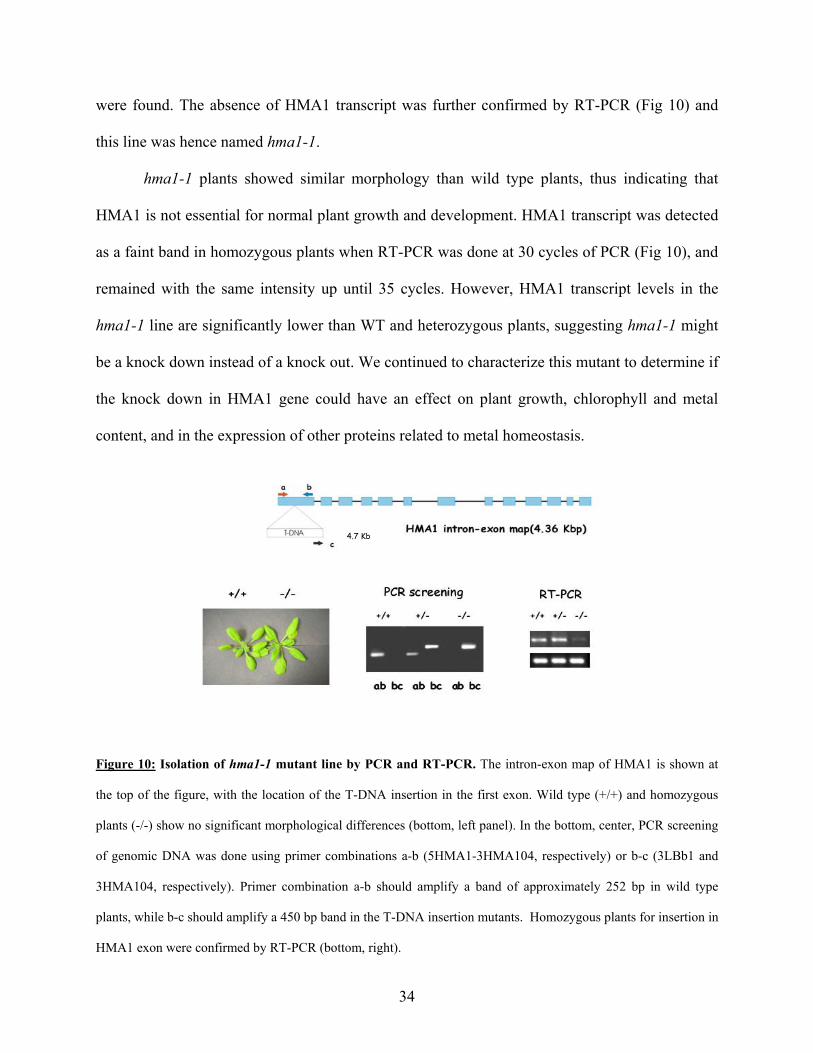
were found. The absence of HMA1 transcript was further confirmed by RT-PCR (Fig 10) and
this line was hence named hma1-1.
hma1-1 plants showed similar morphology than wild type plants, thus indicating that
HMA1 is not essential for normal plant growth and development. HMA1 transcript was detected
as a faint band in homozygous plants when RT-PCR was done at 30 cycles of PCR (Fig 10), and
remained with the same intensity up until 35 cycles. However, HMA1 transcript levels in the
hma1-1 line are significantly lower than WT and heterozygous plants, suggesting hma1-1 might
be a knock down instead of a knock out. We continued to characterize this mutant to determine if
the knock down in HMA1 gene could have an effect on plant growth, chlorophyll and metal
content, and in the expression of other proteins related to metal homeostasis.
4.7 Kb
Figure 10: Isolation of hma1-1 mutant line by PCR and RT-PCR. The intron-exon map of HMA1 is shown at
the top of the figure, with the location of the T-DNA insertion in the first exon. Wild type (+/+) and homozygous
plants (-/-) show no significant morphological differences (bottom, left panel). In the bottom, center, PCR screening
of genomic DNA was done using primer combinations a-b (5HMA1-3HMA104, respectively) or b-c (3LBb1 and
3HMA104, respectively). Primer combination a-b should amplify a band of approximately 252 bp in wild type
plants, while b-c should amplify a 450 bp band in the T-DNA insertion mutants. Homozygous plants for insertion in
HMA1 exon were confirmed by RT-PCR (bottom, right).
34

Characterization of hma1-1 mutant
Macroscopically, hma1-1 plants grow in a similar fashion than wild type plants (Fig 10).
However, hma1-1 shows lower weight and less total chlorophyll content than WT plants (Fig
11).
0
0.2
0.4
0.6
0.8
1
1.2
Col0 hm a1-1
B
0
50
100
150
200
250
300
C ol0 hm a1-1
A
Figure 11: Measurement of fresh weight (A) and total chlorophyll content (B) in Col 0 and hma1-1 plants.
(A) Mean ± SD, n=20. (B) Total chlorophyll content was determined as described by Wu et al (2002). Mean ± SD,
n=3.
To determine if mutant plants could have impaired growth under metal stress or
deficiency, WT and hma1-1 seedlings were grown in plates with MS media in metal deficiency,
or supplemented with excess of either CuSO4, ZnSO4, CdCl2, NiSO4, CoCl2, MnCl2 and AgNO3.
The metal concentrations used were determined based on concentration that would inhibit
seedling root growth significantly in wild type plants. Eight days after germination, root length
of seedlings was measured (Fig 12).
35

0
5
10
15
20
25
30Col0hma1-1
Figure 12: Root length of seedlings grown in metal deficiency or with excess metal in the growth media. Mean
± SD, n=20.
As shown in Fig 12, mutant seedlings grown in control media show longer roots than WT,
and this pattern is observed in all conditions tested except for high Cd2+ concentration, where
root lengths are not significantly different, and in high Co2+, Mn2+ and Zn2+, where roots are
shorter in mutants than in WT plants (P<0.05). This result suggests that absence of HMA1 has an
effect on metal homeostasis mechanisms, and HMA1 could be involved in metal tolerance or
regulation of metal levels.
To determine if metal levels are affected in mutant plants, whole plants (rosette stage),
leaves and roots form WT and hma1-1 plants were analyzed for iron, zinc and manganese
content by atomic absorption spectroscopy. These determinations were done per dry weight of
36

tissue given the differences in fresh weight obtained for WT and hma1-1 plants. Mutant plants
have higher zinc, manganese and iron contents than WT and mutant plants (Fig 13). This
indicates that mechanisms for maintenance of homeostatic iron, zinc and manganese levels are
affected in hma1-1 plants, and that HMA1 could play an essential role in maintaining
physiological levels of a range of different metals in A. thaliana.
0
500
1000
1500
2000
2500
Mn Zn Fe
Col0hma1-1
Figure 13: Manganese, zinc and iron content in whole plants (rosette stage) of WT (Col0) and hma1-1 plants
per dry weight.
Metal concentrations were also determined in leaves and roots of rosette stage plants.
These determinations were done per fresh weight, and then normalized to obtain metal content
per plant (Fig 14). Zinc and manganese accumulation appears to be located mainly in leaves,
while the levels in roots remained the same in WT and mutant plants. This also correlates with
the expression of HMA1 in aerial parts of the plant, being only leaves in rosette stage plants. The
results obtained with iron levels were somewhat striking. Mutant plants accumulate more iron
37
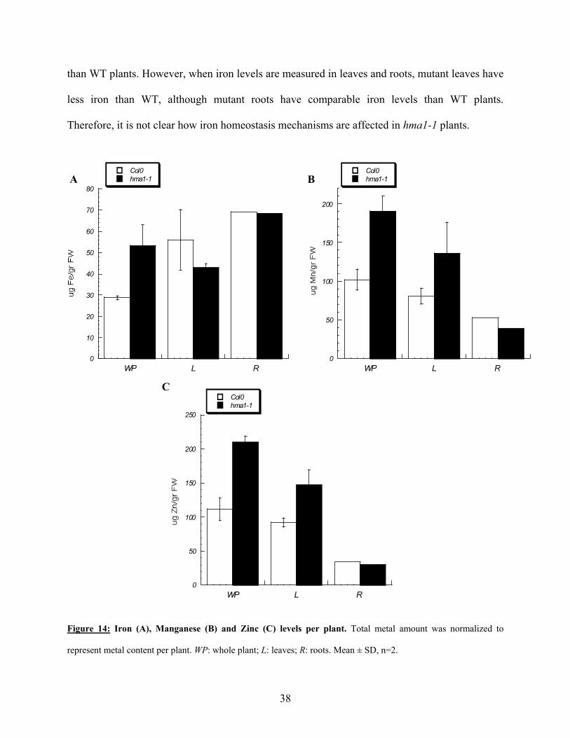
than WT plants. However, when iron levels are measured in leaves and roots, mutant leaves have
less iron than WT, although mutant roots have comparable iron levels than WT plants.
Therefore, it is not clear how iron homeostasis mechanisms are affected in hma1-1 plants.
A
0
10
20
30
40
50
60
70
80
WP L R
Col0hma1-1 B
0
50
100
150
200
WP L R
Col0hma1-1
C
0
50
100
150
200
WP L R
250
Col0hma1-1
Figure 14: Iron (A), Manganese (B) and Zinc (C) levels per plant. Total metal amount was normalized to
represent metal content per plant. WP: whole plant; L: leaves; R: roots. Mean ± SD, n=2.
38

To test the effect of knocking down HMA1 gene on the expression of some Arabidopsis
proteins related to metal homeostasis, expression of the metallothioneins MT1a, MT1c, MT2a
and MT2b; the phytochellatin synthases PCS1 and PCS2, and other related HMAs, HMA3 and
HMA4, was determined by semiquantitative RT-PCR (Fig 15).
MT1a
MT1c
MT2a
MT2b
PCS1
PCS2
HMA4
HMA3
EF1α
WT hma1-1
Figure 15: Transcript levels of proteins related to heavy metal homeostasis in WT and hma1-1 plants.
Expression of metallothioneins 1a, 1c, 2a and 2b, Phytochellatin synthases 1 and 2, and Heavy Metal ATPases 3 and
4 from Arabidopsis, was measured by RT-PCR.
In hma1-1 plants, MT1a and MT2b are upregulated, MT1c levels are lower in mutant
plants while MT2a expression appears not to change compared to expression in WT plants.
PCS1 and PCS2, both are downregulated in the mutant plants. HMA3 and HMA4, two proteins
that apparently transport Zn2+/Cd2+/Pb2+, are upregulated in mutant plants.
It is apparent that knocking down HMA1 gene has a significant effect on many metal
homeostasis components. Mutant plants contain less chlorophyll, are smaller and have increased
levels of metals compared to WT plants. Also, proteins that have been directly involved with
metal tolerance and scavenging in cells have altered expression patterns. However, to interpret
39

these results in the context of the role of HMA1 in all of these processes, functional studies with
isolated protein need to be performed.
40

DISCUSSION
The knowledge of plant metal homeostasis is crucial to understand how plants can grow
in highly polluted soils, or how to enrich mineral content of foodstuffs. In this work we show the
expression of HMA1 and 6 in different plant organs and upon metal stress by RT-PCR analysis,
and the effect of HMA1 mutation on plant metabolism. The results obtained in this work indicate
that HMA1 and HMA6 are essential components of metal homeostasis in Arabidopsis thaliana,
although the mechanism by which metal concentrations are regulated is still to be determined by
complementing the results obtained in this work with functional data.
Cloning of HMA1 and HMA6 genes
HMA1 was expressed in neither bacteria nor yeast. Analysis of the sequence of the
cDNA indicates this protein has the correct frame for translation when present in pBADTOPO or
in pYES2/CT. It could be possible that during or after transcription and/or translation, the
transcript or the protein are targeted for degradation.
HMA6 was successfully expressed in bacteria and in yeast, and located to membranes in
both systems, although protein was non functional. Several assays including ATPase, metal
tolerance, phosphorylation and mutant complementation were performed but no metal stimulated
activity was detected. It could be possible that given HMA6 is a chloroplast bound protein,
presence of chloroplastic cofactors are necessary for its activation. Additionally, lack of removal
of the transit peptide could interfere with proper folding and activity. Future experiments could
aim to clone HMA6 without the transit peptide and determine if the protein is then functional.
41

Expression of HMA1 and HMA6 in organs and upon metal stress
HMA1 was expressed in all organs except for roots. This indicates a role in metal
transport in aerial parts, probably xylem unloading and phloem and leaf cells loading. HMA1
transcript appears not to vary significantly upon stress provoked by different metals in seedlings,
although a decrease in HMA1 expression upon Zn2+ and Ni2+ stress and an increase upon Cu2+
and Co2+ stress was observed by RT-PCR.
HMA6 is proposed to transport Cu+/Ag+. Its transcript was found in all organs tested. Its
presence in roots in levels comparable to those in leaves is striking since this protein is
apparently located in chloroplasts (Shikanai et al., 2003). It could be possible that this protein is
targeted to another cellular compartment in roots, however the mechanisms of targeting should
be elucidated given the presence of a chloroplast signal target peptide in HMA6 structure. The
presence of its transcript in every organ tested indicates that this protein might be a ubiquitous
component of metal homeostasis in the whole plant body. When seedlings were grown in 0.1
mM CuSO4, HMA6 was also overexpressed. This result contradicts the one obtained by Tabata
et al (1997), where they did not observe changes in transcript level in CuSO4 media at 0.01 and 1
mM CuSO4. In their report, plants were treated with CuSO4 in media for 24 h and RNA from
whole plants was extracted. Here, plants were germinated and grown for 12 days in media
containing metal. It is possible the longer metal treatment could account for the increased levels
of transcript found in this work
Together, these results indicate HMA1 and HMA6 to be ubiquitous components of plant
metal homeostasis, and HMA1 to be involved in tolerance to a range of metals when present in
high concentrations. HMA6 appears to be involved in tolerance to high Cu2+ concentrations,
instead.
42

Characterization of hma1-1 insertion mutant
To understand how essential these proteins are for plant metabolism, analysis of insertion
mutants for the genes of interest was initiated. Two insertion lines for HMA1 gene and two for
HMA6 gene were obtained and screened for homozygous in the genes of interest.
When HMA6 insertion lines were screened for homozygous, with one of the lines
(SALK_109629) all wild type genotypes were obtained, while for the other line
(SALK_072581), wild type and heterozygous genotypes were obtained, though no homozygous.
Other investigators working with the same lines had similar results (Torres and Ward, personal
communication). Non-obtaining homozygous could be explained if the homozygous is embryo
lethal or the female is sterile. Siliques were dissected and embryos counted to determine if 25%
of embryos were dead or if female were sterile. All embryos were alive and ovules were viable,
therefore it is apparent that lethality is neither due to embryo lethality nor female sterility.
However, analysis of other insertion lines should be done to confirm these results.
For HMA1 gene, homozygous for one insertion line was obtained, hma1-1
(SALK_088042, this study), while for the other line (SALK_043265), only 5% of plants actually
germinated when planted for initial screening, and those plants were wild type. It has been
observed that the insertion information at the Salk Institute database has a 70% error rate (Ward,
personal communication). Therefore, it is possible this line was not a mutant, or it might have
had more than one insertion, thus affecting plant germination.
Mutant hma1-1 plants show similar morphology, although smaller size and chlorophyll
content than WT plants. hma1-1 plants also contained higher Zn2+, Mn2+ and Fe2+ content than
WT plants, and Zn2+ and Mn2+ accumulation was mainly in leaves, while metal levels in roots
remained unchanged. The results obtained with Fe2+ were somewhat striking, given that the
43

content in whole plants was higher than in WT plants, although in leaves smaller than WT and
unchanged in roots. The mechanisms of iron regulation by HMA1 are still to be determined. This
results suggest that HMA1 act in maintaining metal concentrations in leaves, at least for the
metals tested.
Altered expression of proteins related to metal homeostasis was found in hma1-1 plants
compared to WT plants. Metallothioneins 1a and 2b were overexpressed, while 1c was
underexpressed and 2a remained unchanged. Phytochellatin synthases 1 and 2 had lower
transcript levels than WT plants, and HMA3 and HMA4, two P1B-ATPases belonging to
subgroup 1B-2, are overexpressed in hma1-1 plants. This results show that HMA1 has a pivotal
role in maintaining metal concentrations within homeostatic ranges; knock down of this gene
generated multiple responses in the expression of proteins involved in metal homeostasis.
These results altogether indicate that HMA1 is an essential component of metal
homeostasis in Arabidopsis thaliana, and its role might be direct or through other metal
regulating proteins. In spite the specificity of HMA1 has not yet been determined, results
obtained in this work indicate HMA1 as a necessary component for maintenance of
concentrations of a broad range of metals, and that its function is mainly exerted in leaves.
Functional studies with isolated protein are necessary to elucidate the mechanisms by which this
regulation of metal concentrations is being produced.
44
BIBLIOGRAPHY
Alonso, J.M., Anna N. Stepanova, Thomas J. Leisse, Christopher J. Kim, Huaming Chen,
Paul Shinn, Denise K. Stevenson, Justin Zimmerman, Pascual Barajas, Rosa Cheuk,
Carmelita Gadrinab, Collen Heller, Albert Jeske, Eric Koesema, Cristina C. Meyers,
Holly Parker, Lance Prednis, Yasser Ansari, Nathan Choy, Hashim Deen, Michael
Geralt, Nisha Hazari, Emily Hom, Meagan Karnes, Celene Mulholland, Ral Ndubaku,
Ian Schmidt, Plinio Guzman, Laura Aguilar-Henonin, Markus Schmid, Detlef Weigel,
David E. Carter, Trudy Marchand, Eddy Risseeuw, Debra Brogden, Albana Zeko,
William L. Crosby, Charles C. Berry, and Joseph R. Ecker. 2003. Genome-Wide
Insertional Mutagenesis of Arabidopsis thaliana. Science 301:653-657
Argüello, J.M. 2003. Identification of ion selectivity determinants in heavy metal transport P1B-
ATPases. J Membr Biol 195: 93-108
Axelsen, K.B., Palmgren, M.G. 1998. Evolution of substrate specificities in the P-type ATPase
superfamily. J Mol Evol 46:84-101.
Axelsen, K.B., Palmgren, M.G. 2001. Inventory of the superfamily of P-type ion pumps in
Arabidopsis. Plant Physiol 126:696-706.
Baxter I, T.J., Sussman MR, Boutry M, Palmgren MG, Gribskov M, Harper JF, Axelsen
KB. 2003. Genomic comparison of P type ATPase Ion Pumps in Arabidopsis and rice. Plant
Physiol 132:618-628
Bissig, K.D., Wunderli-Ye, H., Duda, P.W., Solioz, M. 2001. Structure-function analysis of
purified Enterococcus hirae CopB copper ATPase: effect of Menkes/Wilson disease mutation
homologues. Biochem J 357:217-23.
45

Bloss, T., Clemens, S., Nies, D.H. 2002. Characterization of the ZAT1p zinc transporter from
Arabidopsis thaliana in microbial model organisms and reconstituted proteoliposomes. Planta
214:783-91.
Bradford, M.M. 1976. A rapid and sensitive method for the quantitation of microgram
quantities of protein utilizing the principle of protein-dye binding. Anal Biochem 72:248-254
Bull, P.C., Cox, D.W. 1994. Wilson disease and Menkes disease: new handles on heavy-metal
transport. Trends Genet 10:246-52.
Bull, P.C., Thomas, G.R., Rommens, J.M., Forbes, J.R., Cox, D.W. 1993. The Wilson
disease gene is a putative copper transporting P-type ATPase similar to the Menkes gene
[published erratum appears in Nat Genet 1994 Feb;6(2):214]. Nat Genet 5:327-337
Clemens, S. 2001. Molecular mechanisms of plant metal tolerance and homeostasis. Planta
212:475-86.
Cobbett, C.S. 2000. Phytochelatin biosynthesis and function in heavy-metal detoxification. Curr
Op Plant Biol 3:211-216
Cobbett, C.S., Hussain, D., Haydon, M.J. 2003. Structural and functional relationships
between type 1B heavy metal-transporting P-type ATPases in Arabidopsis. New Phytologist
159:315-321
Cohen, C.K., Fox, T.C., Garvin, D.F., Kochian, L.V. 1998. The role of iron-deficiency stress
responses in stimulating heavy-metal transport in plants. Plant Physiol 116:1063-72.
Curie, C., Alonso, J.M., Le Jean, M., Ecker, J.R., Briat, J.F. 2000. Involvement of NRAMP1
from Arabidopsis thaliana in iron transport. Biochem J 347 Pt 3:749-55.
Edwards, K., Johnstone, C., Thompson, C. 1991. A simple and rapid method for the
preparation of plant genomic DNA for PCR analysis. Nucleic Acids Res 19:1349
46

Eide, D., Broderius, M., Fett, J., Guerinot, M.L. 1996. A novel iron-regulated metal
transporter from plants identified by functional expression in yeast. Proc Natl Acad Sci U S A
93:5624-8.
Fan B, R.B. 2002. Biochemical characterization of CopA, the Escherichia coli Cu(I)-
translocating P-type ATPase. J Biol Chem 277:46987-46992
Garcia-Hernandez, M., Murphy, A., Taiz, L. 1998. Metallothioneins 1 and 2 have distinct but
overlapping expression patterns in Arabidopsis. Plant Physiol 118:387-97.
Gravot, A., Lieutaud, A., Verret, F., Auroy, P., Vavasseur A., Richaud, P. 2004. AtHMA3, a
plant P1B-ATPase, functions as a Cd/Pb transporter in yeast. Febs Lett 561:22-28
Grotz, N., Fox, T., Connolly, E., Park, W., Guerinot, M.L., Eide, D. 1998. Identification of a
family of zinc transporter genes from Arabidopsis that respond to zinc deficiency. Proc Natl
Acad Sci U S A 95:7220-4.
Hall, J.L. 2002. Cellular mechanisms for heavy metal detoxification and tolerance. J Exp Bot
53:1-11.
Hamer, D.H. 1986. Metallothionein. Annu Rev Biochem 55:913-51
Hamza, I., Schaefer, M., Klomp, L.W., Gitlin, J.D. 1999. Interaction of the copper chaperone
HAH1 with the Wilson disease protein is essential for copper homeostasis. Proc Natl Acad Sci
U S A 96:13363-8.
Himelblau, E., Mira, H., Lin, S.J., Culotta, V.C., Penarrubia, L., Amasino, R.M. 1998.
Identification of a functional homolog of the yeast copper homeostasis gene ATX1 from
Arabidopsis. Plant Physiol 117:1227-34.
Hirayama, T., Kieber, J.J., Hirayama, N., Kogan, M., Guzman, P., Nourizadeh, S., Alonso,
J.M., Dailey, W.P., Dancis, A., Ecker, J.R. 1999. RESPONSIVE-TO-ANTAGONIST1, a
47

Menkes/Wilson disease-related copper transporter, is required for ethylene signaling in
Arabidopsis. Cell 97:383-93.
Kampfenkel, K., Kushnir, S., Babiychuk, E., Inze, D., Van Montagu, M. 1995. Molecular
characterization of a putative Arabidopsis thaliana copper transporter and its yeast
homologue. J Biol Chem 270:28479-86.
Korshunova, Y.O., Eide, D., Clark, W.G., Guerinot, M.L., Pakrasi, H.B. 1999. The IRT1
protein from Arabidopsis thaliana is a metal transporter with a broad substrate range. Plant
Mol Biol 40:37-44.
Laemmli, U.K. 1970. Cleavage of structural proteins during the assembly of the head of
bacteriophage T4. Nature 227:680-5.
Li, L., He, Z., Pandey, G.K., Tsuchiya, T., Luan, S. 2002. Functional cloning and
characterization of a plant efflux carrier for multidrug and heavy metal detoxification. J Biol
Chem 277:5360-8.
Lin, S.J., Pufahl, R.A., Dancis, A., TV, O.H., Culotta, V.C. 1997. A role for the
Saccharomyces cerevisiae ATX1 gene in copper trafficking and iron transport. J Biol Chem
272:9215-20
Lutsenko, S., Kaplan, J.H. 1995. Organization of P-type ATPases: Significance of structural
diversity. Biochem 34:15607-15613
Lutsenko, S., Petrukhin, K., Cooper, M.J., Gilliam, C.T., Kaplan, J.H. 1997. N-terminal
domains of human copper-transporting adenosine triphosphatases (the Wilson's and Menkes
disease proteins) bind copper selectively in vivo and in vitro with stoichiometry of one copper
per metal-binding repeat. J Biol Chem 272:18939-18944
48

Lutsenko, S., Petris, MJ. 2002. Function and regulation of the mammalian copper- transporting
ATPases: Insights from biochemical and cell biological approaches. J Membr Biol 191:1-12
Mana-Capelli, S., Mandal, A.K., Arguello, J.M. 2003. Archaeoglobus fulgidus CopB Is a
Thermophilic Cu2+-ATPase: functional role of its histidine-rich N-terminal metal binding
domain. J Biol Chem 278:40534-40541
Mandal, A.K., Cheung, W.D., Arguello, J.M. 2002. Characterization of a Thermophilic P-type
Ag+/Cu+-ATPase from the Extremophile Archaeoglobus fulgidus. J Biol Chem 277:7201-7208
Mandal AK., Arguello J.M. 2003. Functional roles of metal binding domains of the
Archaeoglobus fulgidus Cu(+)-ATPase CopA. Biochemistry 42:11040-7
Marschner, H. 1995. Mineral nutrition of higher plants. Academic Press, London ; San Diego
Maser, P., Thomine, S., Schroeder, J.I., Ward, J.M., Hirschi, K., Sze, H., Talke, I.N.,
Amtmann, A., Maathuis, F.J., Sanders, D., Harper, J.F., Tchieu, J., Gribskov, M.,
Persans, M.W., Salt, D.E., Kim, S.A., Guerinot, M.L. 2001. Phylogenetic relationships
within cation transporter families of Arabidopsis. Plant Physiol 126:1646-67.
Melchers, K., Weitzenegger, T., Buhmann, A., Steinhilber, W., Sachs, G., Schofer, K.P.
1996. Cloning and membrane topology of a P type ATPase from Helicobacter pylori. J Biol
Chem 271:446-457
Mills, R.F., Krijger, G.C., Baccarini, P.J., Hall, J.L., Williams, L.E. 2003. Functional
expression of AtHMA4, a P1B-type ATPase of the Zn/Co/Cd/Pb subclass. Plant J 35:164-176
49

Mitra, B., Sharma, R. 2001. The cysteine-rich amino-terminal domain of ZntA, a
Pb(II)/Zn(II)/Cd(II)-translocating ATPase from Escherichia coli, is not essential for its
function. Biochemistry 40:7694-7699
Murphy, A., Taiz, L. 1995. A New Vertical Mesh Transfer Technique for Metal-Tolerance
Studies in Arabidopsis (Ecotypic Variation and Copper-Sensitive Mutants). Plant Physiol
108:29-38.
Odermatt, A., Suter, H., Krapf, R., Solioz, M. 1993. Primary structure of two P-type ATPases
involved in copper homeostasis in Enterococcus hirae. J Biol Chem 268:12775-12779
Ortiz, D.F., Ruscitti, T., McCue, K.F., Ow, D.W. 1995. Transport of metal-binding peptides
by HMT1, a fission yeast ABC-type vacuolar membrane protein. J Biol Chem 270:4721-8.
Outten, C.E., O'Halloran, T.V. 2001. Femtomolar sensitivity of metalloregulatory proteins
controlling zinc homeostasis. Science 292:2488-92.
Paulsen, I.T., Saier, M.H., Jr. 1997. A novel family of ubiquitous heavy metal ion transport
proteins. J Membr Biol 156:99-103
Pence, N.S., Larsen, P.B., Ebbs, S.D., Letham, D.L., Lasat, M.M., Garvin, D.F., Eide, D.,
Kochian, L.V. 2000. The molecular physiology of heavy metal transport in the Zn/Cd
hyperaccumulator Thlaspi caerulescens. Proc Natl Acad Sci U S A 97:4956-4960
Petrukhin, K., Lutsenko, S., Chernov, I., Ross, B.M., Kaplan, J.H., Gilliam, T.C. 1994.
Characterization of the Wilson disease gene encoding a P-type copper transporting ATPase:
genomic organization, alternative splicing, and structure/function predictions. Hum Mol Genet
3:1647-56
50
Pufahl, R.A., Singer, C.P., Peariso, K.L., Lin, S.J., Schmidt, P.J., Fahrni, C.J., Culotta,
V.C., PennerHahn, J.E., Ohalloran, T.V. 1997. Metal ion chaperone function of the soluble
Cu(I) receptor Atx1. Science 278:853-856
Rae, T.D., Schmidt, P.J., Pufahl, R.A., Culotta, V.C., O'Halloran, T.V. 1999. Undetectable
intracellular free copper: The requirement of a copper chaperone for superoxide dismutase.
Science 284:805-808
Rauser, W.E. 1999. Structure and function of metal chelators produced by plants: the case for
organic acids, amino acids, phytin, and metallothioneins. Cell Biochem Biophys 31:19-48
Rensing, C., Mitra, B., Rosen, B.P. 1997. The zntA gene of Escherichia coli encodes a Zn(II)-
translocating P-type ATPase. Proc Natl Acad Sci U S A 94:14326-31
Robinson, N.J., Wilson, J.R., Turner, J.S. 1996. Expression of the type 2 metallothionein-like
gene MT2 from Arabidopsis thaliana in Zn(2+)-metallothionein-deficient Synechococcus PCC
7942: putative role for MT2 in Zn2+ metabolism. Plant Mol Biol 30:1169-79.
Rogers, E.E., Guerinot, M.L. 2002. FRD3, a Member of the Multidrug and Toxin Efflux
Family, Controls Iron Deficiency Responses in Arabidopsis. Plant Cell 14:1787-99.
Rutherford, J.C., Cavet, J.S., Robinson, N.J. 1999. Cobalt-dependent transcriptional
switching by a dual-effector MerR-like protein regulates a cobalt-exporting variant CPx-type
ATPase. J Biol Chem 274:25827-25832
Sambrook, J., Fritsch, E.F., Maniatis, T. 1989. Molecular cloning. A laboratory Manual. Cold
Spring Harbor Laboratory Press, New York
Sarkadi, B.E., A; Foldes-Papp, Z; Gardos, G. 1986. Molecular characterization of the in situ
red cell membrane calcium pump by limited proteolysis. J Biol Chem 261:9552-9557
51

Sharma, R., Rensing, C., Rosen, B.P., Mitra, B. 2000. The ATP hydrolytic activity of purified
ZntA, a Pb(II)/Cd(II)/Zn(II)-translocating ATPase from Escherichia coli. J Biol Chem
275:3873-3878
Shikanai, M.-M., Munekage, Niyogi, Pilon. 2003. PAA1, a P-type ATPase from Arabidopsis,
Functions in Copper Transport in Chloroplasts. Plant Cell 15:1333-1346
Tabata, K., Kashiwagi, S., Mori, H., Ueguchi, C., Mizuno, T. 1997. Cloning of a cDNA
encoding a putative metal-transporting P-type ATPase from Arabidopsis thaliana. Biochim
Biophys Acta 1326:1-6.
Thomine, S., Wang, R., Ward, J.M., Crawford, N.M., Schroeder, J.I. 2000. Cadmium and
iron transport by members of a plant metal transporter family in Arabidopsis with homology
to Nramp genes. Proc Natl Acad Sci U S A 97:4991-6.
Tsai, K.J., Lin, Y.F., Wong, M.D., Yang, H.H., Fu, H.L., Rosen, B.P. 2002. Membrane
topology of the p1258 CadA Cd(II)/Pb(II)/Zn(II)-translocating P-type ATPase. J Bioenerg
Biomembr 34:147-56.
van der Zaal, B.J., Neuteboom, L.W., Pinas, J.E., Chardonnens, A.N., Schat, H., Verkleij,
J.A., Hooykaas, P.J. 1999. Overexpression of a novel Arabidopsis gene related to putative
zinc- transporter genes from animals can lead to enhanced zinc resistance and accumulation.
Plant Physiol 119:1047-55.
Vert, G., Briat, J.F., Curie, C. 2001. Arabidopsis IRT2 gene encodes a root-periphery iron
transporter. Plant J 26:181-9.
Voskoboinik, I., Mar, J., Strausak, D., Camakaris, J. 2001. The regulation of catalytic
activity of the menkes copper-translocating P-type ATPase. Role of high affinity copper-
binding sites. J Biol Chem 276:28620-7.
52

Voskoboinik, I., Strausak, D., Greenough, M., Brooks, H., Petris, M., Smith, S., Mercer,
J.F., Camakaris, J. 1999. Functional analysis of the N-terminal CXXC metal-binding motifs
in the human Menkes copper-transporting P-type ATPase expressed in cultured mammalian
cells. J Biol Chem 274:22008-22012
Vulpe, C., Levinson, B., Whitney, S., Packman, S., Gitschier, J. 1993. Isolation of a candidate
gene for Menkes disease and evidence that it encodes a copper-transporting ATPase. Nat
Genet 3:7-13
Williams, L.E., Pittman, J.K., Hall, J.L. 2000. Emerging mechanisms for heavy metal
transport in plants. Biochim Biophys Acta 1465:104-26.
Woeste, K.E., Kieber, J.J. 2000. A strong loss-of-function mutation in RAN1 results in
constitutive activation of the ethylene response pathway as well as a rosette-lethal phenotype.
Plant Cell 12:443-55.
Wu Z, L.F., Hong B, Young JC, Sussman MR, Harper JF, Sze H. 2002. An endoplasmic
reticulum-bound Ca(2+)/Mn(2+) pump, ECA1, supports plant growth and confers tolerance to
Mn(2+) stress. Plant Physiol 130:128-137
Yoshimizu, T., Omote, H., Wakabayashi, T., Sambongi, Y., Futai, M. 1998. Essential Cys-
Pro-Cys motif of Caenorhabditis elegans copper transport ATPase. Biosci Biotechnol
Biochem 62:1258-1260
53