hole’s essentials of human anatomy &...
TRANSCRIPT

1CopyrightThe McGraw-Hill Companies, Inc. Permission required for reproduction or display.
Chapter 13
Hole’s Essentials of HumanAnatomy & Physiology
David ShierJackie ButlerRicki Lewis
Created by Dr. Melissa Eisenhauer Head Athletic Trainer/Assistant Professor
Trevecca Nazarene UniversityAmended by John Crocker

2
Chapter 13
Cardiovascular System
3
Introduction A. The cardiovascular system consists of
1. Heart 2. Blood vessels
a) Arteriesb) capillaries c) veins
B. Supplies oxygen and nutrients to tissues C. Removes wastes from tissues D. The pulmonary circuit carries deoxygenated blood
to the lungsE. The systemic circuit sends oxygenated blood to all
body cells
CopyrightThe McGraw-Hill Companies, Inc. Permission required for reproduction or display.

4

5
Structure of the HeartA. Hollow, cone-shaped, muscular pump within the
thoracic cavityB. Size and Location
1. Average adult heart is 14 cm long and 9 cm wide2. Lies in the mediastinum under the sternum3. Apex extends to the fifth intercostal space
CopyrightThe McGraw-Hill Companies, Inc. Permission required for reproduction or display.

6
A. Coverings of the Heart1. Pericardium encloses the heart2. It is made of two layers:
a) Tough outer connective tissue fibrous pericardium
b) Surrounds a more delicate visceral pericardium (epicardium) that surrounds the heart
c) At the base of the heart, the visceral pericardium folds back to become the parietal pericardium that lines the fibrous pericardium
d) Between the parietal and visceral pericardia is a potential space (pericardial cavity) filled with serous fluid
CopyrightThe McGraw-Hill Companies, Inc. Permission required for reproduction or display.

7
8
A. Wall of the Heart1. Epicardium
a) Outermost layerb) Made up of connective tissue and epitheliumc) Houses blood and lymph capillaries and coronary arteriesd) Same as the visceral pericardium
2. Myocardium a) Middle layerb) Thickest layerc) Consists of cardiac muscle
3. Endocardium a) Inner layerb) Smooth c) Made up of connective tissue and epitheliumd) Continuous with the endothelium of major vessels joining
the heart
CopyrightThe McGraw-Hill Companies, Inc. Permission required for reproduction or display.

9

10
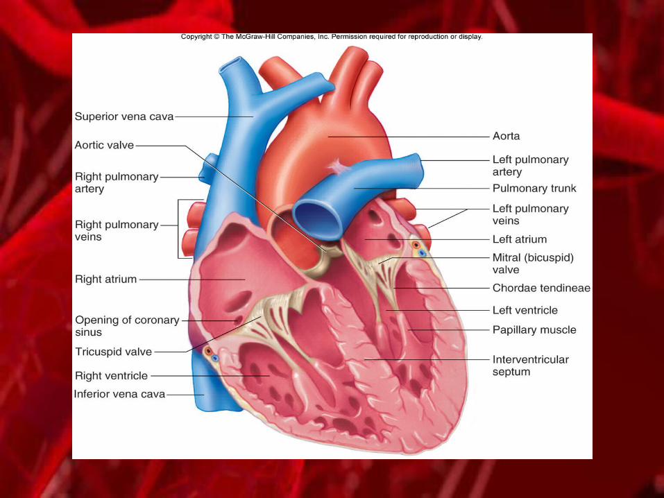
A. Heart Chambers and Valves 1. The heart has four internal chambers:
a) Two atria on top 1) receive blood returning to the heart2) thin walls 3) ear-like auricles projecting from their exterior
b) Two ventricles below1) The thick-muscled 2) Pumps blood to the body
CopyrightThe McGraw-Hill Companies, Inc. Permission required for reproduction or display.

11
1. A septum divides the atrium and ventricle on each side
2. Each also has an atrioventricular (A-V) valve to ensure one way flow of blooda) Right A-V valve (tricuspid) b) Left A-V valve (bicuspid or mitral valve) c) Have cusps to which chordae tendinae attachd) Chordae tendinae attach to papillary muscles in
the inner heart wall 1) Contract during ventricular contraction 2) Prevent the backflow of blood through the A-V valves
CopyrightThe McGraw-Hill Companies, Inc. Permission required for reproduction or display.

12
1. The superior and inferior vena cavae bring blood from the body to the right atrium
2. The right ventricle has a thinner wall than does the left ventricle because it must pump blood only as far as the lungs rather than the entire body
3. Pulmonary valvea) At the base of the pulmonary trunk leading to the lungsb) Prevents a return flow of blood to the ventricle
4. The left atrium receives blood from 4 pulmonary veins5. The left ventricle
a) Pumps blood into the entire body through the aortab) Guarded by the aortic valve c) Prevents backflow of blood into the ventricle
CopyrightThe McGraw-Hill Companies, Inc. Permission required for reproduction or display.

14
A. Skeleton of the Heart1. Rings of dense connective tissue surround the
pulmonary trunk and aorta to provide attachments for the heart valves and fibers
2. These tough rings prevent dilating of tissue in this area
CopyrightThe McGraw-Hill Companies, Inc. Permission required for reproduction or display.

15

16
A. Path of Blood through the Heart 1. Blood low in oxygen returns to the right atrium via
a) Venae cavaeb) Coronary sinus
2. The right atrium contracts3. Blood forced through the tricuspid valve into the
right ventricle4. The right ventricle contracts, closing the tricuspid
valve5. Blood forced through the pulmonary valve into the
pulmonary trunk and arteries
CopyrightThe McGraw-Hill Companies, Inc. Permission required for reproduction or display.

17
1. Pulmonary arteries carry blood to the lungs where it: a) Rids itself of excess carbon dioxide b) Picks up a new supply of oxygen
2. Freshly oxygenated blood is returned to the left atrium of the heart through the pulmonary veins
3. The left atrium contracts4. Blood forced through the left bicuspid valve into the
left ventricle5. The left ventricle contracts closing the bicuspid valve 6. Aortic valve is forced open 7. Blood enters the aorta for distribution to the body
CopyrightThe McGraw-Hill Companies, Inc. Permission required for reproduction or display.


19
A. Blood Supply to the Heart 1. The first branches off of the aorta
a) Right and left coronary arteries b) Feed the heart muscle itselfc) Carry freshly oxygenated blood
2. Branches of the coronary arteries feed capillaries of the myocardium
3. Heart requires a continuous supply of freshly oxygenated blood
4. Smaller branches of arteries often have anastomoses as alternate pathways for blood
5. Cardiac veins a) Drain blood from the heart muscle b) Carry it to the coronary sinusc) Coronary sinus empties into the right atrium
CopyrightThe McGraw-Hill Companies, Inc. Permission required for reproduction or display.

20

21
22
Heart ActionsA. Cardiac cycle consists of
1. Atria beating in unison (atrial systole) 2. Followed by the contraction of both ventricles,
(ventricular systole) 3. Then the entire heart relaxes for a brief moment
(diastole)
CopyrightThe McGraw-Hill Companies, Inc. Permission required for reproduction or display.

23
A. Cardiac Cycle 1. Pressure within the heart chambers rises and falls
with contraction and relaxation of atria and ventricles2. When atria fill pressure in the atria is greater than
that of the ventricles forcing the A-V valves open3. Pressure inside atria rises further as they contract
forcing the remaining blood into the ventricles4. When ventricles contract pressure inside them
increases sharply a) A-V valves forced closed b) aortic and pulmonary valves forced openc) papillary muscles contract pulling on chordae tendinae and
prevent the backflow of blood through the A-V valves
CopyrightThe McGraw-Hill Companies, Inc. Permission required for reproduction or display.

24

25
A. Heart Sounds1. Due to vibrations in heart tissues as blood rapidly
changes velocity within the heart2. Heart sounds can be described as "lubb-dupp" 3. The first sound (lubb) occurs as ventricles
contract and A-V valves are closing4. The second sound (dupp) occurs as ventricles
relax and aortic and pulmonary valves are closing
CopyrightThe McGraw-Hill Companies, Inc. Permission required for reproduction or display.

26
A. Cardiac Muscle Fibers 1. A mass of merging fibers that act as a unit is
called a functional syncytium2. One exists in the atria (atrial syncytium) 3. One in the ventricles (ventricular syncytium)
CopyrightThe McGraw-Hill Companies, Inc. Permission required for reproduction or display.

27
A. Cardiac Conduction System 1. Specialized cardiac muscle tissue conducting
impulses throughout the myocardium 2. A self-exciting mass of specialized cardiac muscle
called the sinoatrial node (S-A node or pacemaker) is located on the posterior right atrium
3. S-A node generates the impulses for the heartbeat4. Impulses spread next to the atrial syncytium and it
contracts
CopyrightThe McGraw-Hill Companies, Inc. Permission required for reproduction or display.

28
1. Impulses travel to the junctional fibers a) Junctional fibers are small b) Lead to atrioventricular node (A-V node) in the
septum c) Allow the atria to contract before the impulse
spreads rapidly over the ventricles2. Branches of the A-V bundle give rise to Purkinje
fibers leading to papillary muscles3. These fibers stimulate contraction of the papillary
muscles at the same time the ventricles contract
CopyrightThe McGraw-Hill Companies, Inc. Permission required for reproduction or display.

29

30
A. Electrocardiogram1. Record of the electrical changes that occur during a
cardiac cycle2. The first wave, the P wave, corresponds to the
depolarization of the atria3. The QRS complex corresponds to the depolarization
of ventricles and hides the repolarization of atria4. T waves end the ECG pattern and corresponds to
ventricular repolarization
CopyrightThe McGraw-Hill Companies, Inc. Permission required for reproduction or display.

31
32
A. Regulation of the Cardiac Cycle 1. The amount of blood pumped at any one time
must adjust to the current needs of the body (more is needed during strenuous exercise)
2. The S-A node is innervated by branches of the sympathetic and parasympathetic divisionsa) CNS controls heart rateb) Sympathetic impulses increase heart ratec) Parasympathetic impulses decrease heart rate
CopyrightThe McGraw-Hill Companies, Inc. Permission required for reproduction or display.

33
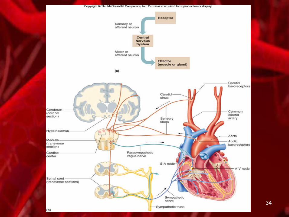
1. Baroreceptors detect changes in blood pressure and signal medulla oblongata
2. The cardiac control center of the medulla oblongata maintains a balance between the sympathetic and parasympathetic divisions
3. Impulses from the cerebrum or hypothalamus may also influence heart rate
4. Body temperature and the concentrations of certain ions may influence heart rate as well
CopyrightThe McGraw-Hill Companies, Inc. Permission required for reproduction or display.
34
35
Blood Vessels A. Form a closed tube B. Carry blood away from the heart, to the cells, and
back againC. Consist of
1. Arteries2. Arterioles3. Capillaries4. Venules5. Veins
CopyrightThe McGraw-Hill Companies, Inc. Permission required for reproduction or display.

36
A. Arteries and Arterioles1. Arteries
a) Strong, elastic vessels b) Adapted for carrying high-pressure blood
2. Arteries become smaller as they divide and give rise to arterioles
3. The wall of an artery consists of a) Endotheliumb) Tunica media (smooth muscle)c) Tunica externa (connective tissue)
4. Vasoconstriction directed by the sympathetic impulses
5. Vasodilation results when impulses are inhibited
CopyrightThe McGraw-Hill Companies, Inc. Permission required for reproduction or display.

37
38
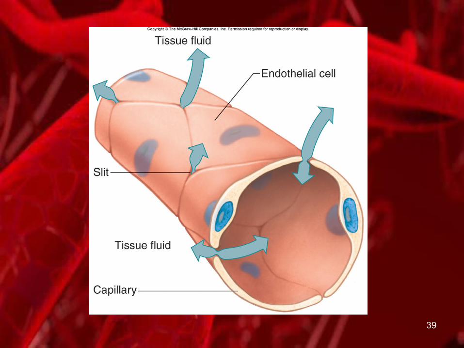
A. Capillaries1. Capillaries are the smallest vessels2. Consist of only of a layer of endothelium
through which substances are exchanged with tissue cells
3. Capillary permeability varies from one tissue to the nexta) Generally more permeability in:
1) Liver2) Intestines3) certain glands
b) Less permeability in: 1) Muscles 2) Brain (blood-brain barrier)
CopyrightThe McGraw-Hill Companies, Inc. Permission required for reproduction or display.
39

40
1. The pattern of capillary density also varies from one body part to the next.
2. Areas with a great deal of metabolic activity have higher densities of capillaries
3. Precapillary sphincters a) Controlled by oxygen concentration in the area b) Regulate the amount of blood entering a
capillary bed c) If blood is needed elsewhere in the body the
capillary beds in less important areas are shut down
CopyrightThe McGraw-Hill Companies, Inc. Permission required for reproduction or display.

41
A. Exchanges in the Capillaries 1. Blood entering capillaries contain:
a) High concentrations of oxygen and nutrients b) Diffuse through capillary walls into the tissuesc) Diffusion driven by hydrostatic pressure
2. Plasma proteins remain in the blood due to their large size
3. Osmotic pressure of the blood causes much of the tissue fluid to return to venules
4. Lymphatic vessels collect excess tissue fluid and return it to circulation
CopyrightThe McGraw-Hill Companies, Inc. Permission required for reproduction or display.

42

43
A. Venules and Veins 1. Venules leading from capillaries merge to form veins
that return blood to the heart.2. Veins
a) Do not carry high-pressure blood b) Thinner and less muscular than arteriesc) Have the same three layers as arteries
1) Endothelium2) Tunica media (smooth muscle)3) Tunica externa (connective tissue)
d) Flap-like valves prevent backflow of bloode) Function as blood reservoirs
CopyrightThe McGraw-Hill Companies, Inc. Permission required for reproduction or display.

44

45
Blood Pressure A. Blood pressure is the force of blood against the
inner walls of blood vessels B. The term "blood pressure" usually refers to arterial
pressure
CopyrightThe McGraw-Hill Companies, Inc. Permission required for reproduction or display.

46
A. Arterial Blood Pressure 1. Arterial blood pressure rises and falls following a
pattern established by the cardiac cycle a) During ventricular contraction, arterial pressure
is at its highest (systolic pressure) b) When ventricles are relaxing, arterial pressure is
at its lowest (diastolic pressure)2. The surge of blood that occurs with ventricular
contraction can be felt as a pulse
CopyrightThe McGraw-Hill Companies, Inc. Permission required for reproduction or display.

47
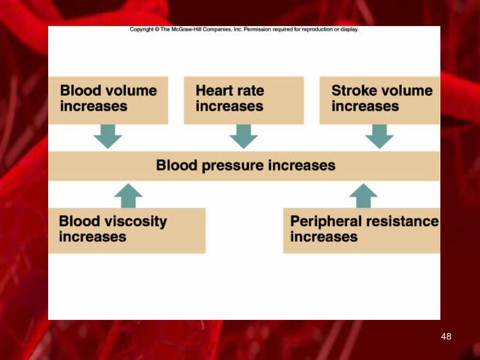
A. Factors that Influence Arterial Blood Pressure 1. Heart action
a) Dependent upon Stroke volume and heart rate (together called cardiac output)
b) Increased cardiac output increases → increased blood pressure
2. Blood volumea) Normally directly proportional to blood pressureb) Varies with age, body size, and gender
3. Resistance to flowa) Friction between blood and walls of blood vessels is
peripheral resistanceb) Peripheral resistance increases → blood pressure increases
4. Blood viscositya) Greater the viscosity → greater resistanceb) Greater resistance → higher blood pressure
CopyrightThe McGraw-Hill Companies, Inc. Permission required for reproduction or display.
48

49
A. Control of Blood Pressure 1. Blood pressure is determined by cardiac output
and peripheral resistance2. The body maintains normal blood pressure by
adjusting cardiac output and peripheral resistance3. Cardiac output depends on
a) stroke volume and heart rateb) a number of factors can affect these actions
4. Blood volume entering the right atrium ~ the volume leaving the left ventricle
CopyrightThe McGraw-Hill Companies, Inc. Permission required for reproduction or display.
50
1. The cardiac center of the medulla oblongata responds to arterial blood pressurea) Arterial pressure increases → parasympathetic
impulses to slow heart rate are sentb) Arterial pressure drops → sympathetic impulses
to increase heart rate are sent2. Emotional upset, exercise, and increased
temperature can result in increased cardiac output and increased blood pressure
3. The vasomotor center of the medulla oblongata can adjust the sympathetic impulses to smooth muscles in arteriole walls, adjusting blood pressure
CopyrightThe McGraw-Hill Companies, Inc. Permission required for reproduction or display.

51

52
1. CO2, O2, and H+ can affect peripheral resistance2. Venous Blood Flow
a) Partially the result of heart action b) Contractions of skeletal muscle squeeze blood
back up veins one valve at a timec) Breathing movements and vasoconstriction of
veinsd) Differences in thoracic and abdominal
pressures draw blood back up the veins
CopyrightThe McGraw-Hill Companies, Inc. Permission required for reproduction or display.

53
Paths of CirculationA. Pulmonary circuit
1. Blood from the right ventricle to the pulmonary arteries
2. To the lungs and alveolar capillaries 3. Pulmonary veins lead from the lungs to the left
atriumB. Systemic circuit
1. The aorta and its branches lead to all body tissues2. Veins return blood to the right atrium
CopyrightThe McGraw-Hill Companies, Inc. Permission required for reproduction or display.

54
Arterial System A. The aorta is the largest arteryB. Principal Branches of the Aorta
1. Ascending aortaa) Right and left coronary arteriesb) Lead to heart
2. Principal branches of the aortic archa) Brachiocephalicb) Left common carotidc) Left subclavian arteries
CopyrightThe McGraw-Hill Companies, Inc. Permission required for reproduction or display.

55
1. Descending aorta (thoracic aorta) gives rise to many small arteries to the thoracic wall and thoracic viscera
2. Branches of the abdominal aorta:a) Celiacb) Superior mesentericc) Suprarenald) Renale) Gonadalf) Inferior mesentericg) Common iliac
CopyrightThe McGraw-Hill Companies, Inc. Permission required for reproduction or display.

56

57
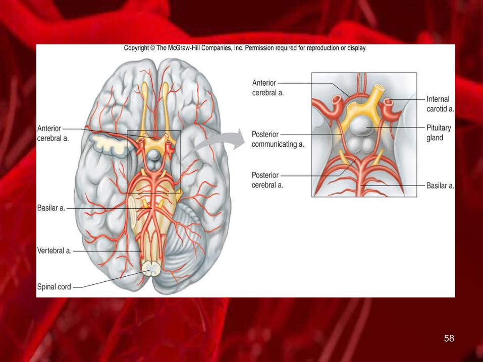
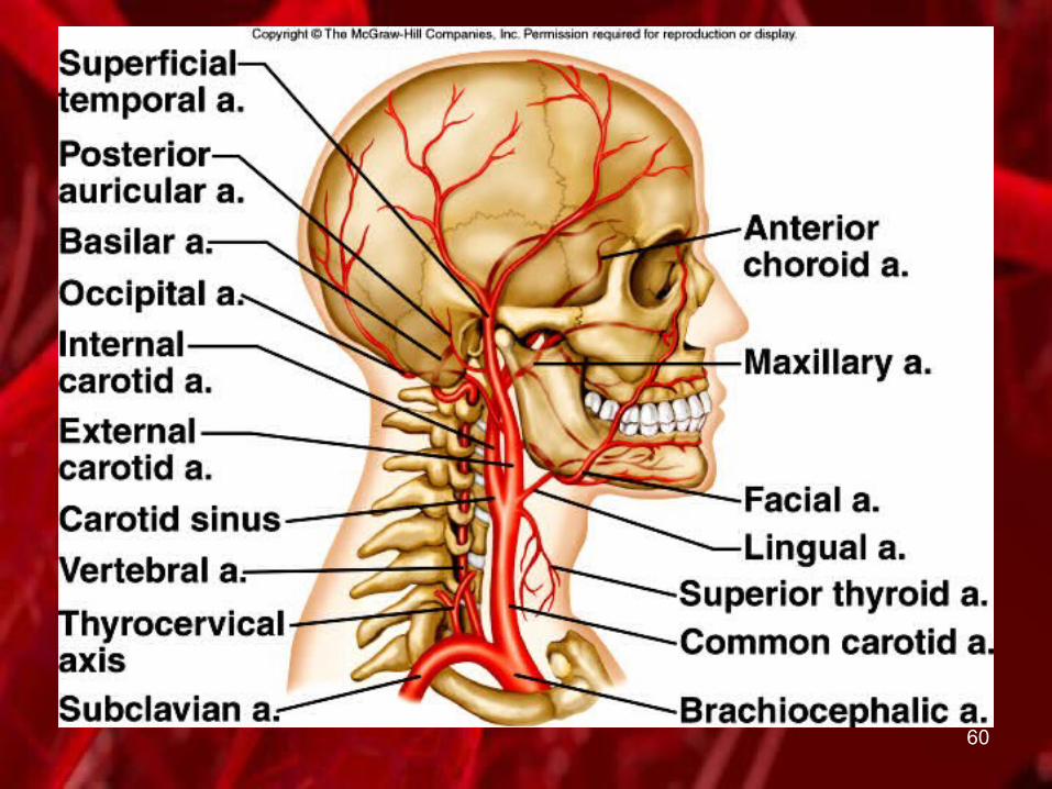
A. Arteries to the Head, Neck, and Brain 1. Include branches of the subclavian and common
carotid arteries2. Vertebral arteries
a) Supply the vertebrae and their associated ligaments and muscles
b) Unite to form a basilar artery which ends as two posterior cerebral arteries in the cranial cavity
3. Posterior cerebral arteries help form the circle of Willis providing alternate pathways through which blood can reach the brain
CopyrightThe McGraw-Hill Companies, Inc. Permission required for reproduction or display.
58

59
1. The right and left common carotid arteries diverge into a) External carotid b) Internal carotid arteries
2. Near the base of the internal carotid arteries are the carotid sinuses
3. Carotid sinuses contain baroreceptors to monitor blood pressure
CopyrightThe McGraw-Hill Companies, Inc. Permission required for reproduction or display.
60

61
A. Arteries to the Shoulder and Upper Limb 1. The subclavian artery continues into the arm where
it becomes the axillary artery2. In the shoulder region, the axial artery becomes the
brachial artery 3. The brachial artery gives rise to the ulnar and radial
arteries
CopyrightThe McGraw-Hill Companies, Inc. Permission required for reproduction or display.

62
63
A. Arteries to the Thoracic and Abdominal Walls 1. Branches of the thoracic aorta and subclavian artery
supply the thoracic wall with blood2. Branches of the abdominal aorta and other arteries
supply the abdominal wall with blood
CopyrightThe McGraw-Hill Companies, Inc. Permission required for reproduction or display.

64
A. Arteries to the Pelvis and Lower Limb1. At the pelvic brim, the abdominal aorta divides to
form the common iliac arteries that supply the pelvic organs, gluteal area, and lower limbs
2. The common iliac arteries divide into internal and external iliac arteriesa) Internal iliac arteries supply blood to pelvic
muscles and visceral structuresb) External iliac arteries lead into the legs, where
they become femoral, popliteal, anterior tibial, and posterior tibial arteries
CopyrightThe McGraw-Hill Companies, Inc. Permission required for reproduction or display.

65

66
Venous System A. Veins return blood to the heart after exchange of
substances in the tissuesB. Venous Pathways
1. Larger veins parallel the courses of arteries and are named accordingly
2. Smaller veins take irregular pathways and are unnamed
3. Veins from the head and upper torso drain into the superior vena cava
4. Veins from the lower body drain into the inferior vena cava
5. The vena cavae merge to join the right atrium
CopyrightThe McGraw-Hill Companies, Inc. Permission required for reproduction or display.
67
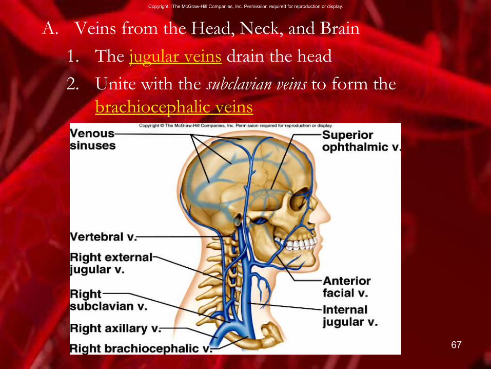
A. Veins from the Head, Neck, and Brain 1. The jugular veins drain the head 2. Unite with the subclavian veins to form the
brachiocephalic veins
CopyrightThe McGraw-Hill Companies, Inc. Permission required for reproduction or display.
68
A. Veins from the Upper Limb and Shoulder 1. The upper limbs are drained by superficial and
deep veins2. Major superficial veins
a) Basilic veins b) Cephalic veins
3. The major deep veins include a) Radialb) Ulnarc) Brachiald) Axillary veins
CopyrightThe McGraw-Hill Companies, Inc. Permission required for reproduction or display.

69

70
A. Veins from the Abdominal and Thoracic Walls Tributaries of the brachiocephalic and azygos veins drain the abdominal and thoracic walls
B. Veins from the Abdominal Viscera 1. Blood draining from the intestines
a) Enters the hepatic portal system b) Flows to the liver then into general circulation
2. The liver processes the nutrients absorbed during digestion and removes bacteria
CopyrightThe McGraw-Hill Companies, Inc. Permission required for reproduction or display.

71
1. Hepatic veins drain the liver2. Gastric veins drain the stomach3. Superior mesenteric veins lead from the small intestine
and colon4. Splenic vein leaves the spleen and pancreas5. Inferior mesenteric vein carries blood from the lower
intestinal area
CopyrightThe McGraw-Hill Companies, Inc. Permission required for reproduction or display.

72

73
A. Veins from the Lower Limb and Pelvis 1. Deep and superficial veins drain the leg and pelvis2. Deep veins include
a) Anterior and posterior tibial veins b) Unite into the popliteal vein and femoral vein
3. Superficial veins include:a) Small saphenous veins b) Great saphenous veins
4. These veins all merge to empty into the common iliac veins
CopyrightThe McGraw-Hill Companies, Inc. Permission required for reproduction or display.

74