hookworm toxocariasis and guinea worm dec2012 final.pdf
TRANSCRIPT
Hookworm
Ancylostoma duodenale, Necator
Americanus
Intestinal Nematode
Page 1
Aliaa Tayea*, Sina Helbig, Akre M Adja, Neil Arya**
Prepared as part of an education project of the Global Health Education Consortium and
collaborating partners
*First author **Corresponding author

4.1 Epidemiology
• Common disease with at least 740 million people
infected globally (estimates up to >1 billion)
• Causes more morbidity than any other geohelminth
principally by consequences of iron-deficiency
anemia
o Hookworm infection is the leading cause of iron
deficiency anemia worldwide
• Largely worldwide distribution, prevalent in all tropical
and subtropical countries, especially sub-Saharan
Africa, South China, Pacific and South-east Asia
Page 2

4.2 Risk factors
• Poor sanitation
• Walking barefoot in soil contaminated with feaces
• Infection by A. duodenale probably also occurs by oral and
transmammary route
Page 3
Photo: http://www.johntyman.com/africa/a442.jpg

4.3 Biology
• Human infection caused by 2 hookworms o Small white-grey or reddish-brown thread like worms
• Ancylostoma duodenale o Buccal capsule containing 2 pairs of teeth for attachment to
the small-intestinal mucosa
o Male: 1 x 0.5 cm
o Female: 1.2 x 0.6 cm
o Maximum egg output 15-18 months after infection
o Interval between infection and disappearance of eggs from stool with death of the worm averages 1 year
o Female produces 25,000-35,000 eggs/day (18-54 million during its lifetime)
o Adult worm lives 1-3 yrs (often longer)
Page 4

• Necator americanus
o Shorter and more slender than A. duodenale (1 x 0.4 cm)
o Smaller buccal capsule than A. duodenale
o Cutting plates instead of teeth
o Eggs slightly larger than A. duodenale (70 x 40 μm)
o Female produces ~20,000 eggs daily
o Adult worm lives 3-10 yrs (3-5yrs for female worm)
• Humans are definitive host
4.3 Biology
Page 5

4.3 Life cycle
• Eggs are deposited in duodenal lumen
• Leave body through feaces
• If deposited in damp shaded soil they hatch into
rhabditiform larva (first stage)
o Feeds on organic debris
o Becomes elongated and fully developed
• Larva moults to form a filariform larva (infective
stage moves away from the feaces into soil)
• Protected from desiccation, they can live in
warm damp soil for up to 2 yrs
• Filariform larva penetrates skin of host when
contact initiated receives host signal to
resume development
• Enters vasculature travels to lung breaks
through alveoli, moves up trachea, is
swallowed and reaches small intestine
• During migration 3rd moult takes place, upon
arrival in intestine 4th moult
• Worm attaches to the small-intestinal mucosa
where it sucks blood
• A. duodenale can also infect by ingestion
Page 6

4.4 Pathology
• 3 stages (first two usually only seen in primary infection, during larva migration phase)
o Invasive phase
Vesiculation and pustulation at entry site
o Migration via venous system through the lungs with small hemorrhages into the alveoli and eosinophilic and leucocytic infiltration Coughing, asthma and bronchitis (Loeffler´s Syndrome)
Larvae moved up, either by coughing or ciliary escalator to pharynx where they are swallowed move to intestines
o Established infection by adult worms in intestine
Seen in the inhabitants of endemic areas
May be asymptomatic or, in case of severe infections, lead to anemia
Page 7

4.4 Symptoms
• Initial invasive stage
o Entry site of larva: ground itch (irritating vesicular rash)
Limited to the area around entry points of the body (usually palms
and soles, between toes)
o Lasts up to 10 days
• Larva migration phase
o Appear 1-2wks after the primary infection, and depend on
worm burden
o Pulmonary symptoms with dry cough, asthmatic wheezing,
fever, high eosinophilia
Wheezing less pronounced than with A. lumbricoides
o Low-grade fever may be present
o Entire episode usually of 2-3m duration – mostly self-limiting
Page 8

• Later, established infection
o Epigastric pain upon worm migration
• Can be relieved by food DDx: duodenal ulcer!
• Symptoms peak at 30-45d after infection and gradually
disappear
o Occult blood in stools to frank melena
• Results when hookworm detaches from one site in intestine to
move to another location
o Iron deficiency anemia
• Occurs after iron stores are depleted
o Protein-depleting enteropathy (hypoalbuminemia): puffiness,
edema
o Retinal hemorrhages
4.4 Symptoms
Page 9

• Effects of anemia: malaise, digestive disturbance, no
wasting
o Each worm consumes .03-.6 mL of blood per day
o Usually 40-160 worms are enough to cause anemia
o 500-1000 = significant blood loss and anemia even in the
presence of iron supplementation
o Shortness of breath, high output heart failure
• Severe infection - persistent anemia in children may have
severe long-term consequences
o Stunting of growth and development, cognitive impairment
4.4 Symptoms
Page 10
4.5 Diagnosis
• Detection in stool o Ova: thin clear shell
o Sometimes also detection of rhabditiform larva in stool (DDx: Strongyloides)
o Ova appears about 42d after infection
o Sensitivity can be increased by examining multiple samples over consecutive days
o Kato-Katz smear provides quantitative estimate (samples should be examined within an hour of preparation or earlier, depending on heat and humidity conditions)
• Serological diagnostic o Multiplex real-time PCR
Page 11

4.6 Management/Treatment
• Treatment of anemia (iron supplementation)
• Treatment is targeted against adult stages
• Albendazole
o 400mg single dose (80% cure rate)
o 200mg daily x 3 days (100% cure rate)
• Mebendazole: only partially active and treatment over
multiple days might be required for severe infections
• Levamisole and pyrantel pamoate are less effective
Page 12

4.7 Control
• Proper disposal of faeces to minimize risk of contact with body o Provision/proper use of sanitation facilities
• Good hygiene to break fecal-oral transmission route
• In some (endemic) areas regular de-worming of children and
examination of lactating mothers
Page 13
Photo:http://artforgorillas.wildlifedirect.org/files/2010/03/Washing-Hand.-
Photo-by-Molly-Feltner.-Staying-Healthy-lessons-by-Art-of-Conservation.-
Rwanda-2010.jpg

Toxocariasis
ROUNDWORMS
Page 14

5.1 Epidemiology
• Result of infection with dog ascarid Toxocara canis (most common) or the cat ascarid Toxocara cati
• Cosmopolitan in distribution
• Often associated with A. lumbricoides and Trichuris
trichiura infection
• Mortality unusual; morbidity largely due to ocular
involvement o Toxocariasis is an important cause of reduced visual acuity in tropical
areas
Page 15
Photo:http://www.cdc.gov/parasites/images/toxocariasis/home_page_image_toxocariasis.jpg

5.2 Risk factors
• Exposure to (particularly young) dog and cat faeces
o Direct contact not necessary, as eggs need weeks of development in
soil to become infective
o Small children; more hand-to-mouth contact during play
• Outdoor parks in urban and suburban environments
o Most likely to be contaminated by animal faeces
o Children playing in the sand/soil higher risk of accidentally ingesting
Toxocara eggs
• Pet ownership (litter)
• Geophagia (both in children and adults)
Page 16

5.3 Biology
• Definitive hosts are dogs (T. canis) and cats (T. cati)
• Humans are incidental hosts; parasite does not undergo normal development in humans after ingestion
• Infected embryonated eggs ingested after exposure from soil/sand contaminated by dog/or cat faeces
• Eggs ruptured in GI tract, releasing larvae; further development is arrested at the larval stage
• Morphology similar to A. lumbricoides
o Male worms: 40 – 60 mm long; Female worms: 65 - 100 mm long
o Eggs: 85 x 75 µm
Page 17

• Larvae survive in humans for months to years, causing damage to
tissues as they wander through body o Complex mechanisms to evade immune system
• 3 recognized syndromes o Covert toxocariasis (long-term exposure to migrating juvenile larvae)
o Visceral larva migrans (VLM)
o Ocular toxocariasis (ocular larva migrans or OLM)
5.3 Biology
Page 18
Photo:http://cms.revoptom.com/handbook/IMA
GES/oct02_sec5_fig7.jpg

5.3 Life cycle
• Life cycle in cats and dogs is similar to A.
lumbricoides infection in humans
• Difference: transplacental infection is
common with offspring shedding numerous
eggs from birth
• Adult animals excrete few eggs
• Dogs/cats are infected by ingesting eggs
from contaminated soil
• Eggs hatch in stomach of humans
• Second stage larvae penetrate mucosa to
enter circulation via the mesenteric vessels
intestinal viscera and liver may stay
there or travel to lungs, brain, eye.
• Larvae are eventually destroyed by
granulomatous reaction blocks further
migration and causes pathology
• Larvae can remain alive for > 11yr in
humans
Page 19
5.4 Pathology
• Visceral larva migrans (VLM) o Stage larvae are arrested mostly in the liver where they cause
few or many lesions. Granulomas form which can be seen as white subcapsular nodules
o Other sites: lungs, kidneys, heart, striated muscle, brain, eye
• Ocular toxocariasis o Granulomatous reaction forms a large subretinal mass with a
superimposed pathology of choroiditis which can closely resemble retinoblastoma
• Tissue damage due more to host inflammatory response to larvae than to parasite itself
Page 20
5.5 Symptoms
• Symptoms depend on intensity of infection; most cases
asymptomatic
• Incubation period dependent on worm burden (weeks to
years)
• VLM can be self limiting to lethal (unusual)
• Ocular lesions can lead to strabismus, decrease in vision
or blindness
• Milder infection more common in adults: ocular
toxocariasis
Page 21
5.5 Symptoms – Covert toxocariasis
• Covert toxocariasis in children (mainly <5yrs) usually
subclinical or mild febrile illness
o May result from long term exposure to migrating juvenile larvae
o Can manifest as cough, behavioural or sleeping problems,
headache, chronic/recurring abdominal pain, anorexia
• May also have lymphadenitis, hepatomegaly
• Toxocara titres lower than in VLM, and eosinophilia less
common and less pronounced
• Long-term exposure of larvae to lungs can lead to asthma
Page 22
5.5 Symptoms - VLM
• Classic VLM syndrome
o Fever, coughing/wheezing, anemia, hepatomegaly, eosinophilia,
positive Toxocara titre
o Most commonly with heavy infection in childhood
• Pulmonary signs (e.g. coughing, wheezing), asthma
• Cardiac dysfunction
• Nephrosis
• CNS involvement: aseptic meningitis, mass lesions causing
seizures*, paresis (spinal cord lesions)
*less common, but may contribute to higher rates of epilepsy in parts of
developing countries with high infection rates
Page 23
5.5 Symptoms – Ocular toxocariasis
• Usually unilateral
• Presenting symptoms is often strabismus due to macular
damage low grade iridocyclitis can progress to general
endophthalmitis and retinal detachment
o Eye pain
• If lesion is central decrease in visual acuity
• Solid retinal tumor close to macula
• In early stages closely mimics retinal neoplasm since it is
raised above the level of the retina
• Later lesion remains a clear-cut circumscribed area of
retinal degeneration
Page 24

5.6 Diagnosis
• SVLM
• Stable persistent eosinophilia (sometimes >70%), leucocytosis, hypergammaglobulinemia
• Decreased albumin:globulin ratio, increase in IgG, IgH, anti-A and anti-B isohemaglutinin titres
• High resolution ultrasonography: hypoechoic areas in liver
• Demonstration of larvae is difficult and seldom achieved, sometimes found partially destroyed in centre of granuloma
• Serology: ELISA using excretory-secretory antigens harvested from second stage larvae in vitro
• Sensitivity > 95%, specificity > 90%
• Can be improved by indirect Ab – competition: e.g. specific IgE, IgG4
• Ocular toxocariasis o Ophthalmological examination; second stage larvae rarely
seen with slit-lamp microscope in anterior chamber of eye
Page 25

5.7 Treatment
• Anti-helminthic therapy for VLM
o Albendazole (preferred), Mebendazole, Thiabendazole
o DEC reportedly more effective than benzimidazoles but more adverse reactions
• In VLM, eosinophilia may persist over months after clinical cure (decrease in hepatomegaly, subsiding fever)
• Possible increased inflammatory reaction during therapy; corticosteroids often beneficial
• Treatment of OLM more difficult, surgical therapy may be needed for severe disease
• Recurrence unlikely, providing risk factors mitigated
Page 26

5.8 Control
• Breaking hand-to-mouth transmission especially in children
o Hand washing before eating, especially after handling pets, pet litter or
soil
• General education on disease, transmission and risk factors for
toxocariasis
• Animal control in public areas
o Fencing, limiting access to
playgrounds
• Regular de-worming of
dogs and cats
Page 27
Photo:http://www.vetbiomed.murdoch.edu.au/numbatnews/conte
nt_images/Deworming-CommunityProject.JPG

Dracunculiasis
Guinea worm disease
Page 28

6.1 Epidemiology
• Dracunculus = Latin “little dragon”; also called “Guinea fire worm”
• Infection caused by nematode Dracunculus medinensis
• Found in abundance in natural freshwater bodies
• Presence is an indicator of extreme poverty
• Mortality is low - associated with untreated secondary infection
• Morbidity is high - associated with months of debilitating pain,
incapacitation
Page 29
Photo:http://plpnemweb.ucdavis.edu/nemaplex/taxadata/Dmedinensis.HTM

6.1 Epidemiology
• > 3.5 million cases, in 20 countries, reported in the 1980s
• Following eradication campaigns since 1986, dramatic
reduction (>99%) in reported cases
o 5,000 in 2008
o <1,800 in 2010 (94% in South Sudan)
• Sustained campaigns (largely by Carter Center) of community
education, safer water provision (especially using appropriate
filters), political mobilization
• Remains endemic in only 6 African countries: Ethiopia, Ghana,
Mali, Niger, Nigeria and Sudan
Page 30
WHO. (2009). Action Against Worms. Newsletter Issue 13/ http://www.who.int/neglected_diseases/preventive_chemotherapy/Newsletter13_En.pdf

6.1 Distribution
Page 31
http://www.who.int/health_mapping/programme_support/guinea_worm/en/index.html

6.2 Risk factors
• Dependence on poor quality drinking water, unsafe water sources
• Drinking water from still freshwater reservoirs
o Ponds, shallow wells, streams, etc.
• Contact of affected individuals with water sources, continuing
transmission cycle
• Civil unrest, hindering other efforts to control disease and associated
risk factors (e.g. S. Sudan)
Page 32
Photohttp://love2others.org/wp-content/uploads/2012/05/33.jpg
6.3 Biology
• Nematode parasite related to filarial worms
• Larvae released into water by adult female worms
• Vectors: cyclopoid copepods (water fleas) – tiny free
swimming crustaceans – swallow larvae after release
o Development within vector, larvae infective after ~ 3wks
• Humans acquire infection by drinking water containing
the copepod vectors infected with guinea worm larvae
• Stomach digestive acids kill copepods, but not larvae
o Larvae migrate through stomach wall into subcutaneous
tissue of abdomen, thorax
Page 33

• After 2-3 months, worms develop and mate, after which males die; females continue their development and migration
• Adult female guinea worm o Up to 60-80cm long and 1.5-2mm thick
o Inhabits the subcutaneous connective tissues of humans
o Located anywhere in the body; in late stage usually attracted to lower extremities (most likely to come into contact w/water)
• Formation of blister, which bursts after ~48hrs
• Female worm protrudes its tip through resulting ulcer, releasing fluid filled with larvae upon contact with water
• Embryos taken up by vector, and cycle begins again
6.3 Biology
Page 34

6.3 Life cycle
• Human is seeking freshwater reservoir for relief blister ruptures discharge of first stage larvae into water
• It remains protruding for the next 2-6 weeks, releasing larvae each time
• Larvae are infective in water for 5-6 days
• For further development must within this period be swallowed by a copepod penetrates gut wall and reaches the infective 3rd stage within 2 weeks
Page 35

6.4 Symptoms
• Usually asymptomatic in prepatent period (interval
between infection of an individual by a parasitic organism
and the first ability to detect from that host a diagnostic
stage of the organism)
• First symptoms occur a few days prior to the worm piercing
the skin, and largely related to hypersensitivity reaction
• If worm is close to joint may also cause arthritis
• Dermis becomes elevated and blister develops
o with intense burning, itching sensation
o ~24-48 hrs later blister bursts
o Intense sensations provoke patient to submerse area in water, which
relieves some of the burning sensation
Page 36

• Further inflammation or calcification of worms may cause stiff
joints in lower limbs crippling of patient
• If secondary bacterial infection of ulcer (common), cellulitis or
tetanus can develop
• If worm is only incompletely extricated, the worms withdraws into
the host causing a severe inflammatory reaction with ulcer
formation and scarring
• Encysting or calcification of worms, sterile subcutaneous
abscess formation
• Rarely migration of worms to vital organs
o Brain - cerebral/subdural abscess can develop
o Eyes - blindness can develop
6.4 Symptoms
Page 37
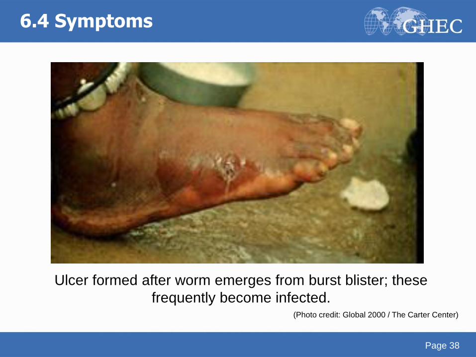
Ulcer formed after worm emerges from burst blister; these
frequently become infected. (Photo credit: Global 2000 / The Carter Center)
6.4 Symptoms
Page 38
6.5 Diagnosis
• Diagnosis usually clinical; cannot diagnose in prepatent period =
first 8-10 months of infection
o Shortly prior to appearance the worm can sometimes be palpated
under the skin
o Later: observing female protruding from the blister
o Typical appearance of blister with local itching, burning pain
• Serology is of no practical use in diagnosis
o Constant exposure in high endemic areas – variably detectable
antibody titers
o No acquired immunity
o People in endemic areas suffer from repeated infections
• High eosinophilia is common
• Dead calcified worms can be seen on radiographic imaging
Page 39

6.6 Treatment
• Affected areas should be kept clean & bandaged
• Most effective: slow extraction of emergent guinea worm
o Protruding part of the adult female worm is attached to a stick, which
is twisted a small amount each day until the whole worm has been
removed (can take up to a month)
o Care should be taken not to break the worm
o Should be accompanied by supportive antibiotics, cleaning and
dressing of ulcers as well as Tetanus vaccination
• Antibiotics for secondary/superinfection
• Analgesics for pain
Page 40
Photo:http://www.parasitemuseum.com/wp-content/gallery/guinea-
worm/guineaworm2j_lores.jpg
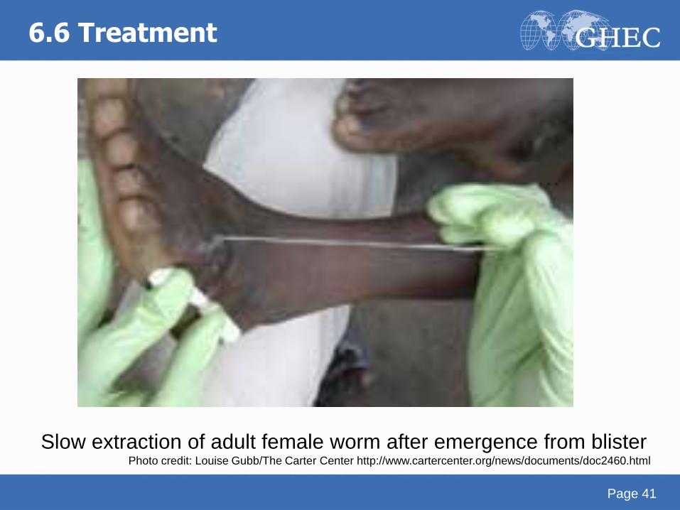
Slow extraction of adult female worm after emergence from blister Photo credit: Louise Gubb/The Carter Center http://www.cartercenter.org/news/documents/doc2460.html
6.6 Treatment
Page 41

• Surgical extraction of the guinea worm prior to eruption – has resulted in less associated disability o However, not widely available in problematic area
• No curative antihelminthic treatment is available o Niridazole has been reported to decrease inflammation
around the worm, allowing for easier extraction
o Metronidazole, thiabendazole (adults) also used as adjunct to stick removal; however to be used with caution due to one study’s finding that these were associated with aberrant migration of worms
6.6 Treatment
Page 42
6.7 Control
• Community education on disease & transmission
o Educating affected individuals not to immerse the affected areas in
water which is used for public consumption
• Promotion and provision of safe drinking water sources
• Boiling water
• Point-of-use filtration of drinking water to “strain” copepods
o Nylon filters, straw filters
o Low-cost methods effective, e.g. filtration through clean cloth
• Larvicide to kill copepods
Page 43
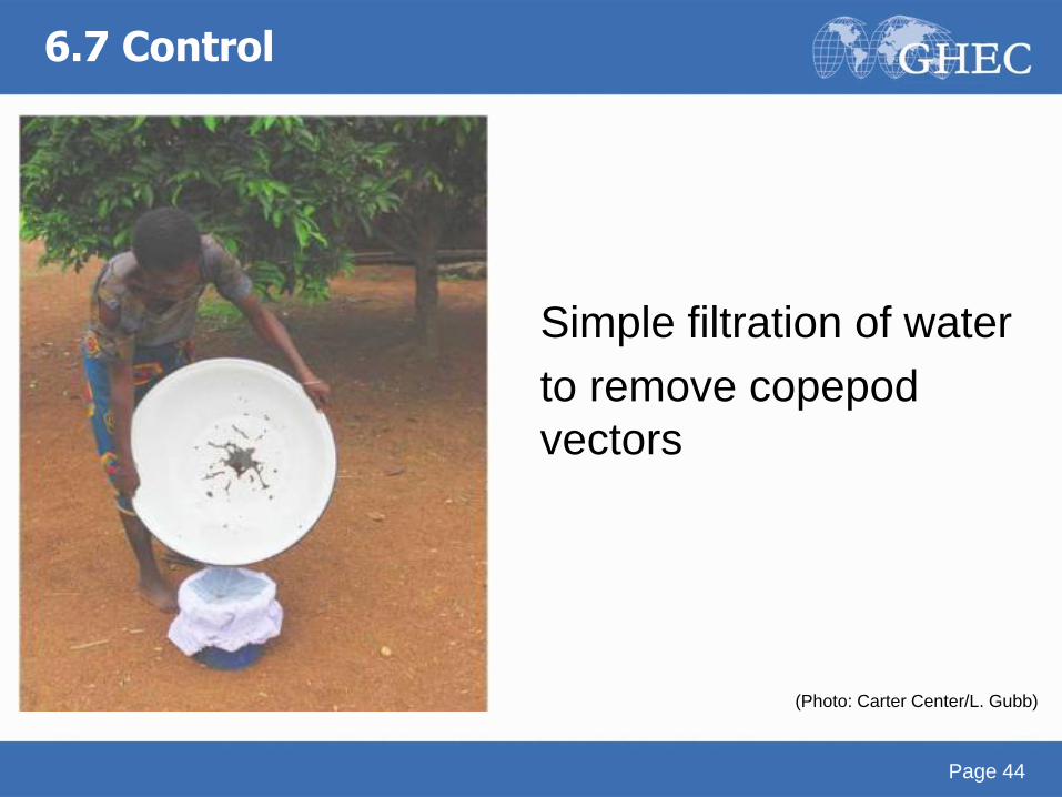
Simple filtration of water
to remove copepod
vectors
(Photo: Carter Center/L. Gubb)
6.7 Control
Page 44

Pipe filters: portable, for use anytime and at any water source available. Photo: Carter Center / L. Gubb
6.7 Control
Page 45

Page 46
Acknowledgments
• Thanks to Jenna Kelly, Shazeen Bandukwala and
Melissa Whaling for critical editing.
• We appreciate Tim Brewer and Jackeline Alger for
thoughtful review.
Page 46

Credits
Akre M Adja1, Sina Helbig2, Alia Tayea3,
Neil Arya4
1: Institut Pierre Richet, Université de Cocody Abidjan
2: Boston University School of Medicine, Division of Infectious
Diseases, Boston, MA, USA
3: Médecins Sans Frontières
4: Western University, University of Waterloo, McMaster University
Contact [email protected]

The Global Health Education Consortium and the Consortium of
Universities for Global Health gratefully acknowledge the support
provided for developing teaching modules from the:
Margaret Kendrick Blodgett Foundation
The Josiah Macy, Jr. Foundation
Arnold P. Gold Foundation
This work is licensed under a Creative Commons Attribution-Noncommercial-No Derivative Works 3.0 United States
License.