how does the cytoskeleton read the laws of geometry in...
TRANSCRIPT

Development Supplement 1, 1991, 55-65Printed in Great Britain © The Company of Biologists Limited 1991
55
How does the cytoskeleton read the laws of geometry in aligning the
division plane of plant cells?
CLIVE W. LLOYD
Department of Cell Biology, John Inncs Institute, John Innes Centre for Plant Science Research, Colney Lane, Norwich NR4 7UH, UK
Summary
Since Robert Hooke observed the froth-like texture ofsectioned plant tissue, there have been numerousattempts to describe the geometrical properties of cellsand to account for the patterns they form. Some aspectsof biological patterning can be mimicked by compressedspheres and by liquid foams, implying that compressionor surface tension are physical bases of patterning.
The 14-sided semi-regular tetrakaidecahedron en-closes a given volume most efficiently and packs to fillspace. However, observations of real plant tissue (and ofsoap bubbles) in the first half of this century establishedthat plant cells only rarely form this mathematicallyideal figure composed predominantly of 6-sided poly-gons. Instead, they tend to form a topologicallytransformed variant having mainly pentagonal facesalthough there is variability in the number of sides andthe angles formed. But the one irreducible component ofnormal cell and tissue geometry is that only three edgesmeet at a point in a plane. In solid space, this gives rise totetrahedral junctions and it is from this that certainlimitations on sidedness flow. For three edges to meet ata point means that there must be an avoidancemechanism which prevents a new cell plate fromaligning with an existing 3-way junction. Sinnott andBloch (1940) saw that the cytoplasmic strands whichprecede the cell plate, predicted its alignment and alsoavoided 3-way junctions in unwounded tissues. Re-cently, F-actin and microtubules have been detected inthese pre-mitotic, transvacuolar strands. The questionconsidered here is why those cytoskeletal elements avoid
aligning with the vertex where a neighbouring cross wallhas already joined the mother wall. An hypothesis isdiscussed in which tensile strands - against a back-ground of cortical re-organization during pre-mitosis -tend to seek the minimal path between nucleus andcortex. In this way, it is suggested that unstable strandsare gradually drawn into a transvacuolar baffle (thephragmosome) within which cell division occurs. Ver-tices are avoided by the strands because they constituteunfavoured longer paths. The demonstrable tendency oftensile strands to contact mother walls perpendicularlywould seem to account for Hofmeister's and Sachs' rulesinvolving right-angled junctions. As others have dis-cussed, such right-angled junctions give way to co-equal120° angles between the three walls during subsequentcell growth. It is this asynchrony of cell division - whereattachment of a cell plate causes the neighbouring wall tobuckle - that forms a vertex to be avoided by subsequentpre-mitotic strands in that neighbouring cell. In thisway, successive division planes would not co-align. It istherefore suggested that the exceptional formation of4-way junctions in wounded tissue results from the factthat adjacent cells divide simultaneously; the lack of pre-buckling of a common wall under these circumstancesmeans that there is no vertex to be avoided by theminimal path mechanism.
Key words: plant cells, packing patterns, cytoskeleton,geometry.
The form of an object is a 'diagram of forces',D'Arcy Thompson (1942)
Honeycombs, compressed spheres (such as lead shotand pea seed), solid and liquid foams and plant cells in atissue show fascinating, attractive and rather similarpacking patterns. Some - like the honeycomb - appearto be highly regular, composed of identical cells. Othersmay be less well ordered, composed of units of differentsize and shape. But the ways in which cells (in the widersense) can pack is limited and the limitations have beendistilled over the years, into various rules suggesting a
basic conformity. The geometrical constraints on fillingspace apply as much to plant cells as to inanimateobjects. However, although patterns may be homolo-gous, and reflect the same underlying laws, the ways inwhich they are achieved may be quite different. Plantcells are more than just compressible bodies and it istheir ability to regenerate packing patterns by theprocess of cell division that is of interest here.
Up until about 1950, there was great interest indescribing cellular geometry by scientists such as,D'Arcy Thompson, 1942; Lewis, 1923-1943; Matzke,1939, 1945; Marvin, 1939. Most of this work dealt with

56 C. W. Lloyd
comparing real cellular polyhedra with mathematicallyideal figures, and with soap bubbles which readilyexemplified principles of surface tension in achievingstability. In a sense, the living, biological dimension wasmissing from such descriptions, since more emphasiswas placed on the shape achieved than on the act ofplacing a cell plate across a mother cell. Nineteenthcentury continental scientists did attempt to crystallizetheir observations on wall placement, into rules. As weshall see, such rules are not always adhered to, but thefact that they are at least good approximations suggeststhe action of a common underlying cellular mechanism.Because daughter cells conform to the restrictions ofpacking space, the mechanism for dividing the cellsmust be sensitive to, and somehow read, thoserestrictions. The empirical rules provide no clue to thecytoplasmic mechanism of establishing the cell plate butthe important and central work of Sinnott and Bloch inthe 1940s (1940, 1941a,b) did show that the alignmentof the plate could be predicted by the orientation of pre-mitotic cytoplasmic strands. The question is therefore,how is the behaviour of cytoplasmic strands compatiblewith the laws of solid geometry that lead to efficient cellpacking?
A reasonable description can now be proposed forhow actin filaments and microtubules, (particularlythose in transvacuolar cytoplasmic strands), reorganizeand predict the plane of cell division. The aim of thisessay is to set these recent advances against thegeometrical pre-occupations of the first half of thiscentury.
The efficient packaging of space: polygons intopolyhedraThe sphere represents the most efficient way (surfacearea/volume ratio) of enclosing space but a collectionof spheres is not, because of the gaps between. But solidbodies can be compressed together to eliminate theinterstices, and bubbles aggregate. As they do, theircircular cross-sections are deformed to make polygons.The solid bodies thus constructed pack together butwhat forms do these polyhedra take and whichrepresents the most efficient packaging of space?
By joining regular polygons at their edges, withoutdistorting them, five regular convex polyhedra can beproduced. Four triangles combine to form a tetra-hedron, eight triangles form the octahedron, twentytriangles can be joined to form the icosahedron.Respectively, these regular polyhedra have 3-way,4-way and 5-way corners. Six squares can be joined toform the cube (with three-way corners) and twelveregular pentagons can be combined to form a dodeca-hedron with 3-way corners. Space cannot be enclosedby combining only 6- or 7- or 8-sided polygons and thusthere are only 5 simple polyhedra. As long as thepolygons, and the angles they form, are kept regular,there can be no more than 5. These regular solids wereknown to the Greeks and are referred to as the Platonicbodies. Joining regular plane figures of more than onetype - but keeping the corners or vertices the same -yields semi-regular polyhedra. Excluding prisms, only
14 such Archimedean bodies can be made (Stevens,1976). Of these, the tetrakaidecahedron is of thegreatest interest. It has eight hexagonal and six squaresides and is perhaps most easily conceived as a cubewith extra facets produced by shaving the corners (atruncated octahedron).
In discussing the theory of polyhedra, Euler's Law(see Thompson, 1942; Gibson and Ashby, 1988) has acentral importance. This states that for every polyhed-ron, the sum of the faces and corners outnumbers theedges by two. Various corollaries flow from this: nopolyhedron can exist without a certain number oftriangles, squares or pentagons in its composition; andno polyhedron can be constructed entirely of hexagons.Furthermore, it follows from Euler's Law that anirregular 3-connected aggregation of polyhedra has anaverage of six sides per face. A 5-sided face can only beintroduced if a 7-sided face is created, for balance.
As D'Arcy Thompson (1942) states, the broadgeneral principle to be learned is that we cannot groupas we please any number and sorts of polygons into apolyhedron, but that the number and kind of facets inthe latter is strictly limited to a narrow range ofpossibilities.
Soap films illustrate the juncture angles of space-fillingbodiesThe apparent similarity between soap bubbles in a foamand plant tissue has long been noted, and gives rise tosome interesting demonstrations.
By sandwiching three drawing pins (thumb tacks)between two plates of glass (Fig. 1), soap films areobserved to form between the pins when the wholeapparatus is removed from a soap solution (see Stevens,1976). The films meet at co-equal angles of 120°(Fig. 1A). Surface tension drives the films to seek theminimal path and V-shaped and A-shaped figures donot form since they would use significantly morematerial.
A fourth thumb tack can be added, defining thevertices of a square (Fig. IB). When removed from thesoap solution, the film does not enclose three sides of
Fig. 1. Soap films formed by thumb tacks sandwichedbetween two glass plates then dipped into a soap solution.In A, the films do not form a triangle around the boundarybut meet in the 3-way junctions of 120°; this uses lessmaterial. Even when the shape of the triangle is altered bymoving the pins, the 3-way junction remains the same. Thesecond illustration (B) demonstrates that soap filmsnaturally form double 3-way junctions within a 4-pinarrangement. Again, this uses less material than any patharound the edges (see Stevens, 1976).

Cytoskeleton and cell geometry 57
r \\ — - -1
Fig. 2. The Belgian physicist Plateau (1801-1883)demonstrated how soap films spanned the contour ofvarious wire cages, forming a minimal surface of least area.The above illustrates the arrangement formed within atetrahedron. This shows that only 3 films meet at an edge,and that only 4 fluid edges meet at a point - all at co-equalangles. As D'Arcy Thompson (1942) noted, this symmetryapplies for any close-packed tetrahedral aggregate of co-equal spheres: a froth of soap suds or a parenchyma ofcells.
the square, nor does it form an X. The X - if formed -would use 6 % less material than needed to enclose thesquare on 3 sides. Instead, the film unerringly forms adouble, 3-way junction (see Fig. IB) which is moreeconomical still, in using 9% less material. Formationof 3-way junctions by elements capable of exertingequal tension is a crucial process in biological patternformation, as we shall see later.
This sandwich system of films is essentiallytwo-dimensional, demonstrating that three films meet apoint at equal angles of 120°. Plateau's beautifuldemonstration (see D'Arcy Thompson, 1942) showswhat happens when films meet in three dimensions(Fig. 2). He constructed a tetrahedral wire cage anddipped it into soap solution. When withdrawn, six filmscould be seen to meet in threes, at four edges. Thisdemonstrates the principles of minimal area that onlythree films meet at an edge, and no more than fouredges meet at a point. The four edges meet at co-equalangles which, in this case, are 109°28' 16". This is thetetrahedral angle which runs throughout the simplehomogeneous partitioning of three-dimensional space.
Bubbles, unencumbered by frames, are free to formfoams; these coarsen with time, demonstrating theconstant readjustment of fluid walls as surface tensionconstantly strives to compartmentalize maximum spacewith minimal surface area. Although we can talk aboutlimits that define the numbers of edges and the size ofangles, such foams are heterogeneous. Contemplatingthe difficulty, in nature, of making a regular foam, LordKelvin (Sir W. Thomson, 1887; 1894) proposed thetetrakaidecahedron to be the ideal volume-containingform with space packing properties (Fig. 3A). Of thespace-filling polyhedra with plane sides of a similarshape, the rhombic dodecahedron possesses the mini-mal surface area/volume ratio but this is improved
Fig. 3. (A) Lord Kelvin demonstrated that the semi-regular polyhedron - the tetrakaidecahedron - mostefficiently divided space with minimal partitional area, ie itencloses a given volume with minimal surface area andclose-packs to fill space. The figure has three pairs of equaland opposite quadrilateral faces, and four pairs of equaland opposite hexagonal faces. This figure - in which all thefacets are plane - is the ortho-tetrakaidecahedron.However, this is not a minimal figure. (B) shows such aminimal 14-hedron in which the quadrilaterals have curvededges and the hexagons have slightly curved surfaces. Thiscurvature meets the stability requirements demonstrated byfroths of bubbles, such that it forms the tetrahedral angleof 109° 28' upon packing. Although these figures representmathematical solutions, they do not embody the sidednessdisplayed by real plant tissues. (C) shows Williams' (1968)/S-tetrakaidecahedron produced by topologicallytransforming the Kelvin body. This has a predominance ofpentagonal faces.
upon by the semi-regular tetrakaidecahedron. How-ever, no planar polyhedron that packs to fill space alsosatisfies the stability requirement that juncture anglesshould be 109°28'. From observing other experimentsof Plateau, showing that congruent soap films gentlycurve, Lord Kelvin proposed the minimal tetrakaideca-hedron (Fig. 3B). By distorting this 14-hedron so that ithas doubly curved hexagonal faces, and quadrilateralfaces with bowed edges, the so-called Kelvin body canbe made to satisfy the requirements of 109°28' junctureangles.

58 C. W. Lloyd
But plant cells are not ideal figuresOf the regular and semi-regular polyhedra, the dodeca-hedron and the tetrakaidecahedron are the mostefficient in packing space. However, there has been along history in attempting to define whether any suchideal figure is actually the one that nature uses tocompartmentalize cellular space.
Stephen Hales, in 1727, compressed pea seed in a potto form pretty regular dodecahedrons. This, of theregular polyhedra with identical faces, has the leastsurface area/volume ratio, but Matzke (1939, 1950)could not confirm the observations of this frequentlycited classical experiment. Matzke suggested that Halesmight have been influenced by the stacking of cannonballs, where 12 surround a central one. Compressingsuch an aggregation (without slippage) would produce arhombic dodecahedron, providing that the regular unitswere first stacked in that configuration and not, aspresumably was the case for Hales' peas, poured intothe container. Marvin (1939) and Matzke (1939)repeated this experiment with lead shot and found thatregular units, when stacked like cannon balls, didcompress to form dodecahedra, but that poured shotproduced irregular 14-sided bodies. Mixing large andsmall shot produced bodies with more or less than 14sides, the average being less than 14. This agreed withLewis' (1923) detailed observations of undifferentiatedplant tissue, showing an average of 14 faces.
In a similar investigative vein, Matzke (1945) re-examined bubbles in a foam and found an average of13.7 contacts. Just as he could not find a single cell withthe 6-0-8 formula (numbers of 4-, 5- and 6-sidedpolygons respectively) required by the Kelvin body,neither did he find a single central bubble amongst 600to have the ideal configuration. Indeed, the commonestwas 1-10-2, revealing a predominance of pentagonalfaces. As Stevens (1976) discusses, polygons meeting atthe tetrahedral juncture angle of 109°28' 16" must have5.1 edges, and in order to make a polyhedron, 13.394 ofthose polygons must join at 22.789 corners. These arethe geometrical constraints necessary to make continu-ous three-dimensional networks. Matzke's direct obser-vations established that the theoretical ideals areapproached in averages - an average of 5-6 edges perface, 13-14 faces and 22-23 corners in a froth ofbubbles. Clearly, bubbles cannot be stacked, bubblesare of different sizes, and unlike pre-arranged cannonballs, bubble walls slip and adjust as the foam degrades.Similarly, plant cells are produced asynchronously, cellsare not of equal size, the network expands and suchgrowth is often polar. For plant cells, therefore, thehistory of divisions is an important aspect of shape sincethe majority of cell facets are inherited by daughters,from the mother cell. Plant walls too, are not producedas a compression of spheres but by the deposition of aninitially non-rigid cell plate.
Searching for minimal tetrakaidecahedra amongstleaf parenchymal cells, in order to record the curvatureof the walls, Macior and Matzke (1951) had greatdifficulty in identifying minimal figures. Again,although the average number of cell faces tended
towards 14, the authors illustrate great variation inachieving the average. And again, the occurrence of5-sided faces was stressed. Williams (1968, 1979)collected these examples, together with those fromuniform bubbles, mixed bubbles and /J-brass grains toillustrate the point that 5-edged faces occur mostfrequently. This detracts from Kelvin's tetrakaideca-hedron as a paradigm but Williams did show that thisfigure could be topologically transformed to another 14-sided figure - the ^3-tetrakaidecahedron - with spacepacking properties (Fig. 3C). The /3-figure has twoquadrilateral, eight pentagonal and four hexagonalfaces. This has an average of 5.143 sides per face. Thisaccommodates the tendency towards 5-sidedness ob-served in the faces of naturally packed polyhedraalthough it requires about 4% more surface than theKelvin body to enclose the same volume.
The message for plant material is that there is no oneway of enclosing space; cells may tend to have 14 sidesbut they vary in their number of edges, five edges beingpreferred to six. There are several reasons whyvegetable polyhedra are variable but the way in whichthey divide is likely to be an important aspect of this. Inthe next section, the impact that division has on thepatterning of plant cells is examined.
Some feature of cell division is concordant with cellpacking principlesLewis (1926) studied the anticlinal division of cells inthe epidermal mosaic of cucumber epidermis. Epider-mal cells are different from those of internal tissue, forthe absence of external neighbours replaces four facetswith a free surface, giving an average of 11 instead of 14sides. Lewis considered the two-dimensional epidermalmosaic as being composed, basically, of hexagons.Although it is clear that the number of cell sides and cellsizes is variable, Lewis was engaged in explaining howdivision tended to restore the hexagonal average. Inepidermis, periclinal divisions are generally suppressed,inhibiting stratification, and so anticlinal (radial)divisions are visible - like hedges across a patchwork offields - as they divide cells in a mosaic. When ahexagonal polygon divides (Fig. 4), avoiding forming4-rayed intersections, it forms two pentagons. Twoadjacent cells are facetted in this process - acquire anextra face - and become heptagons. This maintains theaverage of six sides.
An octagon sometimes replaces two heptagons and asdivision proceeds, the active division of cells and thepassive acquisition of facets by neighbours tends torestore the primary hexagonal form.
This relates to division of polygons within a two-dimensional mosaic but Lewis (1928) does, parentheti-cally, extend the case to solid bodies in a tissue.Division of a 14-hedron adds 14 surfaces to theassemblage of polyhedra. Each half of the divided 14-hedron has 11 surfaces, and 6 adjacent polyhedra eachreceive one added surface; 22+6-14=14. A relatedobservation, known as the Aboav/Weaire Law (seeGibson and Ashby, 1988), and derived from soapbubble honeycombs, is that cells with more sides than

Cytoskeleton and cell geometry 59
Fig. 4. From a study of cucumber epidermis (A), Lewis(1926) concluded that a hexagonal cell was just as likely todivide in planes a, b or c but not in plane d, which wouldresult in a 4-way junction. When such a cell divided (B) itproduced two daughters with pentagonal outer faces.Attachment of the cell plate to two neighbouring cellsdivided the attached walls, giving each an extra facet. Onedivision therefore produced two pentagonal and twoheptagonal cells, maintaining the hexagonal average.
average have neighbours with fewer sides than average.Gibson and Ashby use Euler's Law and the Aboav/Weaire Law in characterizing cellular solids such asfoams, but they also make use of a rule ascribed toLewis (1943): that the area of a cell varies linearly withthe number of edges. These latter two rules helpdescribe the effects of coarsening in foams butbiological cells can mitigate the effects of coarsening byadding new walls, ie biological cells can regenerate. Themessage to be extracted from this is that the passiveacquisition of facets, by cells surrounding a dividingcell, increases the size of neighbours. And, according toLewis (1943), the epidermal cells studied were morelikely to divide perpendicular to the free surface, themore sides they possessed; the net result of dividinglarge cells was to reduce their number of sides towardsthe average.
Lewis (1936, 1943) also developed ideas on howelongating cells in a shaft divide in a manner that retainsparticular cell proportions and relationships withneighbours. He considered a 14-hedral elongate cell asa hexagonal prism (Fig. 5). The edges on the lateralfaces alternate 1/3, 2/3 etc. around the cell thus makingalternating pairs of rectangular hexagons and quadrila-terals. The important feature of this particular configur-ation is that it demonstrates a uniform and maximumavoidance of 4-rayed vertices within the figure itself iecorners are only made as the meeting of three edges.Plotting the level at which transverse walls are actually
, — •
\
4
\
6
\
-. '
6
4
4
6
Fig. 5. Lewis' (1936) prismatic tetrakaidecahedron. The i,\ alternation of lateral cross walls shows maximalavoidance of 4-way junctions (which would form anencircling belt) and maintains the original cell proportions.
attached in 100 cells, Lewis found that this 1/3, 2/3alternation on opposite sides of a cell did exist and it isclaimed that this 1/3, 2/3 succession is the only patternin which bisection maintains hexagonally-sectionedcells in their original proportions. And since divisionaffects the sidedness of neighbours, some rhythm wouldappear to be required in the timing of divisions if theoriginal proportions are to be maintained duringgrowth.
As we have seen, such a prismatic tetrakaideca-hedron of the 6-0-8 formula (a distorted version of theKelvin body) does not seem to be favoured by real cells.Nevertheless, the principle demonstrated by thisillustration seems to hold (and was later taken up bySinnott and Bloch, 1941a), namely, that formation of14-hedra and their maintenance during subsequentdivisions, requires the maximum avoidance of 4-rayedintersections. The latter, if produced, would lead to acheckerboard of cells in ranks and files, instead of thebonded brick appearance of cells packed (in section)with 3-way junctions.
Framing the rules for division plane alignmentThree laws or rules have been drawn from directobservation of the way that cell plates meet existingwalls. The first belongs to Hofmeister (1863) who statedthat the partition wall always stands perpendicular towhat was previously the principal direction of cellgrowth, ie generally perpendicular to the long axis ofthe cell. Sachs (1878) proposed that the cell tends todivide equally, and each new plane of division tends tobe perpendicular to the previous plane. Errera (1888)further proposed that the cell plate is the minimal areafor halving the cell's volume.
In a series of papers, Korn re-examined the utility ofthese rules - a valuable exercise which placed themechanics of cell plate alignment in centre stage ratherthan seeing cell shape as the expression of packingforces or mathematical inevitabilities. In 1974, Kornmodelled cell growth by cutting three-dimensionalblocks according to two rules only: Hofmeister's rulethat cell division is perpendicular to the long axis, and arule ascribed to Sinnott and Bloch (1941a), that cells

60 C. W. Llovd
divide to form only 3-rayed vertices even if it requiresunequal division. Using these rules, blocks could be cutrepeatedly to yield cells whose sides closely correspondto those observed in real tissue. Since the application ofthese two rules was sufficient to yield data comparableto those derived from real tissues, Korn concluded thatproposing further shaping forces was unnecessary.Although he dismissed 'mutual pressure' as a shapingforce in plant tissue, Korn did muse upon why thegeometry of cells formed by progressive halving, andthose of soap bubbles contoured by mutual pressureforces and slippage of films, should both average 14sides.
Perpendicular attachment of cell plates and thegeneration of 120° anglesA common strand running through the laws ofHofmeister, Sachs and Errera is the perpendicularity ofthe cell plate to existing walls. How does this give rise tothe 120° edge angles approximated when three maturewalls meet? D'Arcy Thompson (1942) explained this onthe basis of relative tensions (Fig. 6). A new partitionwall meets an old wall at 90° because its tension is smallcompared to what theirs has become. It is not themeeting of three co-equal partners that occurs whenbubble walls meet. However, with subsequent growth,the tension of the walls becomes equal, realigning thejuncture to co-equal angles of 120°.
Even where expansion is asymmetric, such that thecells markedly elongate to form rectangular hexagons,D'Arcy Thompson illustrates local curvature aroundthe triple junction in an attempt to meet - if onlymicroscopically - the 120° angles. The 120° and 90°angles are found in a wide variety of materials. D'ArcyThompson (1942) notes that fine veins on a dragonfly'swing meet in threes at 120°, whereas such fine veinsmeet the stronger ribs at 90°. Similarly, wax walls in ahoneycomb meet each other at 120°, but meet thewooden walls of the hive at 90°. Soap films meet in
threes at 120°, but contact the containing vessel at 90°.These angles are also the ones in which materials tendto crack. Elastic materials such as rock and mud formcracking patterns based on sudden production of 3-wayjoints (Stevens, 1976). By contrast, inelastic material,such as the glaze on a piece of pottery, cracks to relievethe stress first in one direction and then at right anglesto relieve the perpendicular stress. This latter, essen-tially orthogonal pattern, is produced by sequentialformation of new cracks perpendicular to older ones.This returns us to D'Arcy Thompson's conclusion forbiological materials, that Sachs' rule is limited to caseswhere one wall grows stiff or solid before anotherimpinges upon it. The difference in strength betweenthe mother wall and the cell plate is suggested to yieldperpendicular junctions, which, when the new plategains strength (or can exert equal tension), allows thethree facets to re-arrange, forming co-equal angles of120°.
Korn (1980) offers a variant of this explanation(Fig. 7). He finds that the cell plate does not expand forone generation. Certainly, the new cell plate is callosicsuch that its initial lack of expansion might be attributedto its slow conversion to a regular cellulosic wall. A cellbecomes marked by the external attachment of a cellplate deposited across a dividing neighbour. As the cellexpands, its wall in contact with the cell plate becomesfacetted, that is, the non-expanding plate causes theolder wall to hinge at that junction. Perpendicularjunctions thereby change, and planar surfaces becomecurved as the cell plate acts as a brake on its faster-expanding neighbour.
According to Korn (1980) therefore, this change injuncture angles occurs because of differential expansionwhile the plate is immature, not when the platebecomes strong enough to exert equal tension.
Whatever the reason, lack of expansion or lack oftoughness, the curvature caused by the recession of thecell plate away from the centre of the expandingneighbour provides one explanation for the curvatureof leaf parenchymal cells illustrated by Macior andMatzke (1951), and required of minimal, as opposed toorthic, polyhedra.
Fig. 6. The 120° angles of Fig. 1 depend upon the meetingof 3 films of equal tension. D'Arcy Thompson (1942)observed (A) that the new partition walls generally meetmother walls perpendicularly because the tension in theformer is smaller. However, as the new wall strengthens(B) it is able to exert equal tension and the network adjustto form co-equal angles of 120°.
Fig. 7. Korn (1980) measured new cell walls and concludedthat they did not expand for one generation. Meanwhile,other cell walls continued to expand (A-B), causing themto curve away from the non-expanding attached wall. This'buckling' depends upon asynchrony of division betweenneighbours. It is suggested to generate the curvaturerequired for minimal space-packing polyhedra.

Cytoskeleton and cell geometry 61
Interim summaryAlthough the minimal 14-hedron represents the idealsolution for cellular packing, plant cells evidently fallshort of the ideal. Ideal figures can be produced bycompressing regular stacked spheres but lack ofregularity and asynchronous division frustrate a math-ematical best fit.
Cell plates tend to attach to mother walls perpendicu-larly, but this subsequently changes, if only locally, toform co-equal angles of 120°.
A key feature of actual cell packing is the formationof 3-way junctions. Avoidance of attaching a cell plateto an existing vertex, where three facets already meet, istherefore crucial in generating and maintaining cellulargeometry.
Avoidance of 4-way junctionsMinimally-packed figures have curved edges that meetthe angular stability conditions in liquid films. Observ-ing curved edges and facets in plant tissue, Macior andMatzke (1951) mooted that cell walls must be laiddown in a liquid or semi-liquid state. The argumentagainst a liquid-film-like explanation for division planealignment is that the structure that precedes andpredicts the cell plate is not a continuous structure andshould not therefore be confused with analogues suchas bubble walls (although, as we shall see, similarprinciples do apply). Two structures are known toforetell the position of the cell plate: the phragmosomeand the preprophase band. Only a brief description ofthese structures can be given here; their history andcytoskeletal composition are fully referenced elsewhere(Lloyd, 1991).
The transvacuolar phragmosome predicts the divisionplaneSinnott and Bloch (1940) described the phragmosomeas a pre-mitotic fusion of transvacuolar strands thatsupported the central nucleus across the vacuole andpredicted the eventual division plane. Importantly, theyemphasized that cell plate alignment could be predictedprior to mitosis. Furthermore, they noted that thephragmosome avoided aligning with transverse walls inadjacent cell files. These authors made use of woundedtissue since nucleus-anchoring strands could be seenclearly in large vacuolated cells where the nucleus, inpreparation for mitosis, was induced to migrate into thecentre of the cell. In wounded tissue, however, groupsof cells are induced to divide synchronously in contrastto the asynchronous mitosis in normal tissue. Acorollary of the synchronous divisions is that cell platesin this case line up from cell to cell, parallel to the cutmade in the tissue. In normal tissue this is avoided infavour of staggered 3-way junctions. Sinnott and Bloch(1941a) offered the tentative explanation that if thephragmosome tended to carry on, at its point ofattachment to the wall, an active exchange of nutrientsbetween its own cell and the next, then the junctionwith the adjacent transverse wall, would form a 'blindspot' to be avoided. Korn (1980) later proposed that
diffusion of some substance, ie from the vertex,inhibited placement of the cell plate there.
The preprophase band contains actin and MTs thatradiate from the central nucleusThe preprophase band (PPB) is a cortical ring of MTsthat anticipates where the cell plate will fuse with thematernal wall (Pickett-Heaps and Northcote, 1966). Itdoes this for meristematic cells but, importantly, alsopredicts the division plane in vacuolated cells that areinduced, by wounding, to divide outside the normaldevelopmental programme. A puzzling feature of thePPB is that it disappears well before the cytokineticapparatus deposits the cell plate along the predeter-mined division plane. However, recent studies usingrhodamine-phallotoxins to label actin filaments individing suspension cells have indicated that thesefilaments remain in the division plane throughout; theyanchor the central nucleus and guide the outgrowingphragmoplast to the cortical site once marked by thePPB (Traas et al. 1987; Kakimoto and Shibaoka, 1987;Lloyd and Traas, 1988). One model proposed fromobservations on carrot suspension cells (Lloyd, 1989) isthat the actin filaments, radiating from the nucleus bymeans of transvacuolar strands associate with MTs atthe cortex. As the cortical MTs bunch up during PPBformation, the radial strands re-align so that theyoccupy a plane defined at its perimeter by a PPB of MTsand actin filaments. The cortical cytoskeleton depolym-erizes by metaphase, but the radial F-actin componentof the transvacuolar phragmosome remains to mark thedivision plane until cytokinesis. This model is nowrevised to accommodate the more recent observationsthat MTs also radiate from the pre-mitotic nucleus(Flanders etal. 1990; Katsuta etal. 1990). This indicatesthat PPBs may be constructed, at least in part, fromnascent MTs. However, these radiating MTs, within thetransvacuolar strands, share the same fate as thecortical MTs of the PPB - both classes disappear bymetaphase, leaving the radial actin strands to memorizethe division plane.
As discussed, the characteristic texture ofunwounded plant tissue is heavily dependent upon theavoidance of forming intercrossing septa, and now thatactin filaments and microtubules radiating from thenucleus are established as being part of the predictivesystem, the question reduces to how these cytoskeletalelements avoid lining up with existing 3-way junctions.In Datura stramonium stem epidermal cells, the pre-mitotic cytoskeletal strands, which gradually re-align toform the phragmosome, certainly do avoid contactingvertices. The pattern of strand alignment in differentcells provides additional clues. Stem epidermal cellsform two kinds of pattern, containing cells of differentshape. In one, elongated cells occur in files withtransverse cross walls forming a pattern like a path ofbonded bricks. In the other, isodiametric cells form anirregular patchwork with cross walls at various angles tothe stem axis (Flanders et al. 1989). Both displayavoidance of 4-way junctions but in one, directional cellexpansion is accompanied by transverse partitions,

Preprophaseband
Phragmosomalstrands
Fig. 8. Summarizing the observations of Flanders et al.(1990). (A) During pre-mitosis, the nucleus adopts acentral position in elongated, vacuolated stem epidermalcells of Datura stramonium. The nucleus is suspended bystrands containing MTs and actin filaments. As the corticalcytoskeleton re-organizes to form the preprophase band(B), the strands radiating from the nucleus re-align. Thoseto the end walls remain as polar strands, whilst thosewithin the confines of the PPB establish the transvacuolarphragmosome. Phragmosomal strands avoid contacting3-way junctions. In epidermal cells of hexagonal section(C), the strands align with the mid-edge. These strandsalso avoid vertices but. being equi-axed, constitute morepotential division planes than in elongated cells (comparewith Fig. 4).
whilst in the other cells expand isotropically with noapparent preferred plane of division.
In the case of the elongated cells, the MT-containingstrands radiate in all directions from the central pre-mitotic nucleus. Interphase MTs wind transverselyaround such cells (Flanders et al. 1989) and during thispre-mitotic stage, form initially broad transverse PPBs.But as the PPB bunches up to form a tight transversering (Fig. 8), so the radial strands that contacted thecortex within the confines of the initially broad PPB alsore-align. Eventually, most of the radial strands come tolie within the plane outlined by the PPB, leaving only afew polar strands to run perpendicularly away to endwalls or distant locations on side walls (Fig. 8B).
Such elongated cells occasionally form double PPBsthat straddle the plane that would - if selected - bringthe new cell plate in alignment with cross walls betweencells in neighbouring files. This is not easy tocomprehend in terms of PPB placement per se, sincecortical interphase MTs previously lined these facettedcell edges without showing signs of avoidance. Butfocusing upon the underlying radial, MT-containingstrands reveals that they avoid contacting the existingvertices where neighbouring walls attach. If bandplacement depends upon the alignment of underlyingstrands (particularly if new MTs pass out from thenuclear surface, along the strands, to contribute to the
PPB), then, again, an understanding of division planealignment may be more readily gained from under-standing strand behaviour.
The isodiametric cells are intriguing becauseexamples are readily found in which strands radiatefrom the central nucleus to the mid-edge of subtendingwalls (Fig. SC). This appears to be a stable configur-ation, for strands remain in this star-like configurationinstead of forming a transverse phragmosome withperhaps only two polar strands, as can occur inelongated cells. The PPBs in such cells are weak, notdense and tight; they may even be angled on the outerepidermal wall, following non-diametrical radii. Thisirregularity cannot be attributed to the isodiametricnature of the cells, since non-elongate orthogonal cellsform tight PPBs with the majority of the transvacuolarstrands in a corresponding phragmosome. It seemsinstead to be due to an inability of planar PPBs to formacross opposite cell edges with non-parallel sides; eitherbecause the underlying radial strands do not massivelyre-organize into a single plane and/or because of thegeometrical difficulty of constructing a band aroundpolyhedra without a defined long axis.
Lewis (1926) figured the possible division planes of acell which - like isodiametric D. stramonium epidermalcells - tended to form regular hexagons in section(Fig. 4A). He suggested that whereas planes a,b,c wereequally likely, plane d was unlikely because: (1) itwould give rise to unstable tetrahedral angles at eitherend; (2) it would contravene Errera's rule of least area;or (3) it would place the poles of the mitotic spindle in ashort axis of the cell.
Modelling this situation with springs or bubbles inflexible hexagonal frames produces another answer(Flanders et al. 1990).
Soap films model the avoidance of 4-way junctionsexhibited by transvacuolar strandsIn Fig. 9, glycerol-stabilized soap bubbles are held in aframe arranged as a rectangular hexagon (A). Whenthe central pin-joints down the long edges are pulledslightly outwards (B) to form vertices, bubble wallsslide away from the vertices. When the frame isdistorted further (C) into a regular hexagon, bubblewalls take up the maximal avoidance configuration inwhich they attach to each mid-edge. Although bubblewalls meet in the centre of the frame, in threes, at co-equal angles of 120°, a bubble wall tends to meet thestiffer edge of the frame, perpendicularly (as wasdiscussed earlier for the walls of a honeycomb). Theother feature of minimal area systems demonstratedhere is that elements under tension adopt short paths tothe perimeter, not the longer paths required to meet avertex. In Fig. 9C, for example, it can readily be seenthat the bubble walls to the mid-edge constitute radii ofan in-circle (of which the edges are tangents) whereas alarger radius would be required to reach the verticeswhich lie on the circum-circle. Where cells divideasynchronously, the attachment of a cell plate to aneighbouring wall causes that wall to buckle or to forma facet as the wall (but not the cell plate) subsequently

Cytoskeleton and cell geometry 63
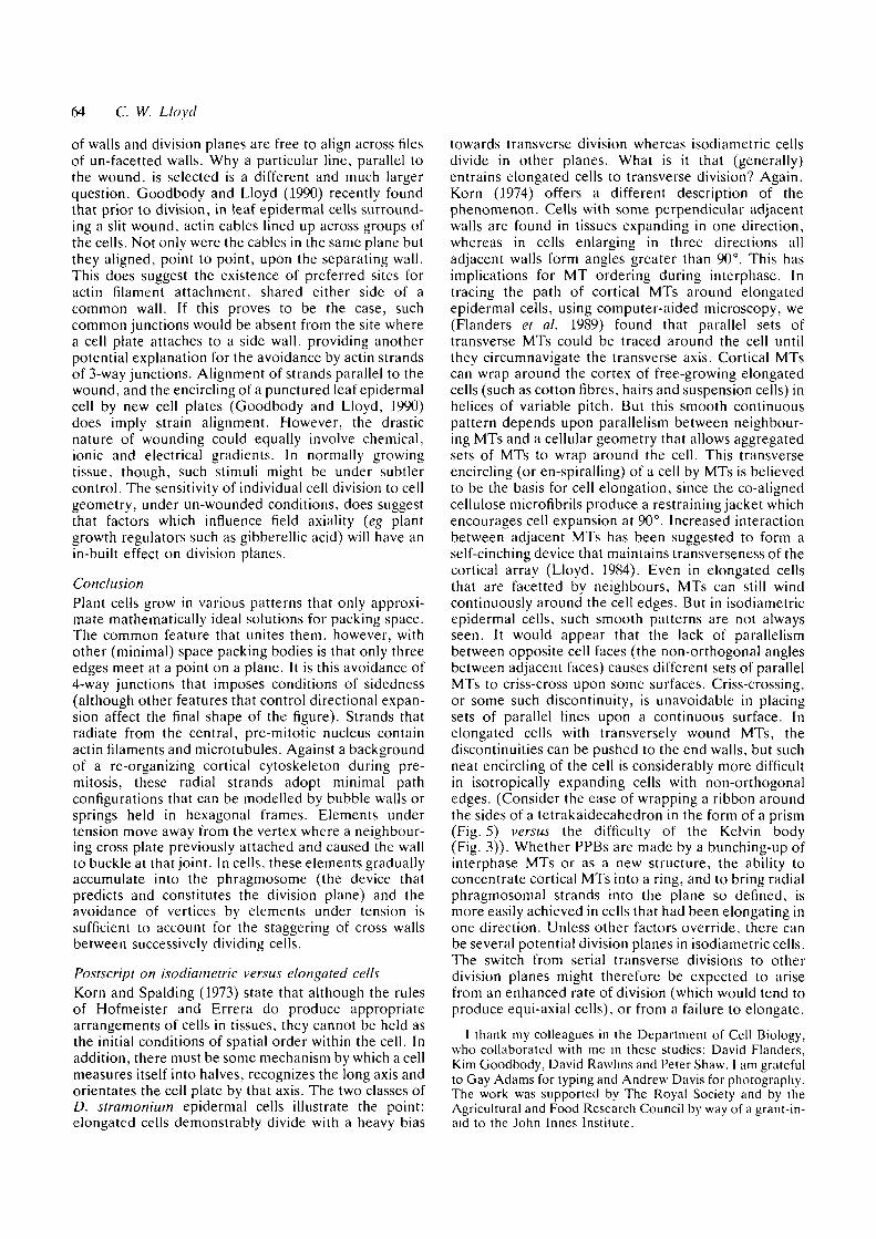
Fig. 9. Modelling the behaviour of cytoplasmic strands with bubble walls. Glycerol-stabilized bubbles are held in a flexible,prismatic hexagonal frame. In A, the frame forms a rectangular hexagon. In B, the long side walls are pulled slightlyoutwards to form vertices. The bubble walls now avoid the pin-joints at the vertices, just as they avoid making contact withthe outer corners of the frame. As the frame is pulled into an isodiametric hexagon, bubble walls show maximumavoidance of vertices by aligning perpendicularly with the mid-edges of the frame. A similar demonstration was shown(Flanders et al. 1990) using springs in a frame. The minimal path avoidance hypothesis is based on the fact that tensileelements, which are free to move, will tend to avoid the long path to a vertex and to contact side walls perpendicularly. Inasynchronously dividing tissue, the pre-buckling of a cell by a neighbouring cross wall (see Figs 6 and 7) forms a vertex tobe avoided when that buckled cell later divides. Thus successive cross walls would not touch. However, this does occur inwounded tissue where cross walls are deposited simultaneously and should also occur in un-wounded tissue where lack ofexpansion has failed to generate the necessary buckling. (Reproduced from Flanders et al. 1990, by permission of theJournal of Cell Biology).
expands (Thompson, 1942; Korn, 1980). This feature ofasynchronous division to form vertices, together withthe tendency for elements under tension (which are freeto move) to seek a minimal path, provides a mechanismfor the avoidance of forming 4-way junctions. Such amechanism, which tends to form tetrahedral angles in3-D, is sufficient to account for the quasi-14-hedralgeometry that follows from such an arrangement.Although strands that connect the nucleus to the cortexare not continuous like bubble walls, the similaritybetween plant tissues and foams is not fortuitous, forboth depend upon minimal path mechanisms. But whatevidence is there that pre-mitotic strands are undertension? They are certainly free to move and this hasbeen observed by time lapse microscopy by Goodbodyand Lloyd (1990) in wound-stimulated Tradescantiaepidermal cells. Hahne and Hoffman (1984) used lasermicrosurgery to demonstrate that transvacuolarstrands, holding the central nucleus in Hibiscusprotoplasts, are under tension and cause in-pullings onthe protoplast surface. We have recently confirmed (K.C. Goodbody, C. J. Venverloo and C. W. Lloyd,unpublished observations) that the pre-mitotic radialstrands in epidermal cells stimulated to divide byexplantation are also under tension: laser ablation of astrand between nucleus and cortex causes the nucleus torecoil towards the opposite cortex and the cut ends ofthe strand to retract.
To summarize, the reorganization of the cortex at
preprophase allows tensile strands, which radiate fromthe nucleus, to adopt minimal paths; such paths aregenerally perpendicular to the cortex. In elongated cellsthe majority of strands will seek the transverse axis, butwill avoid a vertex since this is not a minimal path. Inisodiametric cells, strands adopt a favourable configur-ation on each cell face without the massive long-edgere-distribution observed in elongated cells. This offersalternative axes for division plane alignment as testifiedby the irregular patchwork arrangement of such cells.
Wounding produces 4-way junctionsSinnott and Bloch (19416) induced large, vacuolatedcells to divide by wounding the tissue. They observedthat the new phragmosomes formed parallel to thecourse of the wound and often lined up, from cell tocell, without staggering the joints. A major differencebetween this response (which produces 4-way junc-tions) and normally occurring divisions (producing3-way junctions) is that the former divisions aresynchronous whereas the latter are not. Korn's (1980)concept of cell buckling or facetation depends uponasynchrony: a new cell plate causes the neighbouringattached walls to buckle before the neighbouring cellsthemselves divide. According to the minimal pathexplanation (Flanders et al. 1990) such prior facettationproduces a vertex to be avoided subsequently by pre-mitotic strands in neighbouring cells. The synchronicityof wound-induced divisions allows no such pre-buckling
64 C. W. Lloyd
of walls and division planes are free to align across filesof un-facetted walls. Why a particular line, parallel tothe wound, is selected is a different and much largerquestion. Goodbody and Lloyd (1990) recently foundthat prior to division, in leaf epidermal cells surround-ing a slit wound, actin cables lined up across groups ofthe cells. Not only were the cables in the same plane butthey aligned, point to point, upon the separating wall.This does suggest the existence of preferred sites foractin filament attachment, shared either side of acommon wall. If this proves to be the case, suchcommon junctions would be absent from the site wherea cell plate attaches to a side wall, providing anotherpotential explanation for the avoidance by actin strandsof 3-way junctions. Alignment of strands parallel to thewound, and the encircling of a punctured leaf epidermalcell by new cell plates (Goodbody and Lloyd, 1990)does imply strain alignment. However, the drasticnature of wounding could equally involve chemical,ionic and electrical gradients. In normally growingtissue, though, such stimuli might be under subtlercontrol. The sensitivity of individual cell division to cellgeometry, under un-wounded conditions, does suggestthat factors which influence field axiality (eg plantgrowth regulators such as gibberellic acid) will have anin-built effect on division planes.
ConclusionPlant cells grow in various patterns that only approxi-mate mathematically ideal solutions for packing space.The common feature that unites them, however, withother (minimal) space packing bodies is that only threeedges meet at a point on a plane. It is this avoidance of4-way junctions that imposes conditions of sidedness(although other features that control directional expan-sion affect the final shape of the figure). Strands thatradiate from the central, pre-mitotic nucleus containactin filaments and microtubules. Against a backgroundof a re-organizing cortical cytoskeleton during pre-mitosis, these radial strands adopt minimal pathconfigurations that can be modelled by bubble walls orsprings held in hexagonal frames. Elements undertension move away from the vertex where a neighbour-ing cross plate previously attached and caused the wallto buckle at that joint. In cells, these elements graduallyaccumulate into the phragmosome (the device thatpredicts and constitutes the division plane) and theavoidance of vertices by elements under tension issufficient to account for the staggering of cross wallsbetween successively dividing cells.
Postscript on isodiametric versus elongated cellsKorn and Spalding (1973) state that although the rulesof Hofmeister and Errera do produce appropriatearrangements of cells in tissues, they cannot be held asthe initial conditions of spatial order within the cell. Inaddition, there must be some mechanism by which a cellmeasures itself into halves, recognizes the long axis andorientates the cell plate by that axis. The two classes ofD. stramonium epidermal cells illustrate the point:elongated cells demonstrably divide with a heavy bias
towards transverse division whereas isodiametric cellsdivide in other planes. What is it that (generally)entrains elongated cells to transverse division? Again,Korn (1974) offers a different description of thephenomenon. Cells with some perpendicular adjacentwalls are found in tissues expanding in one direction,whereas in cells enlarging in three directions alladjacent walls form angles greater than 90°. This hasimplications for MT ordering during interphase. Intracing the path of cortical MTs around elongatedepidermal cells, using computer-aided microscopy, we(Flanders et al. 1989) found that parallel sets oftransverse MTs could be traced around the cell untilthey circumnavigate the transverse axis. Cortical MTscan wrap around the cortex of free-growing elongatedcells (such as cotton fibres, hairs and suspension cells) inhelices of variable pitch. But this smooth continuouspattern depends upon parallelism between neighbour-ing MTs and a cellular geometry that allows aggregatedsets of MTs to wrap around the cell. This transverseencircling (or en-spiralling) of a cell by MTs is believedto be the basis for cell elongation, since the co-alignedcellulose microfibrils produce a restraining jacket whichencourages cell expansion at 90°. Increased interactionbetween adjacent MTs has been suggested to form aself-cinching device that maintains transverseness of thecortical array (Lloyd, 1984). Even in elongated cellsthat are facetted by neighbours, MTs can still windcontinuously around the cell edges. But in isodiametricepidermal cells, such smooth patterns are not alwaysseen. It would appear that the lack of parallelismbetween opposite cell faces (the non-orthogonal anglesbetween adjacent faces) causes different sets of parallelMTs to criss-cross upon some surfaces. Criss-crossing,or some such discontinuity, is unavoidable in placingsets of parallel lines upon a continuous surface. Inelongated cells with transversely wound MTs, thediscontinuities can be pushed to the end walls, but suchneat encircling of the cell is considerably more difficultin isotropically expanding cells with non-orthogonaledges. (Consider the ease of wrapping a ribbon aroundthe sides of a tetrakaidecahedron in the form of a prism(Fig. 5) versus the difficulty of the Kelvin body(Fig. 3)). Whether PPBs are made by a bunching-up ofinterphase MTs or as a new structure, the ability toconcentrate cortical MTs into a ring, and to bring radialphragmosomal strands into the plane so defined, ismore easily achieved in cells that had been elongating inone direction. Unless other factors override, there canbe several potential division planes in isodiametric cells.The switch from serial transverse divisions to otherdivision planes might therefore be expected to arisefrom an enhanced rate of division (which would tend toproduce equi-axial cells), or from a failure to elongate.
1 thank my colleagues in the Department of Cell Biology,who collaborated with me in these studies: David Flanders,Kim Goodbody, David Rawhns and Peter Shaw. I am gratefulto Gay Adams for typing and Andrew Davis for photography.The work was supported by The Royal Society and by theAgricultural and Food Research Council by way of a grant-in-aid to the John Innes Institute.

Cytoskeleton and cell geometry 65
References
ERRERA, L. (1888). Uber Zellformen and Siefenblasen. BomnischesCentralblatt, 34, 395-399.
FLANDERS, D. J., RAWLINS, D. J., SHAW, P. J. AND LLOYD, C. W.(1989). Computer-aided 3-D reconstruction of interphasemicrotubules in epidermal cells of Datura stramonium revealsprinciples of array assembly. Development 106, 531-541.
FLANDERS, D. J., RAWLINS, D. J., SHAW, P. J. AND LLOYD, C. W.(1990). Nucleus-associated microtubules help determine thedivision plane of plant epidermal cells: Avoidance of four-wayjunctions and the role of cell geometry. J. Cell Biol. 110,1111-1122.
GIBSON, L. J. AND ASHBY, M. R. (1988). Cellular Solids. Structureand properties. Pergamon Press: Oxford.
GOODBODY, K. C. AND LLOYD, C. W. (1990). Actin filaments line-up across Tradescantia epidermal cells, anticipating wound-induced division planes. Protoplasma (in press).
HALES, S. (1727). Vegetable Staticks. London. Exp. 32, 94-96.HAHNE, G. AND HOFFMAN, F. (1984). The effect of laser
microsurgery on cytoplasmic strands and cytoplasmic streamingin isolated plant protoplasts. Eur J. Cell Biol. 33, 175-179.
HOFMEISTER, W. (1863). Zusatze und Berichtigungen zu den 1851veroffentlichen Untersuchungengen der Entwicklung hohererKryptogamen. Jahrbucher fur Wissenscliaft und Botanik. 3,259-293.
KAKIMOTO, I. AND SHIBAOKA, H. (1987). Actin filaments andmicrotubules in the preprophase band and phragmoplast oftobacco cells. Protoplasma 140, 151-156.
KATSUTA, J., HASHIGUCHI, Y. AND SHIBAOKA, H. (1990). The roleof the cytoskeleton in positioning of the nucleus in pre-mitotictobacco BY-2 cells. J. Cell Sci. 95, 413-422.
KORN, R. W. (1974). The three-dimensional shape of plant cellsand its relationship to pattern of tissue growth. New Phytol. 73,927-935.
KORN, R. W. (1980). The changing shape of plant cells:transformations during cell proliferation. Ann. Bot. 46,649-666.
KORN, R. W. AND SPALDING, R. M. (1973). The geometry of plantepidermal cells. New Phytol. 72, 1357-1365.
LEWIS, F. T. (1923). The typical shape of polyhedral cells invegetable parenchyma and the restoration of that shapefollowing division. Proc. Am. Acad. Arts Sci. 58, 537-552.
LEWIS, F. T. (1926). The effect of cell division on the shape andsize of hexagonal cells. Anat. Rec. 33, 331-355.
LEWIS, F. T. (1928). The correlation between cell division and theshapes and sizes of prismatic cells in the epidermis of Cucunus.Anat. Rec. 38, 341-376.
LEWIS, F. T. (1936). A volumetric study of growth and celldivision in two types of epithelium - the longitudinally prismaticepidermal cells of Tradescantia and the radially prismaticepidermal cells of Cucumis. Anat. Rec. 47, 55-99.
LEWIS, F. T. (1943). The geometry of growth and cell division inepithelial mosaics. Am. J. Bot. 30, 766-776.
LLOYD, C. W. (1984) Toward a dynamic helical model for the
influence of microtubules on wall patterns in plants. Int. Rev.Cytol 86, 1-51.
LLOYD, C. W. (1989). The Plant Cytoskeleton. Current Opinion inCell Biology 1, 30-35.
LLOYD, C. W. (1991). Cytoskeletal elements of the phragmosomeestablish the division plane in vacuolated higher plant cells. InThe Cytoskeletal Basis of Plant Growth and Form (ed. C. W.Lloyd). Academic Press: London (In press).
LLOYD, C. W. AND TRAAS, J. A. (1988). The role of F-actin indetermining the division plane of carrot suspension cells. Drugstudies. Development 102, 211-221.
MACIOR, W. A. AND MATZKE, E. B. (1951). An experimentalanalysis of cell-wall curvatures and approximations to minimaltetrakaidecahedra in the leaf parenchyma of Rhoeo discolor.Am. J Bot. 38, 783-793.
MARVIN, J. W. (1939). The shape of compressed lead shot and itsrelation to cell shape. Am. J. Bot. 26, 280-288.
MATZKE, E. B. (1939). Volume-shape relationships in lead shotand their bearing on cell shapes. Am. J. Bot. 26, 2S8-295.
MATZKE, E. B. (1945). The three-dimensional shapes of bubbles infoams. Proc. natn. Acad. Set. U.S.A. 31, 281-289.
MATZKE, E. B. (1950). In the twinkling of an eye. Bull Torrevbot. Club, 77, 222-227.
PICKETT-HEAPS, J. D. AND NORTHCOTE, D. H. (1966). Organizationof microtubules and endoplasmic reticulum during mitosis andcytokinesis in wheat meristems. J. Cell Sci. 1, 109-120.
SACHS, J. (1878). Uber die Anordnung der Zellen in jungstenPflanzentheilen. Arbeiten des Botanisches Institut Wurzburg 2,46-104.
SINNOTT, E. W. AND BLOCH, R. (1940). Cytoplasmic behaviourduring division of vacuolate plant cells. Proc. natn. Acad. Sci.U.S.A. 26, 223-227
SINNOTT, E. W. AND BLOCH, R. (1941a). The relative position ofcell walls in developing plant tissues. Am. J. Bot. 28, 607-617.
SINNOTT, E. W. AND BLOCH, R. (1941ft). Division in vacuolateplant cells. Am. J Bot. 28, 225-232.
STEVENS, P. S. (1976). Patterns in nature. Penguin Books Ltd.,Harmondsworth, Middx, England.
THOMPSON, D. W. (1942). On Growth and Form. CambridgeUniversity Press, Cambridge.
THOMSON, W. (1887). On the division of space with minimalpartitional area. Phil Mag. 24, 503-514.
THOMSON, W. (1894). On homogeneous division of space. Proc R.Soc. Lond. 55, 1-17.
TRAAS, J. A., DOONAN, J. H., RAWUNS, D. J., SHAW, P. J..
WATTS, J. AND LLOYD, C. W. (1987). An actin network ispresent in the cytoplasm throughout the cell cycle of carrot cellsand associates with the dividing nucleus. J Cell Biol. 105,387-395.
WILLIAMS, R. E. (1968). Space-filling polyhedron: its relation toaggregates of soap bubbles, plant cells, and metal crystallites.Science, NY 161, 276-277.
WILLIAMS, R. E. (1979) The geometrical foundation of naturalstructure. A source design book. Dover Publications Inc., NewYork.