how i treat waldenstrom's macroglobulinemia - wmuk · waldenström s macroglobulinemia (wm) is...
TRANSCRIPT
doi:10.1182/blood-2009-05-174359 Prepublished online Jul 17, 2009;
Steven P. Treon
How I treat Waldenstrom's macroglobulinemia
http://bloodjournal.hematologylibrary.org/misc/rights.dtl#repub_requestsInformation about reproducing this article in parts or in its entirety may be found online at:
http://bloodjournal.hematologylibrary.org/misc/rights.dtl#reprintsInformation about ordering reprints may be found online at:
http://bloodjournal.hematologylibrary.org/subscriptions/index.dtlInformation about subscriptions and ASH membership may be found online at:
. Hematology; all rights reservedCopyright 2007 by The American Society of DC 20036.by the American Society of Hematology, 1900 M St, NW, Suite 200, Washington Blood (print ISSN 0006-4971, online ISSN 1528-0020), is published semimonthly
For personal use only. at Harvard Libraries on July 17, 2009. www.bloodjournal.orgFrom
How I treat Waldenstrom’s Macroglobulinemia.
Steven P. Treon MD, MA, PhD
Bing Center for Waldenstrom’s Macroglobulinemia, Dana Farber
Cancer Institute, Harvard Medical School, Boston, MA 02115,
USA.
Corresponding author:
Steven P. Treon M.D., M.A., Ph.D.
Bing Center for Waldenström’s Macroglobulinemia
Dana-Farber Cancer Institute
M548, 44 Binney Street, Boston, MA 02115 USA
Tel: (617) 632-2681
Fax: (617) 632-4862
Email: [email protected]
Blood First Edition Paper, prepublished online July 17, 2009; DOI 10.1182/blood-2009-05-174359
Copyright © 2009 American Society of Hematology
For personal use only. at Harvard Libraries on July 17, 2009. www.bloodjournal.orgFrom

Abstract
Waldenström’s macroglobulinemia (WM) is a distinct B-cell disorder resulting
from the accumulation, predominantly in the bone marrow, of clonally
related IgM secreting lymphoplasmacytic cells. Genetic factors play an
important role, with 20% of patients demonstrating a familial predisposition.
Asymptomatic patients should be observed. Patients with a disease related
hemoglobin level <10g/L, platelet count<100x109/L, bulky adenopathy or
organomegaly, symptomatic hyperviscosity, peripheral neuropathy,
amyloidosis, cryoglobulinemia, cold-agglutinin disease or evidence of disease
transformation should be considered for therapy. Plasmapheresis should be
considered for symptomatic hyperviscosity, and for prophylaxis in patients in
whom rituximab therapy is contemplated. The use of rituximab as
monotherapy or in combination with cyclophosphamide, nucleoside
analogue, bortezomib or thalidomide based regimens can be considered for
the first line therapy of WM, and should take into account specific treatment
goals, future autologous stem cell transplant eligibility, and long term risks
of secondary malignancies. In the salvage setting, the re-use or use of an
alternative frontline regimen can be considered as well as bortezomib,
alemtuzumab, and stem cell transplantation. Newer agents such as
bendamustine and everolimus can also be considered in the treatment of
WM.
For personal use only. at Harvard Libraries on July 17, 2009. www.bloodjournal.orgFrom
Introduction
Waldenström’s macroglobulinemia (WM) is a distinct B-cell disorder resulting
from the accumulation, predominantly in the bone marrow, of clonally
related lymphoplasmacytic cells which secrete a monoclonal IgM protein1.
This condition is considered to correspond to the lymphoplasmacytic
lymphoma (LPL) as defined by the Revised European American Lymphoma
(REAL) and World Health Organization classification systems.2,3 Most cases
of LPL are WM, with less than 5% of cases made up of IgA, IgG and non-
secreting LPL.
Clinical Features
The clinical and laboratory findings for 356 newly diagnosed patients who
presented to our Institution are depicted in Table 1. Unlike most indolent
lymphomas, splenomegaly and lymphadenopathy are present in only a
minority of patients (<15%). The morbidity associated with WM is typically
mediated by tissue infiltration by neoplastic cells, the physicochemical and
immunological properties of the monoclonal IgM or both. As shown in Table
2, the monoclonal IgM can produce clinical manifestations through several
distinct mechanisms including an effect on serum viscosity, mediation of
For personal use only. at Harvard Libraries on July 17, 2009. www.bloodjournal.orgFrom

auto-antibody activity, interactions with other proteins, precipitation on
cooling, and tissue deposition.4-6
Diagnostic Workup
History taking
There is a strong familial predisposition in WM7-9, therefore a good family
history is important. While the identification of such familiarity does not at
this time influence treatment decisions, it may spawn a discussion in families
with multiple cases of WM or related B-cell disorders to participate in familial
studies aimed at identifying genetic predispositions to WM which are
currently underway at the Dana Farber Cancer Institute and the National
Institutes of Health. Exposure to hepatitis C has been implicated in some,
but not all studies, and evaluation of risks/exposure is important particularly
among patients who have Type II (mixed) cryoglobulinemia.10-12 A thorough
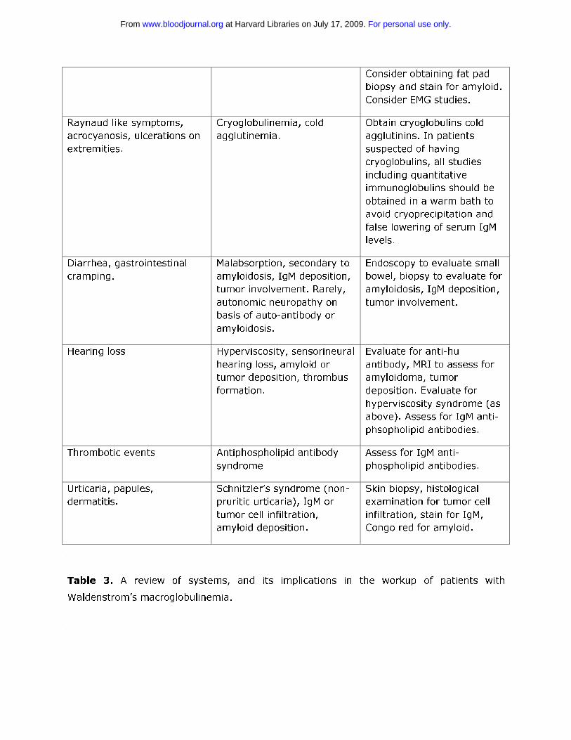
review of systems (ROS) is very important in the workup of WM patients
given the vast array of presenting symptoms and complaints, and may well
impact on treatment considerations. A ROS checklist which we use at our
For personal use only. at Harvard Libraries on July 17, 2009. www.bloodjournal.orgFrom

Institution along with their implications for the care of WM patients is
presented in Table 3, and can be used in the work-up of WM patients.
Laboratory Studies
To establish the diagnosis of WM, it is necessary to demonstrate an IgM
monoclonal protein, along with histological evidence of infiltration of the
bone marrow by lymphoplasmacytic cells.1 There is no minimal serum IgM
level, nor a minimal percentage of bone marrow infiltration to establish the
diagnosis of WM, since patients can be symptomatic and in need of
treatment even at low levels of IgM (<1,000 mg/dL) or bone marrow
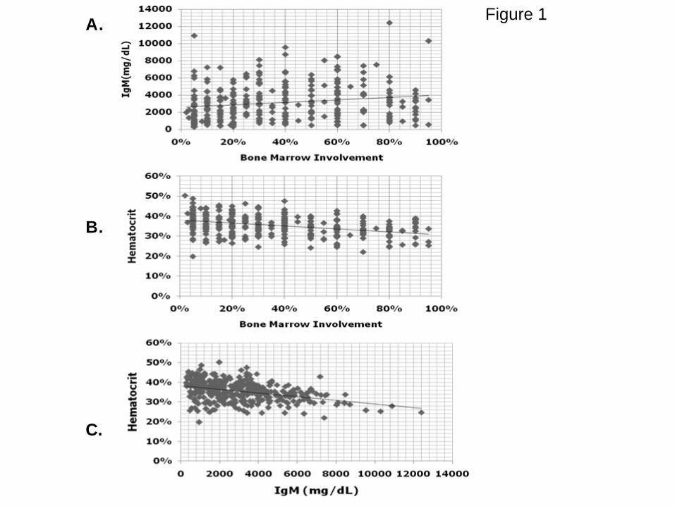
involvement. In fact, there can be great heterogeneity among patients
between their respective serum IgM levels and bone marrow involvement
(Figure 1). In general, for an individual patient, serum IgM levels tend to
show good correlation with disease burden. There are some exceptions to
this point which usually emerge in patients with cryoglobulinemia, as well as
those patients undergoing treatment with certain biological agents (i.e.
rituximab, bortezomib). The assessment of response for patients undergoing
treatment with biological agents is further discussed under Response
Assessment.
For personal use only. at Harvard Libraries on July 17, 2009. www.bloodjournal.orgFrom
Peripheral Blood Testing
Baseline serum protein electrophoresis (SPEP), quantitative
immunoglobulins, complete blood counts, liver function tests, blood urea
nitrogen and creatinine should be obtained. A warm bath collection should
be considered for those patients suspected of having cryoglobulinemia in
order to avoid underestimation of the serum IgM levels. It is important to
note that serum immunoglobulin levels can vary considerably between
institutions, hence for comparative purposes serum immunoglobulin levels
are best followed by the same laboratory so as to avoid misinterpretation.
Since the value of serum free light chain testing remains to be clarified in
the management of WM patients, its inclusion in routine testing of WM
patients is not advised at this time.
While IgM levels will be elevated in almost all WM patients, IgA and IgG
levels are subnormal, and may contribute to recurring sinus and bronchial
infections.13 Serum IgA and IgG levels seldom return to normal, even after
good remissions (including attainment of CR) and may reflect a
constitutional defect in plasma cell development. While liver function tests
should be evaluated, these seldom are abnormal on the basis of disease.
Azotemia can often present in WM on the basis of light chain or amyloid
For personal use only. at Harvard Libraries on July 17, 2009. www.bloodjournal.orgFrom
deposition, as well as parenchymal involvement by lymphoplasmacytic cells.
Therefore, renal function should carefully be evaluated. Complete blood
counts should also be carefully evaluated, and expanded up by evaluation of
the mean corpuscular volume and reticulocyte count for evidence of
underlying autoimmune hemolysis. Lastly, B2M and albumin levels can
obtained for purposes of prognostication, though their use in making
treatment related decisions remains to be clarified.14,15
Selective Blood Testing
There can be considerable heterogeneity between bone marrow disease
involvement and anemia in patients with WM (Figure 1). As such, the
underlying basis for anemia may need to be better delineated, particularly in
patients whose anemia appears to be out of proportion to the level of their
disease involvement. Autoimmune hemolytic anemia (AHA) can commonly
occur either on the basis of cold or warm antibodies in WM patients. The
examination of reticulocyte counts, lactic dehydrogenase (LDH) and
haptoglobin levels can be useful, though most often hemolysis is
extravascular.16,17 Testing for cold agglutinins, and thereafter direct and
indirect Coombs antibody testing if cold agglutinins are negative is advised
as part of the workup of patients presenting with AHA. Iron deficiency
For personal use only. at Harvard Libraries on July 17, 2009. www.bloodjournal.orgFrom
related anemia is also commonly encountered in WM, and often is refractory
to oral but not intravenous iron repletion. As such, iron studies can be useful
in patients presenting with microcytic anemia. For some patients, correction
of the iron deficiency with intravenous iron leads to improvements in
anemia, and may defer the necessity for immediate chemotherapeutic
intervention. At higher serum IgM levels, anemia may also be more
pronounced due to a hemodilutional effect (Figure 1).
Obtaining a serum viscosity (SV) level is helpful in patients in whom
hyperviscosity is suspected. While most WM patients will exhibit an elevated
SV level i.e. >1.8 centipoise (cp), patients typically become symptomatic at
SV levels of > 4.0 cp. However, there can be great variability in the SV level
at which patients become symptomatic. At SV levels as low as 3.0 cp,
patients can exhibit retinal changes including hemorrhages, thereby
warranting intervention at lower SV levels.18 Conversely, patients with SV
levels higher than 4.0 cp can often be asymptomatic suggesting that
variables besides the level of serum IgM alone play a role in producing
symptomatic hyperviscosity, such as the presence of cryoglobulinemia.19 As
such, testing for cryoglobulins should be considered in patients with
symptomatic hyperviscosity who display relatively low serum IgM and SV
levels.
For personal use only. at Harvard Libraries on July 17, 2009. www.bloodjournal.orgFrom
Peripheral neuropathy (PN) is an important morbidity in patients with WM,
with up to 20-25% of patients demonstrating disease related PN, which most
often is sensory in nature.20,21 In patients suspected of having IgM related
PN, the evaluation of anti-myelin associated glycoprotein (MAG), anti-
ganglioside M1 (GM1), and anti-sulfatide IgM antibodies is appropriate.
While the presence of one of these antibodies may support the diagnosis of
IgM related neuropathy, their absence should not exclude the diagnosis
since other myelin associated antigens may be targeted which are not
clinically evaluable at this time. Amyloidosis should also be considered in
patients presenting with a peripheral neuropathy, and a fat pad biopsy with
Congo red staining obtained.22 Electromyography (EMG) may be helpful, and
often shows a demyelinating neuropathy. A sural nerve biopsy should be
avoided due to frequent neuropathic complications. In rare circumstances
where a myopathy may be suspected on the basis of WM, the investigation
for anti-decorin IgM antibodies can be considered.23
Bone Marrow Evaluation
The bone marrow is almost always involved in WM, and as such a bone
marrow biopsy and aspiration should be obtained. Central to the diagnosis of
For personal use only. at Harvard Libraries on July 17, 2009. www.bloodjournal.orgFrom

WM is the demonstration of bone marrow infiltration by a lymphoplasmacytic
cell population manifested by small lymphocytes with evidence of
plasmacytoid/plasma cell differentiation.1 The pattern of bone marrow
infiltration may be diffuse, interstitial or nodular, is usually intertrabecular. A
solely paratrabecular pattern of infiltration is unusual and should raise the
possibility of follicular lymphoma.1 The bone marrow infiltration should be
supported by immunophenotypic studies (flow cytometry and/or
immunohistochemistry) showing the following profile:
sIgM+CD19+CD20+CD22+CD79+.24,25 Up to 20% of cases may express either
CD5, CD10 or CD23.26 In such cases, care should be taken to satisfactorily
exclude chronic lymphocytic leukemia and mantle cell lymphoma.1 An
increased number of mast cells, usually in association with the lymphoid
aggregates is commonly found in WM, and their presence may help in
differentiating WM from other B-cell lymphomas.2,3
Cytogenetic Studies
Multiple studies have been published on cytogenetic findings in WM, and
have demonstrated a great variety of numerical and structural chromosome
abnormalities. Chromosome 6q deletions encompassing 6q21-25 have been
observed in up to half of WM patients, and at a comparable frequency
For personal use only. at Harvard Libraries on July 17, 2009. www.bloodjournal.orgFrom
amongst patients with and without a familial history.7,27,28 Several candidate
tumor suppressor genes in this region are under study including BLIMP-1.29
Despite an earlier study suggesting prognostic significance to 6q deletions in
WM, a more recent study did not confirm such significance.27,30 As such,
routine cytogenetic testing is not advised at this time. An exception,
however, is the use of cytogenetics to clarify the diagnosis of WM from
suspected cases of IgM myeloma. In the latter, IgH switch region
rearrangements (14q32 translocations) are a predominant feature, whereas
these are typically absent in WM.31
Imaging Studies
CT scans of the chest, abdomen, pelvis should be obtained at time of
diagnosis in order to properly stage the patient.32 Up to 20% of WM patients
may have extramedullary disease, and CT scans offer an opportunity to
assess for adenopathy, splenomegaly, and for other extramedullary disease
sites. Follow-up CT scans are only necessary for those patients with baseline
extramedullary disease (or who are later suspected of having extramedullary
disease), and may be used to assess disease progression, as well as
response. There is no role for routine magnetic resonance imaging (MRI); in
For personal use only. at Harvard Libraries on July 17, 2009. www.bloodjournal.orgFrom
as well there is not routine role for PET scanning unless disease
transformation is suspected.
Opthalmological Examination
Patients with WM often exhibit retinal changes due to hyperviscosity related
changes which occur as a consequence of elevated IgM levels. Retinal
findings associated with hyperviscosity can include peripheral dot and blot
like hemorrhages, dilated retinal veins, central hemorrhages, tortuous blood
vessels, venous “sausaging” and/or optic disc edema. In one study, retinal
changes were observed at serum IgM and viscosity levels as low as 3,000
mg/dL and 2.4 cp, respectively.18 Importantly, plasmapheresis can lead to
prompt resolution of hyperviscosity related retinal changes.33 Examination of
the retina may therefore be useful in identifying the symptomatic threshold
of serum viscosity levels in patients with WM, and may be used as an
important gauge for the effectiveness of both plasmapheresis and
chemotherapy. In our practice, we typically recommend a baseline
opthalmological examination in those WM patients whose serum IgM levels
are > 3,000 mg/dL.
For personal use only. at Harvard Libraries on July 17, 2009. www.bloodjournal.orgFrom

Treatment Approaches to WM
Management of the Asymptomatic or Smoldering WM Patient
Patients with a disease related hemoglobin level <10 g/dL, platelet count
<100x109/L, bulky adenopathy or organomegaly, symptomatic
hyperviscosity, moderate to severe, or advancing peripheral neuropathy on
the basis of disease, symptomatic amyloidosis, cryoglobulinemia, or cold-
agglutinin disease should be considered for therapy.14 Initiation of therapy
should not be based on serum monoclonal protein levels per se, and
asymptomatic patients should be observed. Asymptomatic patients with a
low B2-microglobulin (<3 g/dL) and a hemoglobin level of >12 g/dL may
have an indolent course, and not require therapy for a long period of time,
even when their monoclonal protein exceeds 3,000 mg/dL. As such the
identification of the asymptomatic patient is important, and close
observation (i.e. every few months) rather than therapy is appropriate for
these patients.
For personal use only. at Harvard Libraries on July 17, 2009. www.bloodjournal.orgFrom

Management of the Symptomatic WM
Frontline treatment options for WM include oral alkylators (e.g.
chlorambucil), nucleoside analogues (cladribine or fludarabine), the
monoclonal antibody rituximab as well as combinations of these agents.34,35
Individual patient considerations, including the presence of cytopenias, need
for more rapid disease control, age, and candidacy for autologous transplant
therapy should be taken into account in making the appropriate choice of a
first-line agent. A suggested algorithm for the primary therapy of WM is
provided in Figure 2. For patients who are candidates for autologous
transplant therapy, exposure to continuous oral alkylator therapy (such as
chlorambucil) or nucleoside analogues should be limited. The use of
nucleoside analogues in particular should be approached cautiously in
patients with WM since difficulties with stem cell collection, as well as
increased risk of disease transformation, myelodysplasia and acute
myelogenous leukemia have been reported.36-39
For personal use only. at Harvard Libraries on July 17, 2009. www.bloodjournal.orgFrom
Treatment of the WM Patient Requiring Immediate Disease Control
For WM patients presenting with circumstances requiring immediate disease
control, such as symptomatic hyperviscosity, cryoglobulinemia, or moderate
to severe cytopenias due to cold agglutinemia or immune related
thrombocytopenia, emphasis should be placed on achieving rapid
paraprotein reduction. In these circumstances, plasmapheresis can be
initially performed.19 Typically 2 to 3 sessions of plasmapheresis are required
to reduce serum IgM levels by 30-60%. For each plasmapheresis, we usually
perform one complete plasma volume exchange, and use albumin for
replacement product. Great care should be exercised in the timing of red
blood cell transfusions in patients presenting with symptomatic
hyperviscosity, and ideally should follow plasmapheresis so as to not
aggravate whole blood viscosity levels.19 Following plasmapheresis,
treatment should be initiated as soon as possible, as IgM levels will steadily
begin to rise and return to baseline levels in 4-5 weeks.19 For patients
requiring immediate disease control, the use of bortezomib based therapy
such as bortezomib, dexamethasone and rituximab (BDR) is preferable so as
to achieve more rapid disease control.40 The time to at least a minimum
response (TTR) in WM patients treated with BDR was 1.1 months in a
WMCTG study, while the overall response rate with BDR was 96%, with 22%
of patients achieving a CR. With a median follow-up of 2 years, 80% of
For personal use only. at Harvard Libraries on July 17, 2009. www.bloodjournal.orgFrom

patients remained free of disease progression including all patients achieving
a VGPR or better in this study. Herpes zoster prophylaxis should be
instituted with BDR therapy using an oral anti-viral agent such as acyclovir,
famvir or valcyclovir and maintained throughout the course of BDR, and
thereafter for at least 6 months. A close watch for the development of
bortezomib related neuropathy should be maintained on BDR. Treatment
related peripheral neuropathy which occurred at a grade 3 level in 30% of
patients was reversible in most patients, who benefitted by interim support
with pregabalin. A rituximab related flare with BDR was observed in only 9%
of patients, which may reflect the ability of bortezomib to suppress IgM
production independent of tumor cell killing.41,42 As an alternative to the
twice a week schedule of bortezomib used with BDR, the use of once a week
bortezomib at a higher dose (i.e. 1.6 mg/m2) may be considered with
rituximab, and appears in one study to be associated with lower risk of
neuropathy.43,44 Disease control may lag, and the incidence of rituximab
related IgM flare may be higher (20%) with once a week versus twice a
week administration of bortezomib.
As an alternative to bortezomib based therapy, a cyclophosphamide based
rituximab containing regimen can be considered in patients <70 years45-48; a
nucleoside analogue in combination with rituximab can also be considered in
patients 70 years or older, or in younger patients where the use of
For personal use only. at Harvard Libraries on July 17, 2009. www.bloodjournal.orgFrom
bortezomib or cyclophosphamide based therapy may not be an option.49-51
In a recent update of the Southwest Oncology Group (SWOG) directed
Intergroup Trial S9003, the 10 year event free survival (EFS) to single agent
fludarabine was 20%.52 By multivariate analysis, patients with lower levels
of B2M (<3 mg/L) demonstrated significantly better EFS in this series. It is
unclear if the inclusion of cyclophosphamide to a nucleoside analogue
containing rituximab regimen (such as FCR) in WM extends response, and its
addition may contribute to additional toxicity.53 The overall response rates
with cyclophosphamide or nucleoside analogue based rituximab containing
regimens is 70-80%, with CR attainment in about 10% of patients. Cytokine
support following ASCO guidelines should be considered with either
cyclophosphamide or nucleoside analogue based therapy.
There has been considerable debate on the value of including doxorubicin
and vincristine to cyclophosphamide based therapy in WM. Dimopoulos et
al46 reported that the combination of rituximab, cyclophosphamide and
dexamethasone (R-CD) led to overall and complete responses in 78% and
7% of WM patients respectively, and a 2- year progression free survival of
80%, which appear on par with those results achieved in a comparable
population of untreated WM who received CHOP-R.45,47 Ioakimidis et al48
compared the activity and toxicity associated with CHOP-R, CVP-R and CP-R
in patients with WM. The use of CP-R was associated with analogous
For personal use only. at Harvard Libraries on July 17, 2009. www.bloodjournal.orgFrom

response rates to CVP-R and CHOP-R in the frontline treatment of WM, while
treatment related complications including febrile neutropenia,
hospitalizations and vincristine related neuropathy were less. As such, CP-R
or R-CD may be preferable to CVP-R or CHOP-R in the management of WM.
An important consideration in the treatment of WM patients, particularly
those with high IgM levels is the potential for a rituximab mediated IgM flare
which may lead to symptomatic hyperviscosity, as well as worsening of IgM
related neuropathy, cryoglobulinemia and other IgM related complications.54-
60 The occurrence of an IgM flare is quite common following rituximab
therapy in WM patients, with a 40-50% occurrence rate when rituximab is
used as monotherapy.54,55 In combination therapy, the occurrence of the
rituximab mediated IgM flare can vary considerably, and appear dependent
on both the regimen used as well as the sequencing of rituximab
administration.40,43, 48,61-63
Because of concern over a rituximab related IgM flare aggravating serum
viscosity levels or IgM related morbidity, the omission of rituximab can also
be considered for the first 1-2 cycles of treatment. Serum IgM levels should
be closely monitored (at least weekly) during the time patients are receiving
rituximab based therapy. The IgM flare may last for several weeks, and even
For personal use only. at Harvard Libraries on July 17, 2009. www.bloodjournal.orgFrom

months and does not per se herald treatment failure.54,62 The use of
rituximab is best avoided as single agent therapy in patients with high IgM
levels since in two studies considerably lower response rates were observed
in those patients with higher serum IgM levels (>4,000 mg/dL).64,65
Treatment of the WM Patient Requiring Non-Immediate Disease
Control
For those WM patients presenting with disease not requiring immediate
disease control, a number of options can be considered and should ideally
take into account specific disease controlling objectives. The use of CP-R or
R-CD may be considered, and may be particularly preferable in younger
transplant eligible patients. The overall reported response rates with CP-R or
R-CD are 70-80%, and the 2 year progression free survival rates with these
regimens is 70%.46,48 In patients >70 years, nucleoside analogue based
therapy may also be considered.49-51 In a recent study by the WMCTG, the
overall response rate to FR was 96%, and the median progression free
survival was 51.2 months; in patients achieving a VGPR, the median
progression free survival in this series was in excess of 88 months.50
Because of the significant and prolonged myelosuppression observed in the
WMCTG study which utilized 6 cycles of fludarabine, each with 5 day courses
at 25 mg/m2, a more condensed course (i.e. 4 days of fludarabine at
For personal use only. at Harvard Libraries on July 17, 2009. www.bloodjournal.orgFrom

25mg/m2 per cycle for 4 cycles) may be preferable in more indolent
patients.50 The use of thalidomide in combination with rituximab (TR) also
represents an alternative choice in the management of WM patients not
requiring immediate disease control, and is associated with an overall
response rate of 70%, and a median PFS of 3 years.61 TR may be
particularly applicable to those WM patients presenting with significant
myelosuppression. Peripheral neuropathy (> grade 2) was seen in 40% of
WM patients treated with TR, in whom doses of 200-400 mg daily were
used.62 Lower doses of thalidomide (i.e. 100 mg daily) may be more
appropriate in WM patients given the increased propensity for WM patients
to develop treatment related neuropathy. The use of lenalidomide has been
explored in WM, and was associated in one study with an acute drop in
hematocrit and hospitalizations of several patients due to aggravated
anemia and related complications.63 Despite dose reductions, lenalidomide
related anemia persisted in many patients. As such, the use of lenalidomide
should be avoided in WM patients. The use of rituximab as a single agent
can also be considered in select patients with WM, such as those presenting
with a low tumor burden, mild to moderate cytopenias due to bone marrow
involvement or autoimmune related destruction (i.e. cold agglutinemia,
immune mediated thrombocytopenia) or IgM related neuropathy (see
below). Overall response rates with 4 weekly infusions of rituximab is 20-
30%,64-66 whereas the use of extended dose rituximab (i.e. 4 weekly
For personal use only. at Harvard Libraries on July 17, 2009. www.bloodjournal.orgFrom
infusions followed by 4 more weekly infusions at week 12) has been
associated with higher overall response rates (40-50%).67,68 Polymorphisms
in position 158 of the CD16 (FcγRIIIA-158) receptor have been shown in WM
and related indolent lymphomas to predict response.69 In WM patients, a 4
fold higher rate of response has been reported among WM patients bearing
the V/V or V/F polymorphism versus F/F. Testing for the FcγRIIIA-158
polymorphism was recently cleared by the U.S. Food and Drug
Administration and may help in predicting response to single agent
rituximab. In WM patients with the FcγRIIIA-158 F/F polymorphism,
clinicians can consider alternatives to single agent rituximab treatment, such
as the use of rituximab in combination therapy or the use of a non-rituximab
based therapy.
Treatment of Peripheral Neuropathy
The treatment of IgM related neuropathy is usually rituximab based, with
improvements in sensory function accompanying reduction in anti-neuronal
antibody titers observed in several studies, including a recent placebo
controlled trial.70-72 The use of single agent rituximab can be considered in
patients with mild, progressive neuropathy. In patients with moderate to
severe IgM related neuropathy, or where the course of the IgM neuropathy
For personal use only. at Harvard Libraries on July 17, 2009. www.bloodjournal.orgFrom
appears aggressive, the use of CP-R or R-CD may be preferable in order to
achieve more robust paraprotein reductions. There is debate on the role of
novel agents such as bortezomib or thalidomide in combination with
rituximab for the treatment of IgM related neuropathy, since these agents
are associated with treatment related neuropathy. Despite these concerns,
improvements in IgM related neuropathy have been observed in patients
receiving rituximab with either bortezomib or thalidomide.40,62 The
risk/benefit of treatment related neuropathy versus control of IgM related
neuropathy needs to be considered, and the use of either bortezomib or
thalidomide is better considered as a salvage measure for those patients not
responding to CP-R or R-CD. In such cases, an attenuated schedule or
dosing of these agents should be considered to minimize treatment related
neuropathy.
Maintenance Therapy in WM
There is considerable debate on the use of maintenance rituximab in WM
patients. In our practice, we typically use maintenance rituximab in those
patients who have responded to a rituximab containing regimens. While
there have been no prospective, randomized trials addressing the role of
maintenance rituximab in WM patients per se, the lessons learned in related
For personal use only. at Harvard Libraries on July 17, 2009. www.bloodjournal.orgFrom

indolent lymphomas appear applicable to the care of WM patients whose
response characteristics to rituximab therapy closely parallel those attained
in patients with other indolent B-cell lymphomas. Moreover, in two studies
examining the role of extended rituximab in WM patients, improvements in
the overall response rate, and possibly PFS were observed versus earlier
studies examining standard 4 weekly rituximab infusions.67,68 As with other
indolent B-cell lymphomas, the exact schedule and length of maintenance
therapy with rituximab in WM patients remains to be clarified. In our
practice, we typically administer one single infusion of rituximab (at 375
mg/m2) every 3 months for 2 years based on the schedule reported by Van
Oers et al.73 In some patients, the IgM flare can occur in the maintenance
phase of rituximab administration, and can be mistaken for progression. For
this reason, a bone marrow biopsy should be obtained following induction
therapy, and can be repeated if there is ambiguity over whether the patient
is experiencing an IgM flare related to rituximab, or if disease progression is
occurring during maintenance therapy.
Salvage Therapy in WM
A suggested algorithm for the salvage therapy of WM patients is provided in
Figure 3. For patients in relapse or who have refractory disease, the use of
For personal use only. at Harvard Libraries on July 17, 2009. www.bloodjournal.orgFrom
an alternative first-line agent as discussed above can be considered, with the
caveat that in patients for whom autologous transplantation is seriously
being considered, exposure to stem-cell damaging agents such as
chlorambucil or nucleoside analogues should be avoided, and a non-stem-
cell toxic approach considered if stem cells have not been previously
harvested.34,35 In addition to the use of an alternative frontline treatment
regimen, bortezomib based therapy can be considered given overall reported
response rates of 60-80%.41-44,74,75 In our clinic, we typically administer
bortezomib at 1.3 mg/m2 on days 1, 4, 8, 11 as part of a 3 week cycle,
along with 40 mg IV dexamethasone with each bortezomib dosing given
potential synergism of these agents.76 Prophylaxis against herpes zoster
should be strongly considered with bortezomib and steroid combinations40,
and patients should be carefully evaluated for the development of
bortezomib related peripheral or autonomic neuropathy. If disease control is
established after a few cycles of twice a week bortezomib therapy, once a
week therapy with bortezomib can be considered at 1.6 mg/m2/week in
order to attenuate further risk of neuropathy.43,44
As a third line approach to the salvage therapy of WM, alemtuzumab can be
also be considered.34,35 Alemtuzumab targets CD52, which is widely
expressed on both bone marrow WM cells, as well as on mast cells which
provide growth and survival signals to WM cells.77,78 As part of a WMCTG
For personal use only. at Harvard Libraries on July 17, 2009. www.bloodjournal.orgFrom
effort79, 28 patients with LPL (27 with WM) were treated with alemtuzumab.
Twenty three of these patients were previously treated and all had rituximab
previously. All patients received herpes zoster and pneumoncystis carinii
pneumonia prophylaxis. The overall response rate in this study was 76%,
with major responses in 32% of patients. Hematological toxicities, as well as
cytomegalovirus (CMV) reactivation and infection were common among
previously treated patients, with the latter possibly related to one death.
With a median follow-up exceeding 9 months, 11 of 19 responding patients
were free of progression. High response rates with alemtuzumab were also
reported in another series of heavily pretreated WM patients.80 Infectious
complications were common, and CMV reactivation occurred in 3 of 7
patients requiring ganciclovir therapy; 3 patients were also hospitalized for
bacterial infections. Opportunistic infections occurred in two patients, and
were responsible for their deaths. Alemtuzumab should therefore be used
with caution, and patients closely monitored for hematological and infectious
complications.
Stem cell transplantation (SCT) remains an option for the salvage therapy of
WM particularly among younger patients who have had multiple relapses, or
for those patients with primary refractory disease. The European Bone
Marrow Transplant Registry recently reported the largest experience for both
autologous as well as allogeneic SCT in WM.81 Among 202 WM patients,
For personal use only. at Harvard Libraries on July 17, 2009. www.bloodjournal.orgFrom

most of whom had relapsed or refractory disease, the 5 year progression
free and overall survival rates following autologous SCT was 61% and 33%,
respectively. Chemosensitive disease at time of the autologous SCT was the
most important prognostic factor for non-relapse mortality, response rate,
progression free and overall survival in this series. The outcome of
allogeneic SCT transplantation in 106 WM patients was also included in this
study. Included in this series were 44 patients who received a conventional
myeloablative allogeneic SCT, and 62 patients who received reduced
intensity conditioning allogeneic SCT. Most of the patients had advanced
WM. The 3 year non-relapse mortality rate for all patients was 33%. The 5
year progression free and overall survival rates in this series were 48% and
63%, respectively. Forty eight patients in this series developed acute, and
16 and 11 patients developed limited and extensive chronic graft versus host
disease, respectively. The potential role for reduced intensity conditioning
(RIC) allogeneic SCT to induce responses, including complete responses,
among patients with very advanced WM has also been reported by Maloney
and Anderson82 who observed 6 complete, 1 near complete, and 4 partial
responses among 13 patients. The median prior therapies was 5, and the
overall and progression free survival was 60% in this series. Day 100 non-
relapse mortality was 8%, with 54% of patients experiencing at least grade
2 GVHD.
For personal use only. at Harvard Libraries on July 17, 2009. www.bloodjournal.orgFrom

Autologous, as well as RIC allogeneic SCT may therefore be considered as
appropriate salvage modalities for relapsed or refractory WM patients,
though the risks and benefits of these modalities should be carefully weighed
against other available treatment options.34,35 Conversely, myeloablative
allogeneic SCT represents a high risk option given the reported transplant
related mortality, and should only be considered in the context of a clinical
trial. While in general, we opt in our clinic to defer ASCT or RIC allogeneic
SCT as a salvage modality for WM patients who have had multiple relapses,
or refractory disease, the use of ASCT can be considered as a consolidation
strategy for patients presenting with amyloid related organ dysfunction.83
Additional Options for Therapy of WM
The Study Group for Lymphomas (Stil) recently examined the activity of
bendamustine plus rituximab (BR) versus CHOP-R in a large cohort of
previously untreated patients with indolent non-Hodgkin’s lymphoma.84
Included in this study were 42 patients with WM, 40 of whom were available
for response assessment.85 The overall response rate with BR in this study
was similar to CHOP-R (96% versus 94%, respectively). With a median
follow-up of 26 months, progressive disease was documented in 2 of 23
patients treated with BR, while 7 of 17 patients treated with CHOP-R
For personal use only. at Harvard Libraries on July 17, 2009. www.bloodjournal.orgFrom
progressed. BR was associated with a lower incidence of grade 3 or 4
neutropenia, infectious complications, and alopecia in this study. These
results suggest that BR may be a preferable option to CHOP-R in the
frontline therapy of WM. Studies addressing the role of bendamustine in
combination with other active agents, and as a salvage therapy for indolent
NHL patients are currently underway.
Everolimus (RAD001) is an oral inhibitor of the mTOR pathway, which
recently was approved by the United States Food and Drug Administration
for the treatment of renal cell carcinoma. Gene expression profiling and
quantitative RT-PCR analysis of WM tumor cells shows activation of the Akt-
mTOR-p70 pathway, and inhibition of this pathway leads to apoptosis in
primary WM cells, as well as WM cell lines.86,87 A phase II study of
everolimus was recently reported in 50 patients with relapsed/refractory
WM, who had a median prior therapies of 3.88 Patients in this study received
everolimus at 10 mg daily, with dose reduction permitted to 5 mg a day for
toxicity. The overall response rate in this study was 70% with 44% of
patients achieving a major response, and 28% of patients achieved a minor
response. At one year, 67% of patients remain progression free. Tolerance
to therapy in this series was good, and a clinical trial examining the activity
of everolimus in previously untreated patients with WM has recently been
initiated by the WMCTG.
For personal use only. at Harvard Libraries on July 17, 2009. www.bloodjournal.orgFrom

Finally, as has been emphasized by the Consensus Panels on the Treatment
of WM,34,35 patients should be considered whenever possible for participation
in a clinical trial given the paucity of reported clinical trials in WM. A number
of novel clinical trials including novel combination strategies with rituximab,
bortezomib, bendamustine, as well as novel signal inhibitors, proteasome
inhibitors, epigenetic modifiers, and immunomodulators have been initiated
or are contemplated. Details on several these studies, as well as other WM
related clinical trials can be found at www.clinicaltrials.gov.
Response Assessment
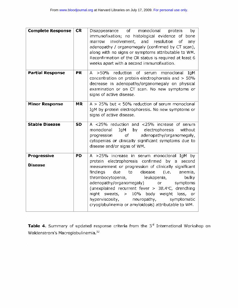
Consensus based uniform response criteria for WM were developed as part
of the International Workshops on WM.32,34,89 The category of minor
response was adopted at the Third International Workshop of WM, given that
clinically meaningful responses have been observed with newer biological
agents such as rituximab and is based on >25 to < 50% decrease in serum
IgM level, which is used as a surrogate marked of disease in WM.32,34 The
term major response is used to denote a response of > 50% in serum IgM
levels, and includes partial and complete responses.32 Response categories
and criteria for progressive disease in WM are summarized in Table 4.
For personal use only. at Harvard Libraries on July 17, 2009. www.bloodjournal.orgFrom
While IgM is commonly used as a surrogate marker of WM disease burden, it
can fluctuate with biological agents such as rituximab and
bortezomib.41,42,54,55 Rituximab can induce a flare in serum IgM levels, which
can occur when used as monotherapy and in combination therapy, and may
last for weeks to months.40,48,54,61-63 Conversely, bortezomib can suppress
IgM levels in some patients independent of tumor cell killing.41,42 Moreover,
Varghese et al90 recently showed that residual IgM producing plasma cells
are spared in patients treated with selective B-cell depleting agents such as
rituximab and alemtuzumab, and may therefore skew response assessment.
Therefore, in circumstances where serum IgM levels may appear out of
clinical context, a bone marrow biopsy should be considered in order to
clarify the patient’s underlying disease burden. Soluble CD27 may serve as
an alternative surrogate marker in WM, and appears to remain a faithful
marker of disease in patients experiencing a rituximab related IgM flare, as
well as plasmapheresis.91,92
For personal use only. at Harvard Libraries on July 17, 2009. www.bloodjournal.orgFrom
Acknowledgements
This work was supported through the Peter and Helen Bing Fund for
Waldenstrom’s macroglobulinemia, the Linda and Edward Nelson Fund, the
Bailey Family Fund for Waldenstrom’s Macroglobulinemia, and the
International Waldenstrom’s Macroglobulinemia Foundation. The author is
grateful to the staff of the Bing Center for Waldenstrom’s Macroglobulinemia,
and in particular Robert J. Manning, Christopher J. Patterson, and Lefkothea
Ioakimidis for the data collection used in this manuscript.
Author Contributions and Conflict of Interest Disclosure
SPT reviewed data and prepared article. The author has received research
support, honoraria and/or consultation fees in connection with products
discussed in this article from Berlex Oncology, Inc., Biogen IDEC Inc.,
Celgene Corporation, Genentech BioOncology Inc., Millenium
Pharmaceuticals Inc. The Takeda Company, and PGX Health, Inc.
For personal use only. at Harvard Libraries on July 17, 2009. www.bloodjournal.orgFrom
References
1. Owen RG, Treon SP, Al-Katib A, et al. Clinicopathological definition of
Waldenström’s macroglobulinemia: Consensus Panel
Recommendations from the Second International Workshop on
Waldenström’s macroglobulinemia. Semin Oncol. 2003; 30 (2):110–
15.
2. Harris NL, Jaffe ES, Stein H, et al. A revised European-American
classification of lymphoid neoplasms: a proposal from the International
Lymphoma Study Group. Blood 1994; 84 (5):1361–92.
3. Harris NL, Jaffe ES, Diebold J, et al. The World Health Organization
classification of neoplastic diseases of the hematopoietic and lymphoid
tissues. Report of the Clinical Advisory Committee meeting, Airlie
House, Virginia, November, 1997. J Clin Oncol. 1999; 155 (1):257–65.
4. Merlini G, Farhangi M, Osserman EF. Monoclonal immunoglobulins with
antibody activity in myeloma, macroglobulinemia and related plasma
cell dyscrasias. Semin Oncol. 1986; 13 (3):350–65.
5. Farhangi M, Merlini G. The clinical implications of monoclonal
immunoglobulins. Semin Oncol. 1986; 13 (3):366–79.
6. Marmont AM, Merlini G. Monoclonal autoimmunity in hematology.
Haematologica 1991; 76 (6):449–59.
7. Treon SP, Hunter ZR, Aggarwal A, et al. Characterization of familial
Waldenstrom’s Macroglobulinemia. Ann Oncol. 2006; 17 (3): 488-94.
For personal use only. at Harvard Libraries on July 17, 2009. www.bloodjournal.orgFrom

8. McMaster ML, Csako G, Giambarresi TR, et al. Long-term evaluation of
three multiple-case Waldenstrom macroglobulinemia families. Clin
Cancer Res. 2007; 13 (17): 5063-9.
9. Kristinsson SY, Bjorkholm M, Goldin LR, et al. Risk of
lymphoproliferative disorders among first-degree relatives of
lymphoplasmacytic lymphoma/Waldenstrom macroglobulinemia
patients: a population-based study in Sweden. Blood 2008; 112 (8):
3052-6.
10. Santini GF, Crovatto M, Modolo ML, et al. Waldenström
macroglobulinemia: a role of HCV infection? Blood 1993; 82 (9):2932.
11. Silvestri F, Barillari G, Fanin R, et al. Risk of hepatitis C virus infection,
Waldenström’s macroglobulinemia, and monoclonal gammopathies.
Blood 1996; 88 (3):1125–6.
12. Leleu X, O’Connor K, Ho A, et al. Hepatitis C Viral Infection Is Not
Associated with Waldenstrom’s Macroglobulinemia. Am J Hematol
2006. Am J Hematol. 2007; 82 (1): 83-4.
13. Treon SP, Hunter Z, Ciccarelli BT, et al. IgA and IgG
Hypogammaglobulinemia Is a Constitutive Feature in Most
Waldenstrom’s Macroglobulinemia Patients and May Be Related to
Mutations Associated with Common Variable Immunodeficiency
Disorder (CVID) Blood 2008; 112 (11): Abstract 3749.
For personal use only. at Harvard Libraries on July 17, 2009. www.bloodjournal.orgFrom
14. Kyle RA, Treon SP, Alexanian R, et al. Prognostic markers and criteria
to initiate therapy in Waldenstrom’s Macroglobulinemia: Consensus
Panel Recommendations from the Second International Workshop on
Waldenstrom’s Macroglobulinemia. Semin Oncol. 2003; 30 (2): 116-
120.
15. Morel P, Duhamel A, Gobbi P, et al. International prognostic scoring
system for Waldenstrom Macroglobulinemia. Blood 2009; 113 (18):
4163-70.
16. Pruzanski W, Shumak KH. Biologic activity of cold-reacting
autoantibodies (first of two parts). N Engl J Med. 1977; 297 (11):538–
42.
17. Pruzanski W, Shumak KH. Biologic activity of cold-reacting
autoantibodies (second of two parts). N Engl J Med. 1977; 297
(10):583–9.
18. Menke MN, Feke GT, McMeel JW, Branagan A, Hunter Z, Treon SP.
Hyperviscosity-related retinopathy in Waldenstrom’s
Macroglobulinemia. Arch Opthalmol. 2006; 124 (11): 1601-6.
19. Menke MN, Treon SP. Hyperviscosity Syndrome. In: Clinical Malignant
Hematology, pgs. 937-41, Sekeres, Kalaycio, Bolwell eds., McGraw Hill
Publishing, New York, 2007.
For personal use only. at Harvard Libraries on July 17, 2009. www.bloodjournal.orgFrom

20. Merlini G, Baldini L, Broglia C, et al. Prognostic factors in symptomatic
Waldenström’s macroglobulinemia. Semin Oncol 2003; 30 (2):211–15.
21. Nobile-Orazio E, Marmiroli P, Baldini L, et al. Peripheral neuropathy in
macroglobulinemia: incidence and antigen-specificity of M proteins.
Neurology 1987; 37 (9):1506–14.
22. Garces-Sanchez M, Dyck PJ, Kyle RA, et al. Antibodies to myelin-
associated glycoprotein (anti-MAG) in IgM amyloidosis may influence
expression of neuropathy in rare patients. Muscle Nerve 2008; 37
(4):490-5.
23. Al-Lozi MT, Pestronk A, Choski R. A skeletal muscle-specific form of
decorin is a target antigen for a serum IgM M-protein in a patient with
a proximal myopathy. Neurology 1997; 49 (6): 1650-4.
24. Owen RG, Barrans SL, Richards SJ, et al. Waldenström
macroglobulinemia. Development of diagnostic criteria and
identification of prognostic factors. Am J Clin Pathol. 2001; 116
(3):420–8.
25. San Miguel JF, Vidriales MB, Ocio E, et al. Immunophenotypic analysis
of Waldenstrom’s macroglobulinemia. Semin Oncol. 2003; 30 (2):187-
95.
For personal use only. at Harvard Libraries on July 17, 2009. www.bloodjournal.orgFrom

26. Hunter ZR, Branagan AR, Manning R, et al. CD5, CD10, CD23
expression in Waldenstrom’s Macroglobulinemia. Clin Lymphoma
2005; 5 (4):246-9.
27. Schop RF, Kuehl WM, Van Wier SA, et al. Waldenström
macroglobulinemia neoplastic cells lack immunoglobulin heavy chain
locus translocations but have frequent 6q deletions. Blood 2002; 100
(8):2996–3001.
28. Ocio EM, Schop RF, Gonzalez B, et al. 6q deletion in Waldenstrom’s
macroglobulinemia is associated with features of adverse prognosis. Br
J Haematol. 2007; 136 (1): 80-6.
29. Leleu X, Hunter ZR, Xu L, et al. Expression of regulatory genes for
lymphoplasmacytic cell differentiation in Waldenstrom
Macroglobulinemia Br J Haematol. 2009; 145 (1): 59-63.
30. Chang H, Qi C, Trieu Y, et al. Prognostic relevance of 6q deletion in
Waldenstrom’s macroglobulinemia. Proceedings of the 5th International
Workshop on Waldenstrom’s macroglobulinemia, Stockholm, Sweden
2008 (Abstract 125).
31. Avet-Loiseau H, Garand R, Lode L, Robillard N, Bataille R. 14q32
translocations discriminate IgM multiple myeloma from Waldenstrom’s
macroglobulinemia. Semin Oncol. 2003; 30 (2):153-155.
For personal use only. at Harvard Libraries on July 17, 2009. www.bloodjournal.orgFrom
32. Kimby E, Treon SP, Anagnostopoulos A, et al. Update on
recommendations for assessing response from the Third International
Workshop on Waldenstrom’s Macroglobulinemia. Clin Lymphoma
Myeloma 2006; 6 (5):380-3.
33. Menke MN, Feke GT, McMeel JW, Treon SP. Opthalmologic techniques
to assess the severity of hyperviscosity syndrome and the effect of
plasmapheresis in patients with Waldenstrom’s Macroglobulinemia.
Clin Lymphoma Myeloma 2009; 9 (1):100-3.
34. Treon SP, Gertz MA, Dimopoulos MA, et al. Update on treatment
recommendations from the Third International Workshop on
Waldenstrom’s Macroglobulinemia. Blood 2006; 107 (9):3442-6.
35. Dimopoulos MA, Gertz MA, Kastritis E, et al. Update on treatment
recommendations from the Fourth International Workshop on
Waldenstrom's Macroglobulinemia. J Clin Oncol. 2009; 27 (1): 120-6.
36. Thomas S, Hosing C, Delasalle KB, et al. Success rates of autologous
stem cell collection in patients with Waldenstrom’s macroglobulinemia.
Proc 5th International Workshop on Waldenstrom’s macroglobulinemia
2008 (Supplemental Abstract).
37. Leleu XP, Manning R, Soumerai JD, et al. Increased incidence of
transformation and myelodysplasia/acute leukemia in patients with
For personal use only. at Harvard Libraries on July 17, 2009. www.bloodjournal.orgFrom

Waldenström macroglobulinemia treated with nucleoside analogs. J
Clin Oncol. 2009; 27 (2): 250-5.
38. Leleu X, Tamburini J, Roccaro A, et al. Balancing risk versus benefit in
the treatment of Waldenstrom’s macroglobulinemia patients with
nucleoside analogue based therapy. Clin Lymph Myeloma 2009; 9
(1):71-73.
39. Rakkhit R, Delasalle KB, Gavino MB, et al. Incidence of transformation
to large cell lymphoma and to second malignancies in symptomatic
patients with Waldenstrom’s macroglobulinemia (WM) treated with
cladribine (2-CdA) combination induction. Blood 2008; 112 (11):
Abstract 3065.
40. Treon SP, Ioakimidis L, Soumerai JD, et al.
Primary Therapy of Waldenstrom’s Macroglobulinemia with
Bortezomib, Dexamethasone and Rituximab. J Clin Oncol. 2009; Epub
ahead of print.
41. Treon SP, Hunter ZR, Matous J, et al. Multicenter Clinical Trial of
Bortezomib in Relapsed/Refractory Waldenstrom’s macroglobulinemia:
Results of WMCTG Trial 03-248. Clin Cancer Res. 2007; 13
(11):3320-5.
42. Strauss SJ, Maharaj L, Hoare S, et al. Bortezomib therapy in patients
with relapsed or refractory lymphoma: Potential correlation of in vitro
For personal use only. at Harvard Libraries on July 17, 2009. www.bloodjournal.orgFrom
sensitivity and tumor necrosis factor alpha response with clinical
activity. J Clin Oncol. 2006; 24 (13): 2105-12.
43. Ghobrial IM, Matous J, Padmanabhan S, et al. Phase II trial of
combination of bortezomib and rituximab in relapsed and/or refractory
Waldenstrom’s Macroglobulinemia. Blood 2008; 112 (11): Abstract
832.
44. Agathocleous A, Rule S, Johnson P. Preliminary results of a phase I/II
study of weekly or twice weekly bortezomib in combination with
rituximab in patients with follicular lymphoma, mantle cell lymphoma,
and Waldenstrom’s macroglobulinemia. Blood 2007; 110 (11):
Abstract 2559.
45. Treon SP, Hunter Z, Branagan A. CHOP plus rituximab therapy in
Waldenström’s macroglobulinemia. Clin Lymphoma Myeloma 2005; 5
(4): 273-7.
46. Dimopoulos MA, Anagnostopoulos A, Kyrtsonis MC, et al. Primary
treatment of Waldenstrom’s macroglobulinemia with Dexamethasone,
Rituximab and Cyclophosphamide. J Clin Oncol. 2007; 25 (22):3344-9.
47. Buske C, Hoster E, Dreyling MH, et al. The addition of rituximab to
front-line therapy with CHOP (R-CHOP) results in a higher response
rate and longer time to treatment failure in patients with
lymphoplasmacytic lymphoma: results of a randomized trial of the
For personal use only. at Harvard Libraries on July 17, 2009. www.bloodjournal.orgFrom
German Low-Grade Lymphoma Study Group (GLSG). Leukemia 2009;
23 (1): 153-61.
48. Ioakimidis L, Patterson CJ, Hunter ZR, et al. Comparative outcomes
following CP-R, CVP-R and CHOP-R in Waldenstrom’s
macroglobulinemia. Clin Lymph Myeloma 2009; 9 (1):62-66.
49. Weber DM, Dimopoulos MA, Delasalle K, et al: 2-chlorodeoxyadenosine
alone and in combination for previously untreated Waldenstrom’s
macroglobulinemia. Semin Oncol. 2003; 30 (2):243-247.
50. Treon SP, Branagan AR, Ioakimidis L, et al. Long term outcomes to
fludarabine and rituximab in Waldenstrom’s macroglobulinemia. Blood
2009; 113 (16): 3673-8.
51. Tam CS, Wolf MM, Westerman D, et al. Fludarabine combination
therapy is highly effective in first-line and salvage treatment of
patients with Waldenstrom’s macroglobulinemia. Clin Lymphoma
Myeloma 2005; 6 (2):136-9.
52. Dhodapkar MV, Hoering A, Gertz MA, et al. Long-term survival in
Waldenstrom macroglobulinemia: 10-year follow-up of Southwest
Oncology Group-directed intergroup trial S9003. Blood 2009; 113
(4):793-6.
For personal use only. at Harvard Libraries on July 17, 2009. www.bloodjournal.orgFrom
53. Tedeschi A, Alamos SM, Ricci F, et al. Fludarabine-based combination
therapies for Waldenstrom’s macroglobulinemia. Clin Lymphoma
Myeloma 2009; 9 (1): 67-70.
54. Treon SP, Branagan AR, Hunter Z, Santos D, Tournhilac O, Anderson
KC. Paradoxical increases in serum IgM and viscosity levels following
rituximab in Waldenstrom's macroglobulinemia. Ann Oncol 2004; 15
(10):1481-3.
55. Ghobrial IM, Fonseca R, Greipp PR, et al: Initial immunoglobulin M
“flare” after rituximab therapy in patients with Waldenstrom
Macroglobulinemia: An Eastern Cooperative Oncology Group Study.
Cancer 2004; 101 (11):2593-8.
56. Ghobrial IM, Uslan DZ, Call TG, Witzig TE, Gertz MA. Initial increase in
the cryoglobulin level after rituximab therapy for type II
cryoglobulinemia secondary to Waldenström macroglobulinemia does
not indicate failure of response. Am J Hematol. 2004; 77 (4): 329-30.
57. Noronha V, Fynan TM, Duffy T. Flare in neuropathy following rituximab
therapy for Waldenstrom's macroglobulinemia. J Clin Oncol. 2006; 24
(1): E3.
58. Broglio L, Lauria G. Worsening after rituximab treatment in anti-MAG
neuropathy. Muscle Nerve 2005; 32 (3):378-9.
For personal use only. at Harvard Libraries on July 17, 2009. www.bloodjournal.orgFrom

59. Kilidireas C, Anagnostopoulos A, Karandreas N, et al. Rituximab
therapy in monoclonal IgM-related neuropathies. Leuk Lymphoma
2006; 47 (5): 859-64.
60. Izzedine H, Bourry E, Amrouche L, et al. Immunoglobulin M 'Flare'
after rituximab-associated acute tubular necrosis in Waldenström's
macroglobulinemia. Int J Hematol. 2009; 89 (2):218-22.
61. Nichols GL, Savage DG. Timing of Rituximab/Fludarabine in
Waldenstrom’s macroglobulinemia may avert hyperviscosity. Blood
2004; 104 (11): Abstract 4612.
62. Treon SP, Soumerai JD, Branagan AR, et al. Thalidomide and rituximab
in Waldenstrom’s Macroglobulinemia. Blood 2008; 112 (12): 4452-7.
63. Treon SP, Soumerai JD, Branagan AR, et al. Lenalidomide and
rituximab in Waldenstrom’s macroglobulinemia. Clin Cancer Res. 2009;
15 (1):355-60.
64. Foran JM, Rohatiner AZ, Cunningham D, et al: European phase II
study of rituximab (chimeric anti-CD20 monoclonal antibody) for
patients with newly diagnosed mantle-cell lymphoma and previously
treated mantle-cell lymphoma, immunocytoma, and small B-cell
lymphocytic lymphoma. J Clin Oncol. 2000; 18 (2):317-24.
65. Treon SP, Agus DB, Link B, et al: CD20-Directed antibody-mediated
immunotherapy induces responses and facilitates hematologic
For personal use only. at Harvard Libraries on July 17, 2009. www.bloodjournal.orgFrom
recovery in patients with Waldenstrom’s macroglobulinemia. J
Immunother. 2001; 24 (3):272-79.
66. Gertz MA, Rue M, Blood E, et al: Multicenter phase 2 trial of rituximab
for Waldenstrom macroglobulinemia (WM): An Eastern Cooperative
Oncology Group Study (E3A98) Leuk Lymphoma 2004; 45 (10):2047-
2055.
67. Dimopoulos MA, Zervas C, Zomas A, et al: Treatment of
Waldenstrom’s macroglobulinemia with rituximab. J Clin Oncol. 2002;
20 (9):2327-33.
68. Treon SP, Emmanouilides C, Kimby E, et al. Extended rituximab
therapy in Waldenström’s Macroglobulinemia. Ann Oncol 2005; 16
(1):132-8.
69. Treon SP, Hansen M, Branagan AR, et al. Polymorphisms in FcγRIIIA
(CD16) receptor expression are associated with clinical responses to
Rituximab in Waldenstrom’s Macroglobulinemia. J Clin Oncol. 2005; 23
(3): 474-81.
70. Pestronk A, Florence J, Miller T, et al. Treatment of IgM antibody
associated polyneuropathies using rituximab. J Neurol Neurosurg
Psychiatry 2003; 74 (4):485-9.
71. Benedetti L, Briani C, Grandis M, et al. Predictors of response to
rituximab in patients with neuropathy and anti-myelin associated
For personal use only. at Harvard Libraries on July 17, 2009. www.bloodjournal.orgFrom

glycoprotein immunoglobulin M. J Peripher Nerv Syst. 2007; 12(2):102-
7.
72. Dalakas MC, Rakocevic G, Salajegheh MD, et al. Placebo-controlled
trial of rituximab in IgM anti-myelin-associated glycoprotein antibody
demyelinating neuropathy. Ann Neurol. 2009; 65 (3):286-293.
73. van Oers MH, Klasa R, Marcus RE, et al. Rituximab maintenance
improves clinical outcome of relapsed/resistant follicular non-Hodgkin
lymphoma in patients both with and without rituximab during
induction: results of a prospective randomized phase 3 intergroup trial.
Blood 2006; 108 (10):3295-301.
74. Chen CI, Kouroukis CT, White D, et al. Bortezomib is active in patients
with untreated or relapsed Waldenstrom’s macroglobulinemia: A phase
II study of the National Cancer Institute of Canada Clinical Trials
Group. J Clin Oncol. 2007; 25 (12):1570-5.
75. Dimopoulos MA, Anagnostopoulos A, Kyrtsonis MC, et al. Treatment of
relapsed or refractory Waldenstrom’s macroglobulinemia with
bortezomib. Haematologica 2005; 90 (12):1655-7.
76. Jagannath S, Richardson PG, Barlogie B, et al. Bortezomib in
combination with dexamethasone for the treatment of patients with
relapsed and/or refractory multiple myeloma with less than optimal
response to bortezomib alone. Haematologica 2006; 91 (7): 929-34.
For personal use only. at Harvard Libraries on July 17, 2009. www.bloodjournal.orgFrom

77. Treon SP, Kelliher A, Keele B, et al: Expression of serotherapy target
antigens in Waldenstrom’s macroglobulinemia: Therapeutic
applications and considerations. Semin Oncol. 2003; 30 (2):248-52.
78. Santos DD, Hatjiharissi E, Tournilhac O, et al. CD52 is expressed on
human mast cells and is a potential therapeutic target in
Waldenstrom's Macroglobulinemia and mast cell disorders. Clin Lymph
Myeloma 2006; 6 (6): 478-83.
79. Hunter ZR, Boxer M, Kahl B, et al. Phase II study of alemtuzumab in
lymphoplasmacytic lymphoma: Results of WMCTG trial 02-079. Proc
Am Soc Clin Oncol 2006; 24 (33): Abstract 7523.
80. Owen RG, Rawstron AC, Osterborg A, et al. Activity of alemtuzumab in
relapsed/refractory Waldenstrom's macroglobulinemia. Blood 2003;
102 (11): 644a.
81. Kyriakou H, on behalf of the Lymphoma Working Party of the European
Group for Blood and Bone Marrow Transplantation. Haematopoietic
stem cell transplantation for Waldenstrom’s macroglobulinemia.
Proceedings of the 5th International Workshop on Waldenstrom’s
macroglobulinemia, Stockholm, Sweden 2008 (Abstract 146).
82. Maloney D. Evidence for GVWM following mini-allo in Waldenstrom’s
macroglobulinemia. Proceedings of the 5th International Workshop on
For personal use only. at Harvard Libraries on July 17, 2009. www.bloodjournal.orgFrom

Waldenstrom’s macroglobulinemia, Stockholm, Sweden 2008 (Abstract
147).
83. Gertz MA, Hayman SR, Buadi FK. Transplantation for IgM amyloidosis
and IgM myeloma. Clin Lymphoma Myeloma 2009; 9 (1):77-79.
84. Rummel MJ, von Gruenhagen U, Niederle N, et al. Bendamustine plus
rituximab versus CHOP plus rituximab in the firstline treatment of pati
ents with follicular, indolent and mantle cell lymphomas: Results of a
randomized phase III study of the Study Group Indolent Lymphomas
(StiL). Blood 2008; 112 (11): Abstract 2596.
85. Rummel MJ, von Gruenhagen U, Niederle N, et al. Bendamustine plus
rituximab versus CHOP plus rituximab in the first-line treatment of
patients with Waldenstrom’s macroglobulinemia-First interim results of
a randomized phase III study of the Studygroup Indolent Lymphomas
(StiL). Proceedings of the 5th International Workshop on
Waldenstrom’s macroglobulinemia, Stockholm, Sweden 2008 (Abstract
139).
86. Hatjiharissi E, Mitsiades CS, Ciccarelli B, et al. Comprehensive
Molecular Characterization of Malignant and Microenvironmental Cells
in Waldenstroms Macroglobulinemia by Gene Expression Profiling.
Blood 2007; 110 (11): Abstract 3174.
For personal use only. at Harvard Libraries on July 17, 2009. www.bloodjournal.orgFrom

87. Leleu X, Jia X, Runnels J, et al. The Akt pathway regulates survival and
homing in Waldenstrom macroglobulinemia. Blood 2007; 110 (11):
4417-26.
88. Ghobrial IM, Chuma S, Sam A, et al.
Phase II Trial of the mTOR Inhibitor RAD001 in Relapsed and/or
Refractory Waldenstrom Macroglobulinemia: The Dana Farber Cancer
Institute Experience. Blood 2008; 112 (11): Abstract 1011.
89. Weber D, Treon SP, Emmanouilides C, et al. Uniform response criteria
in Waldenstrom's macroglobulinemia: Consensus panel
recommendations from the Second International Workshop on
Waldenstrom's Macroglobulinemia. Semin Oncol. 2003; 30 (2):127-
31.
90. Varghese AM, Rawstron AC, Ashcroft J, et al. Assessment of bone
marrow response in Waldentrom’s macroglobulinemia. Clin Lymphoma
Myeloma 2009; 9 (1):53-55.
91. Ho A, Leleu X, Hatjiharissi E, et al. CD27-CD70 interactions in the
pathogenesis of Waldenstrom’s Macroglobulinemia. Blood 2008; 112
(12):4683-9.
92. Ciccarelli BT, Yang G, Hatjiharissi E, et al. Soluble CD27 is a faithful
marker of disease burden and is unaffected by the rituximab induced
For personal use only. at Harvard Libraries on July 17, 2009. www.bloodjournal.orgFrom
IgM flare, as well as plasmapheresis in patients with Waldenstrom’s
macroglobulinemia. Clin Lymph Myeloma 2009; 9 (1):56-58.
For personal use only. at Harvard Libraries on July 17, 2009. www.bloodjournal.orgFrom

Median Range Normal Reference
Range
Age (yr) 58 32-91 NA
Gender
(Male/Female)
215/141 NA
Bone marrow
involvement
30% 5-95% NA
Adenopathy 15% NA
Splenomegaly 10% NA
IgM (mg/dL) 2,620 270-12,400 40-230
IgG (mg/dL) 674 80-2,770 700-1,600
IgA (mg/dL) 58 6-438 70-400
Serum Viscosity (cp) 2.0 1.1-7.2 1.4-1.9
Hct (%) 35.4% 17.2-45.4% 34.8-43.6%
Plt (x 109/L) 275 42-675 155-410
Wbc (x 109/L) 6.4 1.7-22 3.8-9.2
B2M (mg/dL) 2.5 0.9-13.7 0-2.7
LDH (U/mL) 313 61-1,701 313-618
Table 1. Clinical and laboratory findings for 356 newly diagnosed patients with the
consensus panel diagnosis of WM presenting to the Dana Farber Cancer Institute. NA (not
applicable).
For personal use only. at Harvard Libraries on July 17, 2009. www.bloodjournal.orgFrom

Properties of IgM
Monoclonal Protein
Diagnostic Condition Clinical Manifestations
Pentameric Structure Hyperviscosity Headaches, blurred vision,
epistaxis, retinal hemorrhages,
leg cramps, impaired mentation,
intracranial hemorrhage.
Precipitation on cooling Cryoglobulinemia (Type I) Raynaud’s phenomenom,
acrocyanosis, ulcers, purpura,
cold urticaria.
Auto-antibody activity
to Myelin Associated
Glycoprotein (MAG),
Ganglioside M1 (GM1),
Sulfatide moieties on
peripheral nerve
sheaths
Peripheral neuropathies Sensorimotor neuropathies,
painful neuropathies, ataxic gait,
bilateral foot drop.
Auto-antibody activity
to IgG
Cryoglobulinemia (Type II) Purpura, arthralgias, renal failure,
sensorimotor neuropathies.
Auto-antibody activity
to red blood cell
antigens
Cold agglutinins Hemolytic anemia, Raynaud’s
phenomenom, acrocyanosis,
livedo reticularis.
Tissue deposition as
amorphous aggregates
Organ Dysfunction Skin: bullous skin disease,
papules, Schnitzler’s syndrome.
GI: diarrhea, malabsorption,
bleeding.
Kidney: proteinuria, renal failure
(light chain component).
Tissue deposition as
amyloid fibrils
(light chain component
most commonly)
Organ Dysfunction Fatigue, weight loss, edema,
hepatomegaly, macroglossia,
organ dysfunction of involved
organs: heart, kidney, liver,
peripheral sensory and autonomic
nerves.
Table 2. Morbidities mediated by the monoclonal IgM protein in Waldenstrom’s
macroglobulinemia.
For personal use only. at Harvard Libraries on July 17, 2009. www.bloodjournal.orgFrom

Symptom/Complaint Implications Action
Energy level/Changes in
activities of daily life.
Anemia, fatigue without
anemia
Evaluate for anemia,
underlying etiology including
iron deficiency, hemolytic
anemia (warm and cold
antibodies). Consider
amyloidosis. Exclude other
medical causes of anemia.
Constitutional Complaints Tumor related fever, chills,
night sweats.
Recurrent sinus and
bronchial infections.
Chronic sinusitis, usually on
the basis of IgA and IgG
hypogammaglobulinemia.
Antibiotic support, if
refractory to multiple
antibiotic courses,
hospitalizations, or life
threatening, strongly
consider IVIG replacement.
Headaches, blurry vision or
visual loss, confusional
episodes, epistaxis.
Hyperviscosity Funduscopic examination for
hyperviscosity related
changes, obtain serum IgM
and viscosity levels. Consider
emergency plasmapheresis
for symptomatic
hyperviscosity; strongly
consider in patients with
serum viscosity > 4.0 cp
given high risk of
hyperviscosity related
events.
Easy bruising, bleeding
diathesis.
Thrombocytopenia, Acquired
Von Willebrand’s Disorder
(VWD).
Complete blood count,
evaluate for immune
thrombocytopenia or
hypersplenism if indicated;
consider evaluation for VWD;
consider amyloidosis.
Progressive symmetrical
numbness, tingling, burning,
pain feet and hands.
Unsteady gait, deficits in
motor function.
IgM related neuropathy or
myopathy; amyloidosis.
Obtain anti-MAG, anti-GM1,
anti-sulfatidyl IgM antibody
studies; if myopathy
present, consider obtaining
anti-decorin antibodies.
For personal use only. at Harvard Libraries on July 17, 2009. www.bloodjournal.orgFrom
Consider obtaining fat pad
biopsy and stain for amyloid.
Consider EMG studies.
Raynaud like symptoms,
acrocyanosis, ulcerations on
extremities.
Cryoglobulinemia, cold
agglutinemia.
Obtain cryoglobulins cold
agglutinins. In patients
suspected of having
cryoglobulins, all studies
including quantitative
immunoglobulins should be
obtained in a warm bath to
avoid cryoprecipitation and
false lowering of serum IgM
levels.
Diarrhea, gastrointestinal
cramping.
Malabsorption, secondary to
amyloidosis, IgM deposition,
tumor involvement. Rarely,
autonomic neuropathy on
basis of auto-antibody or
amyloidosis.
Endoscopy to evaluate small
bowel, biopsy to evaluate for
amyloidosis, IgM deposition,
tumor involvement.
Hearing loss Hyperviscosity, sensorineural
hearing loss, amyloid or
tumor deposition, thrombus
formation.
Evaluate for anti-hu
antibody, MRI to assess for
amyloidoma, tumor
deposition. Evaluate for
hyperviscosity syndrome (as
above). Assess for IgM anti-
phsopholipid antibodies.
Thrombotic events Antiphospholipid antibody
syndrome
Assess for IgM anti-
phospholipid antibodies.
Urticaria, papules,
dermatitis.
Schnitzler’s syndrome (non-
pruritic urticaria), IgM or
tumor cell infiltration,
amyloid deposition.
Skin biopsy, histological
examination for tumor cell
infiltration, stain for IgM,
Congo red for amyloid.
Table 3. A review of systems, and its implications in the workup of patients with
Waldenstrom’s macroglobulinemia.
For personal use only. at Harvard Libraries on July 17, 2009. www.bloodjournal.orgFrom
Complete Response CR Disappearance of monoclonal protein by
immunofixation; no histological evidence of bone
marrow involvement, and resolution of any
adenopathy / organomegaly (confirmed by CT scan),
along with no signs or symptoms attributable to WM.
Reconfirmation of the CR status is required at least 6
weeks apart with a second immunofixation.
Partial Response PR A >50% reduction of serum monoclonal IgM
concentration on protein electrophoresis and > 50%
decrease in adenopathy/organomegaly on physical
examination or on CT scan. No new symptoms or
signs of active disease.
Minor Response MR A > 25% but < 50% reduction of serum monoclonal
IgM by protein electrophoresis. No new symptoms or
signs of active disease.
Stable Disease SD A <25% reduction and <25% increase of serum
monoclonal IgM by electrophoresis without
progression of adenopathy/organomegaly,
cytopenias or clinically significant symptoms due to
disease and/or signs of WM.
Progressive
Disease
PD A >25% increase in serum monoclonal IgM by
protein electrophoresis confirmed by a second
measurement or progression of clinically significant
findings due to disease (i.e. anemia,
thrombocytopenia, leukopenia, bulky
adenopathy/organomegaly) or symptoms
(unexplained recurrent fever > 38.4°C, drenching
night sweats, > 10% body weight loss, or
hyperviscosity, neuropathy, symptomatic
cryoglobulinemia or amyloidosis) attributable to WM.
Table 4. Summary of updated response criteria from the 3rd International Workshop on
Waldenstrom’s Macroglobulinemia.32
For personal use only. at Harvard Libraries on July 17, 2009. www.bloodjournal.orgFrom

Figure legends
Figure 1. Comparisons of serum IgM, hematocrit and bone marrow disease involvement for
356 newly diagnosed patients with Waldenstrom’s Macroglobulinemia.
Figure 2. Guide to the Primary Therapy of Waldenstrom’s Macroglobulinemia. HV,
hyperviscosity; cp, centipoise; BDR (Bortezomib, Dexamethasone, Rituximab); CPR
(Cyclophosphamide, Prednisone, Rituximab); RCD (Rituximab, Cyclophosphamide,
Dexamethasone); VR (Bortezomib, Rituximab); FR (Fludarabine, Rituximab); R (Rituximab).
1Due to potential risk of stem cell damage and/or secondary malignancies, may consider as
an alternative option if other treatment choices are either unavailable or inappropriate for a
particular patient; 2Consider an attenuated schedule for fludarabine administration in
patients with more indolent disease presentation (see text); 3Avoid as monotherapy in
patients with hyperviscosity and with FcγRIIIA-158 F/F polymorphism. For rituximab based
therapies, consider maintenance rituximab in responding patients (see text). Clinical trials
should be considered for patients whenever possible.
Figure 3. Guide to the Salvage Therapy of Waldenstrom’s Macroglobulinemia. ASCT
(Autologous Stem Cell Transplant); RIC (Reduced Intensity Allogeneic Stem Cell
Transplant); NA; nucleoside analogue based therapy; 1In patients being considered for an
ASCT, stem cell collection should be undertaken before exposure to a nucleoside analogue.
Patients should be considered for clinical trials whenever possible.
For personal use only. at Harvard Libraries on July 17, 2009. www.bloodjournal.orgFrom
A.
B.
C.
Figure 1
For personal use only.
at Harvard Libraries on July 17, 2009.
ww
w.bloodjournal.org
From
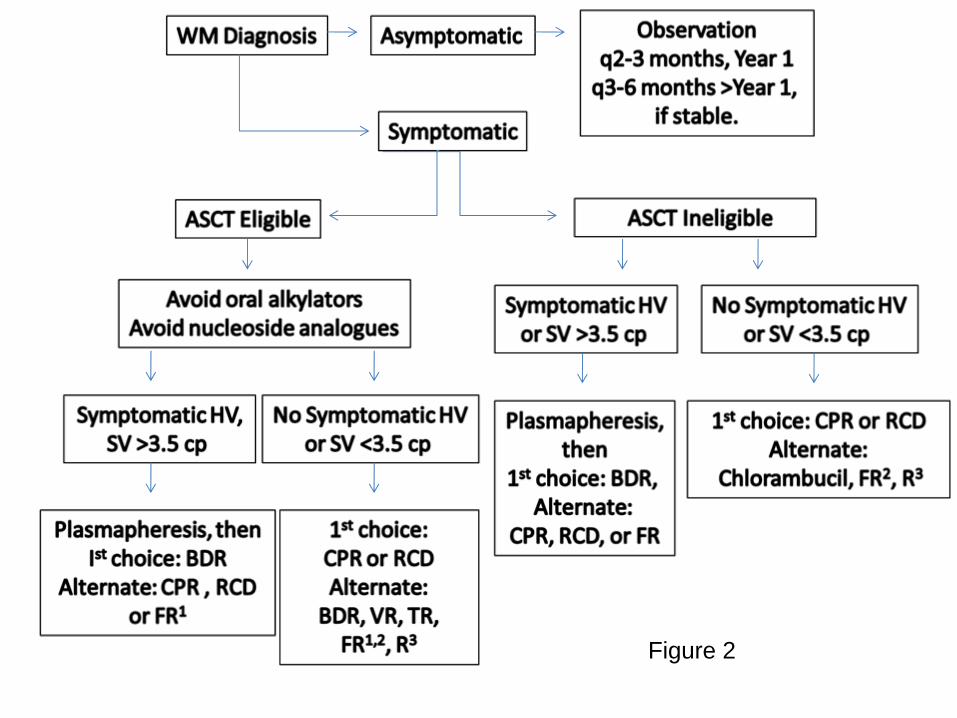
Figure 2
F
or personal use only. at H
arvard Libraries on July 17, 2009. w
ww
.bloodjournal.orgF
rom
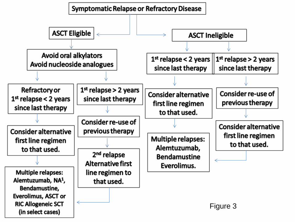
Figure 3
F
or personal use only. at H
arvard Libraries on July 17, 2009. w
ww
.bloodjournal.orgF
rom