how imaging leads to coronary intervention -...
TRANSCRIPT

How Imaging Leads to Coronary Intervention
Lampros K. Michalis, MRCP, FESCProfessor of Cardiology
Medical School, University of Ioannina

Summary
Introduction
Non-invasive imaging modalities
Invasive imaging modalities
Advanced imaging techniques
Conclusions

How We Proceed
• Anatomy
• Functional assessment
– Diagnosis of ischaemia
– Identification of myocardial viability

Non Invasive Imaging Modalities

Echocardiography
Acute MI
identification of certain pathologies, which may need
individualized management
Acute mitral regurgitation
Myocardial rupture
VSD
RV failure
Aneurysm of the ascending aorta

Echocardiography
Stable angina
• stress echocardiography can be used to identify areas of:
• reversible ischemia
• viable myocardial segments

Identification of Reversible Ischaemia

Identification of Reversible Ischaemia

Identification of Myocardial Viability

Myocardial Perfusion Scan
Allows non-invasive detection of myocardial ischemia and myocardial viabiltiy
Myocardial ischaemia protocol
At restDuring stress
Exercise Stress testThe ideal choice even when the ECG in non-diagnositc (digoxin, LVH, LBBB)Patient’s exercise tolerance
We stop: BP >10mmHg, angina, ST >1mm, patient decision, achievement of the goal, ST >2mm, arrythmia, hypertention, tiredness – SOB
Stress
Pharmaceutical stress test Adenosine, dypiridamole, dobutamine
Myocardial viability protocolAfter administration of nitrates (viability)At rest

Myocardial perfusion scan

Computed Tomography Appears to have a high negative predictive value in detecting the presence of
CAD therefore it is the only anatomical test that has indication for symptomatic patients with intermediate likelihood of obstructive CAD (only these patients need diagnostic tests)

Computed Tomography• The problems of low
PPV
Limited positive predictive value especially in stented segments and in calcified arteries
In a recent study that compared FFR with CT the specificity of CT in detecting hemodynamic significant CAD was only 64%

Computed Tomography
Useful in the study of the patency of the grafts
Can be useful in complex procedures

Computed Tomography
CTA: Why useful in complex procedures?
Assessing the course of the vessel in CTO
Detecting the presence of Ca (independent predictor of procedural failure)
Evaluate angulation in bifurcation lesions
• (important for the prediction of carina shift and so for the planning of the procedure)

Magnetic Resonance Imaging• The gold standard for detecting the presence of scar Very useful in discriminating ischeamic cardiomyopathy from other
cardiomyopathies
Where the scar lies
Useful in defining the underlying syndrome in patients with myocardial necrosis and normal angiograms
Acute coronary syndromes vs myocarditis
• Diagnosing myocardial viability A cut-off of 50% scar has been initially used to identify viable myocardium but
recent reports cast doubts about the reliability of this metric
Low-dobutamine stress test can be used to detect myocardial viability
• Diagnosing myocardial ischaemia Pharmacological stress test can be used to detect areas of reversible ischemia
Kim et al. N Eng J Med 2000
Baer et al. JACC 1998
Kaandorp et al. Am J Cardiol 2005

Invasive Imaging Modalities

The acquired cross-sectional images allow:
Identification of the lumen, stent and vessel wall
Quantification of their dimensions and plaque volume
Classification of the plaque type
Detecting plaque erosion, ruptureand the presence of thrombus
Unable to see microstructures related to increased vulnerability
Mintz et al. J Am Coll Cardiol. 2011
Relatively low resolution - limited
capability in:
Intravascular Ultrasound

Intravascular UltrasoundClinical Indications
Bourantas CV, Naka KK, … , Michalis LK. Echocardiography 2010

Intravascular Ultrasound
Assessment of lesion severityWhere we stand

IVUSIVUS: Is this lesion significant?
Mintz G. TCT 2011

Intravascular UltrasoundDiagnosis of Functionally Significant Lesions
Waksman R, et al. JACC 2013
FIRST:
a multinational,
multicenter,
prospective registry
of a large patient
cohort with
intermediate
coronary stenosis
(350 patients; 367
intermediate
lesions)

Intravascular Ultrasound
Guidance of TreatmentWhere we stand

IVUS

201 matched pairs of pts undergoing PCI for unprotected LMS with and without IVUS guidance
At 3 years FU pts had LMS PCI under IVUS guidance had a lower incidence of death and there was a trend for reduced incidence of death-MI


Although meta-analysis and some clinical studies have demonstrated that an IVUS guided PCI is likely to be associated with better clinical outcomes there are no clear indications about the set of patients and the type of lesions in which IVUS imaging should be used

Intravascular UltrasoundResearch Utility
Nissen SE. Am J Cardiol 2002
And to Assess the Effect of Treatment
BASELINE FOLLOW-UP
Nissen SE, et al. JAMA 2006
PLAQUE REGRESSION UNDER STATIN THERAPY

The acquired cross-sectional images allow:
Imaging of micro-features related to plaque vulnerability
Evaluation of stent endotheliazation
Classification of the plaque type
Identification of plaque rupture/thrombus
Tearney et al. J Am Coll Cardiol. 2012
Poor penetration which often does not allow imaging of the entire vessel wall
Cannot “see” behind lipid tissue
Unable to discriminate deeply embeddedlipid-rich from calcific tissue
Limitations of this modality are:
Optical Coherence Tomography

Optical Coherence Tomography
Diagnosis & Guidance of Treatment
Where we stand

OCT allows identification of dissections (N=17)

Anatomy of the LMS, circumflex, LAD, diagona
l and septal branches
Anatomy of the LMS, circumflex, LAD, diagonal and
septal branches
LAD D1
LCx
LAD LAD LAD
LCx
D1D1
LADLAD
PROX
Lets see the anatomy of LMS, LCx, LAD, D1 and S1 in 3 D fashion
Is the evaluation of coronary anatomy better?Is it useful?

LCx
D1
LAD
SEPTAL
View from the LMS
CARINA

Three-dimensional Optical Coherence Tomography Assessment of Coronary
Wire Re-crossing Position duringBifurcation Stenting
Takayuki Okamura, MD*, Jutaro Yamada, MD, Tomoko Nao, MD, Takeshi Suetomi, MD, Takao Maeda, MD, Kohzoh Shiraishi, MD, Toshiro Miura,
MD, Masunori Matsuzaki, MD
Division of Cardiology, Department of Medicine and Clinical Science, Yamaguchi University Graduate School of Medicine, Ube, Japan
EuroIntervention Oct 15, 2011

A novel automatic Strut Detection software (from
Yamaguchi University) enabled a rapid 3-D stent
image reconstruction during bifurcation
stenting procedure.
Napkin-ring Narrowing at the ostium of LCx (0,0,1)

PRE
*
*
PRE
POST
LAD
DIAGONAL
POST
OVERLAPPING SEGMENT
PROTRUDING STRUTS

StillSTENT UNDEREXPANSION*
OVERHANGING STRUT
*

If we needed 20.000 pts to prove that IVUS guided PCI is associated with a better prognosis how many pts do we need in an OCT study?Only 670??

The prognosis of the pts who had OCT guided PCI was compated with the progrnosis of 335 pts with similar demographics that undewent angiography guided PCI
Pts having OCT guided PCI had a
Better prognosis
Stent in case of edge dissection PCI if ΜLA <4mm2
Post-dilation in case of stent underexpansion (ΜLA <90%)
Malapposition when the detachment distance was 200μm
We need a randomized control trial
Prati et al

Optical Coherence Tomography
Assessment of lesion severityWhere we stand

Optical Coherence TomographyDiagnosis of Functionally Significant Lesions
Gonzalo N, et al. JACC 2012
Comparison
with FFR

OCT vs IVUSDiagnosis of Functionally Significant Lesions
Gonzalo N, et al. JACC 2012

Advanced Imaging Techniques
Hybrid Imaging

Advanced Imaging Techniques
Assessment of Lesion Severity

Fusion of SPECT-PET with CT
The fusion of SPECT with Computed tomography appears provide comprehensive imaging and it is likely to be useful in clinical setting:
Gaemperli et al. J Nucl Med. 2007
To estimate the severity of a lesion
To detect the lesion causing symptoms in patients with 2
or 3 vessel disease

HeartFlowTM

CFD in Models derived by 3D QCAFFR assessment

Reconstructed segment
Outlet
Inlet(LM bifurcation)
Virtual FFR: 0.92
Siogkas P, Papafaklis MI, … , Michalis LK, Fotiadis DI. Conf IEEE Eng Med Biol Soc 2013
+
CFD in Models derived by Intravascular Ultrasound
FFR assessment

Advanced Imaging Techniques
Predicting Events and ? Implementing pre-emptive
treatment


Low ESS was associated with plaque progression
Large plaque burden and low ESS appeared as independent predictors of plaque progression
Large plaque burden and low ESS predicted with 41% accuracy disease progression requiring PCI
Stone PH, et al. Circulation 2012
Fusion of Angiography & IVUS:Local Hemodynamics and Disease Progression

Conclusions
Non-invasive imaging modalities have a role in assessing myocardial viability and detecting reversible ischemia
Although intravascular imaging modalities seems to be useful in PCI there are no robust data to support their use in everyday clinical practice
Novel hybrid and computational expensive imaging based techniques have emerged over the last years that appear capable to detect hemodynamic significant lesions and areas at high-risk for future events, and are expected to have a role in the future in the clinical setting


Increasing to
160 ml/min
Increasing to
320 ml/min
Myocardial perfusion scan
Spect with Τh 14mSv Spect with Τc 7mSv + 7mSv Angiogram 14mSv CTA 3mSv
Radiation dose


Introduction
Objective evidence of the presence of a hemodynamic significant lesion is required

Hull & East Yorkshire Hospitals NHS Trust
Stress SA
Stress VLA
Stress HLA


Functional AngioplastyProposed Role of FFR and IVUS
Park, et al. Circulation 2011

Fusion of OCT and Angiography: Enhanced In Vivo Investigations on ESS and High-risk Plaque Features
0.6
0.8
1.0
1.2
1.4
[Pa]
ESS ESS
0.4
0.6
0.8
1.0
1.2
[Pa]
ESS
0.4
0.6
0.8
1.0
1.2
[Pa]
0.6
0.8
1.0
1.2
1.4
[Pa]
ESS
Large calcified plaque Thin-cap atheroma
MΦ accumulation Neovessels
Papafaklis MI, … , Stone PH, Jang IK, Michalis LK. JACC Intv 2013;6:S34

OCT + Angiography:Augmented Lipid Content & Thinner Fibrous Cap in
Areas With Low Endothelial Shear Stress Lipid Arc
Low Higher0
50
100
150 p<0.05
Endothelial Shear Stress
Lip
id A
rc (
o)
FCT
Low Higher0
100
200
300 p<0.05
Endothelial Shear Stress
Lip
id A
rc (
o)
Lip
id A
rc (
o)
Cap
Th
k (μ
m)
Endothelial Shear Stress
Endothelial Shear Stress
Lipid-rich
Plaques
Thinner
Fibrous
Cap
Low ESS
Non-culprit Lesions in Patients With Acute Coronary Syndromes
Vergallo R, Papafaklis MI, … , Michalis LK, Stone PH, Jang IK. AHA Sessions 2013
High-risk plaque: TCFA
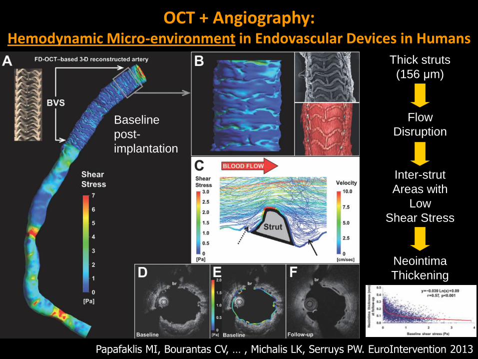
OCT + Angiography: Hemodynamic Micro-environment in Endovascular Devices in Humans
Thick struts
(156 μm)
Flow
Disruption
Inter-strut
Areas with
Low
Shear Stress
Neointima
Thickening
Baseline
post-
implantation
Papafaklis MI, Bourantas CV, … , Michalis LK, Serruys PW. EuroIntervention 2013

Advanced ImagingPrediction of Natural History and Clinical Events
• Prediction of future culprit lesions
• Prediction of coronary regions with rapid worsening
• Prediction of clinical events

697 pts with ACS
3-vessel imaging post PCI
Angiography (QCA of entire coronary tree),
IVUS, Virtual histology, Palpography
At 3y follow-up 107 new symptomatic lesions appeared in 74 pts
Biomarkers-Hs CRP-IL-6-sCD40L-MPO-TNFα-MMP9-Lp-PLA2-others
MSCTSubstudyN=50-100

Fusion of Angiography & IVUS:Local Hemodynamics and Disease Progression
The largest natural history of atherosclerosis study that examined the effect of ESS on plaque growth
506 pts with an ACS had coronary angiography and 3-vessel IVUS
The pts had repeat coronary angiography and IVUS imaging at 6-10 m follow-up
Blood flow simulation was performed and the computed ESS were associated to the changes in plaque burden
Prediction