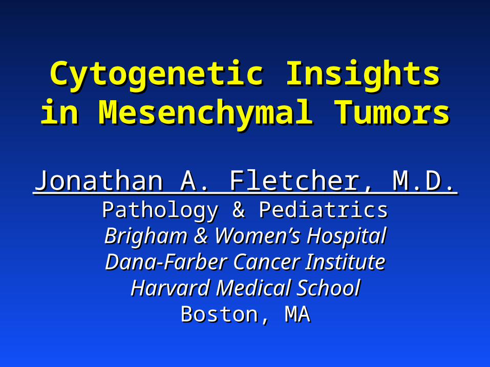
how it is done!
DESCRIPTION
Cytogenetic Insights in Mesenchymal Tumors Jonathan A. Fletcher, M.D. Pathology & Pediatrics Brigham & Women’s Hospital Dana-Farber Cancer Institute Harvard Medical School Boston, MA. How it is done!. Mince, then disaggregate cells by overnight treatment with collagenase. - PowerPoint PPT PresentationTRANSCRIPT
Cytogenetic Insights in Cytogenetic Insights in Mesenchymal TumorsMesenchymal Tumors
Jonathan A. Fletcher, M.D.Jonathan A. Fletcher, M.D.Pathology & PediatricsPathology & Pediatrics
Brigham & Women’s HospitalBrigham & Women’s HospitalDana-Farber Cancer InstituteDana-Farber Cancer Institute
Harvard Medical SchoolHarvard Medical SchoolBoston, MABoston, MA
How it is done!
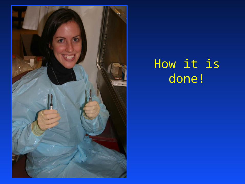
Mince, then disaggregate cells by overnight treatment with collagenase
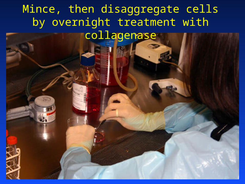
Disaggregated cells are plated as monolayer
cultures on glass slides or in
plastic flasks

All cultures are inspected daily, to determine whether tumor cells are growing, and when metaphase harvests should be performed

Leiomyoma: simple karyotype with t(12;14)
HMGA2(HMGIC)
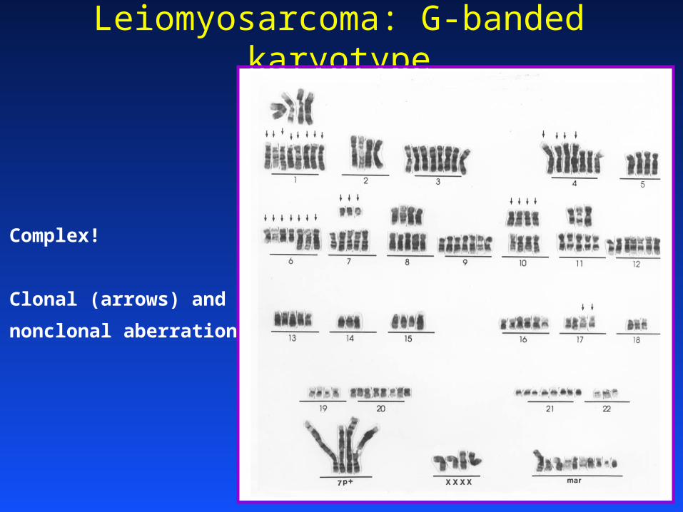
Leiomyosarcoma: G-banded karyotype
Complex!
Clonal (arrows) and
nonclonal aberrations
Example 1
Novel biologic mechanisms revealed through indentification of recurrent
cytogenetic abnormalities in mesenchymal tumors
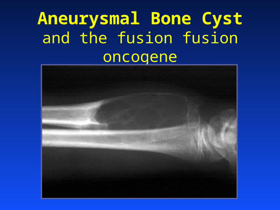
Aneurysmal Bone Cystand the fusion fusion oncogene
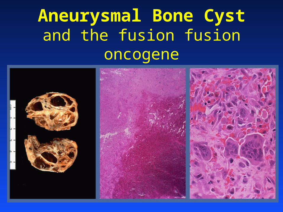
Aneurysmal Bone Cystand the fusion fusion oncogene
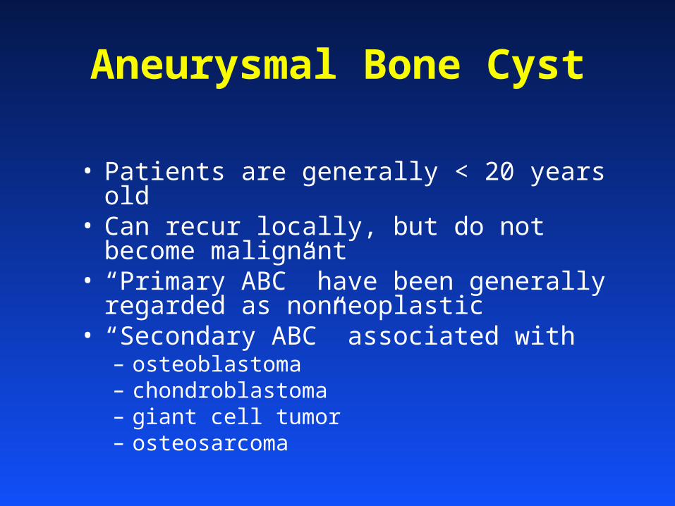
Aneurysmal Bone Cyst
• Patients are generally < 20 years old• Can recur locally, but do not become
malignant• “Primary ABC” have been generally
regarded as nonneoplastic • “Secondary ABC” associated with
– osteoblastoma– chondroblastoma– giant cell tumor– osteosarcoma
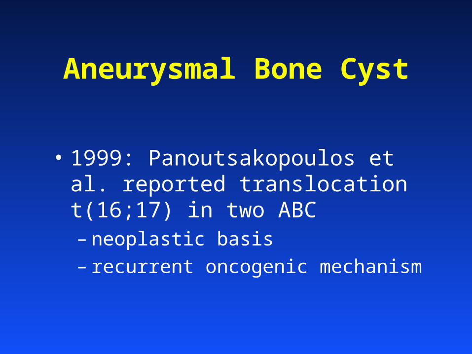
Aneurysmal Bone Cyst
• 1999: Panoutsakopoulos et al. reported translocation t(16;17) in two ABC– neoplastic basis
– recurrent oncogenic mechanism
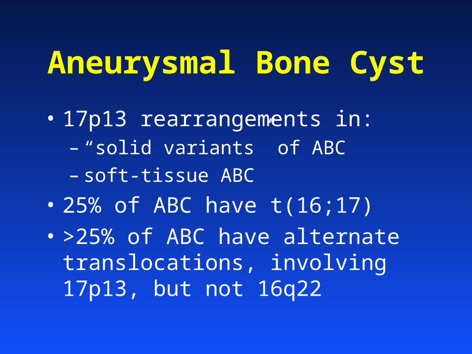
Aneurysmal Bone Cyst
• 17p13 rearrangements in:– “solid variants” of ABC
– soft-tissue ABC
• 25% of ABC have t(16;17)• >25% of ABC have alternate
translocations, involving 17p13, but not 16q22
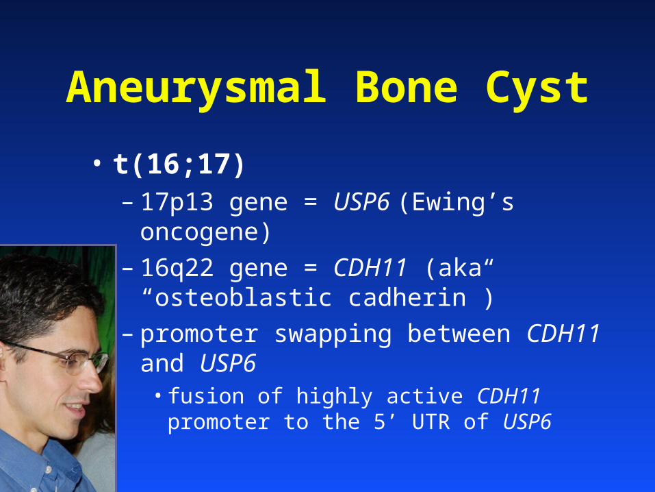
Aneurysmal Bone Cyst
• t(16;17)– 17p13 gene = USP6 (Ewing’s oncogene)
– 16q22 gene = CDH11 (aka “osteoblastic cadherin”)
– promoter swapping between CDH11 and USP6• fusion of highly active CDH11 promoter to the
5’ UTR of USP6
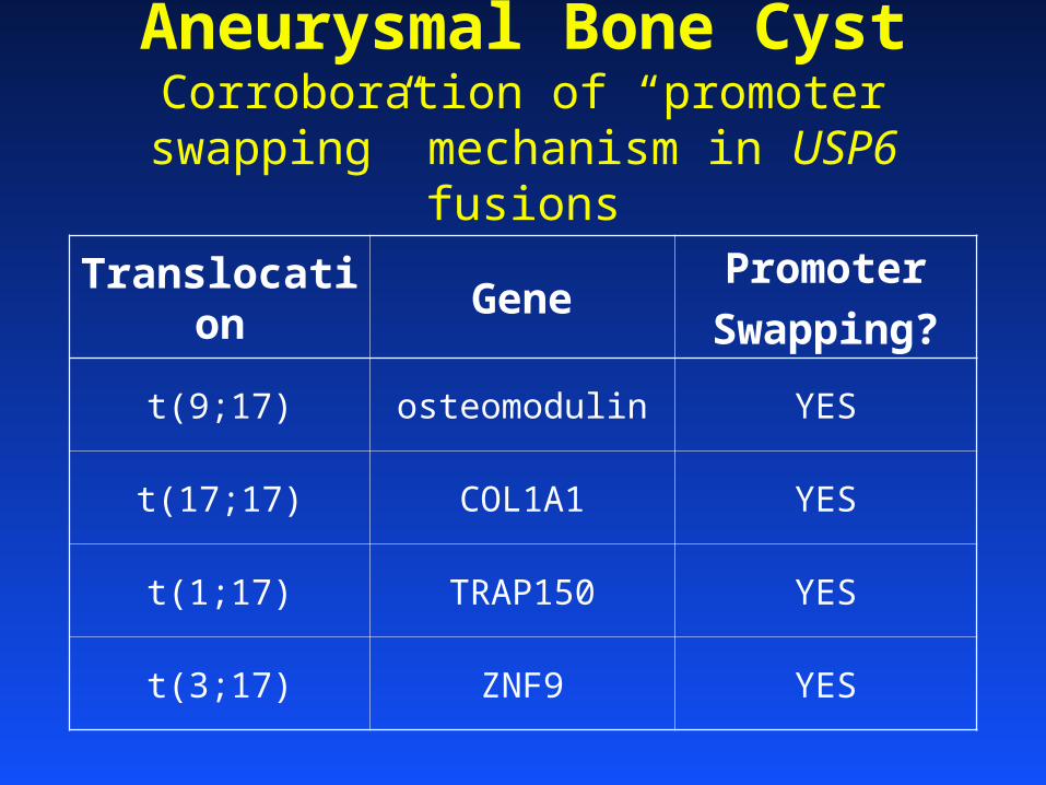
Aneurysmal Bone CystCorroboration of “promoter swapping”
mechanism in USP6 fusions
Translocation GenePromoter
Swapping?
t(9;17) osteomodulin YES
t(17;17) COL1A1 YES
t(1;17) TRAP150 YES
t(3;17) ZNF9 YES
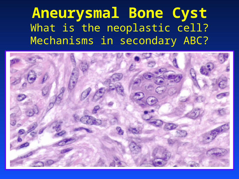
Aneurysmal Bone CystWhat is the neoplastic cell?
Mechanisms in secondary ABC?
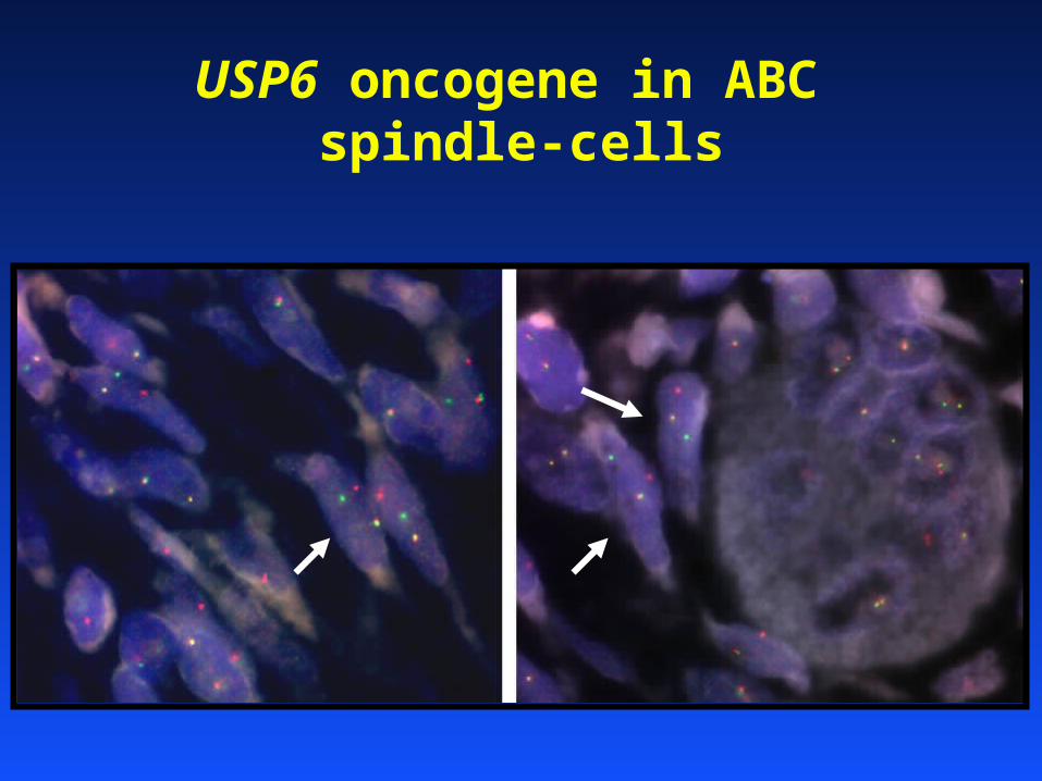
USP6 oncogene in ABC spindle-cells
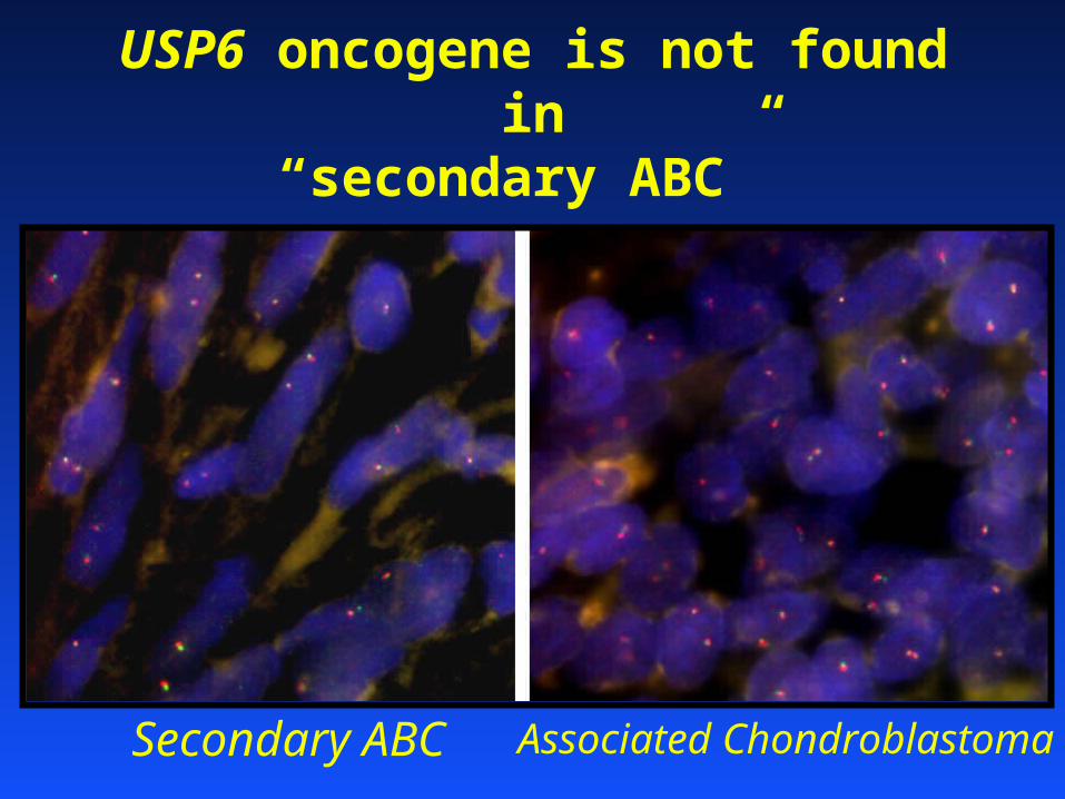
USP6 oncogene is not found in“secondary ABC”
Secondary ABC Associated Chondroblastoma
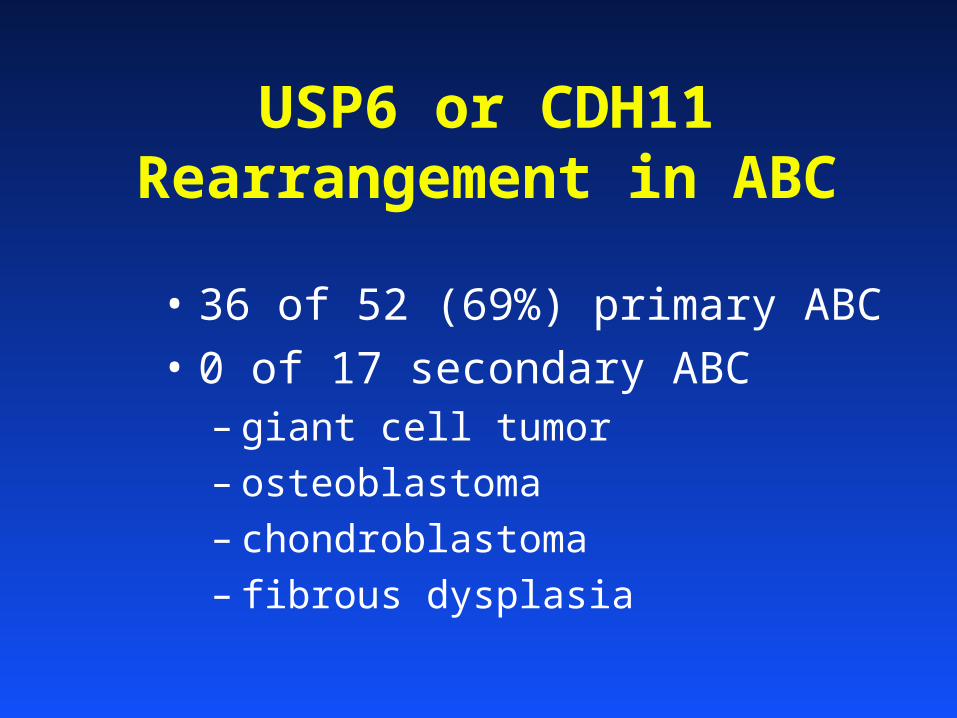
USP6 or CDH11 Rearrangement in ABC
• 36 of 52 (69%) primary ABC• 0 of 17 secondary ABC
– giant cell tumor
– osteoblastoma
– chondroblastoma
– fibrous dysplasia
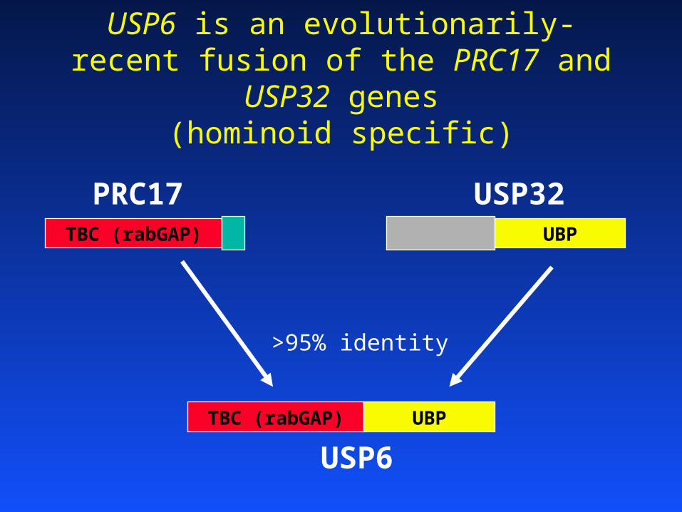
USP6 is an evolutionarily-recent fusion of the PRC17 and USP32 genes
(hominoid specific)
USP6
PRC17 USP32TBC (rabGAP) UBP
TBC (rabGAP) UBP
>95% identity
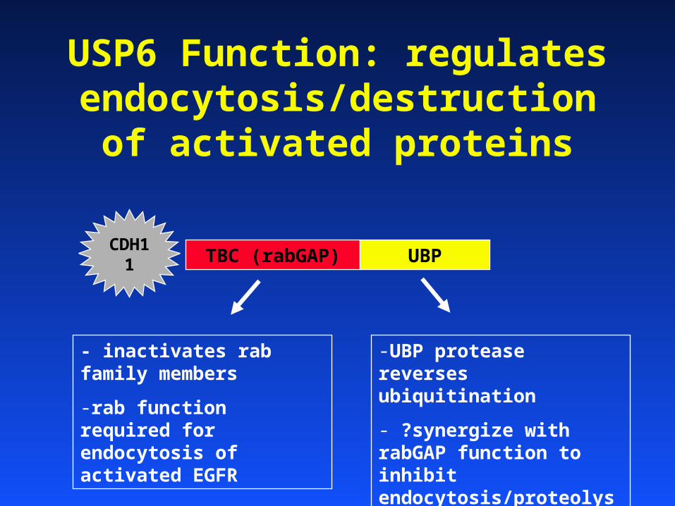
USP6 Function: regulates endocytosis/destruction of
activated proteins
TBC (rabGAP) UBP
- inactivates rab family members
-rab function required for endocytosis of activated EGFR
-UBP protease reverses ubiquitination
- ?synergize with rabGAP function to inhibit endocytosis/proteolysis
CDH11
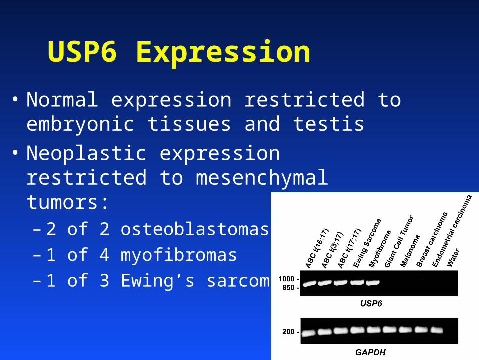
USP6 Expression
• Normal expression restricted to embryonic tissues and testis
• Neoplastic expression restricted to mesenchymal tumors:– 2 of 2 osteoblastomas
– 1 of 4 myofibromas
– 1 of 3 Ewing’s sarcomas
Conclusions
• USP6 is overexpressed due to promoter swapping mechanisms in most primary ABC
• USP6 overexpression may stabilize oncogenic proteins
• USP6 is an evolutionarily recent gene, with likely relevance in sarcoma
• Useful models of mesenchymal tumor biology can come from unlikely places
Example 2Smooth Muscle Tumors
• Use of cytogenetic clues to identify clinically-relevant biologic pathways in a genetically complex disease
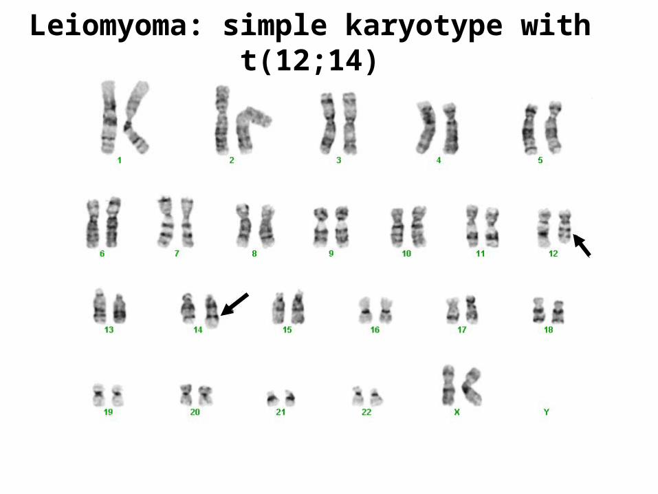
Leiomyoma: simple karyotype with t(12;14)
HMGA2(HMGIC)
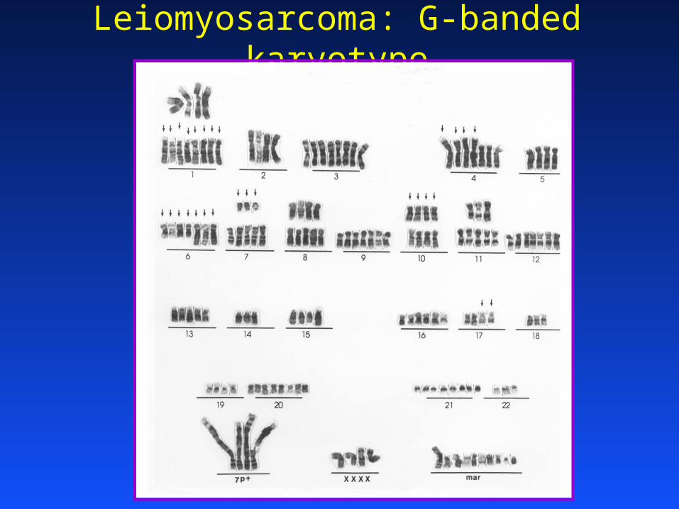
Leiomyosarcoma: G-banded karyotype
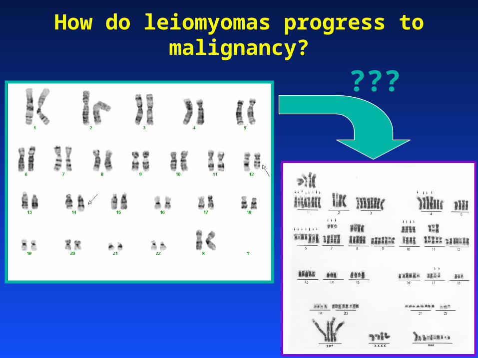
How do leiomyomas progress to malignancy?
???
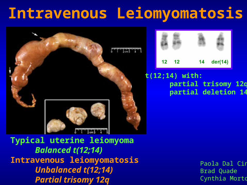
Intravenous Leiomyomatosis
Typical uterine leiomyomaBalanced t(12;14)
Intravenous leiomyomatosisUnbalanced t(12;14)Partial trisomy 12q
Paola Dal CinBrad QuadeCynthia Morton
t(12;14) with:partial trisomy 12qpartial deletion 14q
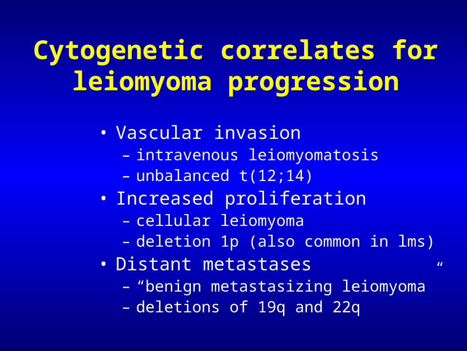
Cytogenetic correlates for leiomyoma progression
• Vascular invasion– intravenous leiomyomatosis– unbalanced t(12;14)
• Increased proliferation– cellular leiomyoma– deletion 1p (also common in lms)
• Distant metastases– “benign metastasizing leiomyoma”– deletions of 19q and 22q
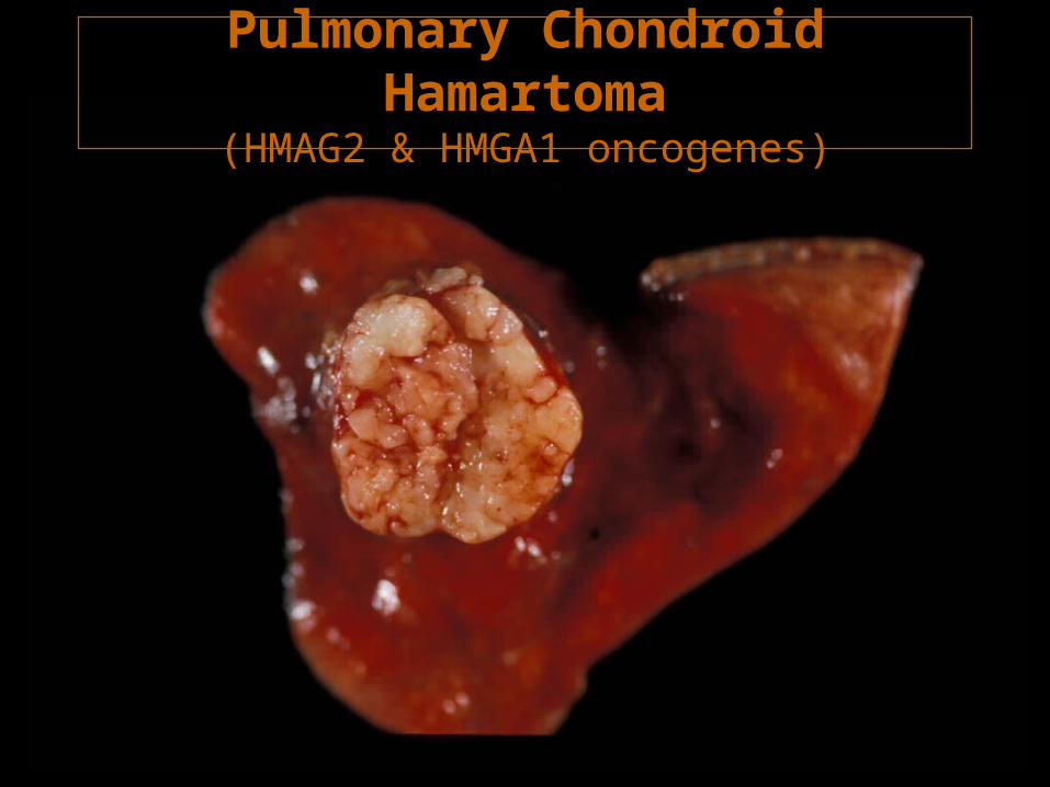
Pulmonary Chondroid Hamartoma(HMAG2 & HMGA1 oncogenes)
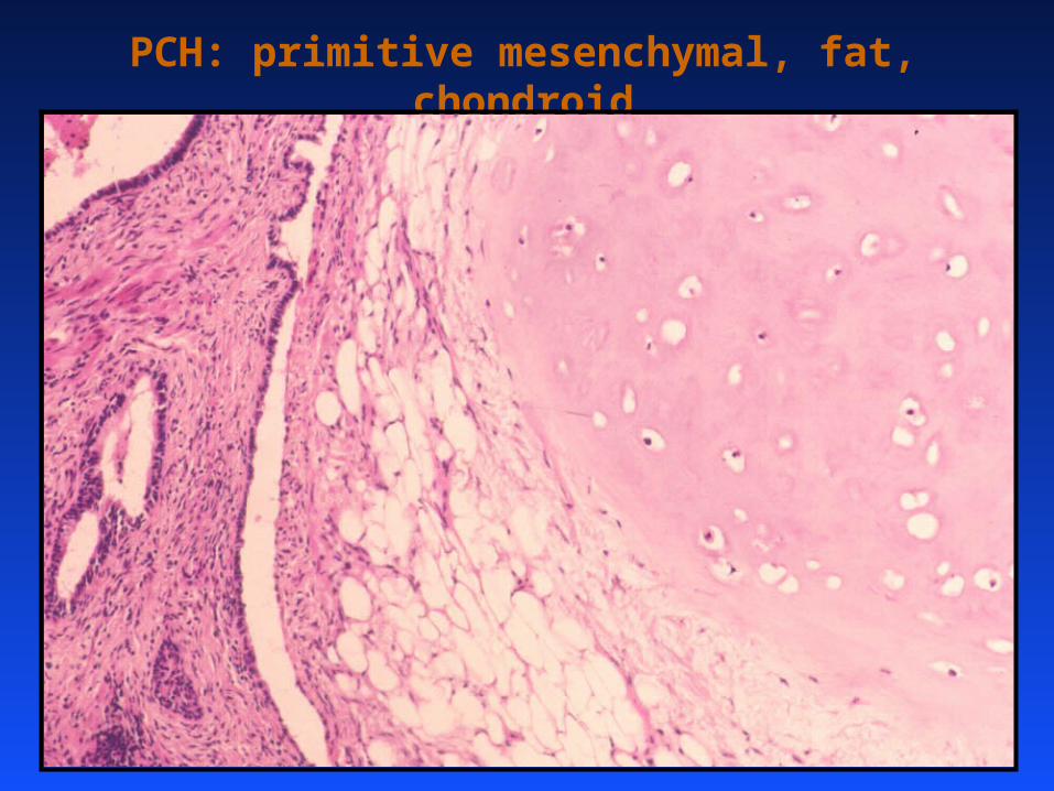
PCH: primitive mesenchymal, fat, chondroid
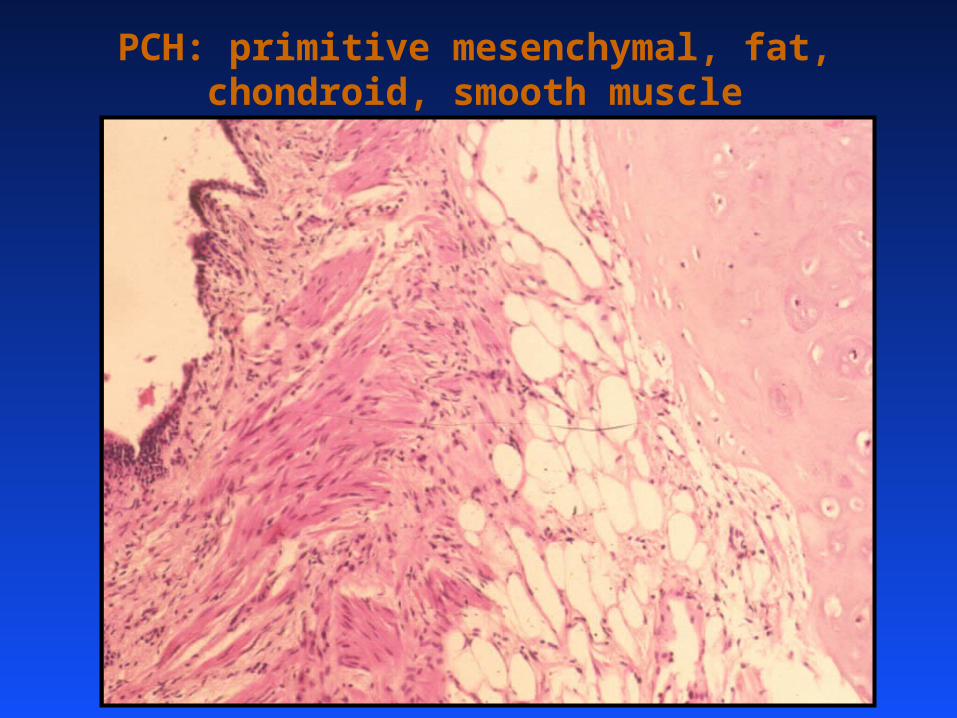
PCH: primitive mesenchymal, fat, chondroid, smooth muscle
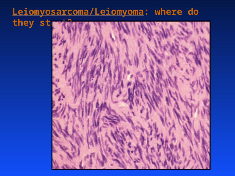
Leiomyosarcoma/Leiomyoma: where do they start?
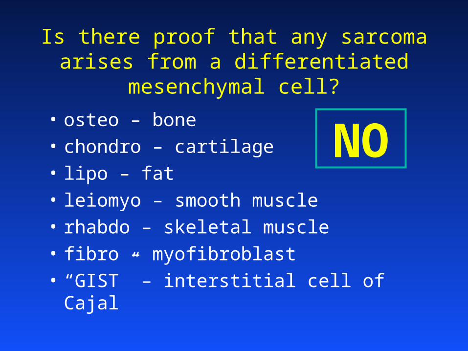
Is there proof that any sarcoma arises from a differentiated mesenchymal cell?
• osteo – bone
• chondro – cartilage
• lipo – fat
• leiomyo – smooth muscle
• rhabdo – skeletal muscle
• fibro – myofibroblast
• “GIST” – interstitial cell of Cajal
NO
Andre Oliveira
Paola Dal Cin
Cynthia Morton
Marisa Nucci
Anette Duensing
Chang-Jie Chen
Nora Joseph
Bryna Mcconarty
Felicity Smith
Lynn Yu
Christopher Hubert
Maureen Thyne
Vicki Derr
Stana Weremowicz
George Demetri
Christopher Fletcher
Sam Singer
Antonio Perez-Atayde
Mark Gebhardt
Andrew Rosenberg
Julia Bridge

THANK YOU!!!