how shelterin solves the telomere end-protection problem · loayza and de lange 2003; lei et al....
TRANSCRIPT
10.1101/sqb.2010.75.017Access the most recent version at doi: 2010 75: 167-177 originally published online January 5, 2011Cold Spring Harb Symp Quant Biol
T. de Lange How Shelterin Solves the Telomere End-Protection Problem
References http://symposium.cshlp.org/content/75/167.refs.html
This article cites 109 articles, 40 of which can be accessed free at:
serviceEmail alerting
click heretop right corner of the article orReceive free email alerts when new articles cite this article - sign up in the box at the
http://symposium.cshlp.org/subscriptions go to: Cold Spring Harbor Symposia on Quantitative BiologyTo subscribe to
Copyright © 2010, Cold Spring Harbor Laboratory Press
Cold Spring Harbor Laboratory Press on August 5, 2011 - Published by symposium.cshlp.orgDownloaded from
QUARTET OF THREATS
In mammalian cells, four distinct pathways threatenchromosome ends: two DNA-damage signaling pathways,transduced by ATM (ataxia telangiectasia mutated) andATR (ataxia telangiectasia and Rad3 related)kinases, and two major double-strand break (DSB) repairpathways, nonhomologous end joining (NHEJ) and ho-mology-directed repair (HDR) (Fig. 1). A short summaryof the key aspects relevant to the telomere end-protectionproblem is given here. For primary references and moreextensive details on these pathways, the reader is referredto excellent recent reviews on these subjects (Hefferin andTomkinson 2005; Harper and Elledge 2007; Cimprich andCortez 2008; Jackson and Bartek 2009; Mahaney et al.2009; Mimitou and Symington 2009; Lieber 2010; Moyn-ahan and Jasin 2010).The ATM kinase pathway, particularly active in mam-
malian cells (more so than in yeast), is activated when theMre11/Rad50/Nbs1 (MRN) complex associates with aDSB. This event helps to recruit the ATM kinase and me-diates its activation in conjunction with the Tip60 acetyl-transferase. The actual molecular mechanism of ATMactivation is not understood. Once active, ATM can phos-phorylate the histone variant H2AX, creating a large do-main of phosphorylated H2AX (γ-H2AX) near the DSB.This chromatin modification functions to recruit MDC1and a host of ubiquitin ligases that generate a cytologicallydefined entity referred to as a DNA-damage focus. Abun-dant residents of DNA-damage foci, such as 53BP1, γ-H2AX, MRN, and MDC1, facilitate detection of the fociby indirect immunofluorescence. The formation of thesefoci is not required for ATM signaling but helps to amplifythe signal, presumably through recruitment of additionalATM kinase molecules. In addition, some of the DNA-damage factors in the foci influence repair reactions. Theoutcomes of ATM signaling are mediated through thephosphorylation of numerous nucleoplasmic proteins thatcan affect events at a distance from the DSB. A key player
is Chk2, which is activated by ATM-mediated phosphory-lation and functions as an effector kinase, enforcing cellcycle arrest in G1/S and G2/M through phosphorylation ofp53, MDM2, and Cdc25 phosphatases.Activation of the ATR kinase pathway at DSBs requires
the binding of replication protein A (RPA) to single-strandDNA (ssDNA). Resection of a DSB can create a 3′ over-hang that, when decorated with RPA, binds the ATR-in-teracting protein (ATRIP)-binding partner of the ATRkinase. The activation of ATR signaling involves addi-tional players, including TopBP1, the 9-1-1 complex, andits Rad17 clamp-loader. Once activated, ATR, like ATM,phosphorylates H2AX in neighboring chromatin andDNA-damage foci are formed. Whether ATM- and ATR-induced foci are identical is not known, but so far, theyhave been indistinguishable in terms of their constituents.However, the DNA-damage foci have not been implicatedin amplification of the ATR signal and may, therefore, beprimarily important for the regulation of DNA repair. TheATR kinase has a large number of targets, including theeffector kinase Chk1, which, when activated by ATR-me-diated phosphorylation, induces cell cycle arrest in G1/Sand G2/M. Depending on the level of damage and the ex-tent of repair, ATM or ATR signaling can lead to an irre-versible arrest accompanied by either apoptosis or senes-cence. NHEJ is an error-prone repair pathway that joins DSBs
regardless of their sequence. This pathway is initiated bythe Ku70/80 heterodimer, which binds DNA only when afree DNA end is available. The reason for this specificityemerged from structural analysis, which showed that theheterodimer is a ring-shaped protein with an opening thesize of double-strand DNA. Once loaded, Ku70/80 medi-ates the synapsis of two DNA ends and facilitates their lig-ation by DNA ligase IV. HDR is, in principle, error free, in particular when DSB
repair takes place after DNA replication so that the sisterchromatid can be used as a template. HDR requires endresection and loading of the Rad51 recombinase on a sin-
How Shelterin Solves the Telomere End-Protection Problem
T. DE LANGELaboratory for Cell Biology and Genetics, The Rockefeller University, New York, New York 10065
Correspondence: [email protected]
The symphony of the human genome concludes with a long Gregorian chant of TTAGGG repeats. This monotonous codarepresents one of the most complex problems in chromosome biology: the question of how cells distinguish their natural chro-mosome ends from double-strand breaks elsewhere in the genome. McClintock’s classic finding of chromosome breakage-fusion-bridge cycles, first reported by her at one of the early Cold Spring Harbor Laboratory Symposia (the ninth), served asa prelude to this question. The 75th Cold Spring Harbor Laboratory Symposium marks the completion of a series of mousegene deletion experiments that revealed DNA-damage-response pathways that threaten chromosome ends and how the com-ponents of the telomeric shelterin complex prevent activation of these pathways.
Cold Spring Harbor Symposia on Quantitative Biology,Volume LXXV. ©2010 Cold Spring Harbor Laboratory Press 978-1-936113-07-1 167
Cold Spring Harbor Laboratory Press on August 5, 2011 - Published by symposium.cshlp.orgDownloaded from

gle-strand 3′ overhang. Rad51 mediates the invasion of thessDNA into homologous sequences. After strand invasion,different further processing reactions can occur. If singleor double Holliday junctions (HJs or dHJs) are formed,their resolution by resolvases Mus81/Eme1 or Gen1 canlead to sequence exchanges (crossovers), whereas theirresolution by BLM/Top3a results in a noncrossover event.Additional outcomes of Rad51-mediated strand invasioninclude a single-strand annealing (SSA) pathway andbreak-induced replication (BIR). These four pathways define the end-protection problem.
Telomeres need to avoid activating the ATM and ATR kinasepathways because this would lead to cell cycle arrest. The“repair” of telomeres by NHEJ would generate lethal chro-mosome fusions, and HDR could change telomere length.Thus, in mammalian cells, telomeres need to block four dis-tinct pathways that are each initiated in different ways.
SHELTERIN: A SEXTET OFPROTECTIVE PROTEINS
Mammalian telomeres are built up on long arrays oftandem TTAGGG repeats, the product of telomerase. Thelength of this repeat region is variable (2—15 kb in humans,up to 100 kb in mice) and ends in a 3′ overhang of 50—400nucleotides (Fig. 2). This DNA protects chromosome endsby recruiting shelterin.Shelterin is a telomere-specific complex composed of
six distinct subunits (Figs. 3 and 4). The complex derivesits specificity for telomeric DNA from TRF1 and TRF2,two related homodimeric proteins that bind to double-strand TTAGGG repeats with Myb/SANT-type DNA-bind-ing domains (Chong et al. 1995; Bilaud et al. 1997;Broccoli et al. 1997). The complex contains a third DNA-binding protein, POT1, that has two oligosaccharide/oligonucleotide binding (OB) folds that associate with sin-
gle-strand TTAGGG repeats (Baumann and Cech 2001;Loayza and de Lange 2003; Lei et al. 2004). The ssDNA-binding activity of POT1 is crucial for telomere protectionbut does not have a role in anchoring shelterin at telomeres(Loayza and de Lange 2003). POT1 is recruited to telo-meres through its interaction with TPP1, another OB-fold-containing protein (Liu et al. 2004; Ye et al. 2004b;Hockemeyer et al. 2007; Wang et al. 2007; Palm et al.2009; Kibe et al. 2010). TPP1 binds to TIN2, which inter-acts with both TRF1 and TRF2 (Kim et al. 1999; Hough-taling et al. 2004; Liu et al. 2004; Ye et al. 2004a,b;O’Connor et al. 2006; Chen et al. 2008). The telomerebinding of POT1 is critically dependent on this TPP1/TIN2link to the proteins that anchor shelterin on double-strand
168 DE LANGE
Figure 1. The end-protection problem in mammalian cells. Schematic shows the four pathways that threaten natural chromosomeends. At chromosome-internal DNA breaks, ATM and ATR signaling can be activated and the lesion can be repaired by NHEJ or HDR.Telomeres must repress all four pathways. Consequences of telomere dysfunction are indicated.
Figure 2. Structure of mammalian telomeres and schematic ofthe DNA component of mammalian telomeres. Micrograph showsa t-loop in isolated chromatin. (Reprinted, with permission, fromNikitina and Woodcock 2004 [©Rockefeller University Press].)
Cold Spring Harbor Laboratory Press on August 5, 2011 - Published by symposium.cshlp.orgDownloaded from
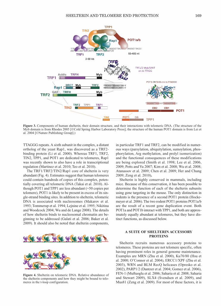
TTAGGG repeats. A sixth subunit in the complex, a distantortholog of the yeast Rap1, was discovered as a TRF2-binding protein (Li et al. 2000). Whereas TRF1, TRF2,TIN2, TPP1, and POT1 are dedicated to telomeres, Rap1was recently shown to also have a role in transcriptionalregulation (Martinez et al. 2010; Teo et al. 2010). The TRF1/TRF2/TIN2/Rap1 core of shelterin is very
abundant (Fig. 4). Estimates suggest that human telomerescould contain hundreds of copies of this complex, poten-tially covering all telomeric DNA (Takai et al. 2010). Al-though POT1 and TPP1 are less abundant (~50 copies pertelomere), POT1 is likely to be present in excess of its sin-gle-strand binding sites. In addition to shelterin, telomericDNA is associated with nucleosomes (Makarov et al.1993; Tommerup et al. 1994; Lejnine et al. 1995; Nikitinaand Woodcock 2004; Wu and de Lange 2008). The detailsof how shelterin binds to nucleosomal chromatin are be-ginning to be addressed (Galati et al. 2006; Baker et al.2009). It should also be noted that shelterin components,
in particular TRF1 and TRF2, can be modified in numer-ous ways (parsylation, ubiquitylation, sumoylation, phos-phorylation, Arg methylation, and prolyl isomerization)and the functional consequences of these modificationsare being explored (Smith et al. 1998; Lee et al. 2006,2009; Potts and Yu 2007; Kim et al. 2008; Wu et al. 2008;Atanassov et al. 2009; Chen et al. 2009; Her and Chung2009; Zeng et al. 2010).Shelterin is highly conserved in mammals, including
mice. Because of this conservation, it has been possible todetermine the function of each of the shelterin subunitsusing gene targeting in the mouse. The only distinction inrodents is the presence of a second POT1 protein (Hocke-meyer et al. 2006). The two rodent POT1 proteins POT1a/bare the result of a recent gene duplication event. BothPOT1a and POT1b interact with TPP1, and both are approx-imately equally abundant at telomeres, but they have dis-tinct functions, as discussed below.
A SUITE OF SHELTERIN ACCESSORYPROTEINS
Shelterin recruits numerous accessory proteins totelomeres. These proteins are not telomere specific, oftenhaving prominent roles in general genome maintenance.Examples are MRN (Zhu et al. 2000), Ku70/80 (Hsu etal. 2000; O’Connor et al. 2004), ERCC1/XPF (Zhu et al.2003), WRN and BLM RecQ helicases (Opresko et al.2002), PARP1/2 (Dantzer et al. 2004; Gomez et al. 2006),FEN-1 (Muftuoglu et al. 2006; Saharia et al. 2008; Sahariaand Stewart 2009), SLX4 (Svendsen et al. 2009), andMus81 (Zeng et al. 2009). For most of these factors, it is
SHELTERIN AND TELOMERE END PROTECTION 169
Figure 3. Components of human shelterin, their domain structure, and their interactions with telomeric DNA. (The structure of theMyb domain is from Rhodes 2005 [©Cold Spring Harbor Laboratory Press]; the structure of the human POT1 domain is from Lei etal. 2004 [©Nature Publishing Group].)
Figure 4. Shelterin on telomeric DNA. Relative abundance ofthe shelterin components and how they might be bound to telo-meres in the t-loop configuration.
Cold Spring Harbor Laboratory Press on August 5, 2011 - Published by symposium.cshlp.orgDownloaded from
known (or suspected) that they interact with shelterin, butthe molecular details are largely unknown. A well-studiedshelterin accessory factor is the Apollo SMN1/Pso2-typenuclease (also referred to as SNM1B), which has a generalfunction in DNA interstrand –cross-link repair. Apollobinds to TRF2 and contributes to the maintenance of theG-strand overhang and the protection of telomeres in Sphase (Freibaum and Counter 2006; Lenain et al. 2006;van Overbeek and de Lange 2006; Chen et al. 2008; Lamet al. 2010; Wu et al. 2010).Unlike most shelterin accessory factors, which are nu-
clear proteins, the tankyrase class of PARPs (Smith et al.1998; Kaminker et al. 2001), which were the first shelterinaccessory factors to be discovered, are now known to havea variety of functions in the cytoplasm (for review, seeHsiao and Smith 2008). However, tankyrases also bind toTRF1 and contribute to telomere length regulation andtelomere resolution in mitosis (Smith and de Lange 2000;Dynek and Smith 2004). Interestingly, the tankyrase–TRF1interaction is not conserved in mouse cells (Donigian andde Lange 2007), providing another example of the slightdifferences between mouse and human telomeres.
MUTING ATM AND ATR
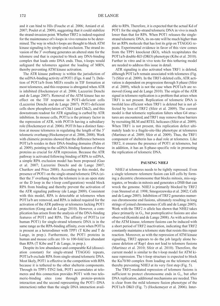
Activation of either the ATM or ATR kinase pathway bythe natural ends of chromosomes is incompatible with cel-lular and organismal survival. Both pathways are kept incheck by shelterin. There is a remarkable division of laborwithin the complex, with TRF2 dedicated to the ATMpathway and POT1 responsible for the repression of ATR(Figs. 5–7) (Lazzerini Denchi and de Lange 2007).
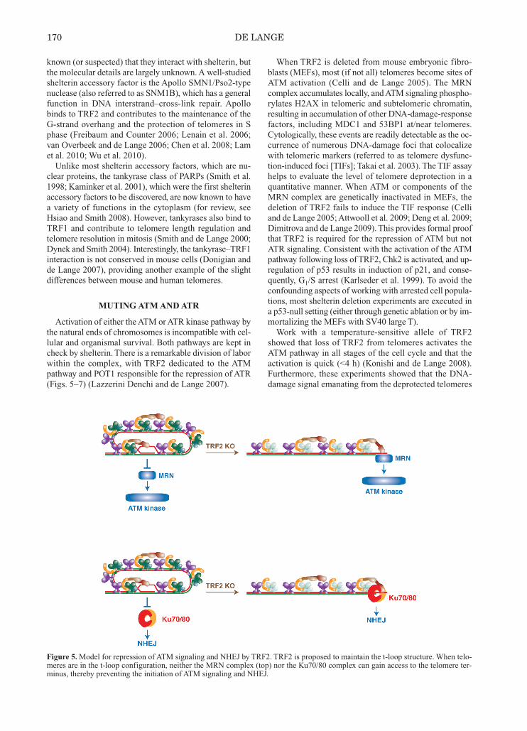
When TRF2 is deleted from mouse embryonic fibro-blasts (MEFs), most (if not all) telomeres become sites ofATM activation (Celli and de Lange 2005). The MRNcomplex accumulates locally, and ATM signaling phospho-rylates H2AX in telomeric and subtelomeric chromatin,resulting in accumulation of other DNA-damage-responsefactors, including MDC1 and 53BP1 at/near telomeres.Cytologically, these events are readily detectable as the oc-currence of numerous DNA-damage foci that colocalizewith telomeric markers (referred to as telomere dysfunc-tion-induced foci [TIFs]; Takai et al. 2003). The TIF assayhelps to evaluate the level of telomere deprotection in aquantitative manner. When ATM or components of theMRN complex are genetically inactivated in MEFs, thedeletion of TRF2 fails to induce the TIF response (Celliand de Lange 2005; Attwooll et al. 2009; Deng et al. 2009;Dimitrova and de Lange 2009). This provides formal proofthat TRF2 is required for the repression of ATM but notATR signaling. Consistent with the activation of the ATMpathway following loss of TRF2, Chk2 is activated, and up-regulation of p53 results in induction of p21, and conse-quently, G1/S arrest (Karlseder et al. 1999). To avoid theconfounding aspects of working with arrested cell popula-tions, most shelterin deletion experiments are executed ina p53-null setting (either through genetic ablation or by im-mortalizing the MEFs with SV40 large T). Work with a temperature-sensitive allele of TRF2
showed that loss of TRF2 from telomeres activates theATM pathway in all stages of the cell cycle and that theactivation is quick (<4 h) (Konishi and de Lange 2008).Furthermore, these experiments showed that the DNA-damage signal emanating from the deprotected telomeres
170 DE LANGE
Figure 5. Model for repression of ATM signaling and NHEJ by TRF2. TRF2 is proposed to maintain the t-loop structure. When telo-meres are in the t-loop configuration, neither the MRN complex (top) nor the Ku70/80 complex can gain access to the telomere ter-minus, thereby preventing the initiation of ATM signaling and NHEJ.
Cold Spring Harbor Laboratory Press on August 5, 2011 - Published by symposium.cshlp.orgDownloaded from
can be readily reversed. When cells are moved back to thepermissive temperature, the TIF response dissipates in afew hours. The ability of TRF2 to establish and maintaina telomere state that hides the chromosome end from theATM pathway does not require its interacting factor Rap1(Sfeir et al. 2010). The role of the second TRF2-bindingprotein in shelterin, TIN2, in the repression of ATM hasnot been established. The TRF2-binding protein Apollohas a minor contribution in the muting of ATM (Wu et al.2010). Deletion of Apollo results in ATM-dependent TIFs,but they occur only in S phase. Assuming that TRF2 is the main player in blocking the
activation of the ATM kinase, the question arises as to howa simple DNA-binding factor such as TRF2 accomplishesthis task. The current model invokes a key role for TRF2in the formation of the t-loop structure (Fig. 5). t-loops arelariats formed by the strand invasion of the 3′ telomericoverhang into the duplex telomeric DNA (Fig. 2). They
were initially observed by electron microscopy (EM)analysis of telomeric DNA isolated from psoralen cross-linked nuclei of human and mouse cells (Griffith et al.1998). The strand invasion displaces the G-rich telomericstrand at the base of the loop, forming a D loop that wasinferred to be present based on coating with an Escherichiacoli single-stranded DNA-binding protein. t-loops are aconserved aspect of telomeres; they have been detected intrypanosomes (Munoz-Jordan et al. 2001), ciliate micronu-clear DNA (Murti and Prescott 1999), plants (Cesare et al.2003), and some strains of Kluyveromyces lactis (Cesareet al. 2008). t-loops have also been demonstrated in iso-lated intact chromatin from chicken erythrocytes andmouse splenocytes (Nikitina and Woodcock 2004). Fur-thermore, TRF2 has biochemical activities in vitro that sug-gest a role in t-loop formation. Recombinant TRF2 cangenerate t-loops when provided with an appropriate telo-meric substrate (Griffith et al. 1999; Stansel et al. 2001),
SHELTERIN AND TELOMERE END PROTECTION 171
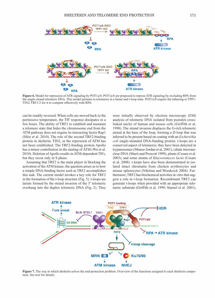
Figure 6. Model for repression of ATR signaling by POT1a/b. POT1a/b are proposed to repress ATR signaling by excluding RPA fromthe single-strand telomeric DNA. This model pertains to telomeres in a linear and t-loop state. POT1a/b require the tethering to TPP1-TIN2-TRF1/2 for it to compete effectively with RPA.
Figure 7. The way in which shelterin solves the end-protection problem. Overview of the functions assigned to each shelterin compo-nent. See text for details.
Cold Spring Harbor Laboratory Press on August 5, 2011 - Published by symposium.cshlp.orgDownloaded from
and it can bind to HJs (Fouche et al. 2006; Amiard et al.2007; Poulet et al. 2009), suggesting that it could stabilizethe strand-invasion point. Whether TRF2 is indeed requiredfor the maintenance of t-loops in vivo remains to be deter-mined. The way in which t-loops are thought to block ATMkinase signaling is by simple end occlusion. The strand in-vasion of the 3′ overhang generates an altered state for thetelomere end that is expected to block any DNA-bindingcomplex that loads onto DNA ends. Thus, t-loops wouldsafeguard the telomeres against the loading of MRN,thereby preventing ATM kinase activation. The ATR kinase pathway is within the jurisdiction of
the ssDNA-binding activity of POT1 (Figs. 6 and 7). Dele-tion of POT1a/b from MEFs results in TIF formation atmost telomeres, and this response is abrogated when ATRis inhibited (Hockemeyer et al. 2006; Lazzerini Denchiand de Lange 2007). Removal of the ATM kinase has noeffect on the TIF response in POT1-deficient cells(Lazzerini Denchi and de Lange 2007). POT1-deficientcells show phosphorylation of Chk1 (and Chk2), and thisdownstream readout for ATR signaling is curbed by ATRinhibition. In mouse cells, POT1a is the primary factor inthe repression of ATR, with POT1b having a subsidiaryrole (Hockemeyer et al. 2006). POT1b has a distinct func-tion at mouse telomeres in regulating the length of the 3′telomeric overhang (Hockemeyer et al. 2006, 2008). Workwith chimeric proteins showed that the difference betweenPOT1a/b resides in their DNA-binding domains (Palm etal. 2009), pointing to the ssDNA-binding features of theseproteins as crucial for ATR repression. Because the ATRpathway is activated following binding of RPA to ssDNA,a simple RPA exclusion model has been proposed (Guoet al. 2007; Lazzerini Denchi and de Lange 2007;Churikov and Price 2008). According to this model, thepresence of POT1 on the single-strand telomeric DNA (ei-ther the 3′ overhang when the telomere is in an open stateor the D loop in the t-loop configuration) would blockRPA from binding and thereby prevent the activation ofthe ATR signaling pathway (de Lange 2009). Consistentwith this model, RPA is detectable at telomeres whenPOT1a/b are removed, and RPA is indeed required for theactivation of the ATR pathway at telomeres lacking POT1protection (Gong and de Lange 2010). However, a com-plication has arisen from the analysis of the DNA-bindingfeatures of POT1 and RPA. The affinity of POT1a (orhuman POT1) for single-strand telomeric DNA is in thesame range as the RPA-binding affinity, even when POT1ais present as a heterodimer with TPP1 (T Kibe and T deLange, in prep.). Furthermore, the POT1 proteins inhuman and mouse cells are 10- to 100-fold less abundantthan RPA (T Kibe and T de Lange, in prep.). Despite its low abundance and comparable Kd (dissoci-
ation constant) for single-strand TTAGGG repeats,POT1a/b exclude RPA from single-strand telomeric DNA.Most likely, POT1 is effective in the competition with RPAbecause it is tethered to the other shelterin components.Through its TPP1-TIN2 link, POT1 accumulates at telo-meres and this connection provides POT1 with two telo-meric-binding sites (one through protein—proteininteraction and the second representing the POT1–DNAinteraction) rather than the single DNA interaction avail-
able to RPA. Therefore, it is expected that the actual Kd ofPOT1 for the single-strand telomeric DNA in vivo is muchlower than that for RPA. When POT1 releases the single-strand telomeric DNA, its on-rate will be much higher thanfor an RPA molecule that has lost its grip on TTAGGG re-peats. Experimental evidence in favor of this view comesfrom the TPP1 knockout (KO), which recapitulates thePOT1a/b double-KO (DKO) phenotype (Kibe et al. 2010).Further in vitro and in vivo tests for this tethering modelare needed to address this issue in detail.ATR signaling is also activated when TRF1 is deleted,
although POT1a/b remain associated with telomeres (Fig.7) (Sfeir et al. 2009). In the TRF1-deleted cells, ATR acti-vation is dependent on progression through S phase (Sfeiret al. 2009), which is not the case when POT1a/b are re-moved (Gong and de Lange 2010). The origin of the ATRsignal in telomere replication is a problem that arises whenTRF1 is not present. Replication of telomeric DNA istwofold less efficient when TRF1 is deleted but is not af-fected by loss of TRF2 (Sfeir et al. 2009). The currentmodel is that telomeric replication falters when G4 struc-tures are encountered, and TRF1 may remove these barriersby recruiting BLM and RTEL helicases (Sfeir et al. 2009).When TRF1 is not present, this replication defect ulti-mately leads to a fragile-site-like phenotype at telomeres(Martinez et al. 2009; Sfeir et al. 2009). Thus, the TRF1component of shelterin has a dual role at telomeres. LikeTRF2, it ensures the presence of POT1 at telomeres, butin addition, it has an S-phase-specific role in promotingthe replication of telomeric DNA.
PAUSING NHEJ
NHEJ at telomeres needs to be tightly repressed. Evena single telomere–telomere fusion can kill cells by form-ing a dicentric chromosome that blocks mitosis, mis-seg-regates, or breaks in mitosis and generates a DSB that canwreck the genome. NHEJ is primarily blocked by TRF2(van Steensel et al. 1998; Smogorzewska et al. 2002; Celliand de Lange 2005). Inhibition of TRF2 generates numer-ous chromosome end fusions, ultimately resulting in longstrings of joined chromosomes (Celli and de Lange 2005).Work with the TRF2 ts allele has shown that NHEJ takesplace primarily in G1, but postreplicative fusions are alsoobserved (Konishi and de Lange 2008). As with activationof the ATM kinase, telomere fusions can be induced witha short period of TRF2 inactivation, indicating that TRF2constantly maintains a telomere state that resists this repairreaction. Moreover, as with the repression of ATM kinasesignaling, TRF2 appears to do the job largely alone be-cause deletion of Rap1 does not lead to telomere fusions(Martinez et al. 2010; Sfeir et al. 2010). Therefore, thecurrent model is similar to the t-loop model for ATM ki-nase repression. The t-loop structure is expected to blockthe Ku70/80 complex from loading on the telomere end,thereby preventing NHEJ at its very first step (Fig. 5).The TRF2-mediated repression of telomere fusions is
sufficient to protect chromosome ends in G1, but afterDNA replication, additional mechanisms are needed. Thisis clear from the mild telomere fusion phenotype of thePOT1a/b DKO (Fig. 7) (Hockemeyer et al. 2006). Inter-
172 DE LANGE
Cold Spring Harbor Laboratory Press on August 5, 2011 - Published by symposium.cshlp.orgDownloaded from

estingly, these fusions primarily involve sister chromatids,indicating a postreplicative event. Thus, after DNA repli-cation, both sister telomeres (one formed by lagging-strand DNA synthesis and one by leading-strand DNAsynthesis) become vulnerable to a joining event, althoughthe incidence of actual fusions is low. Perhaps the loadingof POT1 on the single-strand overhang keeps the repairpathway at bay until the t-loop structure is reformed. Con-sistent with this view, leading-end telomeres (which arepresumably blunt right after DNA replication is com-pleted) require the Apollo nuclease for their proper pro-tection from fusion (Lam et al. 2010; Wu et al. 2010).Apollo has been implicated in the regeneration of the 3′overhang at telomeres after their replication (Lam et al.2010; Wu et al. 2010). A simple scenario can be envisagedin which TRF2 recruits Apollo to regenerate the 3′ over-hang at leading-end telomeres, which then become cov-ered by POT1 so that joining reactions are blocked in theperiod before the t-loop is reformed. The lagging-endtelomere is thought to contain an overhang that is gener-ated by removal of the last RNA primer so that resectionmay not be needed immediately. In contrast, POT1 bind-ing would be needed at both ends, explaining why ApolloKO cells have leading-end fusions, whereas POT1a/bDKO cells show fusions of sister telomeres.Apollo is probably not the only nuclease that mediates
the generation of the 3′ overhang at leading-end telomeres.When Apollo is absent, the leading-end telomere fusionsoccur but are not frequent, suggesting that there is anotherpathway that can generate a 3′ overhang when Apollo ismissing. One possibility is that the ATM signaling attelomeres lacking Apollo is responsible for the resection.MRN-mediated ATM activation can induce resection atDSBs, most likely through the CtIP nuclease. Indicationthat the MRN/ATM-dependent resection can protect lead-ing-end telomeres from NHEJ came from experiments inwhich TRF2 is deleted from MRN- or ATM-deficient cells(Attwooll et al. 2009; Dimitrova and de Lange 2009). Be-cause TRF2 is able to recruit MRN to telomeres, it is alsopossible that the MRN/ATM/CtIP pathway acts in parallelwith Apollo to generate a 3′ overhang at the leading-endtelomeres. It will therefore be interesting to monitor theleading-end telomere fusions in Apollo/MRN or Apollo/ATM double-deficient cells.It should be noted that postreplicative telomere fusions
are not necessarily mediated by NHEJ. Deficiency inDNA ligase IV does not abrogate the postreplicativetelomere fusions occurring when either POT1a/b (Rai etal. 2010) or Apollo (P Wu and T de Lange, unpubl.) is re-moved. This may point to the recently described mam-malian alternative end joining pathway, but geneticevidence in favor of this view is not yet available. There-fore, it is not excluded that the observed “fusions” are, infact, noncovalent associations.
DIRECTING HDR
Compared to the acutely lethal outcomes of NHEJ,HDR may seem less threatening to telomeres. Yet, inap-propriate recombination between telomeres can have direconsequences in the long term. For instance, if two sister
telomeres undergo an unequal exchange, one of thedaughter cells will inherit a shortened telomere. In humansomatic cells without telomerase, this shortened telomerewill likely limit the life span of the lineage established bythe dispossessed daughter. In addition, BIR could inap-propriately extend telomeres and has been invoked as theprimary mechanism by which some telomerase negativecell lines and tumors extend their telomeres. This so-calledalternative lengthening of telomeres (ALT) pathway isthought to involve BIR-like telomere extension eventsusing either telomeres or extrachromosomal telomericDNA as a template (Dunham et al. 2000).HDR can be monitored at telomeres using chromo-
some-orientation fluorescence in situ hybridization (CO-FISH), which labels the telomeres generated by lead-ing- and lagging-strand synthesis with two different fluo-rophores so that sequence exchanges between sistertelo-meres can be visualized. These telomere sister chro-matid exchanges (T-SCEs) are rare in wild-type cells (oneor two per metaphase spread) but can become prominentwhen TRF2 is deleted (Celli et al. 2006). Induction of T-SCEs by loss of TRF2 is only detectable in Ku70/80-de-ficient cells. When Ku70/80 is present, even when NHEJis blocked by the absence of DNA ligase IV, exchangesare rare (Celli et al. 2006). How Ku70/80 acts to preventT-SCEs has not been established, but it may be related tothe general ability of Ku70/80 to repress HDR. Alterna-tively, the association of Ku70/80 with shelterin may beimportant for the repression of HDR. Although T-SCEs are frequent in cells lacking TRF2
(and Ku70/80), TRF2 itself is not involved in regulatingthis reaction. Rather, Rap1 is repressing HDR. As with theTRF2 phenotype, Rap1 KO cells show a minimal level ofT-SCEs (<2%) (Martinez et al. 2010; Sfeir et al. 2010),but in a Ku70-null context, the T-SCEs are very frequent(~10% of chromosome ends) (Sfeir et al. 2010). HowRap1 protects telomeres from HDR is unclear. Rap1 is asmall adapter protein with several domains that could bindother factors, so perhaps it has an interacting partner thatblocks HDR. The view of Rap1 as an adapter can also ex-plain its varied roles at telomeres in other organisms andthe recently established role for Rap1 in NF-κB signaling(Teo et al. 2010).In addition to Rap1, POT1 proteins in shelterin are re-
quired for the repression of HDR. POT1a/b-Ku70 triple-KO cells show the same high frequency of T-SCEs as theTRF2 or Rap1/Ku70 DKO cells (Palm et al. 2009). EitherPOT1a or b is sufficient to repress this phenotype. As withthe Rap1 KO, low levels of T-SCEs have been reported inPOT1a- or b-deficient cells, but it is difficult to evaluatethese minor phenotypes (He et al. 2006; Wu et al. 2006).Thus, the repression of recombination between telomeresby shelterin involves the concerted action of one of thetwo POT1 proteins and Rap1. Shelterin is also required to prevent an HDR reaction that
cleaves the t-loop off the telomere. This reaction producesa shortened telomere and an extrachromosomal circularDNA composed of telomeric repeats (referred to as a t cir-cle) that is detectable on two-dimensional gels (Wang et al.2004). The simplest explanation of these products is the for-mation (through branch migration) of a dHJ at the base of
SHELTERIN AND TELOMERE END PROTECTION 173
Cold Spring Harbor Laboratory Press on August 5, 2011 - Published by symposium.cshlp.orgDownloaded from
the t-loop and a crossover-type resolution by Mus81 orGen1 (Haber 2004). t-loop cleavage is specifically re-pressed by the amino-terminal domain of TRF2, which isrich in Gly/Arg residues and highly basic (referred to as thebasic domain or the Gly/Arg-rich [GAR] domain). TheGAR domain is a signature of mammalian TRF2; it is ab-sent from mammalian TRF1, chicken and Xenopus TRF2,and the TRF-related Taz1p of fission yeast. Recent bio-chemical data have suggested that the GAR domain bindsto HJs (Fouche et al. 2006; Amiard et al. 2007; Poulet et al.2009). It has therefore been proposed that the amino termi-nus of TRF2 prevents t-loop cleavage by physically block-ing resolvases from gaining access to dHJ that might beformed at the base of the t-loop.
ENCORE: TELOMERES AS A TOOL TO STUDYTHE DNA-DAMAGE RESPONSE
Different shelterin subunits repress different branches ofthe DNA-damage response. The corollary of this compart-mentalization is that deletion of individual shelterin proteinscan be used to activate a specific DNA-damage-responsepathway without others becoming involved. The ATM ki-nase pathway can be activated without any signaling byATR or HDR/NHEJ by removing TRF2 from telomeres ofcells that lack DNA ligase IV. In contrast, ATR signalingcan be specifically activated in the absence of ATM signal-ing in POT1a/b DKO cells. Deletion of Rap1 from Ku70-deficient cells generates the unique situation of ongoingHDR at telomeres without NHEJ, ATM signaling, or ATRsignaling. These surgical alterations can be explored to gaininsights into the DNA-damage response that are not readilyobtained from the more popular methods of induction ofDNA damage (e.g., ionizing radiation, ultraviolet [UV], andsite-specific cleavage with ISceI, and V(D)J [variable, di-verse, and joining] recombination).Several insights have already emerged from studying
telomere dysfunction. The rampant NHEJ at telomeres typ-ical of TRF2-deficient cells is largely abrogated when theATM kinase is not present (Lazzerini Denchi and de Lange2007; Dimitrova and de Lange 2009). It is the signaling bythe ATM kinase that is critical for efficient NHEJ. The keytarget of ATM in this setting is 53BP1. Removal of TRF2from telomeres in 53BP1-deficient cells results in hardlyany NHEJ events, although ATM kinase signaling is notdisrupted (Dimitrova et al. 2008). This result was not an-ticipated because 53BP1 had not been identified as a playerneeded for NHEJ in the context of most V(D)J recombi-nation or general DSB repair. The only other settings where53BP1 is required for NHEJ are in class switch recombi-nation (CSR) (Reina-San-Martin et al. 2007) and V(D)Jrecombination between distant sites (Difilippantonio et al.2008). CSR, long-range V(D)J, and telomere fusions allhave DNA ends that are far apart. It is therefore possiblethat 53BP1 functions in mediating synapsis. Studies of de-protected telomeres have suggested a second (not mutuallyexclusive) possibility. 53BP1 was found to greatly stimu-late the mobility of deprotected telomeres in the nucleus,presumably increasing their chance of encountering a fu-sion partner (Dimitrova et al. 2008).
Another new insight into the consequences of the DNA-damage response was obtained with the POT1a/b DKOcells (Davoli et al. 2010). ATR signaling in the POT1a/bDKO cells is unusual compared to most settings in whichATR is activated (e.g., UV) in that the “damage” is ir-reparable. The origin of ATR activation is single-strandtelomeric DNA, and this “lesion” can only be repaired byan NHEJ reaction that joins to two telomeres and cleavesthe overhang in the process. However, in POT1a/b DKOcells, telomere joining is rare, most likely because NHEJcontinues to be inhibited by TRF2, which remains on thetelomeres (Hockemeyer et al. 2006). As a consequence,ATR signaling persists for days if not weeks. Detailedanalysis of the cell cycle and DNA replication behavior ofPOT1a/b DKO cells showed that such persistent DNA-damage signaling induces endoreduplication. The cells,which do not arrest in G1/S due to the absence of p53, dis-play a long G2 phase but ultimately bypass mitosis andreenter a second S phase, thereby generating tetraploidcells (Davoli et al. 2010). This telomere-driven tetraploidization could be relevant
to the development of aneuploidy in human cancer. Telo-mere dysfunction is likely to occur during the develop-ment of a substantial fraction of human cancers. If thetelomere damage is not readily resolved and occurs in ap53/Rb-deficient setting, tetraploid cells are predicted toarise. Tetraploid cells have been observed in the earlystages of human tumorigenesis and are proposed to con-stitute a likely precursor for the genesis of aneuploidy(Storchova and Pellman 2004).
ACKNOWLEDGMENTS
Work in my laboratory is done under the auspices of grantsfrom the National Institutes of Health (OD000379,GM049046, AG016642, CA076027) and with support of theBreast Cancer Research Foundation and the Starr Founda-tion. I am a Professor of the American Cancer Society.
REFERENCES
Amiard S, Doudeau M, Pinte S, Poulet A, Lenain C, Faivre-Moskalenko C, Angelov D, Hug N, Vindigni A, Bouvet P, et al.2007. A topological mechanism for TRF2-enhanced strand in-vasion. Nat Struct Mol Biol 14: 147–154.
Atanassov BS, Evrard YA, Multani AS, Zhang Z, Tora L, DevysD, Chang S, Dent SYR. 2009. Gcn5 and SAGA regulate shel-terin protein turnover and telomere maintenance. Mol Cell 35:352–364.
Attwooll CL, Akpinar M, Petrini JH. 2009. The Mre11 complexand the response to dysfunctional telomeres. Mol Cell Biol 29:5540–5551.
Baker AM, Fu Q, Hayward W, Lindsay SM, Fletcher TM. 2009.The Myb/SANT domain of the telomere-binding protein TRF2alters chromatin structure. Nucleic Acids Res 37: 5019–5031.
Baumann P, Cech TR. 2001. Pot1, the putative telomere end-bind-ing protein in fission yeast and humans. Science 292: 1171–1175.
Bilaud T, Brun C, Ancelin K, Koering CE, Laroche T, Gilson E.1997. Telomeric localization of TRF2, a novel human teloboxprotein. Nat Genet 17: 236–239.
Broccoli D, Smogorzewska A, Chong L, de Lange T. 1997. Humantelomeres contain two distinct Myb-related proteins, TRF1 andTRF2. Nat Genet 17: 231–235.
174 DE LANGE
Cold Spring Harbor Laboratory Press on August 5, 2011 - Published by symposium.cshlp.orgDownloaded from
Celli G, de Lange T. 2005. DNA processing not required for ATM-mediated telomere damage response after TRF2 deletion. NatCell Biol 7: 712–718.
Celli GB, Lazzerini Denchi E, de Lange T. 2006. Ku70 stimulatesfusion of dysfunctional telomeres yet protects chromosome endsfrom homologous recombination. Nat Cell Biol 8: 885–890.
Cesare AJ, Quinney N, Willcox S, Subramanian D, Griffith JD.2003. Telomere looping in P. sativum (common garden pea).Plant J 36: 271–279.
Cesare AJ, Groff-Vindman C, Compton SA, McEachern MJ, Grif-fith JD. 2008. Telomere loops and homologous recombination-dependent telomeric circles in a Kluyveromyces lactis telomeremutant strain. Mol Cell Biol 28: 20–29.
Chen Y, Yang Y, van Overbeek M, Donigian JR, Baciu P, de LangeT, Lei M. 2008. A shared docking motif in TRF1 and TRF2 usedfor differential recruitment of telomeric proteins. Science 319:1092–1096.
Chen YC, Teng SC, Wu KJ. 2009. Phosphorylation of telomericrepeat binding factor 1 (TRF1) by Akt causes telomere short-ening. Cancer Invest 27: 24–28.
Chong L, van Steensel B, Broccoli D, Erdjument-Bromage H,Hanish J, Tempst P, de Lange T. 1995. A human telomeric pro-tein. Science 270: 1663–1667.
Churikov D, Price CM. 2008. Pot1 and cell cycle progression co-operate in telomere length regulation. Nat Struct Mol Biol 15:79–84.
Cimprich KA, Cortez D. 2008. ATR: An essential regulator ofgenome integrity. Nat Rev Mol Cell Biol 9: 616–627.
Dantzer F, Giraud-Panis MJ, Jaco I, Ame JC, Schultz I, Blasco M,Koering CE, Gilson E, Menissier-de Murcia J, de Murcia G, etal. 2004. Functional interaction between poly(ADP-ribose)polymerase 2 (PARP-2) and TRF2: PARP activity negativelyregulates TRF2. Mol Cell Biol 24: 1595–1607.
Davoli T, Denchi EL, de Lange T. 2010. Persistent telomere dam-age induces bypass of mitosis and tetraploidy. Cell 141: 81–93.
de Lange T. 2009. How telomeres solve the end-protection prob-lem. Science 326: 948–952.
Deng Y, Guo X, Ferguson DO, Chang S. 2009. Multiple roles forMRE11 at uncapped telomeres. Nature 460: 914–918.
Difilippantonio S, Gapud E, Wong N, Huang CY, Mahowald G,Chen HT, Kruhlak MJ, Callen E, Livak F, Nussenzweig MC, etal. 2008. 53BP1 facilitates long-range DNA end-joining duringV(D)J recombination. Nature 456: 529–533.
Dimitrova N, de Lange T. 2009. Cell cycle-dependent role of MRNat dysfunctional telomeres: ATM signaling-dependent inductionof nonhomologous end joining (NHEJ) in G1 and resection-me-diated inhibition of NHEJ in G2. Mol Cell Biol 29: 5552–5563.
Dimitrova N, Chen YC, Spector DL, de Lange T. 2008. 53BP1 pro-motes non-homologous end joining of telomeres by increasingchromatin mobility. Nature 456: 524–528.
Donigian JR, de Lange T. 2007. The role of the poly(ADP-ribose)polymerase tankyrase1 in telomere length control by the TRF1component of the shelterin complex. J Biol Chem 282: 22662–22667.
Dunham MA, Neumann AA, Fasching CL, Reddel RR. 2000.Telomere maintenance by recombination in human cells. NatGenet 26: 447–450.
Dynek JN, Smith S. 2004. Resolution of sister telomere associationis required for progression through mitosis. Science 304: 97–100.
Fouche N, Cesare AJ, Willcox S, Ozgur S, Compton SA, GriffithJD. 2006. The basic domain of TRF2 directs binding to DNAjunctions irrespective of the presence of TTAGGG repeats. JBiol Chem 281: 37486–37495.
Freibaum BD, Counter CM. 2006. hSnm1B is a novel telomere-associated protein. J Biol Chem 281: 15033–15036.
Galati A, Rossetti L, Pisano S, Chapman L, Rhodes D, Savino M,Cacchione S. 2006. The human telomeric protein TRF1 specif-ically recognizes nucleosomal binding sites and alters nucleo-some structure. J Mol Biol 360: 377–385.
Gomez M, Wu J, Schreiber V, Dunlap J, Dantzer F, Wang Y, Liu Y.2006. PARP1 is a TRF2-associated poly(ADP-ribose)polymer-ase and protects eroded telomeres. Mol Biol Cell 17: 1686–1696.
Gong Y, de Lange T. 2010. A Shld1-controlled POT1a providessupport for repression of ATR signaling at telomeres throughRPA exclusion. Mol Cell 40: 377–387.
Griffith J, Bianchi A, de Lange T. 1998. TRF1 promotes parallelpairing of telomeric tracts in vitro. J Mol Biol 278: 79–88.
Griffith JD, Comeau L, Rosenfield S, Stansel RM, Bianchi A,Moss H, de Lange T. 1999. Mammalian telomeres end in a largeduplex loop. Cell 97: 503–514.
Guo X, Deng Y, Lin Y, Cosme-Blanco W, Chan S, He H, Yuan G,Brown EJ, Chang S. 2007. Dysfunctional telomeres activate anATM-ATR-dependent DNA damage response to suppress tu-morigenesis. EMBO J 26: 4709–4719.
Haber JE. 2004. Telomeres thrown for a loop. Mol Cell 16: 502–503.
Harper JW, Elledge SJ. 2007. The DNA damage response: Tenyears after. Mol Cell 28: 739–745.
He H, Multani AS, Cosme-Blanco W, Tahara H, Ma J, Pathak S,Deng Y, Chang S. 2006. POT1b protects telomeres from end-to-end chromosomal fusions and aberrant homologous recom-bination. EMBO J 25: 5180–5190.
Hefferin ML, Tomkinson AE. 2005. Mechanism of DNA double-strand break repair by non-homologous end joining. DNA Re-pair 4: 639–648.
Her YR, Chung IK. 2009. Ubiquitin ligase RLIM modulatestelomere length homeostasis through a proteolysis of TRF1. JBiol Chem 284: 8557–8566.
Hockemeyer D, Daniels JP, Takai H, de Lange T. 2006. Recent ex-pansion of the telomeric complex in rodents: Two distinct POT1proteins protect mouse telomeres. Cell 126: 63–77.
Hockemeyer D, Palm W, Else T, Daniels JP, Takai KK, Ye JZ, Kee-gan CE, de Lange T, Hammer GD. 2007. Telomere protectionby mammalian POT1 requires interaction with TPP1. Nat StructMol Biol 14: 754–761.
Hockemeyer D, Palm W, Wang RC, Couto SS, de Lange T. 2008.Engineered telomere degradation models dyskeratosis con-genita. Genes Dev 22: 1773–1785.
Houghtaling BR, Cuttonaro L, Chang W, Smith S. 2004. A dy-namic molecular link between the telomere length regulatorTRF1 and the chromosome end protector TRF2. Curr Biol 14:1621–1631.
Hsiao SJ, Smith S. 2008. Tankyrase function at telomeres, spindlepoles, and beyond. Biochimie 90: 83–92.
Hsu HL, Gilley D, Galande SA, Hande MP, Allen B, Kim SH, LiGC, Campisi J, Kohwi-Shigematsu T, Chen DJ. 2000. Ku actsin a unique way at the mammalian telomere to prevent end join-ing. Genes Dev 14: 2807–2812.
Jackson SP, Bartek J. 2009. The DNA-damage response in humanbiology and disease. Nature 461: 1071–1078.
Kaminker PG, Kim SH, Taylor RD, Zebarjadian Y, Funk WD,Morin GB, Yaswen P, Campisi J. 2001. TANK2, a new TRF1-associated PARP, causes rapid induction of cell death upon over-expression. J Biol Chem 276: 35891–35899.
Karlseder J, Broccoli D, Dai Y, Hardy S, de Lange T. 1999. p53-and ATM-dependent apoptosis induced by telomeres lackingTRF2. Science 283: 1321–1325.
Kibe T, Osawa GA, Keegan CE, de Lange T. 2010. Telomere pro-tection by TPP1 is mediated by POT1a and POT1b. Mol CellBiol 30: 1059–1066.
Kim SH, Kaminker P, Campisi J. 1999. TIN2, a new regulator oftelomere length in human cells. Nat Genet 23: 405–412.
Kim MK, Kang MR, Nam HW, Bae YS, Kim YS, Chung IK. 2008.Regulation of telomeric repeat binding factor 1 binding totelomeres by casein kinase 2-mediated phosphorylation. J BiolChem 283: 14144–14152.
Konishi A, de Lange T. 2008. Cell cycle control of telomere pro-tection and NHEJ revealed by a tsmutation in the DNA-bindingdomain of TRF2. Genes Dev 22: 1221–1230.
Lam YC, Akhter S, Gu P, Ye J, Poulet A, Giraud-Panis MJ, BaileySM, Gilson E, Legerski RJ, Chang S. 2010. SNMIB/Apollo pro-tects leading-strand telomeres against NHEJ-mediated repair.EMBO J 29: 2230–2241.
Lazzerini Denchi E, de Lange T. 2007. Protection of telomeresthrough independent control of ATM and ATR by TRF2 and
SHELTERIN AND TELOMERE END PROTECTION 175
Cold Spring Harbor Laboratory Press on August 5, 2011 - Published by symposium.cshlp.orgDownloaded from

POT1. Nature 448: 1068–1071.Lee TH, Perrem K, Harper JW, Lu KP, Zhou XZ. 2006. The F-boxprotein FBX4 targets PIN2/TRF1 for ubiquitin-mediated degra-dation and regulates telomere maintenance. J Biol Chem 281:759–768.
Lee TH, Tun-Kyi A, Shi R, Lim J, Soohoo C, Finn G, Balastik M,Pastorino L, Wulf G, Zhou XZ, et al. 2009. Essential role ofPin1 in the regulation of TRF1 stability and telomere mainte-nance. Nat Cell Biol 11: 97–105.
Lei M, Podell ER, Cech TR. 2004. Structure of human POT1 boundto telomeric single-stranded DNA provides a model for chromo-some end-protection. Nat Struct Mol Biol 11: 1223–1229.
Lejnine S, Makarov VL, Langmore JP. 1995. Conserved nucleo-protein structure at the ends of vertebrate and invertebrate chro-mosomes. Proc Natl Acad Sci 92: 2393–2397.
Lenain C, Bauwens S, Amiard S, Brunori M, Giraud-Panis MJ,Gilson E. 2006. The Apollo 5′ exonuclease functions togetherwith TRF2 to protect telomeres from DNA repair. Curr Biol 16:1303–1310.
Li B, Oestreich S, de Lange T. 2000. Identification of human Rap1:Implications for telomere evolution. Cell 101: 471–483.
Lieber MR. 2010. The mechanism of double-strand DNA breakrepair by the nonhomologous DNA end-joining pathway. AnnuRev Biochem 79: 181–211.
Liu D, Safari A, O’Connor MS, Chan DW, Laegeler A, Qin J,Songyang Z. 2004. PTOP interacts with POT1 and regulates itslocalization to telomeres. Nat Cell Biol 6: 673–680.
Loayza D, de Lange T. 2003. POT1 as a terminal transducer ofTRF1 telomere length control. Nature 424: 1013–1018.
Mahaney BL, Meek K, Lees-Miller SP. 2009. Repair of ionizingradiation-induced DNA double-strand breaks by non-homolo-gous end-joining. Biochem J 417: 639–650.
Makarov VL, Lejnine S, Bedoyan J, Langmore JP. 1993. Nucleo-somal organization of telomere-specific chromatin in rat. Cell73: 775–787.
Martinez P, Thanasoula M, Munoz P, Liao C, Tejera A, McNeesC, Flores JM, Fernandez-Capetillo O, Tarsounas M, Blasco MA.2009. Increased telomere fragility and fusions resulting fromTRF1 deficiency lead to degenerative pathologies and increasedcancer in mice. Genes Dev 23: 2060–2075.
Martinez P, Thanasoula M, Carlos AR, Gomez-Lopez G, TejeraAM, Schoeftner S, Dominguez O, Pisano DG, Tarsounas M,Blasco MA. 2010. Mammalian Rap1 controls telomere functionand gene expression through binding to telomeric and ex-tratelomeric sites. Nat Cell Biol 12: 768–780.
Mimitou EP, Symington LS. 2009. Nucleases and helicases takecenter stage in homologous recombination. Trends Biochem Sci34: 264–272.
Moynahan ME, Jasin M. 2010. Mitotic homologous recombinationmaintains genomic stability and suppresses tumorigenesis. NatRev Mol Cell Biol 11: 196–207.
Muftuoglu M., Wong HK, Imam SZ, Wilson DM III, Bohr VA,Opresko PL. 2006. Telomere repeat binding factor 2 interactswith base excision repair proteins and stimulates DNA synthesisby DNA polymerase β. Cancer Res 66: 113–124.
Munoz-Jordan JL, Cross GA, de Lange T, Griffith JD. 2001. t-loops at trypanosome telomeres. EMBO J 20: 579–588.
Murti KG, Prescott DM. 1999. Telomeres of polytene chromo-somes in a ciliated protozoan terminate in duplex DNA loops.Proc Natl Acad Sci 96: 14436–14439.
Nikitina T, Woodcock CL. 2004. Closed chromatin loops at theends of chromosomes. J Cell Biol 166: 161–165.
O’Connor MS, Safari A, Liu D, Qin J, Songyang Z. 2004. Thehuman Rap1 protein complex and modulation of telomerelength. J Biol Chem 279: 28585–28591.
O’Connor MS, Safari A, Xin H, Liu D, Z. Songyang Z. 2006. Acritical role for TPP1 and TIN2 interaction in high-order telo-meric complex assembly. Proc Natl Acad Sci 103: 11874–11879.
Opresko PL, Von Kobbe C, Laine JP, Harrigan J, Hickson ID, BohrVA. 2002. Telomere binding protein TRF2 binds to and stimu-lates the Werner and Bloom syndrome helicases. J Biol Chem277: 41110–41119.
Palm W, Hockemeyer D, Kibe T, de Lange T. 2009. Functional dis-section of human and mouse POT1 proteins. Mol Cell Biol 29:471–482.
Potts PR, Yu H. 2007. The SMC5/6 complex maintains telomerelength in ALT cancer cells through SUMOylation of telomere-binding proteins. Nat Struct Mol Biol 14: 581–590.
Poulet A, Buisson R, Faivre-Moskalenko C, Koelblen M, AmiardS, Montel F, Cuesta-Lopez S, Bornet O, Guerlesquin F, GodetT, et al. 2009. TRF2 promotes, remodels and protects telomericHolliday junctions. EMBO J 28: 641–651.
Rai R, Zheng H, He H, Luo Y, Multani A, Carpenter PB, Chang S.2010. The function of classical and alternative non-homologousend-joining pathways in the fusion of dysfunctional telomeres.EMBO J 29: 2598–2610.
Reina-San-Martin B, Chen J, Nussenzweig A, Nussenzweig MC.2007. Enhanced intra-switch region recombination during im-munoglobulin class switch recombination in 53BP1–/– B cells.Eur J Immunol 37: 235–239.
Rhodes D. 2005. The structural biology of telomeres. In Telomeres(ed. T de Lange et al.), pp. 317–344. Cold Spring Harbor Lab-oratory Press, Cold Spring Harbor, NY.
Saharia A, Stewart SA. 2009. FEN1 contributes to telomere sta-bility in ALT-positive tumor cells. Oncogene 28: 1162–1167.
Saharia A, Guittat L, Crocker S, Lim A, Steffen M, Kulkarni S,Stewart SA. 2008. Flap endonuclease 1 contributes to telomerestability. Curr Biol 18: 496–500.
Sfeir A, Kosiyatrakul ST, Hockemeyer D, MacRae SL, KarlsederJ, Schildkraut CL, de Lange T. 2009. Mammalian telomeres re-semble fragile sites and require TRF1 for efficient replication.Cell 138: 90–103.
Sfeir A, Kabir S, van Overbeek M, Celli GB, de Lange T. 2010.Loss of Rap1 induces telomere recombination in the absence ofNHEJ or a DNA damage signal. Science 327: 1657–1661.
Smith S, de Lange T. 2000. Tankyrase promotes telomere elonga-tion in human cells. Curr Biol 10: 1299–1302.
Smith S, Giriat I, Schmitt A, de Lange T. 1998. Tankyrase, apoly(ADP-ribose) polymerase at human telomeres. Science282: 1484–1487.
Smogorzewska A, Karlseder J, Holtgreve-Grez H, Jauch A, deLange T. 2002. DNA ligase IV-dependent NHEJ of deprotectedmammalian telomeres in G1 and G2. Curr Biol 12: 1635.
Stansel RM, de Lange T, Griffith JD. 2001. T-loop assembly invitro involves binding of TRF2 near the 3′ telomeric overhang.EMBO J 20: 5532–5540.
Storchova Z, Pellman D. 2004. From polyploidy to aneuploidy,genome instability and cancer. Nat Rev Mol Cell Biol 5: 45–54.
Svendsen JM, Smogorzewska A, Sowa ME, O’Connell BC, GygiSP, Elledge SJ, Harper JW. 2009. Mammalian BTBD12/SLX4assembles a Holliday junction resolvase and is required forDNA repair. Cell 138: 63–77.
Takai H, Smogorzewska A, de Lange T. 2003. DNA damage fociat dysfunctional telomeres. Curr Biol 13: 1549–1556.
Takai KK, Hooper S, Blackwood S, Gandhi R, de Lange T. 2010.In vivo stoichiometry of shelterin components. J Biol Chem285: 1457–1467.
Teo H, Ghosh S, Luesch H, Ghosh A, Wong ET, Malik N, Orth A,de Jesus P, Perry AS, Oliver JD, et al. 2010. Telomere-indepen-dent Rap1 is an IKK adaptor and regulates NF-κB-dependentgene expression. Nat Cell Biol 12: 758–767.
Tommerup H, Dousmanis A, de Lange T. 1994. Unusual chromatinin human telomeres. Mol Cell Biol 14: 5777–5785.
van Overbeek M, de Lange T. 2006. Apollo, an Artemis-relatednuclease, interacts with TRF2 and protects human telomeres inS phase. Curr Biol 16: 1295–1302.
van Steensel B, Smogorzewska A, de Lange T. 1998. TRF2 pro-tects human telomeres from end-to-end fusions. Cell 92: 401–413.
Wang RC, Smogorzewska A, de Lange T. 2004. Homologous re-combination generates T-loop-sized deletions at human telo-meres. Cell 119: 355–368.
Wang F, Podell ER, Zaug AJ, Yang Y, Baciu P, Cech TR, Lei M.2007. The POT1-TPP1 telomere complex is a telomerase pro-cessivity factor. Nature 445: 506–510.
176 DE LANGE
Cold Spring Harbor Laboratory Press on August 5, 2011 - Published by symposium.cshlp.orgDownloaded from

Wu P, de Lange T. 2008. No overt nucleosome eviction at depro-tected telomeres. Mol Cell Biol 28: 5724–5735.
Wu L, Multani AS, He H, Cosme-Blanco W, Deng Y, Deng JM,Bachilo O, Pathak S, Tahara H, Bailey SM, et al. 2006. Pot1 de-ficiency initiates DNA damage checkpoint activation and aber-rant homologous recombination at telomeres. Cell 126: 49–62.
Wu ZQ, Yang X, Weber G, Liu X. 2008. Plk1 phosphorylation ofTRF1 is essential for its binding to telomeres. J Biol Chem 283:25503–25513.
Wu P, van Overbeek M, Rooney S, de Lange T. 2010. Apollo con-tributes to G overhang maintenance and protects leading-endtelomeres. Mol Cell 39: 606–617.
Ye JZ, Donigian JR, Van Overbeek M, Loayza D, Luo Y, Krutchin-sky AN, Chait BT, de Lange T. 2004a. TIN2 binds TRF1 andTRF2 simultaneously and stabilizes the TRF2 complex on telo-meres. J Biol Chem 279: 47264–47271.
Ye JZ, Hockemeyer D, Krutchinsky AN, Loayza D, Hooper SM,
Chait BT, de Lange T. 2004b. POT1-interacting protein PIP1:A telomere length regulator that recruits POT1 to the TIN2/TRF1 complex. Genes Dev 18: 1649–1654.
Zeng S, Xiang T, Pandita TK, Gonzalez-Suarez I, Gonzalo S, Har-ris CC, Yang Q. 2009. Telomere recombination requires theMUS81 endonuclease. Nat Cell Biol 11: 616–623.
Zeng Z, Wang W, Yang Y, Chen Y, Yang X, Diehl JA, Liu X, LeiM. 2010. Structural basis of selective ubiquitination of TRF1by SCFFbx4. Dev Cell 18: 214–225.
Zhu XD, Kuster B, Mann M, Petrini JH, de Lange T. 2000. Cell-cycle-regulated association of RAD50/MRE11/NBS1 withTRF2 and human telomeres. Nat Genet 25: 347–352.
Zhu XD, Niedernhofer L, Kuster B, Mann M, Hoeijmakers JH, deLange T. 2003. ERCC1/XPF removes the 3′ overhang from un-capped telomeres and represses formation of telomeric DNA-containing double minute chromosomes. Mol Cell 12: 1489–1498.
SHELTERIN AND TELOMERE END PROTECTION 177
Cold Spring Harbor Laboratory Press on August 5, 2011 - Published by symposium.cshlp.orgDownloaded from