how to examine the chest : a practical guide for the use
TRANSCRIPT


HOW TO EXAMINE THE CHEST

HOW TO EXAMINE
THE CHEST
A PKAOTICAL GUIDE FOR THE USE OE STUDENTS
BY
SAMUEL WEST, M.D. Oxon., F.R.C.P.ASSISTANT PHYSICIAN AND MEDICAL TUTOR TO ST. BARTHOLOMEW'S HOSPITAL; SENIOE
PHYSICIAN TO THE ROYAL FREE HOSPITAL? CONSULTING PHYSICIAN TO THE NEWHOSPITAL FOR WOMEN; LATE PHYSICIAN TO THE CITY OF LONDON
HOSPITAL FOR DISEASES OF THE CHEST, VICTORIA PARK
LONDONJ. & A. CHURCHILL
11, NEW BURLINGTON STUEET
1890
mo
flC
UJCO
PEEEACETO THE
SECOND EDITION.
I HAVE taken advantage of a new edition to
recast a few paragraphs wIlicIi I thouglit might
be more concisely or clearly expressed, to make
several verbal alterations, and to introduce
two or three new figures. In all these changes
I have steadily borne in mind the original
object with which the book was written, and I
have not departed from its elementary character
nor added to its size.
SAMUEL WEST.
15 WiMPOLE Street,
Cavendish Squaee, W.October 18th, 1890.


PEEPAOETO THE
FIRST EDITION.
In the following pages I have not aimed at
writing an exhaustive treatise on auscultation
and percussion, but merely an introduction to
the examination of the chest by these and other
methods. Each section is virtually based on
lectures delivered by me at St. Bartholomew's
Hospital, duringthe course of the demonstrations,
which it has been my duty, as medical tutor, to
give to the students during the last few years, by
way of preparation for clinical workin the medical
wards. I have therefore avoided all discussion
of theory, and have adopted in the text, without
argument, that theory in each case which ajopears
to me on the whole to furnish the best explana-
tion of the facts.
I have endeavoured throughout to keep clearly
Vlll PEEPACE TO THE FIRST EDITION.
in view the wants of beginners^ and to write a
simple and concise account of tlie main facts of
prominent importance_, describing what seems to
me the best method of observing these facts^
and showing the use which may be made of them
for the purpose of diagnosis.
I am much indebted to Dr. Andrew and Dr.
Bristowe for their friendly criticism and advice,
which I take this opportunity of gratefully ac-
knowledging.
SAMUEL WEST.'
15 WiMPOLE Steeet,
Cayendish Sqtjaee, W.March, 1883.

CONTEJS'TS.
INTRODUCTORY CHAPTER.The Thorax
The Parts of the Thorax
The Contents of the Thorax
The Position of the Patient
The Methods of Examination
SECTION I.—THE LUNGS.1. Inspection.
a. The Shape of the Chest
Its Measurement
Named Varieties of Thorax
Changes of Shape due to Disease
Deformities .
The Condition of the Superficial Veins
5. The Movements of the Chest on Respira
tion
The Types of Respiration
The Amount of Air respired
Alterations in the Respiratory Movements
The Number of Respirations
Synopsis
2. Palpation.
How to Count the Ribs .
The Shape and Movements of the Chest
Abnormal Sensations
The Vocal Vibrations
Sense of Resistance
Synopsis
I
PAGE
1
1
3
3
5
9
10
12
18
19
19
20
20
21
22
23
26
27
27
28
28
29
30

X CONTENTS.
SECTION I.—THE LUNGS (continued).
3. Peecussion. pa&e
31
34
35
37
39
43
43
45
47
47
49
53
56
59
Method of Percussion
Resonance and Dulness .
The Size of the Lungs
Boundaries of the Lungs .
The Surface-Markings of the Liver
Do. of the Spleen
Do. of the Stomach
Do. of the Lungs
Alterations in the Boundaries of the Lungs
Symmetrical
Unsymmetrical
Varieties of Percussion Sound
Want of Symmetry on Percussion .
Synopsis
4. AtrscrLTATiON.
Of Stethoscopes . . .60Vocal Resonance and its Varieties . . 65
The Sounds of Breathing and their Varieties 70
Use of the Facts ascertained in Diagnosis . 78
Other Pulmonary and Pleural Sounds . 83
Synopsis . . . .91Conclusion . . . .92
Suggestions for the Construction of Diagrams . 93
General Synopsis of the Examination of the Lungs 95
SECTION II.—THE HEART.1. Inspection.
The Shape of the Prsecordial Region . 100
The Movements in the Prsecordial Region
:
Apex Beat
:
Its Normal Position . . 101
Normal Peculiarities . . 102
Displacement . . . 102
Its Character . . .103Changes in Disease , . 104

CONTENTS. XI
SECTION II.—THE HEART (continued), page
Pulsation in Abnormal Places . . 104
Synopsis .... 106
2. Palpation.
How to Fix the Apex . . . 107
Abnormal Sensations . . . 107
Synopsis , , . . 109
3. Peectission.
The Size of the Heart and the Cardiac Dulness 110
Alterations in the Cardiac Dulness . . 114
Synopsis . . . .1184. Auscultation.
The Sounds of the Heart . .121Reduplication . . . . 124
Murmurs . . . .125Their Classification . . . 126
How to Time them . . . 127
Subdivision of . . . 129
Their Cause . . . 134
Their Place . . .135The Position of the Valves . . 137
The Axes of the Heart . . .139The Diagnosis of Valvular Disease . . 143
Inorganic Murmurs—Endocardial . . 147
Exocardial . .151Murmurs Audible in other Parts of the Thorax 153
Synopsis .... 156
General Synopsis of the Examination of the
Heart . . . . .157SECTION III.—THE PULSE.
Its Cause .... 161
The Pulse Rate . . .161The Aeteey.
Its Course . . . .163Its Coats . . . .163Its Tension . . . . .164

Xll CONTENTS.
SECTION III.—THE PULSE (continued).

LIST OF ILLUSTRATIONS.
PAGE
1. The front of the thorax . . .42. The back of the thorax . . .5
Cyrtometer tracings
:
3. of the healthy chest . . 10
4. of an emphysematous chest . 13
5. of a rickety chest . .156. of a pigeon-breast . . 16
7. of an alar or pterygoid chest . 16
8. of an unsymmetrical chest . 19
9. tracing of Cheyne-Stokes' breathing 25
10. Diagram showing position of the thoracic and abdo-
minal organs, with their surface-markings . 36
11. Diagram showing the boundaries of the lungs in
health . . . . .4212. in emphysema . . .4613. in senile emphysema . . .4814. in compensatory hypertrophy . . 50
15. in pneumothorax . » .5216. in pleuritic effusion . . .54
Figure of
:
17. ^— the single stethoscope . . .6218. ——- the binaural and differential stethoscope . 63
19. Diagram showing the position of the bronchi behind 72
20. Diagram showing the real size of the heart, and the
size of the absolute cardiac dulness . . Ill

XIV LIST OF ILLUSTEATIONS.
PAGE
Alterations in the cardiac dulness
:
21. in aortic disease . . . 115
22. in mitral disease . . . 116
23. in pericardial effusion . . . 117
24. Diagram of the heart's action . . . 120
25. Diagram of a systolic murmur . . . 128
26. Diagram of a short prsesystolic murmur . . 130
27. Diagram of a long prsesystolic murmur . . 130
28. Diagram of a short postsystolic murmur . .13129. Diagram of a long postsystolic murmur . . 131
30. Diagram of a double murmur, systolic and postsystolic 132
31. Diagram of a double murmur, prsesystolic and systolic 132
32. Diagram of a mid- diastolic murmur . . 133
33. Diagram of the position of the different parts of the
heart, and of the places at which to examine the
orifices and the axes of the heart . . 136
34. Diagram of the hand on the chest to represent the
heart ..... 138
35. Diagram of the area of cardiac dulness in a case of
mitral stenosis . . . . 145
36. Diagram of the murmurs and of the heart-sounds in
the same case .... 146
Pulse tracings :
37. diagrammatic .... 167
38. 39, 40, and 41. normal and abnormal . 169
42. of mitral incompetence . . . 172
43. of the pulsus paradoxus . . . 174

HOW TO EXAMINE THE CHEST.
INTRODUCTORY CHAPTER.
THE THOEAX.
The Chest or Tliorax is a box, tlie sides of
wliicli are formed by the spine, tlie ribs and
intercostal muscles, and the sternnm.
Above, it is closed by muscles and membrane,
between which the vessels and other structures
pass into, or out of, the cavity of the chest.
Below, it is completely closed by the diaphragm,
which is attached, posteriorly to the spine, ante-
riorly to the sternum, and all round to the free
margin of the ribs, or, as it is called, the Costal
Arch.
The Parts of the Thorax.
Externally, the thorax is mapped out into
certain regions {figs. 1 and 2), named according
to the anatomy of the part
:
1

2 HOW TO EXAMINE THE CHEST.
In the middle line, the Sternal, divided into
the Upper, Middle and Lower Sternal,
with the Episternal above :
On either side of the sternum,
the Parasternal :
Further outwards,
the Clavicular,
the Supraclavicular and Infraclavicular,
the Mammary and Inframammary :
Laterally,
the Axillary, upper and lower
:
Posteriorly,
the Supraspinous and Infraspinous,
the Infrascapular and the Interscapular.
These terms are useful as indicating roughly
the particular region under examination, but
when greater accuracy is required, the locality
should be fixed by reference to the parts of the
bony framework, such as the ribs, sternum, &c.
Measurements are often taken from the nipple
as a fixed point, or from a vertical line passing
through the nipple, and called the Nipple line;
but from the varying position which the nipple
occupies in different persons, especially in women,
such measurements are not satisfactory.
The^line which is usually described as the
nipple line is a vertical drawn from the middle
point of the clavicle downwards. This cuts the
edge of the costal arch, usually at the tip of the

THE THOEAX. 6
eiglitli ribj and in ordinary cases passes through
the nipple.
The Contents of the Thorax.
The thorax contains the Lungs on either side,
and between them the Heart and the other struc-
tures in the Mediastinum.
Closely related as all these organs are to one
another, it is impossible to limit our examination
absolutely to one or other of them ; but for
convenience we may divide our subject in this
way, making reference, in our description of one
organ, to the others, only so far as may be
necessary for clearness.
Position of the Patient.
When the chest is being examined, it should, if
possible, be completely bare.
To examine the front of the chest, the patient
should stand, or sit, straight up, with the arms
hanging down, or, if lying down, should be flat
upon the back, with the arms by the side and the
legs straight.
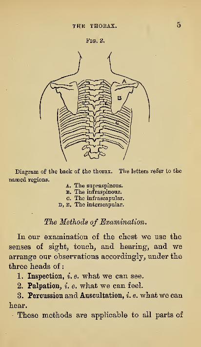
To examine the back of the chest, the patient
should sit or stand with the arms folded, the
shoulders rounded, and the head bent forward,
so as to make the back as broad and round as
possible, and to widen the interscapular spaces.

4 HOW TO EXAMINE THE CHEST,
Fig. 1.
Diagram of the front of the thorax and abdomen.
The vertical lines are the nipple lines. The figures refer to
the'named regions.
1. The supraclavicular.
2. The clavicular.
3. The infraclavicular.
4. The mammary.5. The inframammary.6. The hypochondriac.
7. The episternal.
8, 9, 10. The upper, middle, and lower sternal.
11. The epigastric.
12. The umbilical.
13. The hypogastric.
14. The lumbar.
15. The iliac.
THE THOEAX.
Fl&. 2.
Diagram of the back of the thorax. The letters refer to the
named regions.
A. The supraspinous.
B. The iufraspinous.
C. The infrascapular.
D, E. The interscapular.
The Methods of Examination.
In our examination of tlie chest we use tlie
senses of sights touclij and hearings and wearrange our observations accordingly, under the
three heads of
:
1. Inspection, i.e. what we can see.
2. Palpation, i. e. what we can feel.
3. Percussion and Auscultation, i. e. what we can
hear.
These methods are applicable to all parts of

6 HOW TO EXAMINE THE CHEST.
the body, thougli not in an equal degree, but
they are of cliief importance in tlie examination
of tlie chest.
Any other methods which may be available in
certain cases will be referred to and described as
occasion arises to make use of them.
It is desirable, so far as possible, to represent
in a graphic form all the information we obtain.
Ways of doing this will be suggested as oppor-
tunity offers.
We shall commence with the systematic exa-
mination of the lungs, then proceed to the
examination of the heart, and lastly, to that of
the rest of the mediastinum.
Our observations will be arranged in order
under the heads of Inspection, Palpation, Percus-
sion, Auscultation.

SECTION I.
THE LUNGS.

THE EXAMINATION OF THELUNGS.
INSPECTION.
When we inspect^ or look at^ a chest we have
two sets of facts to observe :
1st. Those, which we can observe as well in a
dead as in a living person, relating to the Shape
or Form of the chest
:
2nd. Those, which are only seen during life,
viz. the Movements of the chest during respira-
tion.
I omit, for the present at any rate, all those
phenomena not associated with the shape or
movements of the chest, such as dilated veins,
&c., as not immediately connected with the exa-
mination of the lungs.
THE SHAPE OF THE CHEST.
This admits of great variation, even within the
limits of health, so that there is no fixed normal
or physiological type.
10 HOW TO EXAMINE THE CHEST.
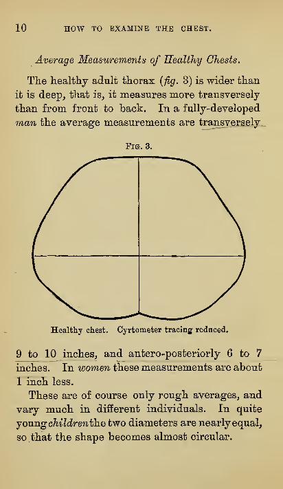
Average Measurements of Healthy Chests.
The healthy adult thorax {fig. 3) is wider than
it is deep, that is, it measures more transversely
than from front to back. In a fully-developed
man the average measurements are transversely
Fig. 3.
Healthy chest. Cyrtometer tracing reduced.
9 to 10 inches, and antero-posteriorly 6 to 7
inches. In women these measurements are about
1 inch less.
These are of course only rough averages, and
vary much in different individuals. In quite
jOMngchildrentlnetwo diameters are nearly equal,
so that the shape becomes almost circular.

THE LUNGS. mSPECTION. 11
The apparatus required for measuring tlie
chest consists of
:
1
.
A measuring tape.
2. A pair of callipers.
3. A cyrtometer.
The Cyrtometer is an apparatus by means of
which a life-size tracing may be obtained of the
shape of the chest.
There are many forms of cyrtometer, but the
most convenient_, and that in ordinary use, is
made of two pieces of composition gas-piping,
each about eighteen inches long, and joined
together by a hinge or piece of gutta-percha
tubing.
The method of using this apparatus is as
follows :
The patient^s chest being bare, a mark is madeat the base of the xiphoid cartilage in front, and
another posteriorly upon the spine, on the same
horizontal level. With a pair of callipers, the
measurement is taken between these two points,
and two marks made upon a sheet of paper,
corresponding with the points of the callipers,
^' spine ^' being written opposite one, and " ster-
num '^ opposite the other.
The '^ cyrtometer ^^ is then taken, the hinge
placed upon the mark upon the spine, and the
soft piping bent round the ribs, until the two
arms meet at the mark in front. A little careful
12 HOW TO EXAMINE THE CHEST.
moulding causes tlie piping to take the form of
tte chest. A mark is now made upon the piping
in frontj to indicate the spots upon the two arms
corresponding withthe middie line of the sternum.
The cyrtometer is then held by the hinge, and
the two arms allowed to fall off the chest by
their own weight, care being taken that they
are not twisted in any way as they are removed.
The whole apparatus is now laid upon the
sheet of paper, so that the hinge corresponds
with the spine mark, and the marks upon the
cyrtometer in front, with the sternum marks.
A pencil is carried round the inside of the arms,
and an exact tracing of the shape of the chest is
thus obtained.
Lastly, the words " Right " and " Left " are
written on the corresponding sides, and the
tracing is complete.*
Named Varieties of Thorax.
The different forms of chest are for the most
part described by ordinary terms, such as long
or short, broad or narrow, deep or shallow, andall of these various forms may be quite consistent
with health.
* By means of a very simple apparatus, such as the pan-
tograph, a cheap form of which may be purchased now for
one shilling, these large tracings may be quickly reduced to a
convenient size for the note-book.

THE LUNGS. INSPECTION. 13
Certain marked deviations from the normal
form havOj however, received special names.
These are
:
1. The Barrel-shaped chest.
2. The Rickety chest.
3. The Pigeon-breast.
4. The Alar chest.
The Barrel-shaped Chest is, as its nameimplies, like a barrel {jig. 4). It is almost
Pia. 4.
Barrel-shaped chest. Cyrtometer tracing from a case of
emphysema reduced.
This is very like the tracing obtained from an infant's
chest, which is also nearly circular in shape.
circular in section. Its transverse and antero-
posterior diameters are almost the same. The
sternum is bowed forwards, and the spine often

14 HOW TO EXAMINE THE CHEST.
backwards, so that, in profile, the outline is
usually distinctly bi-convex.
This form is always associated with a patho-
logical change in the lungs, to which the nameemphysema is given.
The other peculiar forms of chest are not
necessarily associated with any change in the
lungs. They are due to causes which were at
work when the chest was developing in child-
hood, at a time when the ribs were soft and
yielding, and are evidence rather of past than of
present disease, although in all these cases it
commonly happens that the lungs are weak, and
become subsequently affected.
The Rickety Chest gives a tracing such as is
shown i'n.fig. 5. The longitudinal furrow at the
sides of the sternum corresponds with what was
in childhood the ossifying end of the ribs. Most
rickety children suffer much from bronchitis, and
the bronchi in children become easily plugged.
When this is so, the air cannot enter freely into
the air vesicles, and on inspiration the chest walls
are driven in by atmospheric pressure. The
softest parts yield most. These are, of course,
the ossifying ends of the ribs and cartilages,
which are, moreover, in rickets especially soft
and yielding. If this condition lasts for any
length of time, it may become permanent and
give rise to the rickety form found in adults.
THE LUNGS. INSPECTION. 15
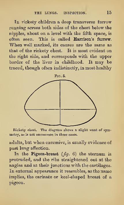
In rickety cliildren a deep transverse furrow
running across botli sides of tlie cliest below the
nipples^ about on a level with the fifth space^ is
often seen. This is called Harrison's furrow.
When well niarked_, its causes are the same as
that of the rickety chest. It is most evident on
the right side, and corresponds with the upper
border of the liver in childhood. It may be
traced, though often indistinctly, in most healthy
Fm. 5.
Rickety chest. The diagram shows a slight want of sym-
metry, as is not uncommon in these cases.
adults, but when excessive, is usually evidence of
past lung affection.
In the Pigeon-breast {fig. 6) the sternum is
protruded, and the ribs straightened out at the
angles and at their junctions with the cartilages.
In external appearance it resembles, as the nameimplies, the carinate or keel-shaped breast of a
pigeon.

16 HOW TO EXAMINE THE CHEST.
ri&. 6.
The Pigeon-breast. Here also is slight want of symmetry.
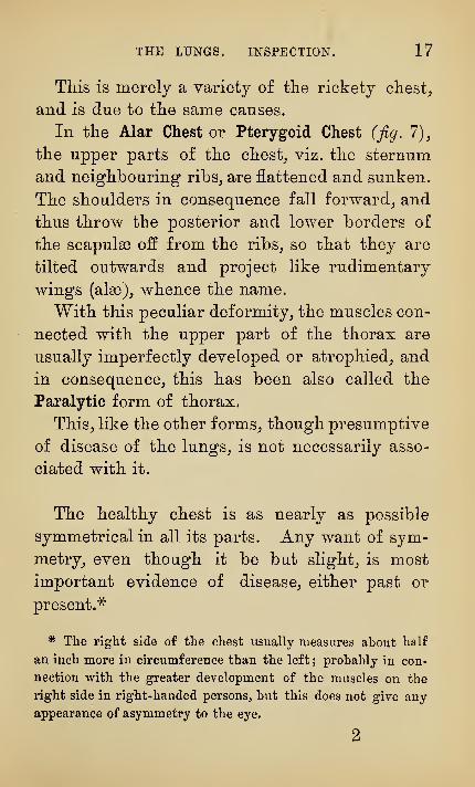
ri&. 7.
The Alar or Pterygoid Chest. This is a good instance of a
tracing of a flat chest.
THE LUNGS. INSPECTION. 17
This is merely a variety of the rickety chesty
and is due to the same causes.
In the Alar Chest or Pterygoid Chest (jig, 1),
the upper parts of the chest_, viz. the sternum
and neighbouring ribs_, are flattened and sunken.
The shoulders in consequence fall forward^ and
thus throw the posterior and lower borders of
the scapulae off from the ribs_, so that they are
tilted outwards and project like rudimentary
wings (alae)^ whence the name.
With this peculiar deformity^ the muscles con-
nected with the upper part of the thorax are
usually imperfectly developed or atrophied^ and
in consequence^ this has been also called the
Paralytic form of thorax.
This, like the other forms, though presumptive
of disease of the lungs, is not necessarily asso-
ciated with it.
The healthy chest is as nearly as possible
symmetrical in all its parts. Any want of sym-
metry, even though it be but slight, is most
important evidence of disease, either past or
present."^
* The right side of the chest usually measures about half
an inch more in circumference than the left; probably in con-
nection with the greater development of the muscles on the
right side in right-handed persons, but this does not give any
appearance of asymmetry to the eye.
2

18 HOW TO EXAMINE THE CHEST.
Changes of Shape due to Disease.
In disease tlie changes of shape may be of two
kinds. There may b(
1st. Increase in size, or as it is usually called,
Bulging;
2nd. Decrease in size, or Contraction.
These changes may affect both sides, i. e. be
bilateral, or only one side, i. e. be unilateral ; and
in either case they may involve either the whole
of the side, or only part of it.
Even where the change is bilateral, it is rarely
absolutely symmetrical.
Symmetrical bilateral increase in size is only met
with in the barrel-shaped chest of emphysema.
Symmetrical bilateral decrease in size occurs
only in the opposite condition, in which both
lungs are uniformly shrunken, and more rarely
also in the paralytic, alar, and other forms of
chest described above.
With these exceptions, changes in shape are
always unsymmetrical, and it is therefore for a
Want of Symmetry, i. e. for a difference between
the corresponding parts of the two sides of the
chest, that we chiefly look as evidence of disease
{fig- 8)-
When there is such a want of symmetry it is
sometimes difficult to say whether this want is
due to a bulging of one side, or to a shrinking of

THE LUNGS. INSPECTION. 19
the other. Further examination only can deter-
mine this question.
Fig. 8.
An extreme instance of want of symmetry. The tracing was
taken from a child in whom the left side was contracted after
an empyema.
Deformities.
In certain trades, for instance, among car-
penters, weavers, and shoemakers, a depression is
often found at the bottom of the sternum, some-
times of considerable depth. This is usually
due to pressure during work (as by the last,
auger, or weavers^ beam), though the deformity
is occasionally congenital.
The Gondition of the Superficial Veins.
Except where patients are very thin, the veins
are rarely visible in health beneath the skin.

20 HOW TO EXAMINE THE CHEST.
In disease they are often dilated and frequently
unsymmetrically so. When this occurs^ the
direction in which the blood is travelling should
be determined. This is done by placing two
fingers upon the most prominent vein, and then
drawing them apart along the vein in order to
press the blood out. By raising first one finger
and then the other^ it will be clear from which
direction the vein fills most easily. This will
be then the direction in which the blood is
travelling."^
THE MOVEMENTS OF THE CHEST ON RESPIRATION.
These are alternately movements of expansion
and contraction^, i. e. inspiratory and expiratory.
On ins]3iration the chest expands in all directions.
The sternum moves forward^ the ribs rise,, the
intercostal spaces widen, and the diaphragm
descends. These movements are freest in the
lower parts of the chest. They are partly
thoracic and partly diaphragmatic.
In women the ribs ntove most, and the respi-
ration is called Thoracic or Costal.
In men and in young children the diaphragm
^ Enlarged subcutaneous veins over the mammse and upper
part of the chest are usual in women who are suckling, or whohave had children. This is, of course, physiological.
THE LUNGS. INSPECTION. 21
moves most, and tlie respiration is called Dia-
phragmatic or Abdominal.
A change of type from costal to abdominal or
vice versa is often an evidence of disease.
The Measurements of the chest vary within
wide limits. The average circumference of a
healthy man^s chest at the level of the nipple is
after expiration about 32 inches, and after in-
spiration about 35^ inches, giving thus a differ-
ence on the average of each respiration of
3 1 inches, or about one twelfth. On forced
respiration the difference can sometimes be
made much greater.
These measurements in women are somewhat
less.
The Amount of Air, which is taken in and out,
will depend upon the amount of the respiratory
movement of the chest and upon its size.
In ordinary breathing it is calculated that in
a healthy man on the average about 30 cubic
inches are drawn in at each inspiration, and
the same quantity emitted at each expiration.
About 100 cubic inches more may be squeezed
out on forced expiration, and about the same
amount more taken in on forced inspiration.
Making the total maximum quantity of air
which can be inspired or expired about 230
cubic inches.
Instruments have been devised for measuring

22 HOW TO EXAMESTE THE CHEST.
the Vital Capacity of the chest,, i.e. the total
amount of air, which can be taken in^ or forced
out, by the deepest possible respiration. They
are known as Spirometers, but hitherto they
have not been found to be of much use in
diagnosis.
Alterations in the Respiratory Movements.
When the respiratorymovements are increased
in range above the normal, we speak of them as
Exaggerated ; when decreased below the normal
we speak of them as Impaired or Deficient.
When the movements are deficient, less air will
enter the lungs than is necessary, and the patient
will suffer from shortness of breath, or as it is
called Dyspnoea (difficulty of breathing)
.
Dyspnoea may be the result of deficient respi-
ratory movements under two opposite conditions,
for the lung's may be prevented either from
expanding, or from contracting, so much as they
should. In the former case the condition is
spoken of as Defective Inspiration or Deficient
Expansion, and the dyspnoea is called Inspiratory;
the latter, as Deficient Expiration, and the dys-
pnoea is called Expiratory.
When the patient cannot lie down on account
of the difficulty in breathing, the term used is
not dyspnoea, but Orthopnoea, i. e. dyspnoea which

THE LUNGS. INSPECTION. 23
obliges the patient to sit up {orihos, upright) to
get breath.
When from any cause there is obstruction to
the entrance of air, the deficient expansion of the
lungs will make itself manifest in the softer parts,
of the thorax, i. e. in the intercostal and supra-
clavicular spaces, and they will sink in somewhat
during inspiration. This is called Inspiratory
Eecession.
When the obstruction is considerable, not only
the soft parts, but also the ribs, especially the
lower ones, yield, and are sucked in during inspi-
ration. In its most extreme form, this is met with
in children suffering from croup, and where the
obstruction is of long standing, or oft repeated,
as has been stated already, it is the cause of
certain deformities, which may be permanent
(p. 14).
The exactly opposite condition to inspiratory
recession of the intercostal and supraclavicular
spaces, viz. Expiratory Bulging, is common in
cases in which the elasticity of the lungs is
reduced, and the expiration obstructed.
It is most marked during a fit of coughing, in
patients suffering from extreme emphysema.
The Number of Respirations is about 14 to 18
in the minute, and bears to the pulse, on the aver-
age, the relation of 1 to 4.
On quiet respiration, the movements occur at

24 HOW TO EXAMINE THE CHEST.
regular intervals^ tliougli they are largely influ-
enced byemotion and excitement^ both as regards
number and regularity.
Except in children and in cases of hysteria^ the
number^ even in disease, rarely- exceeds 40 to
50. As a general rule the more rapid the
respirations^ the more shallow they are.
The movements of respiration, in healthy per-
sons at rest, follow one another at regular
intervals, the rhythm being maintained by the
action of the nerve centres in the medulla
oblongata.
The movements are to a very great extent
under voluntary control, and may therefore be
made to vary much by the action of the will, as in
speaking, singing, &c.j but irregularity is often
independent of the will, and is due then to inter-
ference with the action of the respiratory centre^
usually in response to reflex irritation from some
other part. Thus, mental emotion may lead to
laughing, crying, sobbing, &c., irritation in the
lungs or stomach to coughing, hiccough, &c.
Of all forms of irregular respiration the most
peculiar is that known by the name of Cheyne-
Stokes' Breathing."^
In this form the respiration at times ceases for
some seconds, and then recommences, the move-
* Dr. Cheyne first observed it and Dr. Stokes subsequently
more minutely described it.
THE LUNGS. INSPECTION. 25
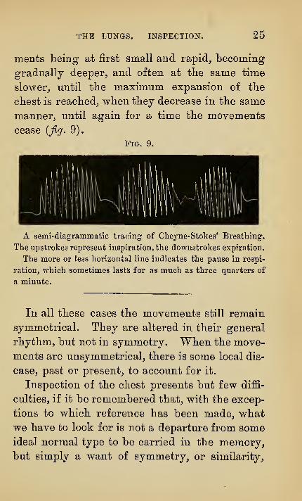
ments being at first small and rapid^ becoming
gradually deeper^ and often at the same time
slower, until the maximum expansion of the
chest is reached, when they decrease in the same
manner^ until again for a time the movements
cease {fig. 9).
Fig. 9.
A semi-diagrammatic tracing of Cheyne-Stokes' Breathing.
The upstrokes represent inspiration, the dovvnstrokes expiration.
The more or less horizontal line indicates the pause in respi-
ration, which sometimes lasts for as much as three quarters of
a minute.
In all these cases the movements still remain
symmetrical. They are altered in their general
rhythm, but not in symmetry. When the move-
ments are unsymmetrical, there is some local dis-
ease, past or present, to account for it.
Inspection of the chest presents but few diffi-
culties, if it be remembered that, with the excep-
tions to which reference has been made, whatwe have to look for is not a departure from someideal normal type to be carried in the memory,but simply a want of symmetry, or similarity.

26 HOW TO EXAMmE THE CHEST.
between tlie two sides of tliat particular chest
whicli we are examining.
When this want of symmetry exists, there
must be some condition of disease, past or pre-
sent, to account for it.
SYNOPSIS.
On Inspection then we note
—
I. The Shape of the Chest
:
Barrel.
Rickety.
Pigeon.
Alar.
Paralytic.
Shoemakers', Weavers', Carpenters'.
Harrison's Furrow.
Defects of Symmetry.
II. The Movements of the Chest :
their number,
their regularity,
their type, costal or diaphragmatic,
if impaired,
or exaggerated.
Defects of Symmetry.
T. f Inspiratory.Dyspncea^^ ^ "^
L Expiratory.
Orthopncea.
THE LUNGS. PALPATION. 27
PALPATION.
The first thing to be done on palpation is to
Count the Ribs, and_, simple as this seems to be^
mistakes are often made. It will be easily and
correctly done, if it be remembered, that the
first rib, which we can get comfortably between
two fingers, is the second. It is easier to count
the spaces than the ribs, and we know, that the
rib corresponding to the space lies above the
space.
Our landmarks, as we shall see, are all deter-
mined with relation to the ribs and spaces.
THE SHAPE AND MOVEMENTS.
Nearly all that can be seen can be also felt,
but sometimes, in case of difficulty, the hand
may help the eye. This is especially the case with
the movements of the chest. For this purpose,
the hands must be placed symmetrically upon
corresponding parts. At the apices, the thumbs
should be placed together upon the sternum, and
the fingers allowed to rest beneath the clavicles,
or, in children, the thumbs may be placed in con-
28 HOW TO EXAMINE THE CHEST.
tact upon tlie spine_, and the fingers bent over
the shoulder^ so as to rest upon the upper part
of the chest in front. In either of these ways
very slight differences in the amount of move-
ment upon the two sides may be detected.
The Widening of the Intercostal Spaces on in-
spiration may be easily observed^ by placing the
hands upon the lower parts of the chest or in the
axillae and spreading the fingers so that they lie
in the intercostal spaces.
"We are able in this way to determine :
1. If the spaces be narrower or wider on one
side than on the other ;
2. If they be retracted or unduly prominent;
3. If the expansion or widening on inspiration
be sufficient in amount^ and equal on the two
sides.
Abnormal Sensations.
Occasionally the grating of Pleuritic Friction
(^. v.)j the wheezing of Rhonchus and Sibilus
(^. v.)y or the crackling of Crepitation {q.v.),
may be felt.
YOCAL VIBEATIONS.
If, while the hand is placed upon the chest, the
patient be made to speak, the vibrations of the
voice will be felt by the hand. They are called
Vocal Vibrations. They may be also heard, if the

THE LUNGS. PALPATION. 29
ear be placed upon the cliestj as we sliall see under
'''Auscultation/^ and then they are spoken of as
Vocal Resonance. There is no real difference
between tliem except one of terms. We feel
vocal vibrations^ and we listen to vocal resonance.
As the ear is more sensitive than the hand^ so wecan occasionally hear the vocal resonance^ whenwe cannot feel the vocal vibrations. This is
especially the case in women and children_, in
whom the vibrations of the voice are not intense.
The louder the voice_, the deeper or more bass
the tone, and the thinner the patient, the more
easily will the vibrations be felt. The other con-
ditions, which alter the vocal vibrations, will be
discussed laterunderthe head of ^^Auscultation,^'
when we speak of vocal resonance.
For the present it is sufficient to say, that the
same want of symmetry in the physical signs,
which we look for on inspection, is to be searched
for also on palpation. It is this want of sym-
metry, which is of the chief practical importance.
Se7ise of Resistance.
If the intercostal spaces be lightly tapped with
the tips of the fingers over the upper part of the
chest, a sensation of elasticity or springiness will
be obtained. If, however, the same thing be done,
where a solid organ lies beneath the chest walls.
30 HOW TO EXAMINE THE CHEST.
as over tlie liver^ the sense of elasticity will be
lost^ and, in its place,, tlie fingers will experience
a feeling of resistance.
The same thing happens, if the lang becomes
solid, or if it be separated from the chest walls
by changes in the pleura. To this feeling the
name Sense of Resistance is given. It is, how-
ever, of no great practical importance, except
when combined with percussion (palpatory per-
cussion).
When, in disease, there is a large collection
of pus in the pleura [Empyema) Fluctuation maysometimes be elicited in the usual way ; but
except when the empyema is pointing, i. e. whenthe pus is close beneath the skin, fluctuation is
extremely rare.
SYNOPSIS.
On Palpation, then, we proceed to count the ribs,
and next to observe :
1 . The Shape and Movements of the Chest.
2. The Vocal Vibrations.
3. The Sense of Resistance.
4. Abnormal Sensations, when present, such as
friction, crepitation, rhonchus, sibilus, or pos-
sibly fluctuation.

THE LUNGS. PEECUIOSSN. 31
PERCUSSION.
By Percussion is meant the metliod of striking
the walls of the bodj^ so as to cause them to
yield a sound.
We must consider^ then^ 1st, the best way of
producing sound by percussion, and 2ndly, the
kinds of sounds, which may be produced, and
what meaning and value may be attached to them.
Percussion may be direct (immediate), when wepercuss upon the skin directly, or indirect (me-
diate), when we percuss upon something placed
upon the skin.
In the examination of the Chest Direct Per-
cussion is not employed now except upon the
sternum, the clavicles and the spine of the
scapula, where, from the absence of much cover-
ing, the sound produced is not interfered with.
For Indirect Percussion we require : 1st, some-
thing to strike with, and 2ndly, something to
strike upon.
Apparatus of various kinds has been devised
for this purpose :—1 . Hammers, or, as they have
been called, Plessors, of various sizes, shapes, and
substances, to strike with. 2ndly. Flat Plates
(Plessimeters), of metal, wood, or ivory, to strike
upon.

32 HOW TO EXAMINE THE CHEST.
These forms of apparatus have been almost
entirely abandoned_, and in their place we are in
the habit of using the fingers on one hand^ as our
plessor^ to percuss with_, and one of the fingers of
the other hand^ as our plessimeter^ to percuss
upon.
In this way we combine; with percussion, those
sensations described in the previous chapter
under the head of " Sense of Kesistance.^^ This
method of percussion has been called Palpatory
Percussion.
The tip or pad of one finger, say the middle,
is the head of the hammer, the rest of the hand,
the handle.
The blow should be light, but firm, produced
by a free action of the wrist, as in playing octaves
upon the piano. The art is not easy to acquire,
but can be well practised by placing the whole
forearm, from the elbow to the fingers, flat upon
a table and then percussing, the forearm being
firmly pressed down with the other hand, to keep
it fixed, and to prevent the wrist being raised
from the table.
The hammer of a piano forms the best illustra-
tion of the kind of movement we require. Whena note is struck upon the key-board, the hammeris driven sharply against the wire, but does not
remain more than an instant upon it, quickly
recoiling and leaving the wire free to vibrate.

THE LUNGS. PEECUSSION. 33
This is wliat the hand should do. The finger
should deliver a short, sharp stroke, and imme-diately return from contact with the chest. It
will require much practice to get this proper
movement.
Sometimes^ instead of the tip of one finger, the
tips of two or three are employed. This has no
special advantage, except^ that as the head of the
hammer is broader, a greater surface is thrown
into vibration, and therefore the sound is some-
what louder ; but, if more fingers than one be
used^ care must be taken that the pads of the
fingers are all upoji the same level,, so that they
may all strike the chest at the same time. This,
again^ can be practised best upon the table, by
pressing first the tips of the fingers firmly downto get them level, then raising them, fixed in that
position, and proceeding to percuss.
In choosing a finger to percuss upon, one should
be selected which is not bandy^ so that it may lie
perfectly flat. It matters little which is chosen.
For convenience^ it is generally either the index
or the little finger.
The object we have in view in percussing the
chest is to throw into vibration the parts beneath
the walls of the thorax. "We must, therefore,
avoid as much as possible all interference with
these vibrations from the walls themselves. This
we do by placing the fingers upon which we per-
3

34 HOW TO EXAMINE THE CHEST.
CUSS perfectly flat upon the chesty and exercising
slight pressure^ so as to condense the tissues
immediately beneath. If the finger be placed
loosely_, instead of firmly, upon the skin_, and still
more if it be not in all parts quite in contact^ the
percussion note will be impaired.
In order that there may be as little of the walls
as possible for the vibrations to pass through
before reaching the organs beneath, we must per-
cuss straight upon the surface, that is, perpendi-
cularly to the walls and not in a slanting direc-
tion.
To sum up
—
1st. Our hands form our only apparatus.
2nd. The finger percussed upon must be placed
flat, and pressed firmly upon the chest.
3rd. The blow must be from the wrist, light,
short, firm, and delivered at right angles to the
chest-walls at the part percussed.
Grood percussion is difficult to acquire, but is
worth all the time and trouble spent upon it.
Resonance and Dulness.
When we percuss upon the walls of a cavity
containing air, for instance over a drum,we obtain
a hollow or, as it is called, a resonant sound.
When we percuss upon a solid mass, like the
thick part of the thigh, the hollow sound is not
THE LUNGS. PEECUSSION. OO
produced, and the sound wliicli is produced is
called non-resonant or dull.
Many varieties of resonance and non-resonance
are described, but it is sufficient for us at pre-
sent to recognise the difference between sounds
which are resonant^ and those which are non-
resonant.
Over the lungs the note is resonant^ because
the lungs contain air. Over a solid organ^ such
as the liver^ the note is non-resonant or dull.
The Size of the Lungs.
We are now in a position to apply these facts
practically.
How large are the Lungs ? This is naturally the
first question of importance^ and percussion alone
enables us to answer it. For the lungs are in
direct relation with solid organs, and, where these
are, the note, which over the lungs has been
resonant, will become non-resonant or dull. If we
mark upon the skin of the chest the places where
this occurs, we obtain certain lines. These are
called Surface-Markings, or Medical Landmarks.
These landmarks indicate certain relations in
which the organs stand to the outer parts of the
body. They must not be confounded with the ana-
tomical boundaries, sizes, and shapes, of these dif-
ferent organs, withwhich they only approximately

36 HOW TO EXAMINE THE CHEST.
Fig. 10.
Diagram showing the position of the great organs of the thorax and abdomen
with their surface-markings.
L, Liver. ST, Storaach. s, Spleen. K, Kidney, c, Colon.
The Lungs are left unshaded. Their upper and anterior boundaries only
are shown by a line of thick dots.
The Heart is represented of its anatomical size, and upon it in black is
indicated the size of tbe absolute cardiac duLness.
The Hepatic dulness is represented in black. The line of small dots above
on the right side, shows tlie position of the upper part of the liver, and there-
fore of the vault of the diaphragm deep within the thorax. Just outside the
right nipple Une, at the edge of the ribs, the little excrescence marks the
position of the Gall-iladder.
The Stomach is indicated partly by dotted and partly by continuous line. Thearea of stomach resonance lies between the dotted line ou the left side and the
margin of the.fiostal arch.
The anteriof^arts of the Spleen and Kidney are indicated in black.

THE LUNGS. PEECUSSION. 37
correspond. For example^ tlie surface-markiDgs
of tlie heart inclose a space (the cardiac area)^
which is small compared with the real size of the
heart [jig. 10), but it is of the greatest importance-
for the reason that^ so long as the organs in
immediate relation with the heart are normal^ the
space varies in size and shape in direct corre-
spondence with any change in the heart itself.
The medical landmarks and anatomical boun-
daries aretherefore not the samethings andthough
closely related must not be confused one with the
other.
The Boundaries of the Lungs.
The lungs are in close contact with the ribs
along their whole length from the sternum to
the spine.
The ribs are^ therefore, the natural External
Boundaries.
The apex of each lung rises as a blunt cone
into the neck as far as an inch and a half above
the clavicle. The curved line, which corresponds
with the apex and sides of this cone, can be easily
percussed out, and gives the Upper Boundary.
The edges of the lungs approximate anteriorly
beneath the manubrium sterni, and come in con-
tact, at a point corresponding with the junction
of the second costal cartilage with the sternum ;
they remain in close approximation doM^n as far

38 HOW TO EXAMINE THE CHEST.
as tlie level of tlie fourth costal cartilage. Fromthis point the anterior margin of the right lung
continues onward down to the bottom of the
sternum_, sloping slightly away to the right side,
while that of the left bends sharply away to the
left side^ to a point about two inches and a half
from the bottom of the sternum. This leaves a
roughly triangular space between the two lungs
in which part of the pericardium is uncovered.
It corresponds roughly speaking with the area of
cardiac dulness [q-v.].
The Middle Boundary cannot be determined by
percussion for the following reason : The sternum
is a solid bone^ which lies for some distance in
close relation with the lungs. When, then, it is
percussed—even in a part, where only solid
structures lie beneath^ as under the upper part
of the manubrium, or under the lower part over
the heart—the vibrations are transmitted to those
parts of it which lie over the lungs^ and so to the
lungs themselves, and, in consequence, the note
in any part of the sternum will be resonant.
The Lowerboundary is of course the diaphragm,
but this is too thin to define by percussion, so
that we can determine its position only by means
of the organs in relation with it ; these are, the
liver, the stomach, and the spleen. Two of these
organs, the liver and the spleen, are solid bodies,
and will give, therefore, a non-resonant sound.
THE LUNGS. PEECUSSION. 39
The stomacli contains air, and will, therefore,
yield a resonant sound.
Percussion will enable us, then, to determine
where these organs are, and in this way where
the diaphragm is. On the right side the termi-
nation o£ the lungs will be marked by a line
of non-resonance, or dulness, corresponding with
the liver, and on the left side by a line of altered
resonance corresponding with the stomach, and
by a line of dulness corresponding with the
spleen.
Before, then, we can determine how large the
lungs are, we require to know what the upper
boundaries of the liver, stomach, and spleen are
in health, I.e. the surface-markings corresponding
with these organs."^
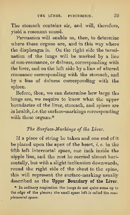
The Surface-Marhmgs of the Liver.
If a piece of string be taken and one end of it
be placed upon the apex of the heart, ^. e. in the
fifth left intercostal space, one inch inside the
nipple line, and the rest be carried almost hori-
zontally, but with a slight inclination downwards,
round the right side of the chest to the spine,
this will represent the surface-marking usually
described as the Upper Boundary of the Liver.
* In ordinary respiration the lungs do not quite come up to
the edge of the pleura; the small space left is called the com-
plemental space.
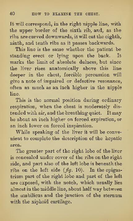
40 HOW TO EXAMINE THE CHEST.
It will correspond, in the right nipple line^ with
the upper border of the sixth rib^ and^ as the
ribs are curved downwards^ it will cut the eighth,
ninth, and tenth ribs as it passes backwards.
This line is the same whether the patient be
standing erect or lying' upon the back. It
marks the limit of absolute dulness, but since
the liver rises anatomically above this line
deeper in the chest, forcible percussion will
give a note of impaired or defective resonance,
often as much as an inch higher in the nipple
line.
This is the normal position during ordinary
respiration, when the chest is moderately dis-
tended with air, and the breathing quiet. It maybe about an inch higher on forced expiration, or
an inch lower on forced inspiration.
While speaking of the liver it will be conve-
nient to complete the description of the hepatic
area.
The greater part of the right lobe of the liver
is concealed under cover of the ribs on the right
side, and part also of the left lobe is beneath the
ribs on the left side {fig. 10). In the epigas-
trium part of the right lobe and part of the left
are exposed, with the notch, which usually lies
almost in the middle line, about half way between
the umbilicus and the junction of the sternum
with the xiphoid cartilage.

THE LUNGS. PEECUSSION. 41
The liver passes under cover of the ribs on the
right side just in the nipple line. This corre-
sponds usually with the tip of the eighth costal
cartilage.
The Lower Boundary of the liver^ then, on the
right side is continuous posteriorlywith the edges
of the costal arch, and comes out from under the
ribs in the right nipple line. It then extends
across the abdomen in a double curve, inter-
rupted by the notch, to the apex of the heart
(/^. 10).
As the lower part of the liver in front lies in
close relation with the stomach and transverse
colon, the transmitted resonance makes it gene-
rally very difficult to ascertain exactly by per-
cussion the lower border of the liver, which is
usually more easily fixed by palpation.
The liver is most conveniently measured in the
nipple line. In this line the Upper Boundary
should be at the level of the upper border of the
sixth rib, and the Lower should cut the margin
of the costal arch. Just outside this part {i.e. at
the tip of the ninth rib) is the position of the
gall-bladder.
The vertical measurement of the hepatic dul-
ness in the nipple line is, in the adult man, on
the average four inches.

42 HOW TO EXAMINE THE CHEST.
Fia. 11.
Diagram showing the normal boundaries of the lungs.
THE LUNGS. PEECUSSION. 43
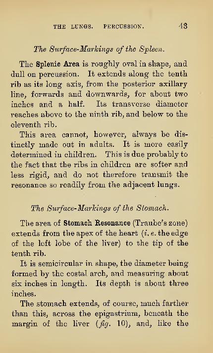
The Surface-Markings of the Spleen.
The Splenic Area is roughly oval in shape^ and
dull on percussion. It extends along the tenth
rib as its long axis^ from the posterior axillary-
line, forwards and downwards, for about two
inches and a half. Its transverse diameter
reaches above to the ninth rib, and below to the
eleventh rib.
This area cannot, however, always be dis-
tinctly made out in adults. It is more easily
determined in children. This is due probably to
the fact that the ribs in children are softer and
less rigid, and do not therefore transmit the
resonance so readily from the adjacent lungs.
The Surface- Markings of the Stomach.
The area of Stomach Resonance (Traube's zone)
extends from the apex of the heart {i. e. the edge
of the left lobe of the liver) to the tip of the
tenth rib.
It is semicircular in shape, the diameter being
formed by the costal arch, and measuring about
six inches in length. Its depth is about three
inches.
The stomach extends, of course, much farther
than this, across the epigastrium, beneath the
margin of the liver {fig' 10), and, like the

44 HOW TO EXAMINE THE CHEST.
coloiij whicli lies in immediate relation with it
beloWj will give a resonant sound there^ but it is
only the limited area described above, which, in
the examination of the chest, is spoken of as the
area of stomach resonance.
The boundaries of the stomach and spleen are
not so constant, or so easy to determine, as those
of the liver, but they are also not of so muchimportance, for the liver reaches so far to the left
side, that its boundaries, taken in conjunction
with the cardiac dulness, are enough to fix, with
sufficient accuracy for ordinary purposes, the size
of the left liing.
We have now ascertained the position of the
diaphragm, and we know, that all that is above
this should be occupied by lung, except in the
mediastinum, where the heart and great vessels
lie. In health we need consider nothing but the
heart, for the rest of the mediastinum gives, as
the lungs do, a resonant note on percussion.
The Area of Cardiac Dulness is roughly tri-
angular in shape, and corresponds approximately
with the space exposed by the left lung as it
recedes from the right. It is represented on the
diagram (2. 'v.)j and will be found fully described
later.

THE LUNGS. PERCUSSION. 45
The Surface-Markings of the Lungs.
Tliese are as follows :
The Upper. A curved line, the apex of whicli
reaches one inch and a half above the clavicle.
The Anterior.
{a) On the Right Side^ the middle line of the
sternum, from the level of the second costal carti-
lage to the base of the xiphoid cartilage.
(h) On the Left Side, the middle line of the
sternum, from the level of the second to the level
of the fourth costal cartilage, and thence to the
apex of the heart.
The lower.
(a) On the Right Side, the upper border of the
liver.
(b) On the Left Side, a line drawn from the
apex of the heart along the upper border of the
stomach resonance and the splenic dulness.
The Posterior.
1. A Vertical Line drawn on each side one inch
from the dorsal spine.
2. A Horizontal Line drawn outwards on each
side from the eleventh dorsal spine. This, on
the right side, is continuous with the upper
boundary of the liver

46 HOW TO EXAMINE THE CHEST.
Fig. 12.
Diagram showing in dark line the actual houndaries of the
lungs in a well-marked ease of emphysema, and in dotted line
the normal houndaries.

THE LUNGS. PEKCFSSION. 47
ALTERATIONS OF BOUNDARIES.
In disease tlie lungs rarely remain of their
normal size. They are either larger or smaller
than they ought to be. These changes^ though
often evident on inspection and palpation_,
are most distinctly indicated by alterations in
the boundary lines.
The diaphragm is freer to move than any part
of the thoracic walls^ and alterations in its posi-
tion are often among the earliest evidences of
changes in the lungs. This can be recognised
only by percussion. Hence the importance of
determining as early as possible in our examina-
tion of the chest the position which the dia-
phragm occupies.
Symmetrical Changes.
Where the lungs are Symmetrically Enlarged,,
as in the disease called emphysema^ there may or
may not be visible enlargement of the thorax,
but there will always be displacement of the
diaphragm. The diaphragm will stand lower
than it ought, often a whole interspace too low.
The cardiac area will also be smaller than it
should be ; for the lungs, as they enlarge, cover
up that part of the prgecordium which, in the
ordinary condition, is exposed.
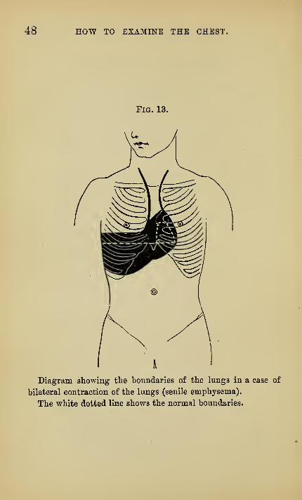
48 HOW TO EXAMINE THE CHEST.
Diagram showing the boundaries of the lungs in a case of
bilateral contraction of the lungs (senile emphysema).
The white dotted line shows the normal boundaries.

THE LUNGS. PEECUSSION. 49
' The percussion signs, then, of emphysema
show displacement downwards of the diaphragm,
and diminution in size, or absence of the cardiac
dulness.
Where the lungs are Symmetrically Contracted,
as often happens in old age (senile emphysema),
the diaphragm will stand at a higher level than
normal. It may be a whole intercostal space too
high, and, in like manner, the prsecordium will
be more uncovered than usual, and the area of
cardiac dulness larger than normal.
Unsymmetrical Changes.
If the changes be limited to one side, the
diaphragm on that side will be displaced. If the
lung on that side be larger, the diaphragm will
stand lower ; if smaller, it will stand higher.
Where a part of one lung is contracted, as
after pleurisy, or where a large cavity has formed
in it, the opposite lung, if it has remained
healthy, often undergoes compensatory enlarge-
ment. This has received the name of Compen-
satory Hypertrophy.* As the one lung is muchsmaller, and the other much larger than it
ought to be, we shall have evidence of a great
* This is also spoken of as compensatory emphysema, but, as
there is probably no true emphysema, this term is misleading,
and should not be used.
4

60 HOW TO EXAMINE THE CHEST.
Fia. 14.
Diagram' showing the displacement of the boundaries in a
case of contraction of the left lung, with compensatory hyper-
trophy of the right.

THE LUNGS. PEECUSSION. 51
dislocation of tlie boundaries as is sliown in
A similar extreme dislocation of boundaries is
seen in cases where one pleural cavity is greatly
distended with, air^ as in pneumothorax [jig. 15),
or with fluid, as in pleuritic effusion (fig. 16)
»
In these latter cases, not only is the diaphragm
pushed down as far as it can go, so as to become
sometimes even concave instead of convex above
and to project below the ribs, but the lateral
boundaries are also dislocated far over towards
the unaffected side.
This we determine by an extension beyond the
sternum, in the one case {pneumothorax) of the
area of resonance, and in the other [pleuritic
effusion) of the area of dulness.
In those cases of pneumothorax, however, in
which there is free communication between the
air inside the pleura and that outside the body,
either by a large opening through the chest-walls
or through the lung, and where, consequently,
there is no distension of the pleura, i. e. no pres-
sure in the pleura, there is still considerable
dislocation of boundaries. This is due to the
elasticity of the lungs, the lungs on each side
contracting, and that on the sound side dragging
over towards itself the mediastinum and the
organs in it.

52 HOW TO EXAMINE THE CHEST.
Fig. 15.
Diagram showing the displacement in a case of pneumo-
thorax of the left side.

THE LUNGS. PEECUSSION. 53
VARIETIES OF PERCUSSION SOUND.
Hitherto we have considered percussion onlya^
the means of determining the size of the lungs,
we must now consider how it enables us to deter-
mine the conditions in which the lungs are.
Wherever the lungs are^ the percussion note
should be resonant. The amount of resonance
varies within wide limits, even in health/ in
different individuals. These variations depend
in great part upon the amount of skin, fat, and
muscle which covers the ribs, i. e. upon the thick-
ness of the walls of the thorax, and is therefore
less in fat than in thin people. Even in a perfectly
healthy chest the resonance varies in different
parts, being greatest in the axilla where there is
least to interfere with the percussion sound.
But, making allowance for all this, the reso-
nance may be greater or less than it ought to be.
In emphysema, where the vesicles of the lungs
are dilated and their walls thinned, where, there-
fore, there is relatively more air and less solid in
the lung, the note becomes deeper, more hollow-
sounding, more drum-like. This is called Tym-
panitic Resonance, and resembles the note which
may be normally obtained on percussing over the
stomach.
A variety of tympanitic percussion is not un-

54 HOW TO EXAMINE THE CHEST.
Fig. 16.
Diagram showing the displacements produced by a large
effusion into the right pleural cavity.
The black area represents the absolute dulness of the fluid.
The heart is displaced so that the apex is outside the left
nipple line. The liver is depressed and twisted, so that the
notch is nearly under the margin of the left costal arch,
instead of being in the middle line.
The white line indicates the probable position of the upper
border of the liver on the right side, and of the edge of the
I'ight pleura on the left side.

THE LUNGS. PEECUSSION. " 55
common in cases, where air-containing lung tis-
sue intervenes between the chest-walls and some
non-resonant substance more deeply seated, as,
for instance, a deep-seated pneumonia, or a
tumour, or even fluid effusion in the pleura.
It is probably due to the relaxation of the lung
tissue, i.e, the loss of its normal tension and
tone above the parts diseased.
A similar hyper-resonance is not rare in acute
fevers, and has probably the same causation and
explanation as that loss of tension and tone in
the intestines, which gives rise, under similar
conditions, to tympanites.
For the opposite condition, in which there is
less air and more solid relatively in the lung,
there is no distinctive name, but the percussion
resonance is spoken of as Impaired or Deficient.
Many other varieties of resonance have been
described and various names given to them, but
they are not really required in actual practice.
For ordinary purposes the following four terms
are all that are necessary
:
1st. Tympanitic.
2nd. Normal.
3rd. Impaired."^
4th. Dull.
* Boxy is a term often used. The term conveys much what
the sound of percussion suggests. It is as though we were per-
cussing an air-containing chamber with dense and rigid walls
56 HOW TO EXAMINE THE CHEST.
One other naraed variety of abnormal percus-
sion sounds must be referred to^ viz. tbe Cracked-
pot Sound (bruit de pot fele). This is a jarring
or jangling sound, like tbat produced when a
cracked china bowl is struck.
It is not uncommon in cases of phthisis over
superficial cavities in the lung, and it is best
elicited by forcible, sharp percussion, the patient
having the mouth wide open and breathing
quietly. It is not constant in phthisis, nor is it
by itself any sign of disease of the lungs, for it
is not rare in children with healthy chests,
and maybe sometimes produced in adults, where
air-containing lung tissue lies between the chest-
walls and some solid mass, either a patch of
pneumonic consolidation, a tumour, or occasion-
ally even an enlarged heart.
It is supposed to be due to the sudden forcing
out by percussion of a stream of air from a por-
tion of the lung into the bronchial tubes, and can
be imitated fairly well by clenching the palms of
the hands loosely together, and striking themsharply upon the knee.
Want of Symmetry on Percussion.
Fortunately, it is not for the most part an
increase or a decrease in tone, as compared with
like a box, and it corresponds frequently with such a condition,
pathologically.

THE LUNGS. PEECUSSION. 57
an ideal typical standard^ wliicli we have to
recognise, but, as on inspection and palpation,
so also on percussion, it is for a want of symmetry,
^. e. for a difference between corresponding parts
on tlie two sides, that we look as evidence of
disease. If, in corresponding parts of the chest,
the percussion resonance is not also symme-
trical, but there is a difference between the two
sides, it is certain that some change has occurred
in the parts beneath.
The only place in health in which want of
symmetry is observed, with the exception of the
cardiac area, to which reference has been already
made, is at the apex. At the right apex the
lung is thicker, stumpier, and is more en-
croached upon by the large vessels than on the
left side. Consequently, there is often a slight
impairment of percussion here, as well as also,
on palpation, and auscultation, a slight increase
in the amount of the vibrations of the voice and
of the breath sounds.
The difference is, however, slight, and yet the
same amount of difference against the left side
would be evidence of disease. It is necessary
to refer to this, although it is not likely to
create difficulty under ordinary circumstances.
Whereverthe percussion note is unsymmetrical,
there is, with the previously mentioned excep-
tions, some change in the condition of the

58 HOW TO EXAMINE THE CHEST.
part beneatlij namely^ in the lungs or in tlie
pleura.
Under '^ Auscultation ^^ we shall learn how to
determine whicli of these it is due to.
For the present it is sufficient to note that by
means of percussion we can establish two sets of
most important facts about the lungs.
1. Their size and their relation to adjacent
organs.
2. In most cases their condition, whether
healthy or not.

THE LUNGS. PEECUSSION. 59
SYNOPSIS.
On percussing a cliest^ it is most important to
proceed systematically.
1st. We must determine the actual boundaries
of tlie lungs, mark tliem carefully, and compare
them with those, which, we know, ought to be
found in health.
2nd. "We must percuss the corresponding parts
of the chest in order, from above downwards,
comparing one side with the other :
1. The supraclavicular
I. In front, <;
regions
;
2. The clavicular re-
gions ;
3. The subclavicular re-
gions;
4. The mammary andinfra-mammary re-
gions ;
II. Laterally, 5. The axillary regions ;
6. The suprascapular re-
gions;
7. The infrascapular re-
gions.
8. The interscapular
spaces.
If these be all symmetrical, the lungs and
pleura are probably healthy. If not, we shall
then proceed to ascertain what is wrong byfurther examination.
III. Posteriorly, <

60 HOW TO EXAMINE THE CHEST.
AUSCULTATION.
Under tMs head we place all facts which wecan ascertain by placing the ear upon the chesty
and listening to the sounds produced. These
are of two kinds.
1. The sounds produced by breathing.
2. The sounds produced by the voice.
Apparatus.
For the purposes of auscultation apparatus is
not generally necessary^ though it is convenient.
Stethoscopes {stethos, the chest ; skopeiuj to
examine) have been devised of all kinds^ some
solidj some hollow, and made of metal^ wood,
ivory, or other substances, of various sizes and
shapes. Habit will accustom us to all, and,
except for convenience, we might do without
any.
The stethoscopes in use at present are of two
kinds, the single, for one ear, and the double,
for both ears—the binaural.
It is best to commence with the single stetho-
scope.
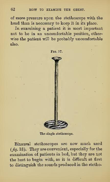
The single stethoscope consists of a cylinder.

THE LUNGS. AUSCULTATION. 61
usually of some tough or light wood, six or eight
inches in length, with a broad flat end on which
to place the ear, and a narrow end to be placed
upon the chest. It is usually perforated by a
hole running from end to end {fig, 17).
In choosing a stethoscope the chief points are
these :—The broad part should be of such a size
and shape that the ear may rest comfortably
upon it. The small end should not be more than
about three quarters of an inch in diameter. It
should have broad, flat, and rounded edges, so
that it may not pinch or cut the skin when it is
placed upon it.
Let us suppose that we are going to examine
the chest with the left ear.
The stethoscope is taken in the right hand near
its small end, and placed upon the part we wish
to examine in such a way that it is everywhere
in close contact with the skin, care being taken
that nothing is touching the stethoscope or
moving upon the walls of the chest. The chest,
if possible, should be bare.
The left hand is laid upon the shoulder or back
of the patient, and the left ear placed upon the
ear-piece of the stethoscope. The right handmay then be taken away, and the stethoscope
will be supported between the ear and the chest.
The hand upon the shoulder will keep the
patient steady, and will prevent the exercising
62 HOW TO EXAMINE THE CHEST.
of more pressure upon tlie stethoscope with the
head than is necessary to keep it in its place.
In examining a patient it is most important
not to be in an uncomfortable position^ other-
wise the patient will be probably uncomfortable
also.
Fig. 17.
The single stethoscope.
Binaural stethoscopes are now much used
[fig. 18). They are convenient,, especially for the
examination of patients in bed/but they are not
the best to begin with^ as it is difficult at first
to distinguish the sounds produced in the stetho-

THE LUNGS. AUSCULTATION. 63
scope; from those produced in tlie lung, and they
have this practical disadvantage, that the chest-
piece must be placed directly upon the skin, for
Fm. 18.
Binaural stethoscopes.
even a covering of thin gauze is sufficient to inter-
fere greatly with the transmission of the sounds.
The differential stethoscope, in which there are
two chest-pieces with separate tubes, is not muchemployed at present, and has, I think, no special

64 HOW TO EXAMINE THE CHEST.
advantage except sometimes for tlie examination
of tlie heart.
The simplest instrument is the best to begin
witb^ and we shall use the single stethoscope
such as is shown in fig. 17.
Every stethoscope has its own intrinsic noises^
which have to be got accustomed to. This is
especially the case with the binaural. A good
auscultator should be equally at home in examin-
ing with either the single or double stethoscope
as well as with the naked ear, for each method
has its own advantages and conveniences.
THE AUSCULTATION OF THE VOICE.
We will deal with the voice sounds first,
because they are less difficult to explain.
Vocal Resonance.
The vibrations of the voice are produced in the
larynx and mouth ; the musical note at the vocal
cords, the words in the mouth and pharynx.
The vibrations are propagated thence in all direc-
tions—outwards through the mouth, and back-
wards along the trachea and bronchial tubes. It
is well to select some simple sound for the patient

THE LUNGS. AUSCULTATION. 65
to produce^ and to use the same always, sucli as
the long vowel ^'ah!'^ or the words '^ninety-
nine/^
When the stethoscope is placed over thelarynx^
and the patient speaks, we hear the voice-sounds
with an intensity which is almost painful. If
the stethoscope be placed lower down upon the
trachea, we hear them less loudly, and over the
alveoli of the lung, though still audible, they are
much diminished in intensity, have lost their
clearness and sharpness, and have become hum-
ming or muffled.
To the vibrations of the voice which we hear
the name Vocal Resonance is given, to distinguish
them from the vibrations which we feel, and
which are called vocal vibrations. Yocal vibra-
tions we feel [Palpation) . Yocal resonance welisten to [Auscultation).
Varieties of Vocal Resonance.
The vocal resonance over the vesicles of the
lung receive the name of pulmonary, muffled, or^
better, Vesicular resonance. Over the larynx it
is called Laryngeal, and over the trachea Tracheal_^
while thatwhich is intermediate between tracheal
and vesicular is called Bronchial, and bears also
the name Bronchophony (phone, voice)
.
5..

6Q HOW TO EXAMINE THE CHEST.
These terms are purely conventional and do
not admit of accurate definition.
The classification is anatomical,, and as the
trachea, for example, passes into the bronchi on
one side and the larynx on the other, so do the
varieties of tracheal resonance pass insensibly
into bronchial or laryngeal.
By laryngeal, tracheal, bronchial, and vesicular
resonance, therefore, is meant resonance of such a
kind as is heard in health over these parts re-
spectively of the respiratory tract, and, when
these terms are used in reference to disease, it is
not meant, that we have necessarily an entirely
new sound such as is never heard in health, but
that sounds, which in health ought only to be
heard in particular places, are in disease heard
somewhere else,where they ought not to be heard=
The sounds of disease are for the most part not
so much abnormal sounds, as normal sounds heard
in abnormal places.
Fortunately it is not the name we give to
these sounds, but the fact itself, which is im-
portant for the purposes of diagnosis.
If, where we should only hear vesicular reson-"
ance, we do not hear it, but some other kind of
resonance, whatever name we call it by, we know
the lung to be in an abnormal condition.
The vibrations of the voice are carried, not by
the walls of the tubes, but by the air within them.

TEE LUNGS. AUSCULTATION. ^1
This is proved by the fact thatj when the column
of air is broken by either a foreign body stick-
ing in a bronchus or by the tubes beiug filled
with mucus^ the vocal resonance is lost in the
corresponding part.
Eveiy time the tubes divide the vibrations
are in part dispersed and lost^ and where the
tubes finally split up into the numerous vesicles of
the lungs, this dispersion becomes so great that
but little of the original vibrations is left to pass
on to the walls of the chest.
How much the alveolar tissue of the lung is
capable of diminishing sound is clearly demon-
strated in the case of emphysema, where the
enlarged lung comes forward and covers up the
heart, muffling the heart-sounds* to such an
extent as to make them almost inaudible.
Hardly anything can muffle sound better than
a pillow, though the actual amount of solid sub-
stance it contains is small. The conditions are
much the same in the lungs, viz. relatively large
air spaces separated from each other by thin,
irregular septa (the feathers)
.
The amount of vocal resonance differs muchin different individuals. This depends in chief
measure :
* It is not uncommon under these circumstances for a dia-
gnosis to be made of a weak heart, though the cause of the
weak heart-sounds lies not in the heart, but in the lungs.
68 HOW TO EXAMINE THE CHEST.
1st. Upon the loudness of the A^oice, i. e. the
amount of sound produced :
2nd. Upon tlie depth of the voice^ for the
deeper the voice is, the coarser are the vibrations;
hence in women and children the vibrations of
the voice are more difficult to hear (as they are
also to feel) than in men :
3rd. Upon the thickness of the walls of the
chest. Thus the vibrations are often absent in
very fat people,, or where the subcutaneous tissue
is oedematous.
When the vocal resonance is listened to over
a portion of consolidated lung, the vibrations are
sometimes carriedwith unusual distinctness along
the stethoscope into the ear, so that not only the
vibrations of the voice, but even the words, are
very distinctly audible. This is called Pectori-
loquy or Direct Vocal Resonance. It is a variety
of bronchophony. In bronchophony there is an
increase in the transmission of the voice or vocal
sounds. In pectoriloquy of the speech or articu-
late sounds.
When the lungs are consolidated, or where
there are large cavities in them, the Heart-
Sounds, like the vocal vibrations, are often carried
with unusual loudness to the ear, and we speak
of them as unduly audible.

THE LUNGS. AUSCULTATION. 69
In cliildren The Cry is as useful as tlie voice in
adultSj and tlie same changes in resonance occur
in it.
Often^ instead of speaking out loudly^ the
patient is made to whisper. The Whisper-Sounds
undergo changes similar to those of the vocal
sounds. They may be diminished or increased.
Thus^ we speak of Bronchial Whisper or Whisper-
ing Bronchophony, of Whispering Pectoriloquy,
and of Cavernous or Amphoric Whisper.
It has been stated that whisper-sounds provide
an easy means of distinguishing between an
effusion of pus and an effusion of serum into the
pleural cavity, the whisper-sound being distinct
when the effusion is serous, and absent when it
is purulent. This is not correct.
When the stethoscope is placed over a part of
the chest where the lung is solid, and the observer
speaks, the resonance of his own voice is some-
times loudly heard in the ear which rests upon
the stethoscope. This has been called Auto-
phony {AutoSj own ; phone, voice) . Its exact
physical causes are not understood, but it is, so
far as is known, only met with over consolidated
lung.
It remains to speak only of one other term often
used, viz. JEgophony. This is a peculiar tremu-
lousness, which is added to the vocal vibrations.

70 HOW TO EXAMINE THE CHEST.
and wliicli lias been compared to the bleating of
a goatj whence the name was taken. It is heard
in cases of moderate pleuritic effusion at the
upper level of the fluid, and usually only poste-
riorly at the inferior angle of the scapula. Occa-
sionally a somewhat similar tremulousness is
heard just above pneumonic consolidation at the
base of the lung.
^gophony is supposed to be due to the irre-
gular conduction of vocal vibrations through
lung tissue which is irregularly compressed and
collapsed, as it lies just above the fluid, ^go-phony is not very common, and when met with
is of little practical importance.
THE SOUNDS OF BREATHINa.
What has been said of the sounds of the voice
is true also to a great extent of the sounds of
breathing".
If the stethoscope be placed upon the larynx
and the patient breathe in and out, sounds will
be heard both on inspiration and on expiration.
These sounds are produced in great measure like
the sounds of the voice by vibrations set up in
and round the glottis, and like them they travel
in all directions, outwards through the mouth
and inwards towards the lungs, and as they

THE LUNGS. AUSCULTATION. 71
travel backwards tliey are progressively dimin-
islied in intensity^ until over the vesicles but
little of tliem is heard.
Now^ if we compare the breathing over the
larynx with that over the vesicles^ for instance^,
at the base of the lung behind^ we shall notice-
that not only is there a difference in loudness^
but that there is also an alteration in character.
At the Larynx the breathing-sounds are clear,
harsh_, and double ; inspiration and expiration
are both loud, and equal in loudness and dura-
tion.
Over the Vesicles the sound is feebler and has
lost its clear character. It has become, like the
voice-sounds, muffled, and instead of two sounds
one only is audible. Expiration is absent, or if
not entirely absent, is only to be heard faintly
and with difficulty.
As in the case of the voice-sounds so with the
breathing-sounds a classification can be madeinto Laryngeal, Tracheal, Bronchial, Vesicular,
and we mean by these terms, when we are
speaking of disease, that the respiratory sounds
audible over the seat of disease resemble the
sounds heard in health either over the larynx^,
trachea, or bronchi, as the case may be.
If we are in doubt what name to give to
a particular kind of breathing we should com-
pare it with that heard over the different parts

72 HOW TO EXAMINE THE CHEST.
Fig. 19.
^hows the position of the trachea and bronchi behind.
The circles mark the spots where bronchial breathing is often
lieard in a healthy chest.

THE LUNGS. AUSCULTATION. 73
of the respiratory tract, and name it accord-
ingly.
The larynx and trachea are superficial and can
be easily reached by the stethoscope. Thebronchi are deeply placed, but bronchial breath-
ing may be heard normally sometimes in front
upon the first piece of the sternum, and behind
either upon the second dorsal spine or right and
left of the spine an inch or two lower in the
interscapular spaces (fig. 19).
Vesicular breathing is soft and muffled in
character, and consists almost entirely of an
inspiratory sound, expiration being either quite
inaudible, or very faint.
The sound is probably made up of two parts.
1. The sound which is conducted along the
bronchi and smaller tubes and modified by having
to pass through the vesicles.
2. A sound produced in the vesicles by the
air as it passes into them from the small air-
tubes.
Whatever explanation is given of its produc-
tion, the sound itself is characteristic. Whatit is, however, cannot be taught by description.
It must be learnt by observation.
Vesicular breathing is, as has been said,
marked off from all other kinds by the absence
or faintness of the expiratory sound.

74 HOW TO EXAMINE THE CHEST.
In laryngeal, tracheal, and bronchial breath-
ing expiration is as loud and long as inspiration,
but in each the sounds have in addition certain
peculiar characters. Thus, in laryngeal breath-
ing it is as if the mouth was in the position to
pronounce the diphthong " Slu/' in tracheal the
vowel ^^ ah/^ and in bronchial the guttural '' ch.^^
. Bronchial breathing is the most important of
all the pathological terms. In it expiration is
as loud and as long as inspiration, and both have
ajeculiar, harsh, grating noise, which is quite
characteristic. No description can convey any
idea of what it is like ; the sound must be heard
to be realized, and once heard will not be easily
forgotten.
Between bronchial breathing and vesicular
breathing come various forms of abnormal
respiration in which expiration is audible, but
in which it is not as long or as loud as inspira-
tion, or if so has not, at any rate, the character-
istic harshness.
A name is required for these varieties of
respiration, which while not vesicular are at the
same time not bronchial, but no name is at
present generally adopted. They might be
termed tubular to indicate that it is the kind of
respiration we might suppose would be heard
over the tubules if we could reach them. Un-

THE LUNGS. AUSCULTATION. 75
fortunately, tubular has been used by some
writers as a general term, to embrace all kinds of
abnormal breathing, and by others to indicate
the same thing as bronchial. Bronchiolar would
be in some respects a better term, but it is not
likely to be adopted. Undefined hreathing'^ was
once suggested, but has fallen out of use.
In the absence of any generally accepted
term to indicate these intermediate forms of
respiration, no choice is left but to describe
what is heard in plain language, and to say that
expiration has such or such a relation in respect
of length and loudness, to inspiration, and that
one or both have such or such a character.
This may be lengthy, but it is not ambiguous.
Much of the confusion in the terminology of
auscultation is due to the use of the term
bronchial by different writers in different senses.
By most teachers and authors bronchial breath-
ing is used to signify a particular kind of
abnormal breathing which is very characteristic,
and in this strict sense it should, in my opinion,
be always and only used. By others, unfortu-
nately, it is used as a general term to mean any
form whatever of breathing which is not normal.
The double use of the term is of course most
perplexing, but by bearing it in mind much con-
fusion will be avoided.
* Unbestimmtes Athmen.

76 HOW TO EXAMINE THE CHEST.
What has been said of tlie vocal resonance
applies with equal force to the breath-sounds^
viz. that none of these technical terms are neces-
sary^ they are only convenient ; and that it is not
the term employed^ but the fact indicated by the
term that is of importance. Moreover^ the
terms used are incapable of exact definition or
description^ for as the trachea passes into the
bronchi^ the bronchi into the tubules, and the
tubules into the vesicles, so will the breathing-
sounds indicated by these terms merge gradually
into one another, and it will be impossible to
agree upon the exact point at which the line
must be drawn in each case between them.
What is meant when these terms are applied
to disease is that over some part of the lung
where, for example, vesicular breathing ought
to be heard, it is no longer heard, but some
other kind of breathing ; and the particular term
used to describe it is taken as indicating roughly
the amount and kind of change which, in our
opinion, the lung has undergone.
Cavernous and amphoric breathing are not
audible anywhere in health. They approximate
in character to the laryngeal sound, hut havejnaddition a greater hollowness, and, in the case
of amphoric breathing, certain superadded semi-
musical (consonant) sounds, such as those heard

THE LUNGS. AUSCULTATION. 77
on breatliing into a jug (amphora), from wMcli
the name is taken. ,
Stridor is a peculiar variety of laryngeal or
tracheal breathing. It is pathognomonic of
narrowing or constriction of some part of these
tubes. It is a loud, harsh, see-saw^ double
grating sound, audible at a distance from the
patient, and associated usually with dyspnoea.
The cough in these cases is also usually stridor-
ous, or as it is sometimes called brassy or metallic.
The constriction is most commonly produced by
the direct pressure upon the trachea of an aneu-
rysm or tumour, but it may have its seat in the
larynx in consequence of local changes there, as
in croup in children, or in stenosis or tumour in
the adult.
Sometimes the breathing-sounds over the
vesicles are normal, so far as their character
goes, i. e. they are vesicular, but their rhythm is
altered. They have become jerky, interrupted
(Saccadee), wavy.
The cause of such an alteration of rhythm
varies. It may be due to irregular action of the
muscles, either in consequence of some change or
defect in the muscle itself, or, as is more common,
in consequence of irregular nervous action
;
hence it is very common in nervous or hysterical

78 HOW TO EXAMINE THE CHEST.
patients. Or its cause may lie in tlie lung itself,
and be due to imperfect and irregular expansion
of tlie lung-tissue ; lience it is often met witli
in tlie early stages of consolidation of the lung.
Sucli alterations in tlie rliytlim of respiration
are usually unsymmetrical and are to be dis-
tinguislied from those symmetrical abnormalities
wMcli are due to irregular respiratory movements
of central origin like sobbings sighing, coughing,
or Cheyne-Stokes breathing (^. v.).
Energetic breathing increases the loudness of
inspiration and makes expiration also audible
over the vesicles. The respiration thus altered
has been named Puerile Breathing because it is
so commonly heard in children, even where the
chest is perfectly healthy. In the adult it is
better called complementary breathing, being
often heard, when a portion of the lungs is
diseased, over the unaffected parts.
USE OF THESE FACTS IN DIAGNOSIS.
Let us now inquire what use we can make of
these facts for the purpose of diagnosis.
The sounds produced at the larynx, whether
by the voice or by breathing, are transmitted, as

THE LUNGS. AUSCULTATION. 79
we have said, almost entirely by the air column
in the trachea, bronchi, and smaller tubes to the
vesicles of the lungs. They then cross the two
layers of the pleura to reach the chest-walls, and
through these are conducted to the ear.
Along their whole course they are progres-
sively diminished;
1st. Every time that the air-tubes divide, and
especially by their terminal expansion into the
vesicles;
2ndly. As the vibrations pass from the vesicles
to the pleura and chest-walls, and thence along
the stethoscope to the ear.
We may regard all these parts, viz. the air-
vesicles, the pleura, and the chest-walls as so manyobstructions placed in the way of the vibrations.
Any alteration in these parts will change the
transmission of the vibrations. It may makethem more easily or less easily heard, and weshall speak then of the voice- or breathing-
sounds.
1. As Increased, i.e. more intense than normal.
2. As Diminished, '\i. e. less intense than
3. As entirely absent, J normal.
And lastly, as Modified (e.g. in segophony).
If, for instance, the walls be very thick, as in
fat persons, or where the subcutaneous tissue is
oedematous, a greater obstruction will be offered
to the vibrations, and they will be diminished.

80 HOW TO EXAMINE THE CHEST.
If we lay outside the walls of the chest several
folds of cloth, we place a fresh obstruction in the
way of the vibrations, and they are therefore
diminished.
If we take a water-cushion and lay it upon the
chest, the vibrations then cease to be audible,
and become completely absent. The effect would
be the same, if we could place the water-cushion
inside the chest-walls^ i. e. in the pleural cavity.
In many forms of disease of the pleura this is
exactly the condition which is produced. Aneffusion of fluid takes place into the pleural sac.
and we have our water-bag inside the chest.
In other cases the pleura itself becomes muchthickened, but in both conditions alike a fresh
obstruction is offered to the passage of vibra-
tions, and they will be either much diminished or
completely absent.
On the other hand^ we have learnt that it is the
breaking up of the air-tubes into the vesicles of
the lung which muffles most the vibrations in
health, for they have to pass through a most
heterogeneous body formed of numerous irre-
gular air chambers separated by thin septa, like
a feather pillow.
Suppose now, that this very heterogeneous
body were made homogeneous by the substitu-
tion for the air in the vesicles of some solid
substance, we should do away at once with what

-THE LUNGS. AUSCULTATION. 81
we know to be one of the cliief obstructions to
tlie passage of vibrations. The other parts then
remaining the same, the vibrations would reach
the chest-walls increased in intensity.
This we find to be the case,, and we have
therefore an easy means of diagnosing between
diseases of the two great structures belonging
to the lung, viz. the lung itself and its pleural
sac, for we have, in affections of the pleura one
obstruction more, and therefore diminished pas-
sage of vibration, and in solidified lung ono
obstruction less, and therefore increased trans-
mission of vibrations.
All that has been just said applies equally to
the vocal resonance, the vocal vibrations, andthe breathing-sounds.
If, then, we find in any part of the chest that
the vocal vibrations, the vocal resonance, and
the breathing-sounds are increased, we may con-
clude that this is due to the lung in that part
having become consolidated. If, on the other
hand, we find the vocal vibrations, vocal reson-
ance, and the breathing-sounds diminished, wemay infer that the pleura is affected. In both
cases alike percussion will be either much im-
paired or absolutely dull, because there is either
but little air-confcaining lung-tissue or none at all
in that part.'":"'"^'~'*' "~
Given then a part of the lung in which the
6
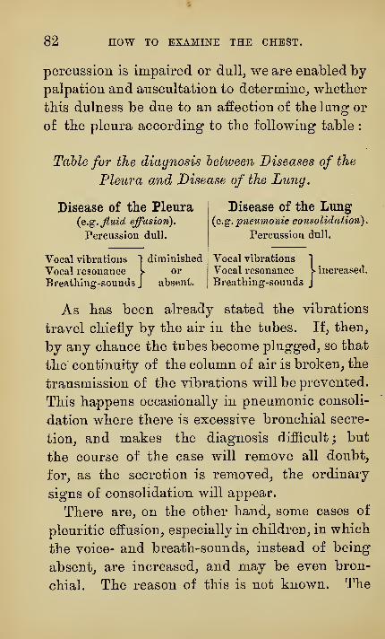
82 HOW TO EXAMINE THE CHEST.
percussion is impaired or dull, we are enabled bypalpation and auscultation to determine, whether
this dulness be due to an affection of the lang or
of the pleura according to the following table ;
Table for the diagnosis between Diseases of the
Pleura and Disease of the Lung.
Disease of the Pleura{e.g. fluid effusion).
Percussion dull.
Disease of the Lung{e.g. pneumonic consolidation).
Percussion dull.
Vocal vibrations "j diminished
Vocal resonance V or
Breathing-sounds J absent.
Vocal vibrations
Vocal resonance >• increased.
Breathing-sounds }
As has been already stated the vibrations
travel chiefly by the air in the tubes. If, then,
by any chance the tubes become plugged, so that
the continuity of the column of air is broken, the
transmission of the vibrations will be prevented.
This happens occasionally in pneumonic consoli-
dation where there is excessive bronchial secre-
tion, and makes the diagnosis difficult ; but
the course of the case will remove all doubt,
for, as the secretion is removed, the ordinary
signs of consolidation will appear.
There are, on the other hand, some cases of
pleuritic effusion, especially in children, in which
the voice- and breath-sounds, instead of being
absent, are increased, and may be even bron-
chial. The reason of this is not known. The

THE LUNGS. AUSCULTATION. 83
fact should be remembered^ but difficulty in
diagnosis is rarely caused by it.
NEW SOUNDS,
We have liitberto spoken of sounds, tbe ana-
logues of most of wMcli may be beard normally
in bealtb. We now come to the class of sounds
which are heard only in disease. These fall into
two groups,
1. Those produced in the lungs, Pulmonary.
2. Those produced in the pleura, Pleural.
To the pulmonary sounds the name Rales was
given by Laennec, who divided them into crepi-
tant, mucous, and sonoro-sibilant ; the two first
are more commonly called Crepitation, and the
latter Rhonchus and Sibilus.
These are the terms which we shall employ.
Crepitation.
Crepitation is produced by the bursting of air-
bubbles in fluid, and it is spoken of as Large,
Medium, or Small, according to the supposed size
of the bubbles which produce it, the larger bub-
bles giving rise to the louder sound.
Excellent crepitation is heard afterwashingthe
hands in warm water, as the little soap-bubbles.

84 HOW TO EXAMINE THE CHEST.
whicli form the lather^ burst. This explanation
has caused crepitation to be often spoken of as
Moist sounds, in contra-distinction to Bhoncbus
and Sibilus, which are called Dry sounds.
One form of crepitation hasprobably a different
explanation from that given above. This is called
Fine Hair Crepitation^ because it sounds like the
crackling of the hair of the head or whisker whenrubbed between the fingers over the ear.
It is probably due to the crackling produced
by the separation of the walls of the vesicles from
each other by inspiration_, when^ as the result of
collapse or pressure, they have been in partial
contact.
Crepitation similar in character^ though prob-
ably not produced in the same way_, is also heard
occasionally over emphysematous lungs.
The noisy bubbling sounds^ which are heard in
the trachea of dying people, and to which the
name of " The Death Rattle '' is given^ are pro-
duced, like the ordinary forms of crepitation, by
the bubbling of air, as it passes through the
mucus which has collected in the trachea.
Rhoiichus and Sihilus.
Rhonchus and Sibilus are sounds often musical
in charact er_, produced by vibrations set up in

THE LUNGS. AUSCULTATION. 85
the air_, as it passes througli tlie broncliial tubes,
tlie mucous membrane of wliicli lias been altered
or roughened by inflammation or secretion.
Rhonchus is produced in the large tubes and
is deeper in tone. It is often described as snor-
ing, cooing, or grating, according to tlie special
characters of the sound.
Sibilus is produced in the small tubes, and is
wheezing and more hissing in character.
One great peculiarity of rhonchus and sibilus
is the rapidway inwhich they alter or occasionally
temporarily disappear, as the mucus which has
caused them is dislodged. A fit of coughing
will often alter the character of the rhonchus
and sibilus, as well as also the places where they
are heard.
Coughing affects also crepitation, occasionally
remoying it, when we may infer that it was pro-
duced by a temporary accumulation of mucus in
a small bronchus, or as is usual rendering it
more evident, and sometimes producing it, where
in ordinary respiration it is not audible. In every
case, where it is at all likely that crepitation may
be produced, the patient should be made to
cough while the chest is being listened to, and
then, either during the cough or, as is more com-
mon, during the deep inspiration which follows
it, crepitation may be heard.

86 HOW TO EXAMINE THE CHEST.
Pleural Friction.
The surfaces of the healthy pleura are smooth,
and move upon each other during respiration
without producing any sound. When the sur-
faces are no longer smooth, but are roughened
by disease, the rubbing of the two rough surfaces
produces a sound, to which the name of Friction
is given.
The character of pleural friction differs muchin different cases. Sometimes it resembles the
creaking of a piece of dry leather when it is
folded. This is called Dry-Leather Creaking'. Atother times it is distinctly rubbing in character.
At other times the sound is hardly a rubbing
sound so much as a minute crackling, audible
usually at the end of inspiration. This it is
often very difficult to distinguish by the sound
alone from the fine small crepitation produced
in the lung.
Characters of the Friction-Sound.
Friction, being produced in the pleura near to
the walls of the chest, will sound " close under
the ear ;'^ as the lungs move to and fro, it will
be '^ double/' being due to the roughening of the
pleura, it will be heard only where the roughen-

THE LUNGS. AUSCULTATION. 87
ing exists, and may therefore be very " limited
in extent ''; and being produced by the move-
ments of the lungs during respiration, it may'^ cease ivhen the breath is held."
Where the pleura is roughened immediately
over the pericardium, the movements of the
heart may be sufficient to produce the rubbing.
This is called Pleuro-Pericardial Friction. Whenas sometimes happens this is not modified bybreathing, it will be difficult to distinguish it
from the friction-sound, produced by a similar
roughening of the pericardium (c/. pericardial
friction).
In inflammation of the pleura, so soon as the
part becomes roughened, a friction-sound is pro-
duced. Soon the two layers are separated byan exudation of fluid, and the sound then dis-
appears. When the inflammation subsides, andthe fluid is reabsorbed, the two layers come once
more into contact, and the friction-sound is
heard again. This is called Redux Friction, i. e.
friction which has come back. The same mayoccur with crepitation in some cases, and then
it is spoken of in like manner as Redux Crepita-
tion.
When from some cause air gets into the
pleural cavity, the case becomes one of Pneumo-
thorax (air in the thorax). The lung then

88 HOW TO EXAMINE THE CHEST.
collapses away from tlie cliest-walls back to-
wards tlie spine round its root_, i. e. the bronclius
and great vessels. As in tlie case of an effusion
of fluid, the lung is separated a long distance
from the walls of the chest, though by air
instead of fluid, and the voice- and breathing-
sounds have to pass through a large air-contain-
ing chamber, before they reach the ribs. In
most cases this has the same effect, as if the
effusion were not air bufc fluid, and the vocal
vibrations, the vocal resonance, and breathing-
sounds are either much diminished or absent.
It might, however, have been expected, that
the air cavity would act like a sounding box, so
that the sounds produced in the bronchi would
be transmitted to the cavity and be thereby
increased in loudness, and being at the same
time altered by consonance become more or less
distinctly amphoric. In some cases this does
actually happen, so that we have two groups of
cases of pneumothorax. In the one the voice-
and breath-sounds are diminished or absent, and
in the other are amphoric in character.
Not only may the breathing- and voice-sounds
become amphoric under these circumstances, but
also crepitation. It has then received the name
of Metallic Crepitation.
Metallic Tinkling has a different explanation.
It is caused by drops of fluid falling from the

THE LUNGS. AUSCULTATION. 89
walls of a cavity into fluid Lelow {gutta cadens),
giving rise to tlie same sound as drops of water
often do, wlien falling in a well or grotto.
Wlien_, over a pneumothorax, a coin is laid
upon tlie chest-wallsj and tapped lightly with
another, while the ear is placed upon the chest,
the sound is often heard like the tapping of a
metal bell or a porcelain dish. To this has been
given the name of Bell-Sound {bruit d'airain) . It
may be audible over any sufl&ciently large cavity.
It is most common, though not constant, in pneu-
mothorax, and in some of these cases ordinary
percussion may be similarly metallic.
When air has been for some time in the
pleura, it is generally associated with an effusion
of fluid, which is sometimes pus and sometimes
serum. The names of Pyopneumothorax {air
and pus in the pleura), and Hydropneumothorax
(serous fluid and air iji the pleura) are then given
to these conditions respectively.
This mixture of fluid and air in a large cavity
gives rise to a new physical sign, which can only
be met with under these conditions {air and
fluid), and which is therefore said to be " patho-
gnomonic.^^ This is Succussion, a splashing
sound, heard when the patient is suddenly
shaken, while the ear is resting upon the chest.

90 HOW TO EXAMINE THE CHEST.
thougli it may sometimes be heard at a distance
from the patient.
Succussion may also be produced in tbe
stomacbj a fact wbicb must be borne in mind
in diagnosing pneumothorax of the left side^
though it can rarely cause any real difficulty.
Theoretically all these sounds, amphoric and
succussion, may be produced quite as well in a
large cavity in the lung as in pneumothorax,
although the cases are in practice rare, in which
confusion would arise. When, however, the
opening into the lung is wide, the physical con-
ditions are almost identical, and the diagnosis
becomes very difficult by auscultation alone.

THE LUNGS. AUSCULTATION. 91
- SYNOPSIS.
On auscultating tlie chest we must proceed
systematically. We must listen :
1. To tlie Voice-Sounds, and determine whether
they are symmetrical_, i. e. whether they are equal
on the two sides ; if there be a difference wemust note it, and ascertain whether it be an
increase on one side, or a diminution on the
other.
2. To the Breathing-Sounds, and ascertain if
they be symmetrical or not; we must listen,
first for the Inspiratory Sound, and next for the
Expiratory Sound; if expiration be audible, wemust note its length, duration, and intensity in
comparison with inspiration, and may express
the changes by the use of such terms as bronchial,
tracheal, &c.
3. We must listen for Superadded Sounds, such
as are never audible in a healthy chest, for Crepi-
tation, Friction, or for sounds characteristic of
large cavities, such as Amphoric or Cavernous
Sounds, Metallic Echo and Tinkling, the Bell-
Sound, Succussion.
92 HOW TO EXAMINE THE CHEST.
CONCLUSION.
The detection of tlie early stages of disease of
tlie lungs is often very difficulty and would be
impossible without careful attention to minute
details.
The normal variations in the physical signs of
tlie chest are so wide^ and the transition between
health and disease so gradual_, tliat no sharp
line of demarcation can be drawn between them.
It is upon a want of symmetry in these physical
signs that we chiefly rely. Most diseases of the
lungs are unsymmetrical, either only one side is
attacked_, or if both, sides are attacked_, they are
rarely affected to the same degree. So that the
physical signs can hardly ever be symmetrical^
and whether we inspect, palpate, percuss, or
auscult, in all cases alike, it is for this want of
symmetry that we are on the watch.
In one place only (with the exception of the
cardiac area) is there normally any want of sym-
metry. This is at the apex. On the right side,
for the reasons already mentioned, percussion is
hardly so resonant, while the vocal vibrations,
the vocal resonance and breathing-sounds are
slightly more intense than on the left side. The

THE LUNGS. AUSCULTATION. 93
difference is only slight^, but tlie same difference
against the left side instead of the right would
be evidence of disease.
Lastly, a diagnosis should never be based upon
a single physical sign. An abnormality in one
respect alone may be physiological. In disease
of the lungs there will certainly be a concurrence
of several physical signs, which separately might
be worth little, but which taken together makethe diagnosis certain.
It is desirable, so far as possible, to represent
all the facts observed in a diagramniatic form.
Dulness may be represented by shading (or
colouring) the corresponding parts of a diagram
of the chest. Vertical lines may be used to indi-
cate that the dulness is due to solid lung, hori-
zontal that it is due to fluid.
Crepitation may be represented by dots or small
circles according to the size (large, medium, or
small).
Cavities by irregular areas, described in the
shaded parts.
Pleuritic Friction by zigzag lines.
Other physical signs by initial letters.
Fl. = Flattening.
Mts. = Movements.
Y. V. = Yocal vibrations.
Y. R. = Yocal resonance.

94 HOW TO EXAMINE THE CHEST,
B-h. = Rlioiiclius.
Sb. = Sibilus.
Pect. = Pectoriloquy,
^g. = ^gopliony.
I. = Inspiration.
B, = Expiration.
Bi. = Respiration.
R. V. = Vesicular. ->.
R. t. = Tubular.
R. br. = Broncliial. ^Respiration.
R. tr, = Tracbeal.
R. c. = Cavernous.
IorE. + . = Prolonged inspiration or expi-
ration.
I or E— . = Deficient ,, ,,
I = E. = Inspiration as long as expiration.
I < E, = J, shorter than „I > E. =
J Jlonger than j.

THE LUNGS. AUSCULTATION. 95
SYNOPSIS OF THE EXAMINYTION OF THE LTJNOS.
In the accompanying table I have given the
chief subdivisions of the examination of the luogs;
the details are given in the text^ and are summa-rised at the end of each chapter.
InsjpGction.
The chest at rest. Its shape.
The chest in motion. The movements of respi-
ration.
Paljpation,
The vocal vibrations.
The sense of resistance.
Abnormal sensations (friction^ &c.).
Percussion.
The normal boundaries of the lungs.
The resonance of the various parts of the
chest.
Auscultation,
The vocal resonance.
The breathing-sounds.
Superadded Sounds.

SECTION II.
THE HEAET,


THE EXAMINATION OF THEHEART.
The examination of tlie liearfc is conducted upoin
the same system as tliat of the lungs.
We consider
:
First—What we can see (Inspection).
Secondly—What we can feel (Palpation).
And lastly—What we can hear (Percussion.
and Auscultation).
100 HOW TO EXAMINE THE CHEST.
mSPECTIOK
Tliat part of the thorax whicli is over the
heart is called the Praecordium or the prascordial
region.
On Inspection we consider
First—its shape
;
Secondly—the movements visible there.
THE SHAPE.
In healthy there is nothing in the shape of the
praecordium to indicate the position of the heart,
and even in many forms of disease there is no
perceptible change in it.
Where, however, the heart is very large, espe-
cially in young people, the praecordium may be
unduly prominent, and it is then said to be
Bulging.
THE MOVEMENTS.
When the heart is healthy, the only movementvisible under ordinary circumstances is that of
THE HEART. INSPECTION. 101
the apex. In the full-grown adult this movement
is seen in the fifth intercostal space, one inch
inside_, and usually about one inch and a half
below the left nipple.
Measurements, however, taken with the nipple-
as a fixed point are unsatisfactory, because the
nipple is not always found in exactly the sam&
place. For this reason it is better to take instead
of the nipple The Nipple Line. This is a line
drawn from the middle point of the clavicle ver-
tically downwards. It passes often through the
nipple.
Position of the Ajpex.
The Apex then is seen in the fifth intercostal
space, one inch inside the nipple line on the left
side.
In health, it is identical with the true ana-
tomical apex of the heart, but in disease this is
often not the case, and the impulse which we see
frequently corresponds with a part of the left
ventricle some distance from the anatomical
apex.
When we speak therefore " clinically " of the
apex, we agree to mean that point on the left
hand side of the chest farthest outwards anddownwards, at which the movement of the heart
can be seen or felt. This is often two or three
inches from the normal place.

102 HOW TO EXAMINE THE CHEST.
No7'mal Peculiarities of the Apex.
In cliildreiij the apex is often normally slightly
"higher and farther out than in adults, and mayfceat in the fourth interspace or under the nipple.
In adults, when lying over on the left side the
apex is often seen in the nipple line. Hence in
fixing the apex, the patient must either stand
erect, or, if lying down, must lie straight upon
the back.
The contraction of the abdominal muscles on
standing up counteracts the tendency of the heart
to fall, so that the apex is not displaced. The
same occurs in the case of the liver, the upper
border of which is if anything, a little higher
when a person is standing up, than when he is
lying down.
Respiration makes no perceptible difference in
the position of the apex.
Displacement of the Apex.
When the apex is not in its normal place, it is
said to be Displaced.
Even a very slight displacement may be of
great importance, for there is hardly any disease
of the heart or pericardium, in which the apex
preserves its normal position.
Thus, in disease of the mitral valve, and in all

THE HEART. INSPECTION. 103
cases of enlargement of tlie riglit side of tlie
hearty tlie apex is displaced outwards {i. e. to
the left side) but it still usually remains in tlie
fifth, space. In disease of the aortic valves^ and
in all cases of enlargement of the left ventricle,
the apex is displaced not only outwards but also
downwards, and is often found in the sixth or
even the seventh space.
In pericarditis what seems to be the apex
beat is often raised so as to be seen in the fourth
space. This is especially the case in children.
Where the heart itself is displaced, the apex
will of course be displaced also.
In some rare cases all the organs are placed
upon the wrong side, e.g. the heart on the right
and the liver on the left. This is spoken of as
Transposition of Viscera. It is a very rare con-
dition.
The Character of the A'pex Beat.
In health this is a simple slight bulging, dueto the contraction of the ventricles, affecting a
circle of about half an inch in diameter, such as
we might expect to produce by tapping with one
finger on the inside of the intercostal space.
In disease the apex beat is frequently altered
in character.
When it is visible over a much larger area

104 HOW TO EXAMINE THE CHEST.
than normal^ it is described as Extended Cardiac
Impulse.
When it is not distinctly localised, it is spoken
of as Diflfased Impulse.
When it is not a simple bulging, but has a
wave-like movement, it is called Undulatory
Impulse.
When the intercostal space corresponding with
the apex is drawn in, instead of bulging, each
time the heart contracts, this alteration is des-
cribed as Systolic Recession of the apex.
SystoHc recession is also occasionally seen in
the second, third, or fourth spaces, but only
when the patient is very thin, and is then com-
monly associated with some contraction of the
upper part of the left lung.
Occasionally an impulse is seen at the apex,
not only when the heart contracts, but also whenit commences to dilate. This is called ^^ Diastolic
Impulse " (or the back stroke of Hope).
Pulsation in Abnormal Places.
In disease impulse synchronous with the move-
ments of the heart is often seen at other parts of
the chest beside the apex
;
1. Below the xiphoid cartilage in the epigas-
trium j this is called Epigastric Pulsation :^
* Pulsation is occasionally observed in the hepatic region

THE HEART. INSPECTION. 105
2. In the second intercostal space, right and
left of the sternum, in relation with the aorta on
the right side, and with the pulmonary artery on
the left j this commonly occurs in cases in which
the lungs are contracted at the apices :
3. Occasionally on the right side in the fourth
or fifth, intercostal spaces, even as far as the right
nipple j this is due to displacement of the heart,
but it is not the apex which beats here, for it is
not a part of the left ventricle, but of the right
;
4. Under the manubrium sterni ; this is gener-
ally due to disease of the great vessels (aneurysm):
5. In the vessels of the neck, either in the
arteries {arterial pulsation) , orin the veins {venous
pulsation) .
Arterial Pulsation is systolic, and due to thevery
forcible projection of blood into the vessels.
Venous Pulsation is usually systolic, but maybe also diastolic.
When the beating of the heart is very forcible,
the whole praecordium is often lifted by it. This
is known as General Heaving.
below the lower ribs on the right side. This usually is asso-
ciated with heart disease, and is called Hepatic Pulsation,

106 HOW TO EXAMINE TEE CHEST.
SYNOPSIS.
On inspection tlien we look for
1st. Changes in the Shape of the Prsecordium.
Bulging
;
2nd. Changes in the Movements,
I. At the Apex,
{a) Change in the position of the
apex beat_,
(6) Change in its character.
Increased,
Extended,
Diffuse,
Undulatory,
Diastolic impulse.
Systolic recession
:
II. In other Places,
Greneral heaving,
Epigastric pulsation.
Pulsation over the aorta or pul-
monary artery.
Pulsation in other parts of thorax,
(Aneurysmal)
.
Pulsation in sides of neck,
(a) Venous.
(b) Arterial.
Pulsation in the hepatic region.

THE HEART. PALPATION. 107
PALPATION.
As in tlie case of tlie examination of tlie lungs,
speaking generally, all that can be seen can be
also felt, both as regards the shape of the prse-
cordium and the movements of the heart.
The movements are often more easily recog-
nised by the hand than by the eye, and the
position of the apex is always finally fixed by
palpation.
To Fix the Apex,
This should be done with the tip of one finger,
the object being to find out the exact point
farthest outwards and downwards at which the
heart can be felt beating.
Abnormal Sensations.
Next the whole hand should be placed upon
the prsecordium, first below and then above, in
order to detect either undue movement in this
region, or abnormal sensations such as do not
occur when the heart is healthy.
The most peculiar of these abnormal sensations

108 HOW TO EXAMINE THE CHEST.
are described as Thrills. They are due to coarse
vibrations, set up by eddy-like currents in the
bloody in consequence usually of some obstruction
to the course of its current.
Thrills may be systolic or diastolic, i. e. mayoccur when the ventricle is contracting or
dilating.
Of all thrills the most striking occurs at the
apex, and has been aptly compared to the sensa-
tion felt when the hand is placed upon the back
of a cat purring, and from this it has received the
name of Purring Tremor or Fremissement Cataire.
This form of thrill occurs immediately before
the contraction of the ventricle, and is there-
fore called prsesystolic.
Thrills are also sometimes felt in the second
or third intercostal spaces on the right or left
side. They are most common on the right side,
and are connected with affections of the aortic
valves. Occasionally the vibrations occur at
such regular intervals as to produce a musical
note. To this reference will be made again
under auscultation.
When the two layers of the pericardium are
roughened, the rubbing of the two surfaces
together may be easily heard, but it is some-
times also felt. It is called Pericardial Friction
or Friction Rub.
Thrills, as well as also friction, are sometimes

THE HEART. PALPATION. 109
felt under tlie manubrium sterni. In these cases
they are due usually to aneurysm of tlie arcli of
tlie aorta.
In the neck, thrills may be felt both in the
arteries and veins. In the arteries, they are
usually propagated from some point below,
generally from the aortic valves. In the veins,
they are commonest in cases of extreme ansemia,
e. g. chlorosis.
When the valves of the pulmonary artery close
forcibly in thin people, their closure may be fre-
quently felt by the hand as a sharp flap or shock
in the second left intercostal space. This is
often to be seen, as well as to be felt, and has
already been referred to under Inspection. It
has received the name of Valve Shock.
SYNOPSIS.
Palpation enables us to confirm most of the
facts recognised on inspection, and further to
ascertain the occurrence
1. Of Thrills,
{a) Systolic,
(6) Diastolic,
(c) Presystolic
;
2. Of Valve Shock;
8. Of Friction.
110 HOW TO EXAMINE THE CHEST.
PEECUSSION.
The Size of the Heart.
How Large is the Heart ? As witli the lungs^
so with the hearty this is naturally the first
important qiiestion_, and it can only be answered
by means of percussion.
It has been already stated_, that part of the
heart is uncovered by the left lung, and that this
part lies just above the diaphragm to the left of
the middle line of the sternum (fig. 20). If we
percuss this part we obtain a non-resonant sounds
for the reason that there is no air-containing
lung beneath.
The Cardiac Area is^ roughly speaking, a tri-
angular space. To describe a triangle three
points are required, and these three points are
:
1. The apex, which is to be found in the fifth
intercostal space one inch inside the left nipple
line j
2. The junction of the fourth left costal car-
tilage with the sternum
;
3. The bottom of the sternum at its junction
with the xiphoid cartilage.

THE HEART. PEECUSSION. Ml
Fig. 20.
Diagram showing the real size of the heart, and the size of
the absolute cardiac dulness.
1, 2, 3, are the three points described in the text.
112 HOW TO EXAMINE THE CHEST.
The area of cardiac dulness may be then
described by drawing
1. One line (the Upper Boundary) slightly
curved outwards from the apex to the junction
of the fourth costal cartilage with the sternum
on the left side
;
2. A second (the Lower Boundary) from the
apex to the base of the xiphoid cartilage ; this
is nearly horizontal ;
3. A third (the Right Boundary) from the base
of the xiphoid cartilage vertically upwards along
the left border of the sternum to the fourth
costal cartilage.
Within this area the percussion-sound will be
dullj or^ if not absolutely dull_, will be much im-
paired. This area is called the area of Absolute
—or Superficial—Cardiac Dulness. It does not,
of course, represent the real size of the heart,
which we know occupies a much larger space in
the thorax {fig. 20). If we percuss forcibly
outside the upper boundary, we obtain a note,
which, though more resonant than within the
area described, is still less resonant than the note
in other parts of the chest, for by forcible per-
cussion we throw into vibration the deeper parts
of the chest, and these are, of course, less
resonant where the heart is, than where there is
nothing but lung-tissue. This increased area of
impaired resonance is called the Deep—or Rela-
THE HEAET. PEECUSSION. IIB
tive—Cardiac Dulness. Et extends about three
quarters of an incli beyond tlie upper border of
tlie area of absolute dulness along the fourth
ribj and curves down to fuse with it at the apex
{fig. 20).
The Lower Boundary cannot be determined bypercussion^ for the heart lies here upon the
diaphragm, and the diaphragm upon the liver^
and both the heart and the liver give a dull note
to percussion. This boundary may be, however,,
accurately represented by drawing a horizontal
line from the apex of the heart to the base of
the xiphoid cartilage. Tbis line is in direct
continuation with the upper border of the area
of hepatic dulness described previously.
The right border of the heart cannot be shownby percussion to extend beyond the left edge of
the sternum, and for this reason. The sternum
is a solid body, lying above and to the right side
immediately upon, the lungs. When it is per-
cussed we obtain vibrations, not only from the
parts immediately beneath, but also by conduc-
tion from the lungs in relation with it at a
distance from the part percussed, and the sound
is then resonant, even on parts of the sternum
under which no lung lies.
When from any cause, as for instance byenlargement of the right side of the heart, the
lung is pushed away from the sternum, the
8

114 HOW TO EXAMINE THE CHEST.
resonance over it will become impaired^ or even
absolutely dull.
If tbe lungs and tbe pleura are normal, any
increase in tbe size of tbe beart is attended by a
correlative increase in tbe size of tbe cardiac
dulness, but, if tbe lungs and pleura be not
bealtby, a difficulty may arise in one of three
ways.
1. If tbe lungs be too large, as in empbysema,
tbey will encroacb upon tbe cardiac area, and
cover up tbe beart, eitber wbolly or in part, and
tbe cardiac dulness will then be eitber wbolly
absent, or be mucb reduced in size [fig. 12).
2. If tbe lungs be contracted, as tbey dften are
in cbronic affections, more of tbe beart will be
exposed, and tbe cardiac dulness will be propor-
tionately increased {fig. 13).
3. Lastly, if tbe lungs be consolidated, or tbe
pleura be diseased, in tbose parts wbicb are in
immediate relation witb tbe beart, we sball obtain
on percussion a dull note, as over tbe beart itself,
and our means of determining tbe beart^s dulness
will be lost.
Alterations in the Cardiac Dulness.
Diseases of tbe beart, in almost every case
except atropby,—and tbis cannot be clinically
demonstrated— are attended by increase in

THE HEART. PEECUSSION. 115
Fig. 21.
Diagram showing the shape of the absolute cardiac dulness in
aortic disease (enlargement of the left ventricle).
Apex displaced downwards and outwards. The white dotted
line shows the normal area of absolute cardiac dulness.

116 HOW TO EXAMINE THE CHEST.
Fig. 22.
©
Diagram showing the shape of the absolute cardiac dulness
in mitral disease (enlargement of the left auricle and of the
right side of the heart).
Apex displaced outwards and not downwards. Great in-
crease of dulness to the right.
The white dotted line shows the normal area of absolute
cardiac dulness.

THE HEART. PERCUSSION. 117
Fig. 23.
Diagram showing the shape of the absolute cardiac dulness
in a case of large pericardial effusion.
The white dotted line indicates the normal area of absolute
cardiac dulness.

118 HOW TO EXAMINE THE CHEST.
size^ and therefore by increase in tlie size of tlie
cardiac dulness. This is easily determined unless
the lungs and pleura be also affected.
When the cardiac dulness is increased^ its shape
varies much^ according to the kind of disease with
which the heart is affected.
The shape may remain triangular and be simply
enlarged. This is the rule in all cases of hyper-
trophy of the left ventricle, of which aortic disease
may be taken as the type {fig. 21).
Or, it may cease to be triangular, and become
either irregularly quadrilateral, as in mitral
disease {fig. 22), or roughly globular, as in cases
of great general enlargement or of pericarditis
(fig.n).Lastly, the cardiac dulness will be displaced
when the heart is displaced, or be altered in size
and shape in consequence of deformities in the
thorax.
SYNOPSIS.
By percussion, then,
1. We determine the size of the cardiac
dulness,
And 2. Ascertain any alterations in its size, shape,
or position.

THE HEART. AUSGDLTATION. 119
AUSCULTATION.
When the ear is placed upon the cardiac
region of a healthy man, the two sounds of thte
heart become audible. They differ somewhat in
character, and have been compared to the twa
syllables 'Mub" and ^^dup/' the first being longer
and heavier (lub), the second shorter and sharper
(dup).
The heart, we know, is in constant movement,
either contracting or dilating, and this is true of
the auricles as well as the ventricles, but, inas-
much as it is the contraction of the ventricles
which is the most important, we agree, when the
terms contraction and dilatation are used, to refer
to what the ventricles are doing, unless we dis-
tinctly specify some other part.
The ventricle then is continually moving, never
absolutely at rest, but always either contracting
or dilating. Directly contraction ends, dilatation
begins, and vice versa.
We may represent the hearths action diagram-
matically in the form of a circle {fig. 24), once
round the circle representing one complete cycle
of the heart's action.
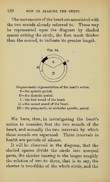
120 HOW TO EXAMINE THE CHEST.
The movements of the heart are associatedwith
the two sounds already referred to. These maybe represented upon the diagram by shaded
spaces cutting the circle^ the first much thicker
±hau the second, to indicate its greater length.
Diagrammatic representation of the heart's action.
S= the systolic period.
D= the diastolic period.
l= the first sound of the heart.
11 = the second sound of the heart.
111= the prsesystolic, or auricular systolic, period.
We have, then, in investigating the hearths
action to consider, first the two sounds of the
heart, and secondly the two intervals by which
these sounds are separated. These intervals in
health are periods of silence.
It will be observed in the diagram, that the
shaded spaces divide the circle into unequal
parts, the shorter bearing to the longer roughly
the relation of two to three, that is to say, the
shorter is two-fifths of the whole circle, and the

THE HEART. AUSCULTATION. 121
longer three-fiftlis. This is approximately tlie
relation, wliicli has been found by measurement
to exist, between the length of the period of con-
traction and of the period of dilatation.
To the shorter of these periods, the period of
contraction, the name Systole is given, and to the
longer, the period of dilatation^ the name Diastole.
Systole and diastole are therefore periods of time,
and, though they stand in a certain relation to the
sounds of the heart, are to be carefully distin-
guished from them. We have therefore, whenlistening to the heart, to observe, first the two
heart- sounds, and secondly the two periods of
silence.
The Sounds of the Heart.
The chief cause of the second sound of the
heart is the closure of the semilunar valves in the
aorta and in the pulmonary artery.
The first sound is more complex, and although
it is due in part to the closure of the auriculo-
ventricular valves, other factors appear to be
necessary for its normal production, such as a
healthy condition of the muscular tissue of the
heart and of the blood.
As the result of observation of the heart-sounds
in disease, it is probable that, even for the second
sound, the simple theory of its production by the

122 HOW TO EXAMINE THE CHEST.
closiDg of valves does not by itself afford a suffi-
cient explanation in all cases.
The valves_, liowever_, play so mucli tlie most
important part in tlie production of tlie sounds
of tlie lieart_, that it will not be necessary in this
place to consider the other possible causes^ but
we may assume for our present purpose_, that the
heart-sounds are due in all cases alike to the
closure of valves.
The first sound is long, rather dragging, and
well represented by the syllable ^^ lub." This is
indicated upon the diagram by a broad shaded
space. The second sound is flapping in character,
shorter, and sharper, and is well represented by
the syllable " dup." It is indicated upon the
diagram by a narrow shaded space.
We have no means at present of measuring the
length of the heart-sounds. Tlie figure given
is diagrammatic^ and indicates their duration
only approximately.
The first sound of the heart is produced, whenthe ventricle begins to contract, and marks there-
fore the commencement of the period of systole.
The second sound is produced, when the ventricle
begins to dilate, and marks therefore the com-
mencement of the period of diastole. Systole
therefore extends from the commencement of the
first sound up to the second; diastole from the
second up to the first.

THE HEART. AUSCULTATION. 123
The first sound is sometimes spoken of as tlie
systolic sound^ and tlie second as the diastolic
sound. These terms are confusing and should
not be used.
The characters of the sounds in health are sufii-
ciently distinct^ so that there is little risk of
mistaking them, but this is not so in disease, and
it becomes necessary to have some means of
determining, which sound it is that we hear, or
if, as occurs in many cases, the sounds be absent,
to determine when the heart is contracting, and
when it is dilating, i. e. " To time the Heart.^^
This is easily done by means of the pulse.
The pulse beat is due to the sudden driving of
blood into the vessels by the contraction of the
left ventricle. Near the heart the pulse beat
and the systole of the ventricle are practi-
cally synchronous, but at a distance from the
heart this is not so, for the wave is delayed in
reaching a distant vessel, as for instance the
radial at the wrist, and in disease this retarda-
tion is often considerable. Some vessel must
therefore be selected as near as possible to the
heart. The most suitable for the purpose is the
carotid.
This will be conveniently reached by grasping
the side of the neck with one hand, the fingers
being behind, and by pressing the thumb down in
124 HOW TO EXAMINE THE CHEST.
front between the sterno-mastoid muscle and tlie
larynx or trachea. The carotid will then be felt
pulsating beneath the thumb.
The pulse wave in the carotid is a long one^
and lasts as long as the period of contraction.
Reduplication and Accentuation of Sounds.
The heart is a complex organ formed of manyparts. It might be supposed, then, that we should
get many sounds instead of only two, and yet in
health the two sides of the heart act in such per-
fect harmony together, that theyproduce only one
first sound and only one second sound. This is by
no means so in disease, for, when this accurate
adjustment is disturbed, the two sides cease to
act synchronously together, and the sounds
become double, or as they are called Reduplicated.
Instead of a clear ^'^lub-dup, lub-dup,^^ we hear
as it were ^^ lur-rub-dur-rup, lur-rub-dur-rup."
This has also been called Cantering [Bruit de
Galop)
.
Either sound may be reduplicated, though it is
more commonly the second.
Sometimes, where the want of harmony is not
so marked as this, the relative loudness of the
sounds on the two sides of the heart may be
altered. These louder sounds are called Accen-
tuated.
THE HEART, AUSCULTATION. 125
Murmurs.
We have stated_, that in a healthy heart we hear
two sounds onlj^ and that thej are separated by
two periods of silence. In disease we often hear
during these periods of silence a fresh sounds and
this we agree to call a murmur.
A Murmur may be defined to be a sounds pro-
duced by the movements of the heart, occurring
in those intervals, during which in health no
sound is audible.
Murmurs must not be confounded with heart-
sounds, nor must they be spoken of as simply
replacing the heart-sounds. They may, it is true,
replace the heart-sounds, but then they always
do something more, and the two things are essen-
tially distinct. Murmurs may be heard, when
both the sounds are present, or when one of the
sounds is absent, or even both of them, and
lastly, the heart-sounds may be modified, or even
absent, without any murmur at all.
The terms, employed to describe the characters
of murmurs, are those in ordinary use, such
as soft, blowingJ
loud, faint, harsh, grating,
rasping, 8fc.
Some are musical in character, and are then
spoken of as musical murmurs. Commonly deep
in tone, but occasionally high pitched, such
murmurs will be described as cooing, piping,

126 HOW TO EXAMINE THE CHEST.
lohistling, &c., according to tlie kind of musical
sound produced. They frequently change their
tone and character, and occasionally the musical
part may even for a time disappear. These
variations depend upon varying conditions of
the circulation.
The vibrations, which produce these musical
murmurs, are often also easily felt, and give rise
to some of the more striking forms of thrills.
Heart murmurs, even when distinctly musical,
are not often heard by the patient, however loud
they may appear to the person auscultating.
Classification of Muvinurs.
Cardiac murmurs are divided into two groups,
according as they are produced inside, or outside
the heart, and they are named accordingly Endo-
cardial and Exocardial.
The exocardial murmurs will be discussed
later. Of the endocardial murmurs there are two
classes,
I. Those which depend upon some pathological
change in the heart, and which are therefore
called Organic;
II. Those, in which no such change exists,
and which are therefore called Inorganic.
Murmurs may also be classified according to
the movements of the heart.

THE HEAET. AUSCULTATION. 127
The heart is always either contracting or dila-
ting, and murmurs_, therefore, fall naturally into
two groups,
I. Those which occur during the period of
contraction (systole) ; these are called Systolic
murmurs;
II. Those which occur during the period of
dilatation (diastole) ; these are called Diastolic
murmurs.
Sow to Time Murmurs.
We are able, as we have seen, to determine,
by feeling the pulse, the time at which the left
ventricle contracts.
If, then, we place the ear upon the chest andthe thumb or the finger upon the carotid, and
hear a murmur, we know, should the murmurcome at the same time as the pulse beat, that it
occurs when the ventricle is contracting, and
that it, therefore, is a systolic murmur. On the
other hand, should the murmur not come at the
same time as the pulse beat, we know that it is
produced when the ventricle is not contracting,
but dilating, i. e. during diastole, and that the
murmur is therefore diastolic.
We may indicate the murmurs which we hear
upon the diagrams, by shading lightly (or in a
different colour) the spaces left between the
darker shadings, which represent the heart-

128 HOW TO EXAMINE THE CHEST.
sounds^ varying tlie deptli of shading witL. tlie
intensity of the murmur. Tiius Jig. 25, repre-
sents a systolic murmur. The shading runs
directly into that corresponding with the first
sound. This indicates, that the murmur in this
casewas continuous with the first sound, and could
not be separated from it. The shading is darkest
towards the first sound, and becomes lighter
towards the end of systole. This indicates, that
Fia. 25.
Diagram of a systolic murmur with botli heart-souuds
audible.
The gradually decreasing intensity of the murmur is indi-
cated by lighter shading.
the murmur was loudest at the commencement,
and that it gradually became fainter towards the
end, of systole.
In a similar way most varieties of murmurs
may be diagrammatically represented.
THE HEART. AUSCULTATION. 129
Subdivision of Endocardial Murmurs.
Systole is a mucli shorter period of time than
diastole^ and we do not find it possible to sub-
divide tlie class of Systolic Murmurs. We con-
tent ourselves with speaking of them as Long and
Short.
Diastole has a longer period and^ moreover,
when change occurs in the rate of beating of the
hearty it is at the expense of the diastole that
this chiefly takes place.
Diastolic Murmurs are easily subdivided into
groups^ according as they occur at the commence-
ment, in the middle, or at the end of the diastolic
period, and have been named accordingly early,
middle, and late diastolic murmurs.
The murmur at the end of Diastole, late Dias-
tolic, gradually increases in intensity up to the
first sound, and is terminated abruptly by it
;
it comes immediately before the systole, or, if
we have one finger upon the carotid, immediately
before the pulse beat there. It is, therefore,
usually called a Praesystolic murmur (before the
systole) {fig. 26). It is frequently associated
with the peculiar purring thrill previously de-
scribed.
A prassystolic murmur may be long or short.
The short occupies aboat the last third of dias-
tole, and is the same as the late diastolic {fig. 26)
.
9

130 HOW TO EXAMINE THE CHEST.
The long occupies tlie last two-thirds of diastole,
and is equivalent to the mid plus the late dias-
tolic [fig, 27).
Fig. 26.
Diagram of a short prsesystolic murraur (auricular systolic
—
late diastolic). At tlie apex the first sound is longer than
normal, and the second sound is absent.
The murmur increases in intensity up to the first sound, and
is abruptly terminated by it.
Fig. 27.
Diagram of a long prsesystolic murmur (late + mid-
diastolic).
Now, what is happening in tlie heart at the
time at which this murmur is produced ?
We know that immediately before the ven-
tricle contracts, the auricle contracts, so as to

THE HEART. AUSCULTATION. 131
ensure the complete filling of the ventricle with,
blood. But this is just the time at which the
murmur is heard. It has for this reason also
received the name of Auricular Systolic^ by which
Fig. 28.
Diagram of a sliort postsystolic murmur (early diastolic),
immediately following and continuous with the second sound,
which is still audible.
Fig. 29.
Diagram of an ordinary diastolic murmur (long postsystolic
or early + mid-diastolic).
The second sound is lost in the murmur.
is meant_, that it is due to the contraction of the
auricle. It is better^ however^ to use the more
common term_, and call it Prsesystolic.

132 HOW TO EXAMINE THE CHEST.
Diastolic murmurs heard at the commencement
of Diastole may be spoken of as Long or Short.
The very short murmur of this kind^ which
occurs immediately after the pulse beat^ or if the
second sound be present, immediately after the
Fig. 30.
Diagram of a double murmur (systolic and long postsvs-
tolic).
Both sounds are lost in the murmurs.
Fig. 31.
Diagi'am of a double murmur (prsesystolic and systolic).
The first sound is audible and the second absent.
second sound, has received the name of Post-
systolic {after the systole) {jig. 28).
The most common kind is usually described as

THE HEAET. AUSCULTATION. 133
Diastolic^ or^ in order to distinguisli it from tlie
praesystolic murmur^ Ordinary Diastolic. ThiSj
liowever_, often leads to confusion and seems to
imply, tliat a prassystolic murmur is not diastolic,
as it clearly is.
It would be better, I think, to speak of all those
murmurs which, occur at the commencement of
diastole as Postsystolic, and subdivide them into
Long and Short, the long answering to what
is usually called the ordinary diastolic, the
short to what is more commonly called the
postsystolic.
The mid-diastolic murmur, i. e. the murmurwhich is heard only in the middle portion of
the diastolic period and has a distinct interval
before and after separating it from the heart-
FiG. 32.
Diagram of mid-diastolic murmur.
sound {fig. 32), is the rarest of all the murmurs.
It is I believe met with only under the same con-
ditions, as give rise to the prgesystolic murmur.
134 HOW TO EXAMINE THE CHEST.
The murmurs may be tabulated tbus
Murmurs <
Systolic {^^'°^^-
pearly 1 = short|p„^^^y^^„,,^_
Diastolic <{ middle:: = Mid-diastolic.
> =long "1^ Prsesystolic or
l^late J = short J Auricular systolic.
The Cause of Endocardial Murmurs.
Murmurs in the heart (or vessels) are due to
eddies set up in the blood. These will be pro-
duced_, whenever a current of fluid has to pass
through a narrow aperture into a larger space^
the eddies thus set up generating a sound.
It might be supposed_, with suchan explanation,
that murmurs would be constantly produced,
even in a healthy heart, inasmuch as the blood
has to pass through the narrow valvular orifices
into the larger cavities of the ventricles or vessels,
but, so long as the normal relation exists between
the size of the orifices and that of the cavities or
vessels, we know that murmurs are not heard
;
so soon, however, as this normal relation is dis-
turbed, murmurs are likely to arise.
Theoretically they may depend upon changes of
two kinds : first, dilatation of the cavities into
which the blood is passing, the orifices being
unaltered; or, secondly, alteration in the condition

THE HEART. AUSCULTATION. 135
of these orifices^ the cavities remaining normal.
These conditions are usually associated. We meet
with both kinds of murmurs clinically, but inas-
much as it is most common to find, when endo-
cardial murmurs have been heard^ that the
orifices are altered, we will for the present con-
fine our attention to those murmurs which are
due to valvular lesions, and consider the means
we have of determining at which orifice of the
heart the disease exists.
ENDOOAEDIAL MUEMURS DUE TO VALVULAR
DISEASE,
The Place of Murmurs.
Murmurs^re transmitted in the direction in
which the blood is passing at the time of their
production. Let us take an instance. If a
murmur is produced at the aortic orifice at the
time when the blood is being driven through it
by the contraction of the ventricle, the murmurwill be carried along the aorta upwards, and will
be heard some distance above the valves. In the
same way, if the murmur is produced when the
heart is dilating, the valves being incompetent,
and allowing a stream of blood to pass back
through them into the ventricle, the murmurs

136 HOW TO EXAMINE THE CHEST.
¥ia. 33.
Diagram of the heart, showing the anatomical position of the
valves, the axes of the aorta and pulmonary artery, and the
places where to put the stethoscope when examining the dif-
ferent orifices of the heart.
The dark lines A, P, M, te, indicate the actual position of the
aortic, pulmonary, mitral, and tricuspid valves respectively
;
The thin lines the axes of the aortic and pulmonary orifices;
The circles, the places upon which to put the stethoscope,
when examining the corresponding orifices.

THE HEART. AUSCULTATION. 137
will be heard below the valves, that is to say, on
the ventricular side of them. Murmurs have,
therefore, been spoken of as Onward and Back-
ward murmurs, that is, murmurs which are pro-
pagated onwards, in the direction of the normal
current, or backwards, contrary to the normal
current. This fact is of the greatest importance,
because it gives us the means of fixing with
certainty the place at which a given murmur is
produced.
We require to know first the normal position
of the valves of the heart.
The Position of the Valves,
Anatomically they lie very close together, as
is shown in the diagram [fig. 33), so that if the
mouth of a stethoscope were placed at the junc-
tion of the third left costal cartilage with the
sternum at its upper border, it would cover the
middle points of three out of the four valves,
the one not covered being the tricuspid.
At this point, then, it would be impossible to
separate the murmurs with certainty, for the pul-
monary valve lies immediately above the aortic,
and the mitral behind and a little to the left of
them both.
If the left hand be loosely clenched, and placed
upon the chest, so that the knuckles of the middle

138 HOW TO EXAMINE THE CHEST.
Fig. 34.
Diagram of the left hand placed upon the chest to repi'esent
the size of the heart and the position of its various parts.
Dotted lines indicate the size of the heart (j%. 20) and of
the absolute cardiac dulness.
For description see text.

THE HEART. AUSCULTATION. 139
phalaDges of the fingers rest upon the sternum^
we shall obtain a rough estimate of the size of
the heart and of the relative position of its dif-
ferent parts in that individual, as the diagram
shows {fig- 34).
The row of knuckles indicates the line of the
tricuspid valve, and what is seen of the fingers
towards the right represents the right auricle.
A line, drawn from the knuckle of the first finger
to the carpal end of the ulna, divides the back of
the hand into two unequal parts ; the larger and
lower represents the anterior surface of the right
ventricle, the upper and smaller that of the left
ventricle, while the line itself corresponds with
the septum. The knuckle of the thumb marksthe position of the left auricle. If the thumb be
now straightened, it will indicate the direction
which the aorta takes in the first part of its
course; and if a pencil be grasped by the hand,
as in the diagram, it will cross the thumb, and
lie in a similar position, directed towards the
opposite side, and will represent the direction
which the pulmonary artery takes in the first
part of its course.
The Axes of the Heart.
We may draw two lines, one in the direction of
the thumb and the other of the pencil, and these
we may speak of as the Axes, or lines of direction.

140 HOW TO EXAMINE THE CHEST.
of the aorta and of tlie pulmonary artery re-
spectively.
Now, it is along tliese axes, onwards or back-
wards, as the case may be, that murmurs pro-
duced at these orifices will be propagated.
By observing, then, the direction in which a
murmur travels, we are enabled to say with accu-
racy at which orifice the murmur is produced.
We may define the Axis of the Aorta to be a
line drawn from the apex of the heart to the right
sterno-clayicular articulation, and the Axis of the
Pulmonary Artery to be a line drawn from the
middle of the xiphoid cartilage to the left sterno-
clavicular articulation [fig. 33).
These lines it will be observed, cross the end
of the second costal cartilage close to the sternum,
and if we place the stethoscope upon them in the
second intercostal space, right or left of the
sternum, we have placed it upon points respec-
tively nearest to one of the valves, and at the
same time farthest from the other. We say, there-
fore, that in order to examine the condition of the
aortic valves, we place the stethoscope, in the posi-
tion indicated (a) in the diagram, in the second
right intercostal space, and of the pulmonary
valves (p) in the second left intercostal space.
We will now consider more particularly the
aortic murmurs.
THE HEART. AUSCULTATION. 141
Systolic aortic murmurs, tliat is to say, mur-
murs whicli are produced at the aortic orifice,
when the blood is being driven out of the left
ventricle, will travel in the axis of the aorta
upwards towards the right sterno-clavicular joint,
and will be heard above the valves here or even
further in the course of the large arteries. Dia-
stolic aortic murmurs, on the other hand, which
are produced by the blood passing back again
into the ventricle, will be propagated in exactly
the opposite directiou, that is to say, towards
the apex of the heart. They are often trans-
mitted with great intensity along the whole
length of the sternum, even as far as the xiphoid
cartilage.
Where these murmurs are very faint, they are
often not intense enough to reach the apex, and
may then be heard at a point in the axis of the
aorta short of the apex, the commonest place being
on the level of the fourth left costal cartilage.
Sometimes it is only in this place that these
murmurs are audible. /.
What has been said of the aorta is true,
mutatis mutandis, also of the pulmonary artery.
In the case of the mitral valve the difl&culties
are greater. The line of direction or Axis of the
Mitral orifice may be roughly taken to be a line
drawn from the apex of the heart to the left

142 HOW TO EXAMINE THE CHEST.
interscapular space posterioi^ly_, on a level witli
the sixth, dorsal vertebra. If a long skewer
were taken, and thrust into the chest at the apex
of the heart_, and brought out at the point men-
tioned behind, it would represent the axis of
the mitral orifice, and give therefore the line of
direction, which the blood follows in passing
from the left auricle into the left ventricle.
If, then, a murmur is produced at the orifice
when the ventricle is contracting, it would be
caused by a stream of blood passing backwards
from the left ventricle into the left auricle in a
direction contrary to the normal current of
blood. We should expect, therefore^ the ven-
tricle being in front and the auricle behind, to
hear such murmurs posteriorly in the left inter-
scapular space.
If, however, the murmur is produced when
the ventricle is dilating, it would be caused by a
stream of blood passing in the opposite direc-
tion, that is, in the same direction as the normal
blood current, and we should expect not to hear
it behind.
Now this is just what occurs. Both kinds of
murmurs are audible at the apex, but the systolic
only behind.
The mitral valve, however, lies so deep down,
that the points along the axis, which we can
reach, nearest to the valve itself are the apex of

THE HEAET. AUSCULTATION. 143
the heart in fronts where the left ventricle be-
comes superficial, and the left interscapular space
behind ; and these are the spots we choose to
listen at, when we wish to examine the condition
of the mitral valve.
The fourth valve, the Tricuspid, lies superficial.
Murmurs produced at this orifice we hear best
immediately over the valve, that is, along the
lower part of the sternum.
IThe places, then, at which we listen for
. murmurs at the different orifices are
1. The aortic, in the second right inte^^costal
space close to the sternum
;
2. The pulmonary, in the second left inter-
costal space close to the sternum
;
3. The mitral, at the apex of the heart;
4. The tricuspid, at the bottom of the sternum.
But it is not sufficient to note the presence
only of a murmur at these places. We must
also carefully trace the direction in which it is
propagated, and determine especially the spot
at which it is heard loudest, i. e. its place of
maximum intensity.
The Diagnosis of Valvular Diseases.
Now, at each of these places we have to
consider, as has already been stated, first, the

144 HOW TO EXAMINE THE CHEST.
two sounds of tlie heart, and, secondly, any
inurmurs wMcli are heard between them, and wemay therefore construct for each of these spaces
a diagram similar to that already described.
The heart being thus a complex organ, all the
various parts of which stand in such close rela-
tion in action to each other^ we should expect to
find, that it would be almost impossible to meet
with any change in one part, which was not
associated with correlative changes in other
parts. Hence the rule to examine each and
every part of the heart, and to note carefully
every one of the changes there detected, before
venturing to make a diagnosis of any disease.
We have therefore four places to listen at,
and at each of these places we have two sounds
and two intervals to consider.
The sounds may be both present, or one or
both absent. They may be accentuated or
reduplicated.
If murmurs be present, we have then to deter-
mine to which of the intervals they belong, i. e.
whether they be systolic or diastolic ; and if dias-
tolic in which part of the diastole they occur.
Examination of the heart, then, is a compli-
cated undertaking, and must be conducted sys-
tematically, if a correct diagnosis is to be made.
It is well to represent, so far as possible,, all

THE HEAET. AUSCULTATION. 145
the information we can obtain in a diagrammatio
form.Fia. 35.
Complete diagram of a case of mitral stenosis. The black
area shows the size and shape of the absolute cardiac dulness.
A. Valve shock.
B. Epigastric pulsation.
C. Venous pulsation. Veins dilated.
Dotted circle indicates the area within which the
murmur is heard and a prajsystolic thrill felt.
X. Shows the point of maximum intensity.
For the purpose of illustration a simple and
well-marked case of mitral stenosis lias been
10

146 HOW TO EXAMINE THE CHEST.
selected^ tliat disease wliicli gives rise to tlie prae-
systolic murmur already frequently referred to.
Fia. 36.
A is the representation of what is heard in the second rightj
p in the second left intercostal space, and M at the apex.
The pulmonary sounds are louder than the aortic. This is
indicated by darker shading, and the second sound is redupli-
cated.
At the apex the first sound is prolonged and loud, the second
sound is absent, and there is a long prsesystolic murmur.
SucL. diagrams are mucli easier to read tlian
long verbal descriptionSj and the construction of
them tends to clearness of conception.

THE HEAET. AUSCULTATION. 147
ENDOCARDIAL MURMURS NOT DUE TO VALVULAR
DISEASE.
We are now in a position to consider some of
the other kinds of murmurs audible in the car-
diac region.
The remaining organic endocardial murmurs
are due for the most part to roughenings or little
outgrowths (vegetations) from some part of the
Inner walls of the heart, and occasionally to local
pouchings [aneurysms) of the heart walls.
These conditions are all of them rare, and are
sometimes impossible to diagnose. The points
upon which chief stress would be laid, are the
evidences of some organic heart disease, but
without the physical signs which indicate any of
the ordinary forms of valvular lesion.
148 HOW TO EXAMINE THE CHEST,
INOEGANIO MURMURS.
The Inorganic Murmurs, i. e. those not asso-
ciated with gross pathological change in the
hearty form a large and important class.
They are rarely lond, harsh^ murmurs^ but for
the most part soft and blowings and are nearly
always systolic.
They may be heard in almost any part of the
cardiac area^ but they are not propagated^ as a
rule^ along the various axes, as organic murmurs
are, and they vary much in different individuals,
and even in the same individual at different
times.
Their varying character, and their usual disap-
pearance in the course of time, are points of dia-
gnostic importance, but they are frequently very
difficult to distinguish from some of the murmurs
due to organic lesions.
The commonest form of inorganic murmur is
heard in the second left intercostal space near the
sternum and a little below this place. It is called
a Pulmonary Systolic murmur.
A similar systolic murmur is often heard lower
down in the third space just within the left
nipple line ; this is possibly produced in the left
auricle : or again, between the left nipple and

THE HEART. AUSCULTATION. 149
the apex of tlie hearty and this is probably pro-
duced in the left ventricle : and lastly over the
right ventricle in the region of the Tricuspid valve.
All these murmurs, as has been already stated,
are systolic. So far as we know at present, dias-
tolic inorganic murmurs do not occur in the car-
diac region.
The causes of these murmurs are not yet quite
established.
From the fact that they are very commonlyfound in conditions of great anaemia, they have
been attributed to an altered condition of the
blood, and have been called Anaemic, Hsemic,
or Blood Murmurs. As the condition of the blood
is only one of the factors in their causation, andinasmuch as there is also in most of these cases
dilatation of the heart, it seems probable, that
many of these murmurs are due to this cause,
and they are now often called Dilatation Murmurs.
Such murmurs have for a long time been
referred bymany writers to regurgitation through
the auriculo-ventricular orifices, although it is
diflBcult to understand, if this be the true explana-
tion, why anaemic murmurs on the left side of the
heart are not, like the ordinary regurgitant
mitral murmurs, audible behind.
When dilatation is extreme, the auriculo-
ventricular orifices certainly become permanently
stretched, and too wide for the valves to close
150 HOW TO EXAMINE THE CHEST.
them completely. In this way regurgitation of
the blood will occur_, and a loud murmur be pro-
ducedj which may be impossible to diagnose from
the murmur due to regurgitation from organic
disease of the valves. Such regurgitation from
stretching may occur on both sides of the hearty
but it is commoner on the right side, and is the
ordinary cause of systolic tricuspid murmurs.

.THE HEAET. AUSCULTATION. 151
EXOCARDIAL MURMURS.
Of the exocardial murmurs the most important
is that^ wMcli is due to the rubbing together of
the two layers of an inflamed pericardium.
The healthy pericardium is smooth, and the
movements of the heart within produce no
sound. So soon^ however, as the pericardium
becomes roughened, as it does in pericarditis, a
sound is produced.
This has received the name of Pericardial
Friction.
It is roughJ
rubbing, grating in character,
nearly always double {to and fro) . It appears
superficialj i, e. close beneath the ear. It is
usually not audible over a wide area, i. e. it is
localised or limited in extent. It does not bear
the same strict relation to the heart-sounds^ and
is not propagated in the way that endocardial
murmurs are. It frequently alters with the posi-
tion of the patient, a phenomenon not so fre-
quently observed with other murmurs.
In most cases pericardial friction is easy to
diagnose from the character of the sound alone^
but in doubtful cases the correct diagnosis will
be made by observing the relation of the mur-

152 HOW TO EXAMINE THE CHEST.
mur to tlie intervals of tlie hearty tlie effects of
position^ and tlie direction of propagation.
Tlie chief difficulty will arise, wlien tlie pleura
is inflamed over the pericardium_, from the fact
that the movements of the heart may then cause
a friction sound by rubbing the two layers of
the roughened pleura together. Such friction is
called Pleuro-pericardial, its seat being not in the
pericardium, but in the pleura over the peri-
cardium.
Pleuro-pericardial friction is often altered by
deep respiration, the lung being forced between
the two layers of roughened pleura and separa-
ting them, so that the friction may disappear for
the time, to return again on expiration. This
does not always happen, and in some cases re-
spiration has no effect upon the sound. The
difficulties of diagnosis will then be very great.
Murmurs are occasionally heard over the prge-
cordium, which have their seat not in the heart,
but in the lung. They are probably produced
by air being suddenly driven out by the move-
ments of the heart from a portion of the lung
in immediate relation with it, with force sufficient
to produce a distinct blowing sound.
Such murmurs are rare.
THE HEART. AUSCULTATION. 153
MUEMURS AUDIBLE IN OTHER PAETS OF THE
THORAX.
Aneurysms or dilatations of the aorta, or occa-
sionally the pressure of a mediastinal tumour
upon the large vessels, may give rise to mur-
murs, audible under the first piece of the sternum,
or to one side of it.
They are generally systolic, though some-
times double, and are not easy to diagnose from
murmurs due to disease of the aortic valves.
In children a continuous murmur is sometimes
heard in this place, when the head is thrown
back, produced, it is stated, in the veins, by the
pressure of large lymphatic glands in the medias-
tinum.
Beneath the clavicle, a systolic murmur is
frequently heard, the Subclavian Murmur.
Its seat is in the subclavian artery, though its
causes vary in different cases.
It is sometimes due to compression of the
artery by the muscles or by the stethoscope,
and is then altered, or made to disappear, by
changing the position of the arms or the
pressure of the stethoscope. In other cases it
occurs in connection with phthisis, and has been
then referred to pinching of the arteries by

154 HOW TO EXAMINE THE CHEST.
adhesions at the apex of the lung. Lastly^ it
may be due to aneurysmal dilatation of the
artery.
A similar murmur, associated with vascular
dilatation_, is common in cases of exophthalmic
goitre over the thyroid gland, and is sometimes
heard over other large vessels.
_ Murmurs are frequently heard, also, in the
Vessels of the Neck, both in the arteries and the
veins. The commonest place for them is at the
root of the neck, just above the clavicles.
In the Arteries, the murmur is systolic, and
occurs in cases of ansemia. Such angemic
arterial murmurs are often difficult to diagnose
from the murmurs propagated upwards along
the vessels in cases of disease of the aortic
valves.
Venous Murmurs are sometimes heard in healthy
people but are commonest in that form of extreme
anaemia, which is called chlorosis.
When in such a patient the stethoscope is
placed above the clavicle over the origin of the
sterno-mastoid muscle, a continuous blowing or
rushing sound is heard, often mingled with hum-
ming, semi-musical sounds, which have been
compared with the buzzing of a fly.
This is the Bruit du Diable or Nonnen-Gerausch.
It is loudest usually on the right side. It is in-
creased by pressure, although pressure alone is

THE HEART. AUSCULTATION. 155
not sufficient to produce it^ and may destroy it. It
increases in intensity with whatever increases tlie
rapidity of circulation through the veins. Hence
it is loudest in the erect position^ during the dias-
tole of the heartj and during inspiration.
The seat of the murmur is in the veins^ for
compression of the vessel with the finger above
stops the murmur^ with a pressure too small to
have any effect upon the artery.
Various explanations have been given of the
murmur. It has been attributed to the peculiar
condition of the blood (spanaemia)^ but as it is
not heard in all cases of anaemia, ifc is more satis-
factory to refer it_, as also those similar murmurs
in the hearty to a condition of dilatation of the
vein. The ansemia produces from malnutrition a
relaxed condition of the muscular tissue, the
relaxed vessel dilates, wherever it can. Where,
however, it passes through dense fascia, as at the
root of the neck, this dilatation is prevented, and
we have in this way a dilatation produced above
and below, with an apparent constriction in the
middle, the exact physical conditions requisite
for a murmur.

156 HOW TO EXAMINE THE CHEST.
SYNOPSIS.
On Auscultation then we have to investigate,
1
.
The Sounds of the heart :
2. The Intervals between them :
3. If Murmurs be present in the Cardiac Region;
1 . Whether they are systolic^ or diastolic,
and if diastolic^ to which part of the
diastole they belong;
2. Their place of maximum intensity ;
3. The direction in which they are propa-
gated;
4. Any special peculiarities they maypresent
:
4. Murmurs elsewhere;
1. Under the Manubrium Sterni
;
2. In the vessels of the neck,
1
.
Arteries
;
2. Veins;
3. Beneath the clavicle.

THE HEAET. AUSCULTATION. 157
SYNOPSIS 0¥ THE EXAMINATION OF THE HEART.
Inspection.
1. The Shape of the prsecordial region.
2. The Movements, a, the apex beat.
hj impulse elsewhere.
Palpation.
1. The position of The Apex.
2. Impulse elsewhere.
3. Thrills, Valve-shock, Friction.
Percussion.
The Cardiac Dulness, its size, shape, andposition.
Auscultation.
1. The Sounds of the heart.
2. Murmurs.
3. Friction.
4. Murmurs elsewhere in thorax or neck.


SECTION III.
THE PULSE.

THE EXAMINATION OF THEPULSE.
When tlie left ventricle contracts^ the blood,
wliicli is driven into the aorta distends it and
travels along the arteries in the form of a wave.
It is this wave of distension, which is felt, whenthe finger is placed upon the artery, and which is
spoken of as the pulse.
The Pulse may be examined in any superficial
artery, but the Radial at the wrist is usually
selected, because it is easily accessible, lying, as
it does, close under the skin, and upon a solid
backing of bone.
In health, every contraction of the heart
produces a beat at the wrist, and we therefore
commence the examination of the pulse by count-
ing the Number of Beats in the minute. This is
called the Pulse Rate.
The Pulse Rate varies much in different individ-
uals according to age, sex, temperament, &c.,
and also in the same individual under different
11

162 HOW TO EXAMINE THE CHEST.
coiiditions_, e.g. rest;, exercise^ emotion^ &c.j but
tlie average in a healtliy young man at rest is
aboQt 70^ and in a woman about 80. In young
cbildren it is more rapid^ and readies 90 or
100_, while in infants it is about 120 (c/. Table).
PULSE EATE AT DIFFERENT AGES.(From Carpenter's 'Physiology.')
Beats per minute,
140—150
130—140115—130
In the foetus in utero
Newly-l3orn infant
During 1st year
„ 2nd year
„ 3rd year
From 7th to 14th year
„ 14th to 21st year
„ 21st to 60th year
Old age
100—11590—10080—90.75—80
70—7575—80
In disease, especially in children, the numbermay rise to 140 or 150, and even higher.
The beats are then often so rapid, that they
appear to run into one another, and cannot be
counted, and tbe pulse is called Running. Thenumber rarely falls below 50, although cases are
recorded, in which it did not exceed 40 or occa-
sionally 30, and even less.
The character of the wave will of course be
much affected, not only by the force with which
the heart contracts, and the amount of blood
driven into the vessels, but also by the condition
of the vessels themselves.

. THE PULSE. 163
- These different factors must be clearly dis-
tinguished in making an examination of the
pulse, and; before considering the characters of
the pulse wave, we require to know all that we
can ascertain about the condition of the artery
itself,, and the amount of blood which it contains.
The Course of the Artery.
A healthy radial artery runs a straight course
down the forearm, but in disease and old age it
is often much twisted, or, as it is called, Tortuous.
The only superficial artery which is normally
tortuous is the temporal, and this only slightly.
The Coats of the Artery.
To estimate the thickness of the walls of an
artery the tip of one finger, usually the index, is
placed upon it, and pressed down so as to flatten
it against the bone, and then moved slowly over
it from side to side.
In a healthy young person, the coats are thin
and smooth, and flatten out underpressure, so as
to lie like a piece of folded tape, and to be hardly
detectable under the finger.
With increasing age, they normally becomesomewhat thicker, and are, therefore, more easily
felt in the middle-aged than in young persons.
In disease and old age, the Thickening is often

164 HOW TO EXAMINE THE CHEST.
SO considerable^ tliat the artery seems like a piece
of thick straw^ or even in extreme cases like a
small pipe stem, and may be made to roll from
side to side distinctly under the finger.
This thickening is usually due to a disease of
the inner coats called atheroma^ which sometimes
affects the whole coat Uniformly_, at other times
only parts of it, occurring then in Patches, which
can often be distinctly felt.
In other cases, as in granular kidney, the
thickening is due to a Hypertrophy of the mus-
cular coat of the artery, and is therefore uniform
and general.
Apart from pathological change, the coats
will appear to be thicker or thinner, according
as the vessel is contracted or dilated. What the
normal relation is between the thickness of the
coats of an artery and its calibre, can only be
learnt by experience, but in all cases alike, so
long as the walls are of normal thickness, the
vessel can be flattened out by pressure so as to
be not more distinctly felt than a piece of folded
tape would be.
The Tension of the Artery,
The ease with which an artery with healthy
-coats can be compressed gives us a measure of
the Tension in the vessel.
Easily compressible, incompressible, soft, and
THE PULSE. 165
hardj are terms often used in descriptions of the
pulse, but they lead to confusion, for they mayapply to different conditions. Thus, for example,.
a cannon ball and a football are both hard and
not easily compressible, but for different reasons..
In the one case the walls are rigid, and in the
other the walls are tense. In describing the-
pulse it is most important to bear in mind this
distinction.
The blood in an artery is under pressure and
tends, therefore, to dilate the vessel. This
tendency is resisted by the muscular and elastic-
tissues in its coats, and the balance of these two-
opposing forces constitutes the tone of the vesseL
Alterations in tension are in most cases due to
alterations in the muscular resistance, and thus
the tension is taken as the index of the muscular
condition of the vessel—increased tension being
regarded as indicating increased muscular resist-
ance, and diminished tension diminished mus-
cular resistance. Increased tension is, therefore,,
usually associated with a contracted vessel, and
diminished tension with a dilated vessel. When^as sometimes happens, the converse is met with,
viz. high tension with a dilated vessel, or low
tension with a contracted vessel, the cause is to-
be sought elsewhere than in the muscular coat
of the vessel, and will be found either in the
heart or in the capillaries.

166 HOW TO EXAMINE THE CHEST.
Alterations in tension will affect not only tlie
calibre of the vessel^ but also tlie size and
character of the pulse wave. Thus high tension
usually goes with a low wave and low tension
with a high wave.
The Pulse Wave.
To examine the pulse wave^ one finger^ say the
indeX; is placed upon the artery with just suffi-
cient pressure to feel the vessel when no wave is
passing. When it passes, the finger will be jerked
up, kept up for a short time, and then allowed
to fall, and we shall be able to form an estimate
of the force, size, speed, and form of the wave.
By means of the Sphygmograph all these various
facts may be recorded in a graphic form, the
pulse or sphygmographic tracing {Jig. 37) ; but
it is well to premise that no instrument can take
the place of the finger. More can be learnt by
the careful and intelligent examination of the
pulse by the finger than by any sphygmographic
tracing.
In the diagram a white line indicates what is
probably the real curve, and it differs in many
respects from the sphygmographic tracing, which
is represented by a dotted line.
These differences are due chiefly to defects in
the instrument, the lever being jerked up too
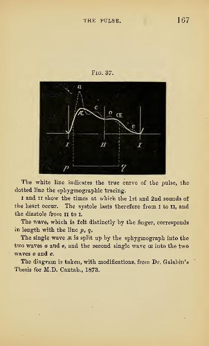
THE PULSE. 167
Fig. 37.
The white line indicates the true curve of the pulse, the
dotted line the sphygmographic tracing.
I and II show the times at which the 1st and 2nd sounds of
the heart occur. The systole lasts therefore from i to ii, and
the diastole from ii to i.
The wave, which is felt distinctly by the finger, corresponds
in length with the line ^, q.
The single wave m is split up by the sphygmograph into the
two waves a and e, and the second single wave ce into the two
waves and e.
The diagram is taken, with modifications, from Dr. Galabin's
Thesis for M.D. Cantab., 1873.
168 HOW TO EXAMINE THE CHEST.
higli and then falling too low^ and in this waygiving a double wave where there ought only to
be one.
The sensation conveyed to the fingers repre-
sents more nearly the real curve. This^ we see
rises sharply^ then subsides slowly for some
distance^ until it is interrupted by another slight
rise^ after which it finally falls more rapidly down
to the level from which it started.
The only part which we feel distinctly is the
top of the wave between m and (E in the diagram^
and it is in this part that the most characteristic
changes occur.
The whole pulse wave extends of course from
I to i^ that is^ from one beat to the next. Along wave would then be the same thing as a slow
pulse.
When we speak of a long wave in this sense_,
it will be better to call it a rapid or slow pulse^
because we really refer to its frequency^ and this
will be discussed later^ and because long and
short are more commonly used in reference to
the top or head of the wave (corresponding with
the line p, q) than to its total length.
The parts of the wave to examine most
attentively are the upstroke or rise^ and the top
or head.
The Upstroke may be abrupt (fig. 41) or gradual
(fig. 42), high {fig. 41) or low {fig. 39).

THE PULSE.
Fm. 38.
Normal pulse tracing.
169
Fig. 39.
Tracing of a pulse of increased tension from a case of gran-
ular kidney (square top). -
Fig. 40.
Tracing of a dicrotic pulse.
Fig. 41.
Pulse tracing from a case of Aortic Incompetence. Water-
hammer pulse.
(Sudden rise and sudden fall.)

170 HOW TO EXAMINE THE CHEST.
The Top of the Wave {p to q) represents tlie
maximum distension in the vessel. If the tension
be well sustained {figs. 38 and 39)^ the subsidence
will be gradual ; if ill sustained_, it will be abrupt
(/?.41).
Where the subsidence is gradual^ the wave
will appear long, and seem to pass slowly under
the finger, and where it is abrupt, it will appear
short, and seem to pass rapidly under the finger.
This is what is meant generally by a Long and
Short Wave.
When the tension is very low, the second wave,
or ^^ recoil wave ^^ as it has been termed ((e), is
often well marked, and may even be as large
and as distinct as the primary wave. The pulse
is then felt as a double beat, and is called Dicrotic
{twice heating) {fig. 40).
In some other rarer cases, instead of two dis-
tinct} waves, several smaller ones are felt, and the
pulse is called Thrilling.
These thrills may in some conditions of the
artery be produced by pressure, and on this
account it is important to feel the pulse only with
one finger, lest by a little unequal pressure vibra-
tions maybe set up, and the pulse made artificially
thrilling.
One of the most peculiar forms, and at the
same time one of the most important clinically.

THE PULSE. 171
is known as tlie Waterhammer Pulse{fig. 41).
This is pathognomic of incompetence of the
aortic valves. It is characterised by a very high
upstroke and by very short duration. It has a
short sudden wave^ and conveys to the finger
the sensation of a sharp forcible jerk^ the
artery seeming to empty itself and collapse,
almost immediately after the beat has been
felt.
The high upstroke is caused by the great force
with which the blood is driven into the arteries^
for the leffc ventricle is hypertrophied.
The short duration or sudden collapse is due
to the fact that the aortic valves do not close^ so
that the blood flows back from the aorta into the
ventricles, and the tension in the arteries is not
maintained.
The position of the arm makes a great differ-
ence in the distinctness with which the characters
of this pulse are felt at the wrist. If the arm
hang down, the force of gravity tends to keep the
artery fairly full, but, if the hand be raised above
the head, it helps to empty it, and the peculiarity
becomes more marked.
,
This pulse is also known by other names :
1. Corrigan^s Fidse, from Dr. Corrigan whodescribed it
;
2. The Locomotor Pulse, from the peculiar way,
in which the impulse seems to travel down the
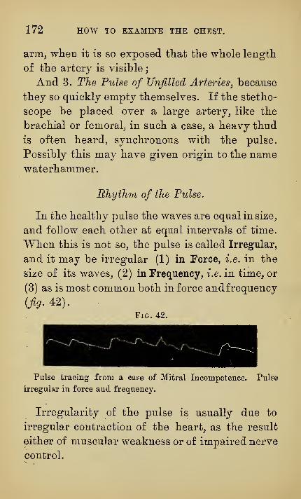
172 HOW TO EXAMINE THE CHEST.
arnij wheD it is so exposed that the whole length
of the artery is visible
;
And 3. The Pulse of Unfilled Arteries, because
they so quickly empty themselves. If the stetho-
scope be placed over a large artery^ like the
brachial or femoral^ in such a case^ a heavy thud
is often heard, synchronous with the pulse.
Possibly this may have given origin to the namewaterhammer.
Rhythm of the Pulse.
In the healthy pulse the waves are equal in size,
and follow each other at equal intervals of time.
When this is not so, the pulse is called Irregular,
and it may be irregular (1) in Force, i.e. in the
size of its waves, (2) in Frequency, i.e. in time, or
(3) as is most common both in force andfrequency
(Jig- 42).
Fig. 42.
Pulse tracing from a case of Mitral Incompetence. Pulse
irregular in force and frequency.
Irregularity of the pulse is usually due to
irregular contraction of the heart, as the result
either of muscular weakness or of impaired nerve
control.

THE PULSE. 173
Any nervous excitement may make botli lieart
and pulse irregular for the time. And in some
acute diseases of tlie brain and upper part of tlie
spinal cord tlie rhythm of the pulse is often of
diagnostic importance. In these cases the beats
both of heart and pulse follow one another appar-
ently with regularity, but the frequency varies
much from time to time even to the extent of 10
or 15 beats in consecutive minutes.
As a matter of routine then the pulse should
be counted two or three times at short inter-
vals.
Muscular weakness of the heart often shows
itself in the pulse by irregularity of force, i.e. in
the size of the waves ; but this is usually associ-
ated also with irregularity in time. In such
cases any slight extra work thrown upon the
heart, even, for example, the effort of standing
up, may increase the difference between the
number of beats at the heart and at the wrist.
When this weakness is great, some of the
beats may fail to reach the wrist and the pulse
is then called ** Intermittent " and in extreme
cases, the pulse may be entirely absent at the
wrist, the heart not having power to drive a
single wave so far.
Although in most cases irregularity of the
pulse is due to irregularity of heart action.

174 HOW TO EXAMINE THE CHEST.
tliere are cases in which the pulse is markedly
irregular, while the heart continues to beat regu-
larly.
This irregularity is generally synchronous with
the respiratory movement s_, and in this wayregular oscillations in the pulse tracings are pro-
duced.
In health this is not detected except by deli-
cate apparatus in the large vessels near the
heart, but, where the circulation is feeble, it maybe observed at the wrist in an irregularity, not
only of force but also of time, synchronous with
the inspiratory movements of the chest.
In the most extreme form, this gives rise
to the Pulsus Paradoxus {fi,cj. 43), or Pulsus cum
Fig. 43.
Pulsus Paradoxus. Pulsus cum luspiratione intermittens.
Taken from a case of Purulent Pericarditis.
The pulse is very dicrotic, the second wave being almost as
large as the first. The horizontal line indicates the remissions
due to inspiration, and lasting for two or three heart beats.
Inspiratione intermittens. The pulse beat then
disappears entirely during inspiration, and often
hardly a trace of vibration can be detected even

THE PULSE. 175
upon an enlarged sphyg-mograpliic tracing. In
the most typical form tlie heart remains perfectly
regular, both in force, as judged by the loud-
ness of the heart-sounds, and in time.
The typical pulsus paradoxus is very rare. Its
causes vary. In some few cases, the heart seems
to be in such an extreme condition of muscular
feebleness, that it is unable to overcome even
the very slight increase of work, which inspira-
tion throws upon it.
In most cases, however, the pulsus paradoxus is
found to be caused by adhesions or bands, which
surround the larg'e vessels either in the medias-
tinum or within the pericardium, in such a waythat, when the chest expands on inspiration, they
are tightened, the vessels compressed or pinched,
and the pulse wave stopped.
Want of Symmetry.
The pulses in the two wrists in health are
exactly symmetrical, both in time and force, the
only allowance, that may have possibly to be
made, is for an anatomical difference in the size
of the vessel, but this does not often present any
practical difficulty.
In disease of various kinds, the artery of one
side may be twisted, pressed upon, stretched, or

176 HOW TO EXAMINE THE CHEST.
displaced in sucli a way as to obstruct tlie circu-
lation through it^ and thus a Want of Symmetry
will be produced.
Usually this shows itself in an alteration in
the Character of the Wave on the affected side,
which may be recognised by the finger and de-^
monstrated by a sphygmographic tracing, but
besides this the wave is often Retarded, so that it
reaches the wrist on the affected side at a per-
ceptibly later time.
These, like all other defects of symmetry, are
of very great practical importance in the dia-
gnosis of disease.

THE PULSE. 177
SYNOPSIS OF THE EXAMINATION OF THE PULSE.
I. The Artery.
, 1. Its course, straight^ tortuous.
2. Its coats J -!^-^^ * fa. Uniformly.
1 tnickenedi , t i. i,^\^b. In patches.
II. The Calibre of the Vessel, i. e. the size of the
column of blood it contains, in relation
to the size of the vessel.
III. The Tension, whether increased or dimin-
ished.
IV. The Pulse wave.
1. Its frequency, ^. e. the number of beats
in the minute.
rirregular ( ^^ ^^^^^^
2
.
Its rhythm -l^orm frequency
.
(^Intermittent.
3. The wave. 1. Its rise, high or low.
2. Itshead/1^^? ^^' ^^^^*^
Lrapid or slow.
3. Its character (dicrotic,
thrilling, waterham-
mer, &c.).
4. Its symmetry, in time and
force.
12


SECTION IV.
THE MEDIASTINUM,


THE EXAMINATION OF THEMEDIASTINUM.
The Mediastimim is the irregular space^ extend-
ing from tlie first rib to tlie diaphragm, and lying
between tbe sternum in front, the spine behind,
and the lungs on either side.
Part of this space is occupied by the heart, the
rest contains the large arteries and veins con-
nected with the heart, the trachea and the roots
of the lungs, the oesophagus, and thoracic duct,
with numerous nerves, small vessels, and glands.
Displacement of the Mediastinum.
All these structures are intimately connected
together, and form a mass which is firmly fixed
below by its attachment to the diaphragm, and
posteriorly and above by its attachment to the
spinal column, so that vertical displacement
is hardly possible. With the sternum, however,
the connections are loose, so that Lateral Displace-
ment may occur by a kind of rotation, as a door
182 HOW TO EXAMINE THE CHEST.
swings upon its hinge^ tlie hinge in this case
being the aorta^ where it is fixed to the spine.
The mediastinum remains in the middle line in
health. Not because it is "fixed" there^ but
because of tho equal balancing of the forces,
which tend to displace it on either side. The
chief of these is the elasticity of the lungs.
If air be admitted freelyinto one pleural cavity,
the lung on that side collapses, and the elasticity
of the other lung, being unopposed, comes into
play. This lung also contracts, and in doing
so pulls over the mediastinum.
If one pleural cavity be greatly distended by
air or fluid, the mediastinum may be pushed over
by the pressure and be still further displaced
{figs. 15 and 16).
On the other hand, if one pleural cavity be
contracted, as with the fibroid thickening, which
often occurs after chronic pleurisy, the medias-
tinum will be pulled over, just as the ribs on that
side are pulled in(fig. 14)
.
The Physical Signs of Displacement of the Medi-
astinum en masse are :
1. Those of displacement of the heart (q.v.) ;
2. Those of displacement of the anterior boun-
daries of the lungs {q. v.).
These have been already discussed, and need
not be further referred to here.

THE MEDIASTINUM. 183
The parts of the Mediastinum.
The Heart, although strictly included in the
mediastinum_, has been already described in a
separate chapter.
The Rest of the Mediastinum is not so easily
accessible.
The places where it is examined are :
1. In front, beneath the upper part of the
sternum
;
2. Behind, on either side of the dorsal vertebrae
in the interscapular spaces.
Diseases of the Mediastinum.
In front, the mediastinal region extends from
the episternal notch down the sternum to the
level of the fourth costal cartilage.
In health this part is only slightly prominent,
moves slightly with respiration, is resonant to
percussion, and beneath it, tracheal or bronchial
breathing and resonance are not infrequently
audible.
Apart from affections of the lungs and pleura,
which may modify the physical signs here, the
diseases of this part are chiefly two, each
attended by the formation 0^ a Tumour, viz.
Aneurysm and New Growth.
In either case the lungs are pushed aside by a

184 HOW TO EXAMINE THE CHEST.
Fi&. 44.
Diagram of a Mediastinal Tumour. The shaded area heneath
the manubrium and upper ribs indicates the area of impaired
percussion resonance.
The edges of the lungs are retracted, or pushed aside by the
mass beneath.

THE MEDIASTINUM. 185
mass, and their place beneatli the sternum is
taken by the non-resonant tumour (jig. 44). The
percussion, therefore, is impaired or dull, and
the physical signs of consolidation are present
as in the lungs, viz. increased vocal vibration, in-
creasedvocal resonance, and increased breathing-
sounds. In addition there may be present in some
cases Bulging, Pulsation, Thrills, or Friction, andoccasionally also Vascular Murmurs.
Posteriorly, in the interscapular spaces, the
same two diseases are met with, and may give
rise to the same physical signs. In this region,
however, impairment of percussion is much moredifficult to make out, because of the thick
muscular covering to the ribs, the percussion
note here being normally deficient in tone, but
of course symmetrical. Want of symmetry is
what we look for, and this is often very distinct,
so far as vocal vibrations, vocal resonance, andbreathing-sounds are concerned, although the
other physical signs which are occasionally
present in front, viz. bulging, pulsation, thrills,
friction and murmurs, are but rarely obtained
behind.
Where the tumour is large, not only the lungs
but also the heart and diaphragm may be pushed
out of place, and we shall then have, in addition_,
the ordinary physical signs of displacement of
these organs.

186 HOW TO EXAMINE THE CHEST.
The Diagnosis of Diseases of the Mediastinum.
The diagnosis of mediastinal affections^ by
means of tlie physical signs alone^ is often very
difficulty especially when the disease is not exten-
sive, and is deeply seated, and it is then rather
upon the secondary symptoms, to which the
disease gives rise, than upon the physical signs^
that the diagnosis depends.
These Secondary Symptoms are for the most
part due to mechanical interference, by pressure,
with the function of the various organs in the
thorax.
What these symptoms may be, depends upon
the part pressed upon, and varies according to
the seat of the disease, and, to determine this,
an intimate knowledge of the relationship of
the different parts in the mediastinum is essential.
These are given in detail in any good work on
anatomy. It will be sufficient for our present
purpose to group the various symptoms together
with reference to their cause, and to indicate in
this way the use, which may be made of them
for diagnosis.
I. Pressure upon the Vessels will lead to inter-
ference with the circulation through them.
When the Veins are compressed, the part of

THE MEDIASTINUM. 187
the vessel on the side away from the heart will
be distended, and an attempt will be made to
establish the circulation through collateral chan-
nels. This will give rise then, first, to Distension
of the Large Veins above the seat of pressure, as
is especially common in the lower part of the
neck, or on the shoulders ; secondly, to abnormal
Dilatation of the Subcutaneous Veins usually over
the front of the chest ;"^ and lastly, to oedema of
those parts supplied by the obstructed veins.
In both cases the distension is usually unsyni-
metrical.
It is important to determine in all these cases
the direction in whicli the blood is travelling in
the dilated vessels, in the way described at p. 20.
The circulation through, the Arteries may be
interfered with, either as the result of direct
pressure upon them,- or in consequence of the
vessels being so twisted or stretched at their
origin, that their mouths are obstructed.
In either case the Pulse, carotid or radial, will
become unsymmetrical. There will be a differ-
ence either in force or in time, that is to say, one
pulse will be either Smaller than the other, or
will reach the wrist later than the other, i. e. be
^eiarded.
* The physiological dilatation of the mammary veins re-
ferred to at p. 20 must not be confounded with this patho-
loofical dilatation.
188 HOW TO EXAMINE THE CBEST.
Pressure upon either arteries or veins may pro-
duce a MurmurJ audible of course near tlie seat
of pressure. In the arteries it is usually SystoUcj
and in the veins often Continuous.
II. Pressure upon the Respiratory Tract will
lead to interference with respiration^ and give
rise to Dyspnoea, Stridor, or Gough, all of which
are frequently Paroxysmal.
Dyspnoea may be due to pressure upon the
trachea or bronchus, or directly upon the lung
itself, and similarly cough may be the result of
irritation of any of these parts.
Stridor, whether in breathing, speaking, or
coughing, on the other hand, is always evidence
of pressure upon the trachea or bronchi, if there
be not a local affection of the larynx to account
for it, as can easily be determined by laryngo-
scopic examination.
Where the pressure is upon the trachea, the
air will have difficulty in entering both lungs
equally, but where it is upon the bronchus or
lung of one side, the obstruction is unilateral,
and, therefore, we shall find a want of symmetry
in the physical signs ; the vocal vibrations and
the vocal resonance may be weaker than on the
unaffected side, and the respiratory murmur may
be feebler or often altered in character, expira-
tion especially becoming prolonged and occasion-
ally wheezing.

THE MEDIASTINUM. 189
The place to examine for these altered physical
signs is as far away as possible from the root of
the lungs^ where, as a rule, the obstruction exists;
and the most convenient part to select is low
down at the base of the lungs posteriorly, i. e. in
the infrascapular region.
In such cases as these a slight want of sym-
metry may be of great importance.
III. The symptoms of Pressure upon the Nerves
vary with the nerve affected, and with the
amount of pressure, slight pressure producing
the results of irritation, and considerable pres-
sure those of paralysis.
The nerves are the Pneumogastric, the Phrenic,
the Sympathetic, and the Intercostal.
1. Of these, the most important is the Pneumo-
gastric, distributed as it is to the larynx, lungs,
and heart.
The Recurrent Laryngeal Branch on the left
side, where it winds round the arch of the aorta,
is especially exposed, and is therefore often
affected.
Irritation of this branch, as also of the pulmo-
nary branches of the pneumogastric, commonlyproduces attacks of Spasmodic Dyspnoea, which
are sometimes fatal.
Paralysis, on the other hand, often leads to
affection of the voice, in consequence of paralysis
of the muscles which move the vocal cords.

190 HOW TO EXAMINE THE CHEST.
The effect upon tlie heart is usually to produce
IrregularityJPalpitation, and Paroxysmal PaiUy
and occasionally attacks of Syncope which mayprove fatal.
2. Pressure upon the Intercostal Nerves gives
rise to Pains, referred usually to their peripheral
distribution on the side or front of the thorax_, or
occasionally following the course of the intercosto-
humeral nerve^ down the inner side of the arm as
far as the elbow.
3. The Phrenic Nerves usually escape, or at all
events the diaphragm rarely gives evidence of
any affection, when only one phrenic is in-
volved.
4. The Sympathetic Nerves enter largely with
the spinal nerves into the various plexuses in the
thorax, and cannot be separated in their action
from the spinal nerves already referred to, unless
certain alterations in the rate of beating of the
heart be attributed to their influence.
lY. Pressure upon the Thoracic Duct produces
no symptoms, by which it can be diagnosed,
although theoretically it might be the cause of
anaemia and malnutrition.
V. Lastly, Pressure upon the (Esophagus causes
ByspJiagia, i.e. difficulty in swallowing either
solids or liquids, and occasionally, where the
obstruction is considerable, it may lead to Regur-
gitant Vomiting.
THE MEDIASTINUM. 191
It is the combination and association of these
various symptoms^ wliicli make it possible to
determine in a given case tlie locality of a tumour
in tlie thorax.
All that the physical signs enable us to estab-
lish in many cases is the presence of a tumour
in some part of the mediastinum. Its nature^
whether aneurysm or new growth^ has often to
be decided rather by the general or constitutional
condition^ than by the physical signs. In young or
old people, the probabilities are in favour of newgrowth ; especially if the cachexia, usual with
malignant disease, be present. On the other
hand, in middle-aged persons, especially in men,,
the probabilities are in favour of aneurysm.
This diagnosis would be confirmed by evidence
of vascular change in other parts, as for example
by thickened arteries, or by the history of causes
likely to produce vascular degeneration, such as
laborious work, drink, and syphilis.
In the majority of cases, however, the diagno-
sis does not present any great difficulties.

192 HOW TO EXAMINE THE CHEST.
SYNOPSIS.
In making the diagnosis of a mediastinal
tumour, we liave to consider
1. The evidence of a mass in the mediastinum,
as given
(a) By the physical signs;
(6) By the pressure symptoms;
2. The facts pointing to the nature of the
mass
;
{a) Special physical signs
;
(6) The age, history, and constitutional
condition of the patient.

INDEX.
A.
PAGE
Abdominal respiration . 21
Absolute cardiac dulness . 112
Accentuation of the heart-
sounds . . . 125
>3Egophony . . .70Air, amount of, on respira-
tion . . . .21Alar chest, the . , .17Amount of air on respira-
tion . . . .21Amphoric breathing . . 76
— whisper . . .69Anaemic murmurs . . 149
Aneurysm, thoracic . . 183
Aortic disease, alteration
- of cardiac dulness in . 118
— murmurs . . .141Apex of the heart, the . 101
— displacements of the . 102
Arch, the costal . . 1
Area, the cardiac . .110— the splenic . . .43— of stomach resonance . 43
Arterial murmurs in neck . 154— pulsation . . . 104
Arteries, calibre of . . 164
PAGE
Arteries, changes in coats
of
— pulse of unfilled .
— tortuous
Asymmetry of thorax
disease of the lungs
Auricular systolic murmurs 131
Auscultation . . 5, 60
— of the breathing sounds 64
. 163
. 172
. 163
in
18,25
—r of the heart .

194 INDEX.
PAGE
Blood-current, in dilated
superficial veins, direc-
tion of . . .20Blood murmurs . . 149
Boundaries of the lungs . 37
— displacement and dislo-
cation of . . .51— of the liver . . .41Breathing, alteration of, in
disease . . .79— Cheyne-Stokes' . . 24
— sounds . . . .70— varieties of . . .71Bronchial breathing . . 171
— resonance . . .65— whisper . . .69Bronchophony . . .65— whispering . . .69Bruit d'airain . . .89— de pot fele . . .56— du diable . . . 154
Bulging, expiratory . . 23
— of chest in disease of
lungs . . . .18— of the priBcordium . 100
C.
Callipers . . . .11Cantering action of the
heart. . . .124
Capacity of the chest, the
vital . . . .22Cardiac dulness, the altera-
tions in . . .114
PAGE
Cardiac dulness, the area
of absolute . . 112
the area of relative . 112
— impulse . . . 104
Carinate chest . . .15Carpenter's chest . . 19
Cataire, fremissement . 108
Cavernous breathing . . 74
— whisper . . . 69
Chest, the.... 1
— alar . . . .17— barrel-shaped . . 13
— change of shape of, in
disease . . .18— measurements of . .10— paralytic . . .17— pigeon-breast . . 15
— position of patient on
examining the . .3— pterygoid . . .17— rickety . . . .14— varieties of . . .12— variations in shape of . 9
Cheyne-Stokes' breathing . 24
Clavicular region, the . 2
Compensatory hypertrophy,
or emphysema . . 49
Complemental space . . 39
Consolidation of lung, the
diagnosis of . .80Contents of the thorax, the 3
Contraction of chest in dis-
ease of the lungs . 18
— of lungs . . .49Corrigan's pulse . .171

INDEX. 195
PAGE
Costal arch, the . . 1
— respiration . . .26Count the ribs, how to . 27
Cracked-pot sound . . 56
Crepitation . . .83— fine hair . . .84— metallic . . .88— redux . . . .87— to be felt ... 28
Croup, chest in . 12, 23
Crying, resonance of . 69
Cyrtometer . . .11
D.
Death rattle, the . . 84
Deep cardiac dulness, the
area of . . . 112
Defective inspiration . 22
Deficient expansion . . 22
— expiration . . .22— resonance on percussion 57
— respiratory movement . 22
Deformities of chest . . 19
Diagram of heart-sounds . Ill
Diaphragmatic respiration 20
Diastole . . . .127
Diastolic impulse . . 104
— murmurs . . .127
Dicrotic pulse . . . 170
Difficulty in breathing . 22
Diffused cardiac impulse . 104
Dilatation murmurs . . 149
— of superficial veins . 20
Direct percussion . . 31
PAGE
Direct vocal resonance . 68
Direction of blood-current
in superficial veins . 20
Displacement of boundaries
of the heart . . 98
— of the lungs . . .49— of the mediastinum . 181
Dry sounds . . .84Dulness, percussion . . 34
Dyspnoea, expiratory . . 22
— inspiratory . . .22
E.
Early diastolic murmur . 131
EfEusion, boundaries of
lungs in pleuritic . 57
Emphysema, compensatory 49
— expiratory bulging in . 23
— physical signs of . .49— senile . . . .49— shape of chest in . . 13
Endocardial murmurs . 126
Enlargement of the lungs . 47
Epigastric pulsation . . 104
Episternal region, the . 2
Exaggerated resonance . 55
— respiratory movements . 22
Examination, methods of . 3
Exocardial murmurs . 126, 151
Expansion, deficient . . 22
Expiration, deficient . . 22
Expiratory bulging . . 23
— dyspnoea . . .22Extended cardiac impulse . 104

196 INDEX.
F.
PAGE
Fine hair crepitation . . 84
Fluctuation . . .20Fluid in pleura, diagnosis of 82
Forms of chest... 9
Fremissement cafcaire . 108
Friction felt on palpation . 28
— pericardial . . 108, 151
— pleuritic . . .86— pleuro-pericardial 87, 152
— redux . . . .87Furrow, Harrison's . .15
G.
Gall-bladder, position of . 41
General heaving of prsecor-
dium.... 105
H.
Hsemic murmurs

INDEX. 197
PAGE
Inspiratory recession . . 23
Intercostal spaces, bulging
of . . . .23— contraction of . .28— recession of . . .23— widening of . . .28Intermittent pulse . . 173
Interrupted breathing . 77
Interscapular region, the . 2
Irregular pulse . . .172— respiration , . .24
J.
Jerky breathing . . 77
L.
Landmarks, medical (see
Surface-markings) . 35
Laryngeal breathing . . 71
•— resonance . . .65Late diastolic murmur . 180
Line, the nipple . . 2
Liver, surface-markings of 39
Locomotor pulse, the . 171
Lungs, auscultation of the 60
— boundaries of the . . 37
— consolidation of the . 81
— contraction of the . 49
— diagnosis between dis-
ease of the pleura and
of the ... 82
— enlargement of the . 47
— inspection of the . . 9
PAG-E
LungSj palpation of the . 27
— percussion of the . . 31
— size of the . . .35
M.
Mammary region, the . 2
Markings, surface . . 35
Measurements of chests . 10
— varying with respiration 21
Mediastinum . . . 181
— boundaries of . . 181
— diagnosis of diseases of . 186
— diseases of . . .183— dislocation of . . 181
— parts of . . . 183
— places to examine . . 183
— synopsis of examination
of ... . 190
Mediate percussion . . 31
Medical landmarks . . 35
Metallic crepitation . . 88
— tinkling . . .88Methods of examination . 3
Mid-diastolic murmur . 131
Mitral disease, alteration of
cardiac dulness in .118— murmurs . . . 142
— stenosis, case of . . 145
Moist sounds . . .84Movements of respiration,
alterations in . . 22
— how to examine, by
palpation . . .27— of chest in respiration . 20

198 INDEX.
PAGE
Movements of the prse-
cordium . . . 100
Murmurs, anaemic, hsemic,
blood . . . .149— aortic .... 141
— auricular systolic . . 131
— backward . . . 137
— cause of endocardial . 134
— classification of . . 126
— definition of. . .125— diastolic . . . 129
— dilatation . . . 149
— endocardial and exo-
cardial . . .126— endocardial, not due to
valvular disease . .147— exocardial . . . 151
— heart . . . .125— how to time . . . 127
— inorganic, characters of 148
— in vessels of neck . .154— mitral .... 142
— musical . . . 126'— onward.... 137
— organic and inorganic . 126
— place of . . . 135
— postsystolic . . .132— prsesystolic . . . 129
— pulmonary systolic . 148
— subclavian . . . 153
— systolic and diastolic . 127
— table of . . . 134
— tricuspid . . 143, 149
— venous . . . 154
Musical heart murmurs . 126
N..
Nipple line, the
Nonnen-gerausch
Number of respirations
O.
Onward murmurs
Orthopncea
P.
PAGE
. 2
. 154
. 23
137
22
Palpation .

INDEX. 199
PAGE
Percussion of lungs . . 31
— palpatory . . .32— sounds, varieties of . 51
Pericardial effusion, altera-
tion of cardiac dulness
in . . . . 118
—^friction . . 108, 151
cliaracters of . . 151
Pigeon-breast, the . . 15
Plessimeters . . .31Plessor . . . .31Pleura, diagnosis between
diseases of lung and
of . . . .82Pleuritic effusion . . 51
diagnosis of . .82— friction . . .86
felt on palpation . 28
Pleuro-pericardial friction
67, 152
Pneumothorax, ausculta-
tion in . . .87— boundaries of lungs in . 51
Position of patient for exa-
mination of the chest . 3
Postsystolic murmurs . 132
Prsecordium, the . . 98
— bulging of . . .98— the movements of. .98— the shape of . . .98Prsesystolic murmurs . 129
Pterygoid chest, the . . 17
Puerile breathing . . 78
Pulmonary systolic mur-
murs .... 148
Pulsation, arterial
— epigastric
— hepatic.
— venous .
Pulse, the .
— Corrigan's .
PAGE
. 104
. 104
. 105
. 105
. 161
. 171
— cum inspiratione in-
termittens . . .174— dicrotic . . . 170
— hard, soft, &c. . . 164
— incompressible . . 164— intermittent . . . 173— irregular . . .172— locomotor . . .171— of unfilled arteries . 172— paradox . . . 174— relation to respiration . 23— retardation of . .176— rhythm of . . . 172— synopsis of the . .175— tension 160
— thrilling . . .170— want of symmetry . 175— waterhammer . . 171
Pulse rate, the . . . 161
conditions affecting
the . . . . 162
Pulse wave, the . . . 166
Purring tremor . . . 108
Pyopneumothorax . . 89
R.
Rales
Rate of respiration
Rattle, the death
83
23
84

200 INDEX.
PAGE
Recession, inspiratory . 23
— of apex beat systolic . 104
Reduplication of the heart-
sounds . . . 124
Redux crepitation . .87— friction . . .87Regions of chest . . 2
Relative cardiac dulness • 112
Resistance, sense of . .29Resonance, percussion . 34
— area of stomach . .43— deficient . . .55— dull .... 55
— impaired . . .55— normal . . . .55— tympanitic . . .53— varieties of . . . 53
— vocal, varieties of . .65Respiration, amount of air on 21
— Cheyne-Stokes' . . 24
— costal or thoracic . . 20
— diaphragmatic or abdo-
minal . . .21— effect of emotion on . 24
— interrupted . . .77— irregular . . .24— jerking. . . .77— movements of . .20— Saccadee . . .77— variations in movements of 22
— voluntary control over . 24
— wavy . . . .77Respirations, effect of ex-
citement and disease
,upon number of . .23
PAGE
Respirations, number of . 23
— relation to pulse . . 23
Respiratory movements,
how to examine (pal-
pation) . . .27in men, women, and
children . . .20Rhonchus . . . ,83— felt . . . . 28
Ribs, how to count the . 27
Rickety chest, the . .14
S.
Senile emphysema . . 49
Sense of resistance . . 29
Shape of the healthy thorax 10
— varieties of . . .12Shoemakers' chest . . 19
Short breath (dyspnoea) . 22
Sibilus . . . .83— felt . . . .28Solidification of lung, dia-
gnosis of . . .81Sound, cracked-pot . . 56
— the bell . . .89— the splashing . . 89
Sounds, dry and moist . 84
— of breathing . . .70— of the heart, the (see
Heart-sounds) . . 120
whisper . . .69Space, complemental . . 39
INDEX. 201

202 INDEX.
Time the heart, to
Tinkling, metallic
Tracheal breathing .
— resonance
Tracing, cyrtometer,
chest .
— sphygmographic .
Transposition of viscera
Traube's zone .
Ti'emor, purring
Tricuspid murmurs .
Tumour, mediastinal
.
Tympanitic resonance,
percussion .
Types of respiration .
U.
PAGE
. 123
. 88
. 71
. 65
of
or
11
167
103
43
108
149
182
53
26
Undulatory cardiac impulse 104
V.
Valve shock . . .109
Valves of heart, position of 137
Valvular disease, diagnosis
of . . . .143
Varieties of chests . . 12
Veins, dilatation of . .20Venous murmurs in neck . 154
— pulsation . . . 105
Vesicular breathing . . 73
PAQ-E
Vesicular resonance . . 65
Vibrations of voice in
women and children . 29
Vibrations, want of sym-
metry in . . .29— vocal . . . .28Viscera, transposition of . 103
Vital capacity of the chest 21
Vocal resonance . 29, 64
varieties of . .65— vibrations . . .28Voice, auscultation of the . 64
W.
Want of symmetry in

No. 1.
London, 11, New Burlington Street,
August, 1895.
A SELECTIONFEOM
J. k A. CHURCHILL'S CATALOGUE,COMPRISIKG
MOST OF THE EECENT WORKS PUBLISHED BY THEM.
N.B.—J. & A. Churchill's larger Catalogue, which contains over 600 ivories,
ivith a Complete Index to their Subjects, will he sent on application.
Human Anatomy : a Treatise by variousAuthors. Edited by Henry Morris, M.A., M.B. Lond., F.E.C.S.,
Surgeon to, and Lectiirer on Surgery at, the Middlesex Hospital.
Eoy. 8vo, with 791 Illustrations, nearly all original, and many of
them in several colours, 40s. (In one vol. or in three parts.)
Heath's Practical Anatomy : a Manual ofDissections. Eighth Edition. Edited by William Anderson,
P.R.C.S., Surgeon and Lecturer on Anatomy at St. Thomas's Hospital
;
Examiner in Anatomy for E.G. P. and S. Crown *vo, with 3i}9 En-
gravings. 158.
Wilson's Anatomist's Vade-Mecum. Eleventh
Edition, by Henry E. Clark, M.E.C.S. Eng., F.F.P.S. Glasg.,
Examiner in Anatomy F.P.S., and Professor of Surgery in St.
Mungo's College, Glasgow. Crown 8vo, with 492 Engravings and 26
Coloured Plates, 18s.
An Atlas of Human Anatomy. By RickmanJ. Godlee, M.S., F.E.C.S., Surgeon and late Demonstrator of
Anatomy, University College Hospital. With 48 Imp. 4to Plates
(112 figures), and a volume of Explanatory Text. Svo, £4 14s. 6d.
Human Osteology. By Luther Holden, Con-suiting Surgeon to St. Bartholomew's Hospital, Seventh Edition,
edited by Charles Stewart, Conservator of the Museum E.C.S.,
and Egbert W. Eeid, M.D., F.E.C.S., Professor of Anatomy in the
University of Aberdeen. Svo, with 59 Lithographic Plates and 75
Engravings, 16s.
By the same Author.
Landmarks, Medical and Surgical. FourthEdition. Svo, 3s. 6d.
11, NEW BURLINGTON STREET.
J. 8^ A. ChurcMlVs Recent Works.
The Student's Guide to Surgical Anatomy.By Edward Bellamy, F.E.C.S., and Member of the Board of Exami-ners. Third Edition. Fcap. 8vo, with 81 Engravings, 7s. 6d.
Diagrams of the Nerves of the Human Body,exhibiting their Origin, Divisions, and Connections, with their Dis-
tribution to the Various Eegions of tlie Cutaneous Surface, and to
all the Muscles. By Sir W. H. Flower, K.C.B., F.E.S., F.E.C.S.
Third Edition, with 6 Plates. Eoyal 4to, 12s.
Pathological Anatomy of Diseases. Arrangedaccording to the nomenclature of the E.C.P. Lond. (Student's Guide
Series). By Norman Moore, M.D., F.E.C.P., Assistant Physician
and Lecturer on Pathological Anatomy to St. Bartholomew's Hos-
pital. Pcap. Svo, with 111 Engra^^ngs, 8s. 6d.
A Manual of Clinical and Practical Pathology.By W. E. Wynter, M.D., M.E.C.P., F.E.C.S., Medical Eegistrar to
Middlesex Hospital, and F. J. Wethered, M.D., M.E.C.P., Assistant
Physician to Victoria Park Hospital. With 4 Coloured Plates and 67
Engravings. 8vo, 12s. 6d.
Lectures on Pathology : delivered at the Lon-don Hospital. By the late Henry Gawen Sutton, M.B., F.E.C.P.,
Physician to, and Lecturer on Pathologj^ at, the London Hospital.
Edited by Maurice B. Paul, M.D., and Revised by Samuel Wilks,M.D., LL.D., F.E.S. Svo, 15s.
General Pathology (an Introduction to). ByJohn Bland Sutton, F.E.C.S., Sir E. Wilson Lecturer on Patho-
logy, E.C.S.; Assistant Surgeon to, and Lecturer on Anatomy at,
Middlesex Hospital. Svo, Avith 149 Engravings, 143.
Atlas of Pathological Anatomy. By Dr.Lancereaux. Translated by W. S. Greenfield, M.D., Professor
of Pathology in the University of Edinburgh. Imp. Svo, with 70
Coloured Plates, £5 5s.
Index Pathologicus, for the Registrations ofthe Lesions recorded in Pathological Eecords or Case-books of
Hospitals and Asylums. By James C. Howden, M.D., Superinten-
dent of the Eoyal Lunatic Asj-lum, Montrose. Fcap. folio, 6s.
Atlas of the Central Nervous System. Fromthe larger work of Hirschfeld and Leveille. Edited by Howard H.
Tooth, M.D., F.E.C.P., Assistant Phj-sician to the National Hos-
pital for the Paralysed and Epileptic. With 37 Plates carefully
coloured by Hand. Large Imp. Svo, 40s.
11, NEW BURLINGTON STREET,

J. ^ A. ChurchilVs Recent Works.
The Human Brain : Histological and CoarseMethods of Eesearch. A Manual for Students and Asylum. Medical
Officers. By W. Bevan Lewis, L.E.C.P. Lond., Medical Super-
intendent, West Eiding Lunatic Asylum. 8vo, with Wood Engravings
and Photographs, 8s.
Elements of Human Physiology. (Student'sGuide Series.) By Ebnest H. Stakling, M.D., M.E.C.P., Joint
Lecturer on Physiology at Guy's Hospital. Fcap. Svo, with 94 En-gravings, 6s. 6d.
Manual of Physiology : for the Use of Juniorstudents of Medicine. By Gerald F. Yeo, M.D., F.E.S., EmeritusProfessor of Physiology in King's College, London. Third Edition.
Crown Svo, with 254 Engravings (many figures), and Coloured Plate of
Spectra, 14s.
Principles of Human Physiology. By W. B.Carpenter, C.B., M.D., F.E.S. Ninth Edition, by Henry PowerM.B„ F.E.C.S. 8vo, with 3 Steel Plates and 377 Wood Engravings,
31s. 6d.
Practical Lessons in Elementary Biology, forJunior Students. By Peyton T. B. Beale, F.E.C.S., Lecturer onElementary Biology and Demonstrator in Physiology in King's
College, London. Crown 8vo, 3s. 6d.
Medical Jurisprudence : its Principles andPractice. By Alfred S. Taylor, M.D., F.E.C.P., F.E.S. Fourth
Edition, by Thomas Stevenson, M.D., F.E.C.P., Lecturer on Medical
Jurisprudence at Guy's Hospital. 2 vols. Svo, with 189 Engravings,
31s. 6d.
By the same Authors.
A Manual of Medical Jurisprudence. TwelfthEdition. Crown Svo, with 55 Engravings, 14s.
The Student's Guide to Medical Jurisprudence.By John Abercbombie, M.D., F.E.C.P., Physician to Charing Cross
Hospital. Fcap. Svo, 7s. 6d.
Sanitary Examinations of Water, Air, andFood. A Vade-Mecum for the Medical Officer of Health. By Corne-
lius B. Fox, M.D., F.E.C.P. Second Edition. Crown Svo, with 110
Engravings, 12s. 6d.
Microscopical Examination of Drinking Waterand of Air. By J. D. Macdonald, M.D., F.E.S., Ex-Professor of
Naval Hygiene in the Army Medical School. Second Edition. Svo,
with 25 Plates, 7s. 6d.
11, NEW BURLINGTON STREET,

J. 8f A. ChurchilVs Recent Works.
Hygiene and Public Health : a Treatise byvarious Authors. Edited by Thomas Stevenson, M.D., F.E.C.P.,
Lecturer on Chemistry and Medical Jurisprudence at Guy's Hospital
;
Ofl&cial Analyst to the Home OflBce ; and Shirley F. Mubphy, Medical
Officer of Health of the County of London. In 3 vols., royal 8vo, fully
Illustrated. Vol. I., 28s, ; Vol. II., 32s. ; Vol. III., 20s.
A Manual of Practical Hygiene. By the lateE. A. Paekes, M.D., F.E.S. Eighth Edition, by J. Lane Nottee,A.M., M.D., F.E.S. , Professor of Military Hygiene in the ArmyMedical School. 8vo, with 10 Plates and 103 Engravings, 18s.
A Handbook of Hygiene and Sanitary Science.By Geo. Wilson, M.A., M.D., LL.D., D.P.H. Camb. Medical
Officer of Health for Mid-Warwickshire, Seventh Edition. Crown8vo, with Engravings, 12s. 6d.
Elements of Health : an Introduction to theStudy of Hygiene. By Louis C. Parkes, M.D., D.P.H. Lond.,
Medical Officer of Health for Chelsea, Lecturer on Public Health at
St. George's Hospital. Post 8vo, with 27 Engravings, 3s. 6d.
The Prevention of Epidemics and the Con-struction and Management of Isolation Hospitals. By EogeeMcNeill, M.D. Edin., D.P.H. Camb., Medical Officer of Health for
the County of Argyll. 8vo, with several Hospital Plans, 10s. 6d.
Hospitals and Asylums of the World : theirOrigin, History, Construction, Administration, Management, andLegislation. By Heney C. Buedett. In 4 vols., super-royal 8vo, andPortfolio. Complete, 168s. Vols. I. and II.—Asylums, 90s. Vols.
III. and IV.—Hospitals, with Plans and Portfolio, 120s.
Mental Diseases : Clinical Lectures. By T. S.Clouston, M.D., F.E.C.P. Edin., Lecturer on Mental Diseases in the
University of Edinburgh. Third Edition. Cr. 8vo, with 13 Plates, 14s
Illustrations of the Influence of the Mindupon the Body in Health and Disease : Designed to elucidate the
Action of the Imagination. By D. Hack Tuke, M.D., F.E.C.P., LL.D.Second Edition. 2 vols, crown 8vo, 15s.
The Insane and the Law : a Plain Guide forMedical Men, Solicitors, and Others as to the Detention and Treat-
ment, Maintenance, Eesponsibility, and Capacity either to give
evidence or make a will af Persons Mentally Afflicted. With Hints to
Medical Witnesses and to Cross-Examining Counsel. By G. Pitt-
Lewk, Q.C, E. Peecy Smith, M D., F.E.C P., Eesident Phj^sician,
Bethlem Hospital, and J. A. Hawke, B.A., Barrister-at-Law. 8vo, 14s.
11, NEW BURLINGTON STREET,

J. 8f A. Churchill *s Uecent Works.
A Dictionary of Psychological Medicine, givingthe Definition, Etymology, and Synonyms of the Terms used in
Medical Psychology ; with the Symptoms, Treatment, and Patho-
logy of Insanity ; and The Law of Lunacy in Great Britain andIreland. Edited by D, Hack Tuke, M.D., LL.D., assisted bynearly 130 Contributors, British, Continental and American. 2 vols.
1,500 pages, royal 8vo, Illustrated, 42s.
Mental Physiology, especially in its Relationto Mental Disorders. By Theo. B. Hyslop, M.D., Assistant Physician
to the Bethlem Eoyal Hospital. 8vo, 18s.
Lunacy Law for Medical Men. By CharlesMercier, M.B., Lecturer on Neurology and Insanity to the West-minster Hospital Medical School, and to the Medical School fur
Women. Crown 8*^o, 5s.
The Journal of Mental Science. PublishedQuarterly, by Authority of the Medico-Psychological Association.
8vo, 3s. 6d.
Mental Affections ot Childhood and Youth(Lettsomian Lectures for 1887, etc.). By J. Langdon-Down, M.D.,
F.E.C.P., Consulting Physician to the London Hospital. 8vo, 6s.
Manual of Midwifery, including all that islikely to be required by Students and Practitioners. By Alfred L.
GrALABiN, M.A., M.D., F.K.C.P., Obstetric Physician and Lecturer
on Midwifery and Diseases of Women to Guy's Hospital. Third
Edition. Crown 8vo, with 261 Engravings, 15s.
The Student's Guide to the Practice of Mid-wifery. By D. Lloyd Roberts, M.D., F.R.C.P., Lecturer on Clinical
Midwifery and Diseases of Women at the Owens College ; Obstetric
Physician to the Manchester Eoj^al Infirmary. Fourth Edition.
Fcap. 8vo, with Coloured Plates and Wood Engravings.[^Preparing.
Obstetric Aphorisms : for the Use of Studentscommencing Midwifery Practice. By Joseph G. Swayne, M.D.,Lecturer on Midwifery in the Bristol Medical School, Tenth Edition,
Fcap, 8vo, with 20 Engravings. 3s. 6d.
Clinical Lectures on Diseases of Women :
delivered in St. Bartholomew's Hospital, by J. Matthews Duncan,M.D., LL,D„ F,R.C.P„ F,R,Ss, L, & E„ late Obstetric Physician
to St. Bartholomew's Hospital. Fourth Edition. 8vo, 16s.
11, NEW BURLINGTON STREET,
J. 8f A. ChurcMlVs Recent Works.
Lectures on Obstetric Operations : includingthe Treatment of Haemorrhage, and forming a Guide to the Manage-
- ment of Difficult Labour. By Egbert Barnes, M.D., P.E.C.P.,Consulting Obstetric Physician to St. George's Hospital. FourthEdition. 8vo, with 121 Engravings, 12s. 6d.
By the same Author.
A Clinical History of Medical and SurgicalDiseases of Women. Second Edition. 8vo, with 181 Engravings, 28s.
Gynaecological Operations (Handbook of).By Alban H. G. Doran, F.K.C.S., Surgeon to the Samaritan Hospital.8vo, with 167 Engravings, 15s.
Diseases of Women. (Student's Guide Series.)By Alfred L. Gaxabin, M.A., M.D., F.E.C.P., Obstetric Phy-sician to, and Lecturer on Midwifery and Diseases of Women at,
Guy's Hospital. Fifth Edition. Fcap. 8vo, with 142 Engravings, 8s. 6d.
Manual of the Diseases peculiar to Women.By James Oliver, M.D., F.K.S.E., M.E.C.P., Physician to the
Hospital for Diseases of Women, London. Fcap. 8vo, 3s. 6d.
By the same Author.
Abdominal Tumours and Abdominal Dropsyin Women. Crown 8vo, 7s. 6d.
A Practical Treatise on the Diseases ofWomen. By T. Gaillard Thomas, M.D. Sixth Edition, by PaulF. MuNDE, M.D., Professor of Gyneecology at the New YorkPolyclinic and at Dartmouth College. Koy. 8vo, with 347 Engrav-ings, 25s.
Notes on Diseases of Women : specially de-signed to assist the Student in preparing for Examination. ByJames J. Eeynolds, L.E.C.P., M.E.C.S. Fourth Edition, Fcap.
8vo, 3s. 6d.
Abdominal Surgery. By J. Greig Smith, M.A.,F.E.S.E., Surgeon to the Bristol Eoyal Infirmary, and Lecturer on
Surgery in the Bristol Medical School. Fifth Edition. Svo, with
Engravings. [In the Press.
The Physiology of Death from TraumaticFever; a Study in Abdominal Surgery. By John D. Malcolm,M.B., CM., F.E.C.S.B., Surgeon to the Samaritan Free Hospital.
8vo, 3s. 6d.
Notes on Gynaecological Nursing. By JohnBenjamin Hellier, M.D., M.E.C.S. Lecturer on the Diseases of
Women and Children in the Yorkshire College, and Surgeon to the
Hospital for Women, etc., Leeds. Crown 8vo, Is. 6d.
11, NEW BURLINGTON STREET.

J. 8f A. ChurchilVs Recent Works,
A Manual for Hospital Nurses and others en-gaged in Attending on the Sick, with a Glossary. By Edward J.
D0MVII.LE, Surgeon to the Exeter Lying-in Charity. SeventhEdition. Crown 8vo, 2s. 6d.
A Manual of Nursing, Medical and Surgical.By Charles J. Cullingworth, M.D., F.R.C.P., Obstetric Physicianto St. Thomas's Hospital. Third Edition. Fcap. 8vo, with Engrav-ings, 2s. 6d.
By the same Author.
A Short Manual for Monthly Nurses.Third Edition. Fcap. 8vo, Is. 6d.
A Practical Treatise on Disease in Children.By Eustace Smith, M.D., F.R.C.P., Physician to the King of theBelgians, and to the East London Hospital for Childien, etc. SecondEdition. Svo, 22s.
By the same Author.
Clinical Studies of Disease in Children.Second Edition. Post Svo, 7s. 6d.
Also.
The Wasting Diseases of Infants andChildren. Fifth Edition Post Svo, 8s. 6d.
The Diseases of Children. (Student's GuideSeries.) By Jas. F. Goodhart, M.D., F.R.C.P., Physician to Guy'sHospital. Fifth Edition. Fcap. Svo, 10s. 6d.
Manual of Diseases of Children, for Prac-titioners and Students. By W. H. Day, M.D., Physician to theSamaritan Hospital. Second Edition. Crown Svo, 12s. 6d.
A Practical Manual of the Diseases of Chil-dren, With a Formulary. By Edward Ellis, M.D. Fifth Edition,
Crown Svo, 10s.
Materia Medica : a Manual for the Use ofStudents. By Isambard Owen, M.D., F.R.C.P., Lecturer on MateriaMedica, etc., to St. George's Hospital. Second Edition. Crown Svo,
63. 6d.
Materia Medica, Pharmacy, Pharmacology,and Therapeutics. By W. Hale White, M.D., F.E.C.P., Physician
to, and Lecturer on Materia Medica and Therapeutics at, Guy'sHospital ; Examiner in Materia Medica on the Conjoint Board of
the Royal Colleges of Physicians and Surgeons. Fcap. Svo, 7s. 6d.
11, NEW BURLINGTON STREET.

J. 8f A. ChurchilVs Recent Works.
Materia Medica and Therapeutics. By CharlesD. F. Phillips, M.D., LL.D., F.E.S. Edin.
Vegetable Kingdom—Organic Compounds—Animal Kingdom. 8vo, 25s.
Inorganic Substances. Second Edition. 8vo, 21s.
Galenic Pharmacy : a Practical Handbook tothe Processes of the British Pharmacopoeia. 'Qy E. A Cripps, M.P.S.
8vo, with 76 Engravings, 8s. 6d.
The Galenical Preparations of the BritishPharmacopoeia; a Handbook for Medical and Pharmaceutica.1 Students.
By C. O. Hawthorne, M.B., CM., Lecturer on Materia Medica andTherapeutics, Qaeen Margaret's College, University of Glasgow. 8vo,
4s. 6d.
Practical Pharmacy. By Barnard S. Proctor,formerly Lecturer on Pharmacy at the College of Medicine, New-cas'tle-on-Tyne. Third Edition. 8vo, with 44 Wood Engravings and'62 Lithograph Fac-Simile Prescriptions, 14s.
Selecta e Prescriptis : containing Lists ofTerms, Phrases, Contractions and Abbreviations used in Prescrip-
tions, with Explanatory Notes, etc. Also, a Series of Abbreviated
Prescriptions and Key to the same, with Translations. By Jona-
than- Pereira, M.D., F.R.S. Eighteenth Edition, by Joseph Ince,
F.C.S., F.L.S. 24mo, 5s.
Pocket Formulary and Synopsis of the Britishand Foreign Pharmacopoeias. By Henry Beaslet. Eleventh
Edition. 18mo, 6s. 6d.
By the same Author,
Druggist's General Receipt-Book. Tenth Edition.
18mo, 6s. 6d.
Also.
Book of Prescriptions : containing upwards of3,000 Prescriptions collected from the Practice of the most eminentPhysicians and Surgeons, English and Foreign, Seventh Edition,
18mo, 6s. 6d.
A Companion to the British Pharmacopoeia.By Peter Squire, Eevised by his Sons, P. W. and A. H. Squire,
Sixteenth Edition. 8vo, 12s. 6d.
By the same Authors.
The Pharmaeopoeias of the London Hospitals,arranged in Groups for Easy Eeference and Comparison. Sixth
Edition. 18mo, 6s.
1], NEW BURLINGTON STREET.

J. ^ A. ChurchilVs Recent Works.
The National Dispensatory : Containing theNatural History, Chemistry, Pharmacy, Actions and Uses of Medi-cines, etc. By Alfred Stille, M.D., LL.D., John M. Maisch,Phar.D., Chas. Caspari, jun., Ph.G., and Henry C. C. Maisch,Ph.G., Ph.D. Fifth Edition, with 320 Engravings. Imp. 8vo, 36s
Royle's Manual of Materia Medica andTherapeutics. Sixth Edition, including additions and alterations
in the B.P. 1885. By John Harley, M.D., Physician to St. Thomas'sHospital. Crown 8vo, with 139 Engravings, 15s.
The Prescriber's Pharmacopoeia : the Medi-cines arranged in Classes according to their Action, with their Composi-tion and Doses. By Nestor J. C. Tirard, M.D., F.E.C.P., Pro-fessor of Materia Medica and Therapeutics in King's College, London.Sixth Edition. 32mo, bound in leather, 3s.
Recent Materia Medica. Notes on theirOrigin and Therapeutics. By P. Harwood Lescher, F.C.S., PereiraMedallist. Fourth Edition. 8vo, 2s. 6d.
Year-Book of Pharmacy: containing the Trans-actions of the British Pharmaceutical Conference. Annually. 8vo, 10s.
Manual of Botany. Vol. i. Anatomy andMorphology. By J. Eeynolds Green, Sc.D., M.A., F.E.S., Professorof Botany to the Pharmaceutical Society. Crown Svo, with 778Engravings. 7s. 6d.
The Student's Guide to Systematic Botany,including the Classification of Plants and Descriptive Botany. ByEgbert Bentley, late Emeritus Professor of Botany in King'sCollege and to the Pharmaceutical Society. Fcap. Svo, with 350Engravings, 3s. 6d.
Medicinal Plants : being Descriptions withoriginal figures, of the Principal Plants employed in Medicine, andan account of their Properties and Uses. By Prof. Bentley and Dr,H. Trimen, F.E.S. In 4 vols., large 8vo, with 306 Coloured Plates,bound in Half Morocco, Gilt Edges, £11 lis.
Practical Therapeutics : a Manual. ByEdward J. Waring, C.I.E., M.D., F.E.C.P., and Dudley W.Buxton, M.D., B.S. Lond. Fourth Edition, Crown 8vo, 14s.
By the same Author.
Bazaar Medicines of India, and CommonMedical Plants. With Full Index of Diseases, indicating their Treat-ment by these and other Agents procurable throughout India, etc.
Fourth Edition. Fcap. Svo, 5s.
11, NEW BURLINGTON STREET.
J. 8f A. Churchill's Recent Works.
Climate and Fevers of India, with a Seriesof Cases (Croonian Lectures, 1882). By Sir Joseph Fayber,K.C.S.I., M.D. 8vo, with 17 Temperature Charts, 12s.
By the same Author.
The Natural History and Epidemiology orCholera : being the Annual Oration of the Medical Society of London,
1888. 8vo, 3s. 6d.
A Manual of Family Medicine and Hygienefor India. Published under the Authority of the Government of
India. By Sir William J. Moore, K.C.I.E., M.D., late Surgeon-
General with the Government of Bombay. Sixth Edition. Post 8vo,
with 71 Eugra\"ings, 12s.
By the same Author.
A Manual of the Diseases of India : with aCompendium of Diseases generally. Second Edition. Post 8vo,
10^-Also.
The Constitutional Requirements for TropicalClimates, etc. Crown Svo, 4s.
The Prevention of Disease in Tropical andSub-Tropical Campaigns. (Parkes Memorial Prize for 1886.) ByAndbew Duncan, M.D., B.S. Lond., F.E.C.S., Surgeon, Bengal
Army. Svo, 12s. 6d.
A Commentary on the Diseases of India. ByNoRMAif Ckeveks, C.I.E., M.D., F.K.C.S., Deputy Surgeon-General
H.M, Indian Army. Svo, 24;S.
Hooper's Physicians' Vade-Mecum : a Manualof the Principles and Practice of Physic. Tenth Edition. By W. A.
Guy, F.E.C.P., F.E.S., and J. Harley, M.D., F.R.C.P. With 118
Engravings. Fcap. 8vo, 12s. 6d.
The Principles and Practice of Medicine.(Text-book.) By the late C. Hilton Fagge, M.D., and P. H.Pye-Smith, M.D., F.E.S., F.E.C.P., Physician to, and Lecturer onMedicine at, Guy's Hospital. Third Edition. 2 vols. Svo, cloth, 40s.
;
Half Leather, 46s.
Manual of the Practice of Medicine. ByFrederick Taylor, M.D., F.E.C.P., Physician to, and Lecturer
on Medicine at, Guy's Hospital. Third Edition, Cr. Svo, with
Engravings, 15s.
11, NEW BURLINGTON STREET,JO
J. (^ A. ChurchilVs Recent Works.
A Dictionary of Practical Medicine. Byvarious writers. Edited by Jas. Kingston Fowlek, M.A., M.D.,
F.K.C.P., Ptiysician to Middlesex Hospital and the Hospital for Con-
sumption. 8vo, cloth, 21s. ; half calf, 25s.
The 'Practice of Medicine. (Student's GuideSeries.) By M. Charteris, M.D., Professor of Therapeutics and
Materia Medica in the University of Glasgow. Seventh Edition.
Fcap. 8vo, with Engravings on Copper and Wood, 10s.
How to Examine the Chest : a PracticalGuide for the use of Students. By Samuel West, M.D., F.E.C.P.
Assistant Physician to St. Bartholomew's Hospital. Second Edition.
With Engravings. Fcap. 8vo, 5s.
The Bronchi and Pulmonary Blood-vessels :
their Anatomy and Nomenclature. By Sir William Ewart, Knt.,
M D., F.E.C.P., Physician to St. George's Hospital. 4to, with 20
Illustrations, 21s.
An Atlas of the Pathological Anatomy of theLungs. By the late Wilson Fox, M.D., F.R.S., F.R.C.P.,
Physician to H.M. the Queen. With 45 Plates (mostly Coloured) and
Engravings. 4to, half-bound in Calf, 70s.
By the same Author.
A Treatise on Diseases of the Lungs andPleura. Edited by Sidney Coupland, M.D., F.R.C.P., Physician to
Middlesex Hospital. Eoy. 8vo, with Engravings ; also Portrait andMemoir of the Author, 36s.
The Student's Guide to Diseases of the Chest.By Vincent D. Harris, M.D. Lond., F.R.C.P., Physician to the City
of London Hospital for Diseases of the Chest, Victoria Park. Fcap.
8vo, with 55 Illustrations (some Coloured), 7s. 6d.
The Schott Methods of the Treatment ofChronic Diseases of the Heart, with an account of the Nauheim Baths,
and of the Therapeutic Exercises. By W. Bezly Thoene, M.D.,M.E.C.P. 8vo, with Illustrations, 5s.
Guy's Hospital Reports. By the Medical andSurgical StafiE. Vol. XXXV. Third Series. 8vo, 10s. 6d.
St. Thomas's Hospital Reports. By the Medicaland Surgical Staff. Vol. XXII. New Series. Svo, »b. tid.
Westminster Hospital Reports. By the Medicaland Surgical Staff. Vol. IX. Svo, 6s.
11, NEIV BURLINGTON iSTRJ^'.^^
11

J. ^ A. ChurchilVs Recent Works.
Medical Diagnosis. (Student's Guide Series.)Bj- Samuel Fenwick, M.D., F.E.C.P., Physician to the LondonHospital. Seventh Edition. Fcap. 8vo, with 117 Engravings, 7s.
By the same Author.
Outlines of Medical Treatment. Fourth Edition.Crown 8vo, with 35 Engravings, 10s.
Also.
Clinical Lectures on some Obscure Diseasesof the Abdomen. DeUvered at the London HospitaL 8vo, withEngravings, 7s. 6d.
Also.
The Saliva as a Test for Functional Diseasesof the Liver. Crown 8vo, 2s.
The Microscope in Medicine. By Lionel SBeale, M.B., F.E.S., Physician to King's College Hospital. Fourth
Edition. 8vo, with 86 plates, 21s.
By the same Author,
The Liver. With 24 Plates (85 Figures). 8vo, 5s.
Also.
On Slight Ailments : and on Treating Disease.Third Edition. 8vo, 5s.
Myxoedema and the Thyroid Gland. By JohnD, GiMLETTE, M.E.C.S., L.E.C.P. Crown 8vo, 5s.
The Physiology of the Carbohydrates ; theirApplication as Food and Eelation to Diabetes. By F. W. Pa\'Y, M.D.,LL.D., F.E.S., F.E.C.P., Consulting Phj-siciau to Guy's Hospital.Eoyal 8vo, with Plates and Engravings, 10s. 6d.
Medical Lectures and Essays. By Sir G.Johnson, M.D., F.E.C.P., F.E.S., Consulting Phj'sician to King'sCollege Hospital. 8vo, with 46 Engravings, 25s.
By the same Author.
An Essay on Asphyxia (Apnoea). 8vo, 3s.
The Climate of Rome and the RomanMalaria. By Professor Tomisiasi-Ceudeli. Translated by CharlesCramond Dick. Crown Bvo, 5s.
Uric Acid as a Factor in the Causation ofDisease. By Alexander Haig, M.D., F.E.C.P. Physician to the
Metropolitan Hospital and the Eoyal Hospital for Children andWomen. Second Edition. 8vo, with Illustrations, 10s. 6d.
11, NEW BURLINGTON iiTREET,
J. 8f A. ChurchilVs Recent Works.
Bronchial Asthma : its Pathology and Treat-ment. By J. B. Berkatit, M.D., late Physician to the City of
London Hospital for Diseases of the Chest. Second Edition, with 7
Plates (35 Figures). 8vo, 10s. 6d.
Bronchial Affections: Pneumonia and FibroidPneumonia (their Pathological Histology) An Original Investiga-
tion. By A. G. AuLD, M.D., Assistant Physician to the GlasgowRoyal Infirmary. 8vo, with Illustrations, 7s. 6d.
Vaccinia and Variola : a Study of their LifeHistory. By John B. Buist, M.D., F.E.S.E., Teacher of Vaccination
for the Local Government Board. Crown 8vo, with 24 Coloured
Plates, 7s. 6d.
Treatment of Some of the Forms of ValvularDisease of the Heart. By A. E. Sansom, M.D., F.E.C.P., Physician
to the London Hospital. Second Edition. Fcap. Svo, with 26 Engrav-ings, 4s. 6d.
Medical Ophthalmoscopy: a Manual and Atlas.By W. E. GOWERS, M.D., F.E.C.P., F.E.S., Physician to the National
Hospital for the Paralyzed and Epileptic. Third Edition. Editedwith the assistance of Marcus Gunn, M.B., F.E.C.S., Surgeon to
the Eoyal London Ophthalmic Hospital. With Coloured Plates andWoodcuts. Svo, 16s.
By the same Author.
A Manual of Diseases of the Nervous System.YoL. I.—Diseases of the Nerves and Spinal
Cord. Second Edition. Eoy. Svo, with 179 Engravings, 15s.
YoL.II.—Diseases of the Brain and CranialNerves: General and Functional Diseases of the Nervous System.Second Edition. Eoy. Svo, with 182 Engravings, 20s.
Also.
Diagnosis of Diseases of the Brain. SecondEdition. Svo, with Engravings, 7s. fid.
Also.
Syphilis and the Nervous System : being aEevised Eeprint of the Lettsomian Lectures for 1890. Delivered before
the Medical Society of London. Svo, 4s.
The Nervous System, Diseases of By J. A.Ormerod, M.D., F.E.C.P., Physician to the National Hospital for th©Paralysed and Epileptic. With 66 Illustrations, Fcap. Svo, 8s. 6d.
11, NEW BURLINGTON STREIiT.

J. 8^ A. ChurchilVs Recent Works.
Diseases of the Nervous System. Lecturesdelivered at Guy's Hospital. By Samuel Wilks, M.D., F.K.SSecond Edition. 8vo, 18s.
Handbook of the Diseases of the NervousSystem. By James Eoss, M.D., F.E.C.P., late Professor of Medicine
in the Victoria University, and Physician to the Eoyal Infirmary,
Manchester. Eoy. Svo, with 184 Engravings, 18s.
By the same Author.
Aphasia : being a Contribution to the Subjectof the Dissolution of Speech from Cerebral Disease. 8vo, with En-gravings, 4s. 6d.
Stammering : its Causes, Treatment, andCure, By A. G. Bernard, M.E.C.S., L.E.C.P. Crown 8vo, 2s.
Secondary Degenerations of the Spinal Cord(Gulstonian Lectures, 1889). By Howarb H. Tooth, M.D., F.E.C.P.,
Assistant Physician to the National Hospital for the Paralysed andEpileptic. With Plates and Engravings. Svo, 3s. 6d.
Diseases of the Nervous System. ClinicalLectures. By Thomas Buzzard, M.D., F.E.C.P., Physician to the
National Hospital for the Paralysed and Epileptic. With Engravings.
Svo, 15s.
By the same Author.
Some Forms of Paralysis from PeripheralNeuritis ; of Gouty, Alcoholic, Diphtheritic, and other origin. CrownSvo, 5s.
Also.
On the Simulation of Hysteria by OrganicDisease of the Nervous System. Crown Svo, 4s. 6d.
Gout in its Clinical Aspects. By J. MortimerGranville, M.D. Crown Svo, 6s.
Diseases of the Liver : with and withoutJaundice. By George Harley, M.D. , F.E.C.P., F.E.S. Svo, with 2
Plates and 36 Engravings, 21s.
Rheumatic Diseases (Differentiation in). Bj'
Hugh Lane, Surgeon to the Eoyal Mineral Water Hospital, Bath,
and Hon. Medical Oflacer to the Eoyal United Hospital, Bath. Second
Edition, much Enlarged, with 8 Plates. Crown Svo, 3s. 6d.
11, NEW BURLINGTON STREET.14

J. ^ A. ChurchilVs Recent Works.
Diseases of the Abdomen, comprising thoseof the Stoinach and other parts of the Alimentary Canal, (Esophagus,
Caecum, Intestines, and Peritoneum. By S. 0. Habershon, M.D.,F.K.C.P. Fourth Edition. 8vo, with 5 Plates, 21s.
On the Relief of Excessive and DangerousTympanites by Puncture of the Abdomen. By John W. Ogle,M.A., M.D., F.R.C.P., Consulting Physician to St. George's Hospital.
8vo, 5s. 6d.
Headaches : their Nature, Causes, and Treat-ment. By W. H. Day, M.D., Physician to the Samaritan Hospital.
Fourth Edition. Crown 8vo, with Engravings, 7s. 6d.
Health Resorts at Home and Abroad. ByM. Charteris, M.D., Professor of Therapeutics and Materia Medicain Glasgow University. Second Edition. Crown 8vo, with Map,5s. 6d.
The Mineral Waters of France, and itsWintering Stations (Medical Guide to). With a Special Map. By A.ViNTKAS, M.D., Physician to the French Embassy, and to the FrenchHospital, London. Second Edition. Crown 8vo, 8s.
Health Resorts of the Canary Islands in theirClimatological and Medical Aspects. By J. Cleasby Taylor, M.D.,M.R.C.S. 8vo, 3s. 6d.
Homburg-Spa : an Introduction to its Watersand their use. By Dr. Arnold Schetelig. Crown 8vo, with Sy-noptical Table, 2s. 6d.
Surgery : its Theory and Practice. By WilliamJ. Walsham, F.R.C.S., Senior Assistant Surgeon to, and Lectureron Anatomy at, St. Bartholomew's Hospital. Fifth Edition. Crown8vo, with 380 Engravings, 12s. 6d.
Surgical Emergencies : together with theEmergencies attendant on Parturition and the Treatment of Poison-ing. By W. Paul Swain, F.R.C.S., Surgeon to the South Devonand East Cornwall Hospital. Fourth Edition. Crown 8vo, with 120Engravings, 5s.
Illustrated Ambulance Lectures : to which isadded a Nursing Lecture in accordance with the Regulations of theSt. John's Ambulance Association for Male and Female Classes. ByJohn M. H. Martin, M.D., F.R.C.S., Honorary Surgeon to theBlackburn Infirmary. Fourth Edition. Crown 8vo, with 60 En-gravings, 2s.
11, NEW BURLINGTON STREET.15
J, & A. Churchill's Recent Works.
Operations on the Brain (a Guide to). ByAlec Fraser, Professor of Anatomy, Eoyal College of Surgeonsin Ireland. Illustrated by 42 life-size Plates in Autotype, and 2
Woodcuts in the text. Folio, 63s.
Surgery. By C. W. Mansell Moullin, M.A.,M.D. Oxon., F.E.C.S., Surgeon and Lecturer on Physiology to the
London Hospital. Large 8vo, with 497 Engra\'ings, 34s.
A Course of Operative Surgery. By Chris-TOPHER Heath, Surgeon to University College Hospital. Second
Edition. "With 20 Coloured Plates (180 figures) from Nature, byM. Leveille, and several Woodcuts. Large Svo, 30s.
By the same Author.
The Student's Guide to Surgical Diagnosis.Second Edition. Fcap. Svo, 6s. 6d.
Also.
Manual of Minor Surgery and Bandaging. Forthe use of House-Surgeons, Dressers, and Junior Practitioners. Tenth
Edition. Fcap. Svo, with 158 Engravings, 6s.
Also.
Injuries and Diseases of the Jaws. FourthEdition. By Henry Percy Dean, M.S., F.E.C.S., Assistant Surgeon
to the London Hospital. Svo, with 187 Wood Engravings, 14s.
Also.
Lectures on Certain Diseases of the Jaws.Delive^-ed at the K.C.S., England, 1S87. Svo, with 64 Engravings,
2s. 6d. Also.
Clinical Lectures on Surgical Subjects. De-livered in University College Hospital. Fcap. Svo, with 23 En-
gravings, 6s.
The Practice of Surgery : a Manual. ByThomas Bryant, Consulting Surgeon to Guy's Hospital. Fourth
Edition. 2 vols, crown Svo, with 750 Engravings (many being
Coloured), and including 6 chromo plates, 32s,
By the same Author.
On Tension : Inflammation of Bone, and HeadInjuries. Hunterian Lectures, 1888. Svo, 6s.
Diseases of Bones and Joints. By CharlesMacnamara, F.E.C.S., Surgeon to, and Lecturer on Surgery at, the
Westminster Hospital. Svo, with Plates and Engravings, 12s.
11, NEW BURLINGTON STREET,16

J. 8f A. ChurchilVs Recent Works.
The Surgeon's Vade-Mecum : a Manual ofModern Surgery. By E. Druitt, F.R.C.S. Twelfth Edition. ByStanley Boyd, M.B., F.R.C.S., Assistant Surgeon and Pathologist
to Charing Cross Hospital. Crown 8vo, with 373 Engravings, 16s.
The Operations of Surgery : intended for useon the Dead and Living Subject alike. By W. H. A. Jacobson,M.A., M.B., M.Ch. Oxon., F.R.C.S., Assistant Surgeon to, andLecturer on Anatomy at, Guy's Hospital. Third Edition. 8vo, withIllustrations. [In the Press.
On Anchylosis. By Bernard E. Brodhurst,F.R.C.S., Surgeon to the Royal Orthopaedic Hospital. FourthEdition. 8vo, with Engravings, 5s.
By the same Author.
Curvatures and Disease of the Spine. FourthEdition. 8vo, with Engravings, 7s. 6d.
Also.
Talipes Equino- Varus or Club -Foot. 8vo,
with Engravings, 3s. 6d.
Surgical Pathology and Morbid Anatomy.By Anthony A. Bowlby, F.R.C.S., Assistant Surgeon to St.
Bartholomew's Hospital. Third Edition. Crown Svo, with 183
Engravings, 10s. 6d.
By the same Author.
Injuries and Diseases of Nerves, and theirSurgical Treatment. Svo, with 20 Plates, 14s.
Illustrations of Clinical Surgery. ByJonathan Hutchinson, F.R.S., Senior Surgeon to the LondonHospital. In 23 fasciculi. 6s. 6d. each. Fasc. I. to X. bound, withAppendix and Index, £3 10s. Fasc. XI. to XXIII. bound, with Index,
£4 10s.
The Human Foot : its Form and Structure,Functions and Clothing. By Thomas S. Ellis, Consulting Surgeonto the Grloucester Infirmary. With 7 Plates and Engravings (50
Figures). Svo, 7s. 6d.
11, NEW BURLINGTON STREET,17

J, ^ A. ChurchilPs Recent Works*
Clubfoot: its Causes, Pathology, and Treat-ment, By Wm. Adams, F.E.C.S., Consulting Surgeon to the Great
Northern and other Hospitals. Second Edition. 8vo, with 106
Engravings and 6 Lithographic Plates, 15s.
By the same Author.
Lateral and other Forms of Curvature of theSpine : their Pathology and Treatment. Second Edition. 8vo, with5 Lithographic Plates and 12 Wood Engravings, 10s, 6d.
Also.
Contraction of the Fingers (Dupuytren'sand Congenital Contractions) : their Treatment by Subcutaneous
Divisions of the Fascia, and Immediate Extension. Also on HammerToe ; its Curability by Subcutaneous Division, And on The Oblitera-
tion of Depressed Cicatrices by a Subcutaneous Operation, 8vo, with
8 Plates and 31 Engravings, 6s. 6d.
Short Manual of Orthopaedy. By HeatherBigg, F.E.C.S. Ed., Part I, Deformities and Deficiencies of the
Head and Neck. 8vo, 2s. 6d.
Face and Foot Deformities. By FrederickChuechiu., cm, 8vo, with Plates and Illustrations, 10s, 6d,
Royal London Ophthalmic Hospital Reports.By the Medical and Surgical Staff, Vol, XIII,, Part 4. 8vo, 5s,
Ophthalmological Society of the United King-dom, Transactions, Vol, XIV. 8vo, 12s. 6d;
The Diseases of the Eye. (Student's GuideSeries,) By Edward Nettleship, F.E.CS,, Ophthalmic Surgeonto St. Thomas's Hospital. Fifth Edition. Fcap. 8vo, with 164
Engravings and a Coloured Plate illustrating Colour-Blindness.
7s. 6d.
Diseases and Refraction of the Eye. ByN. C. Macnamara, F.E.C.S., Surgeon to Westminster Hospital, andGusTAVus Hartridge, F.E.C.S., Surgeon to the Eoyal WestminsterOphthalmic Hospital. Fifth Edition. Crown 8vo, with Plate, 156
Engravings, also Test-types, 10s. 6d.
On Diseases and Injuries of the Eye: a Courseof Systematic and Clinical Lectures to Students and Medical
Practitioners. By J. E. Wolfe, M.D., F.E.C,S,E. With 10 Coloured
Plates and 157 Wood Engravings. 8vo, 21s.
11, NEW BURLINGTON STREET,18

(T. 8f A. Churchills Recent Works.
Normal and Pathological Histology of theHuman Eye and Eyelids. By C. Fred. Pollock, M.D., F.E.C.S.,
and F.R.S.E., Surgeon for Diseases of the Eye to Anderson's College
Dispensary, Glasgow. Crown 8vo, with 100 Plates (230 drawings), 15s.
Diseases of the Eye : a Handbook of Ophthal-mic Practice for Students and Practitioners. By Gr. E. de Schweinitz,
M.D., Professor of Diseases of the Eye in the Philadelphia Polyclinic.
With 216 Illustrations, and 2 Chromo-Lithographic Plates. 8vo, 18s.
Atlas of Ophthalmoscopy. Composed of 12Chromo-lithographio Plates (59 Figures drawn from nature) andExplanatory Text. By Richard Liebreich, M.R.C.S. Translated byH. ROSBOROUGH SWANZY, M.B, Third Edition, 4to, 40s.
Refraction of the Eye : a Manual for Students.By GusTAvus Hartrid&e, F.R.C.S., Surgeon to the Royal West-minster Ophthalmic Hospital. Seventh Edition. Crown 8vo, with
98»niustrations, also Test-types, etc., 6s.
By the same Author.
The Ophthalmoscope : a.Manual for Students.Second Edition. Crown 8vo, with 67 Illustrations and 4 Plates, 4s. 6d.
Glaucoma : its Pathology and Treatment. ByPriestley Smith, Ophthalmic Surgeon to the Queen's Hospital,
Birmingham. 8vo, with 64 Engravings and 12 Zinco-photographs.
7s. 6d.
Hints on Ophthalmic Out-Patient Practice.By Charles Higgen^s, Ophthalmic Surgeon to Guy's Hospital.
Third Edition. Fcap, 8vo, 3s.
Methods of Operating for Cataract andSecondary Impairments of Vision, with the results of 500 cases.
By G. H. Fink, Surgeon-Captain in H.M. Indian Medical Service,
Crown 8vo, with 15 Engravings, 5s.
Diseases of the Eye : a Practical Handbookfor General Practitioners and Students, By Cecil Edward Shaw,M.D. , M.Ch., Ophthalmic Surgeon to the Ulster Hospital for Children
and Women, Belfast. With a Test-Card for Colour-Blindness. Crown8vo, 3s. 6d.
Eyestrain (commonly called Asthenopia). ByErnest Clarke, M.D,, B.S. Lond., Surgeon to the Central LondonOphthalmic Hospital, Surgeon and Ophthalmic Surgeon to the
Miller Hospital. 8vo, with 22 Illustrations, 5s,
11, N:EW BURLINGTON STREET.19

J, 8^ A. Churchiirs Recent Works.
Diseases and Injuries of the Ear. By SirWilliam B. Daxey, F.E.C.S., M.B., Consulting Aural Surgeon to
St. George's Hospital. Fourth Edition. Crown 8vo, with 8 Coloured
Plates and 38 Wood Engravings. 10s. ed.
By the same Author.
Short Contributions to Aural Surgery, between1875 and 1889. Second Edition. 8vOj with Engravings, 3s. 6d,
Diseases of the Ear, including the Anatomyand Physiology of the Organ, together with the Treatment of the
Affections of the Nose and Pharynx, which conduce to Aural Disease
(a Treatise). By T. Mark Hoveix, F.E.C.S.E., M.E.C.S. ; Aural
Surgeon to the London Hospital, and Lecturer on Diseases of the
Throat in the College, etc. 8vo, with 122 Engravings, 18s,
A System of Dental Surgery. By Sir JohnTomes. F.E.S., and C. S. Tomes, M.A., F.E.S. Third Edition. Crown8vo, with 292 Engravings, 1.5s.
Dental Anatomy, Human and Comparative
:
A Manual. By Charles S. Tomes, M.A., F.E.S. Fourth Edition.
Crown 8vo, with 235 Engra^nngs, 12s. 6d.
A Manual of Nitrous Oxide Anaesthesia.By J. Frederick W. Silk, M.D. Lond., M.E.C.S., Assistant
Ansesthetist to Guy's Hospital, Anesthetist to the Dental School of
Guy's Hospital, and to the Eoyal Free Hospital. 8vo, with 26 En-gravings, OS.
Notes on Dental Practice. By Henry C.Quinsy, L.D.S.I., late President of the British Dental Association.
Second Edition. 8vo, with 92 Illustrations, 8s.
Elements of Dental Materia Medica andTherapeutics, with Pharmacopceia. By James Stocken,L.D.S., E.C.S., Pereira Prizeman for Materia Medica, and ThomasGaddes, L.D.S. Eng. and Edin. Third Edition. Fcap. 8vo, 7s. 6d.
Practical Treatise on Mechanical Dentistry.By Joseph Eichardsox, M.D., D.D.S. Sixth Edition, revised andedited by George W Warre>% D.D.S. Eoyal 8vo. With 600 Engrav-
ings, 21s.
Leprosy in British Guiana. By John D. Hillis,F.E.C.S., M.E.I. A., Medical Superintendent of the Leper Asylum,British Guiana. Imp. 8vo, with 22 Lithographic Coloured Plates and
Wood Engravings, £1 lis. 6d.
11, NEW BURLINGTON STREET.20

J. 8f A. ChurchilVs Recent Works.
Diseases of the Skin (Introduction to thestudy of). By P. H. Pye-Smith, M.D., F.E.S., F.R.C.P., Physician
to Guy's Hospital. Crown 8vo, with 26 Engravings, 7s. 6d.
Papers on Dermatology. By E. D. Mapother,M.D., Ex-Pres. E.C.S.I. 8vo, 3s. 6d.
Atlas of Skin Diseases. By Tilbury Fox,M.D., r.R.C.P. With 12 Coloured Plates. Eoyal 4to, half morocco,
£6 6s.
Diseases of the Skin : a Practical Treatise forthe Use of Students and Practitioners. By J. N. Hyde, A.M.,
M.D., Professor of Skin and Venereal Diseases, Eush Medical College,
Chicago. Second Edition. 8vo, with 2 Coloured Plates and 96 En-gravings, 20s.
Sarcoma and Carcinoma : their Pathology,Diagnosis, and Treatment. By Henry T. Butlin, F.E.C.S., Assistant
Surgeon to St. Bartholomew's Hospital. 8vo, with 4 Plates, 8s.
By the same Author.
Malignant Disease of the Larynx CSarcomaand Carcinoma). 8vo, with 5 Engravings, .5s.
Also.
Operative Surgery of Malignant Disease. 8vo,14s.
Cancers and the Cancer Process : a Treatise,Practical and Theoretic. By Herbert L. Snow, M.D., Surgeon to
the Cancer Hospital, Brompton. 8vo, with 15 Plates. 15s.
By the same Author.
The Re-appearance (Recurrence) of Cancerafter apparent Extirpation. 8vo, 5s. 6d.
Also.
The Palliative Treatment of Incurable Cancer.Crown 8vo, 2s. 6d.
Diagnosis and Treatment of Syphilis. ByTom Eobinson, M.D., Physician to St. John's Hospital for Diseases of
the Skin. Crown 8vo, 3s. 6d.
By the same Author,
Eczema : its Etiology, Pathology, and Treat-ment. Crown 8vo, 3s. 6d.
Also.
Illustrations of Diseases of the Skin andSyphilis, with Remarks. Fasc. I. with 3 Plates. Imp. 4to, 5s.
11, NIJW BURLINGTON STREET.21

J. ^ A. ChurchilVs Recent Works.
Cancerous Affections of the Skin (Epitheliomaand Eodent Ulcer). By George Thin, M.D. Post 8vo, with 8 Engrav-
^^^®' ^^- By the same Author:
Pathology and Treatment of Ringworm.8vo, with 21 Engravings, 5s.
On Cancer: its Allies, and other Tumours:their Medical and Surgical Treatment. By F. A. Puecell, M.D.,
M.C., Surgeon to the Cancer Hospital, Brompton. 8vo, with 21
Engravings, 10s. 6d.
Urinary and Renal Derangements and Calcu-lous Disorders. By Lionel S. Beaxe, F.E.C.P., F.E.S., Physician to
King's College Hospital. 8vo, os.
Chemistry of Urine : a Practical Guide to theAnalytical Examination of Diabetic, Albuminous, and Gouty Urine.
By Alfeed H. Alle^-, F.I.C, F.C.S., Public Analyst for the WestEiding of Yorkshire, &c. 8vo, with Engravings, 7s. 6d.
Clinical Chemistry of Urine (Outlines of the).By C. A. MacMunn, M.A., M.D. 8vo, with 6i Engravings and Plate
of Spectra, 9s.
Diseases of the Male Organs of Generation.By W. H. A. Jacobsox, M.Ch.Oxon., F.E.C.S., Assistant-Surgeon to
Guy's Hospital. 8vo, with 88 Engravings, 22s.
Atlas of Electric Cystoscopy. By Dr. EmilBuRCKHARDT, late of the Surgical Clinique of the University of
Bale, and E. Hurry Fexwick, F.E.C.S., Surgeon to the LondonHospital and St. Peter's Hospital for Stone. Eoyal 8vo, with 34
Coloured Plates, embracing 83 Figures. 21s.
Electric Illumination of the Bladder andUrethra, as a Means of Diagnosis of Obscure Vesico-Urethral Diseases.
By E. Hurry Fenwick, F.E.C.S., Surgeon to London Hospital andSt. Peter's Hospital for Stone. Second Edition. 8vo, with 54 En-gravings, 6s. 6d.
By the same Author.
The Cardinal Symptoms of Urinary Disease :
their Diagnostic Significance and Treatment. 8vo, with 36 Illustra-
tions, 8s. 6d.
11, NEW BURLINGTON STREET,22

J. 8f A. ChurcMlVs Recent Works.
By S/R HENRY THOMPSON, F.R.C.S.
Diseases of the Urinary Organs. ClinicalLectures. Eighth Edition. 8vo, with 121 Engravings, 10s. 6d.
Diseases of the Prostate : their Pathologyand Treatment. Sixth Edition. 8vo, with 39 Engravings, 6s.
Some Important Points connected with theSurgery of the Urinary Organs. Lectures delivered in the K.C.S.
8vo, with 44 Engravings. Student's Edition, 2s. 6d.
Practical Lithotomy and Lithotrity ; or, anInquiry into the Best Modes of Eemoving Stone from the Bladder.
Third Edition. 8vo, with 87 Engravings, 10s.
The Preventive Treatment of Calculous Dis-ease, and the use of Solvent Eemedies. Third Edition. Cr. 8vo, 2s. 6d.
Tumours of the Bladder : their Nature, Sym-ptoms, and Surgical Treatment. 8vo, with numerous Illustrations, 5s.
Stricture of the Urethra, and Urinary Fistulse :
their Pathology and Treatment. Fourth Edition. 8vo, with 74 En-gravings, 6s.
The Suprapubic Operation of Opening theBladder for Stone and for Tumours. 8vo, with Engravings, 3s. 6d.
Introduction to the Catalogue; being Notesof 1,000 Cases of Calculi of the Bladder removed by the Author, andnow in the Museum of E.C.S. 8vo, 2s.6d.
The Surgical Diseases of the Genito-UrinaryOrgans, including Syphilis. By E. L. Keyes, M.D., Professor of
Genito-Urinary Surgery, Syphiology, and Dermatology in Bellevue
Hospital Medical College, New York (a revision of Van Buren andKeyes' Text-book). Eoy. 8vo, with 114 Engravings, 21s.
Lectures on the Surgical Disorders of theUrinary Organs. By Eeginald Harrison, F.E.C.S., Surgeon to St.
Peter's Hospital. Fourth Edition. 8vo, with 156 Engravings, 16s.
Syphilis. By Alfred Cooper, F.R.C.S., SeniorSurgeon to St. Mark's Hospital for Fistula. Second Edition. Edited
by Edward Cotterell, F.E.C.S., Surgeon (out-patients) to the
London Lock Hospital. Kvo, with 21 Full-page Plates (12 coloured),
ISs.
11, NEW BURLINGTON STREET.26

J. ^ A. ChurchilVs Recent Works.
Diseases of the Rectum and Anus. By AlfredCooper, F.E.C.S., Senior Surgeon to St. Mark's Hospital for
Fistula; and F. Swinford Edwards, F.E.C.S., Senior Assistant
Surgeon to St. Mark's Hospital. Second Edition, with Illustrations.
8vo, 12s.
Diseases of the Rectum and Anus. ByHarrison Cripps, F.E.C.S., Assistant Surgeon to St. Bartholomew's
Hospital, etc. Second Edition. 8vo, with 13 Lithographic Plates and
numerous Wood Engra-vnngs, 12s. 6d.
By the same Author.
Cancer of the Rectum. Especially consideredwith regard to its Surgical Treatment. Jacksonian Prize Essay.
Third Edition. 8vo, with 13 Plates and several Wood Engravings, 6s.
The Diagnosis and Treatment of Diseases ofthe Eectum. By William Allingham, F.E.C.S., Surgeon to St.
Mark's Hospital for Fistula. Fifth Edition, By Herbert Wm.Allingham, F.E.C.S., Surgeon to the Great Northern Central Hospital,
Demonstrator of Anatomy at St. George's Hospital. Svo, with 53
Engravings, 10s. 6d.
A Medical Vocabulary : an Explanation of all
Terms and Phrases used in the various Departments of Medical Science
and Practice, their Derivation, Meaning, Application, and Pronuncia-
tion. By E. G. Mayne, M.D., LL.D. Sixth Edition, by W. W.Wagstafee, B.A., F.E.C.S. Crown Svo, 10s. 6d.
A Short Dictionary of Medical Terms. Beingan Abridgment of Mayne's Vocabulary. 64mo, 2s. 6d.
Dunglison's Dictionary of Medical Science.Containing a full Explanation of its various Subjects and Terms,
with their Pronunciation, Accentuation, and Derivation. Twenty-
first Edition. By EiCHARD J. DuNca.isoN, A.M., M.D. EoyalSvo, 30s,
Terminologia Medica Polyglotta : a ConciseInternational Dictionary of Medical Terms (French, Latin, English,
German, Italian, Spanish, and Eussian). By Theodore Maxwell,M.D., B.Sc, F.E.C.S. Edin. Eoyal Svo, 16s.
A German- English Dictionary of MedicalTerms. By Frederick Treves, F.E.C.S., Surgeon to the LondonHospital ; and Hugo Lang, B.A. Crown Svo, half-Persian calf, 12s.
11, NEW BURLINGTON STREET.2i

J. 8^ A. ChurehilVs Recent Works.
Chemistry, Inorganic and Organic. With Ex-periments. By Charles L. Bloxam. Eighth Edition, by John
Millar Thomson, Professor of Chemistry in King's College, London,
and Arthur G. Bloxam, Head of the Chemistry Department, the
Goldsmiths' Institute, New Cross. 8vo, with 281 Engravings, 18s. 6d.
By the same Author.
Laboratory Teaching ; or, Progressive Exer-cises in Practical Chemistry. Sixth Edition, by Arthur G. Bloxam.
Crown 8vo, with 80 Engravings, 6s. 6d.
Watts' Manual of Chemistry, Theoretical andPractical. Edited by William A. Tilden, D.Sc, F.R.S., Professor of
Chemistry Normal School of Science, South Kensington. Second
Edition.
Inorganic Chemistry. Crown 8vo, 8s. 6d.
Organic Chemistry. Crown 8vo, iOs.
Practical Chemistry, and Qualitative Analysis.By Frank Clowes, D.Sc. Lond., Professor of Chemistry in the
University College, Nottingham. Sixth Edition. Post 8vo, with 84
Engravings and Frontispiece, 8s. 6d,
Quantitative Analysis. By Frank Clowes.D.Sc. Lond., Professor of Chemistry in the University College,
Nottingham, and J. Bernard Coleman, Assoc. E. C. Sci. Dublin;
Head of the Chemical Department, South-West London Polytechnic.
Third Edition. Post 8vo, with 94 Engravings, 9s.
By the same Authors.
Elementary Qualitative Analysis. W^ith 40Engravings, Post 8vo, 2s. 6d.
Qualitative Analysis. By R. Fresenius. Trans-lated by Charles E. Groves, F.R.S. Tenth Edition. 8vo, with
Coloured Plate of Spectra and 46 Engravings, 15s.
By the same Author.
Quantitative Analysis. Seventh Edition.
Vol. I., Translated by A. Vacher. 8vo, with106 Engravings, 15s.
Vol. II., Parts 1 to 3, Translated by C. E. Groves,F.R.S. 8vo, with Engravings, 2s. 6d. each.
Practical Chemistry, including Analysis. ByJohn E. Bowman and Charles L. Bloxam. Fcap. 8vo. EighthEdition, with 90 Engravings, 5s. 6d.
11, NEW BURLINGTON STREET.25
J. Sf A. Churchill's Recent Works,
Inorganic Chemistry. By Edward Frankland,Ph.D., D.C.L., LL.D., P.E.S., Professor of Chemistry in the NormalSchool of Science, and Francis K. Japp, M.A., Ph.D., F.I.C., F.E.S.,
Professor of Chemistry in the University of Aberdeen. 8vo, with
numerous Illustrations on Stone and Wood, 24s.
Inorganic Chemistry (A System of). ByWilliam Eamsay, Ph.D., F.E.S., Professor of Chemistry in the
University College, London. 8vo, with Engravings, 15s.
By the same Author.
Elementary Systematic Chemistry for the Useof Schools and Colleges. With Engravings. Crown 8vo, is. 6d.
;
Interleaved, 5s. 6d.
Valentin's Qualitative Chemical Analysis.Eighth Edition. By Dr. W. E. Hodgkinson, F.E.S.E., Professor of
Chemistry and Physics at the Eoyal Military Academy, and Artillery
College, Woolwich 8vo, with Engravings and Map of Spectra,
8s. 6d.
Analytical Chemistry. Notes for Students inMedicine. By Albert J. Bernays, Ph.D., F.C.S., F.I.C., late Pro-
fessor of Chemistry, etc., at St. Thomas's Hospital Medical School.
Third Edition. Crown 8vo, 4s. 6d.
Commercial Organic Analysis : a Treatise onthe Properties, Modes of Assaying, Proximate Analytical Examination,etc., of the various Organic Chemicals and Products employed in the
Arts, Manufactures, Medicine, etc. By Alfred H. Allen, F.I.C,
F.C.S., Public Analyst for the West Eiding of Yorkshire, etc.
Vols. I. & II.—Third Edition. [Preparing.
Vol. III.—Part I. Aromatic Acids, Tannins,Dyes, and Colouring Matters. Second Edition. 8vo, 14s.
Part II. Amines and Ammonium Bases,Hydrazines, Bases from Tar, Vegetable Alkaloids. Second
Edition. 8vo, 18s.
Volumetric Analysis (A Systematic Hand-book of) ; or the Quantitative Estimation of Chemical Substances by
Measure, applied to Liquids, Solids, and Gases. By Francis Sutton,
F.C.S., F.I.C, Public Analyst for the County of Norfolk. Sixth
Edition. 8vo, with 102 Engravings, 17s. 6d.
11, n:EW BURLINGTON STREET.26

J. ^ A. ChurchiWs Recent Works.
Chemical Technology : or, Chemistry in its
Applications to Arts and Manufactures. Edited by Charles E.
Groves, F.E.S., and William Thorp, B.Sc.
Vol. I.—Fuel and its Applications. By E.J.Mills, D.Sc, F.R.S., and F. J. Rowan, C.B. Royal 8vo, with606 Engravings, 30s.
Vol. II.—Lighting, Fats and Oils, by W. Y.Dent. Stearine Industry, by J. McArthur. Candle Manu-facture, by L. Field and F. A. Field. The PetroleumIndustry and Lamps, by Boverton Redwood. Miners' SafetyLamps, by B. Redwood and D. A. Louis. Royal 8vo, with 358
Engravings and Map, 20s.
Cooley's Cyclopaedia of Practical Receipts,and Collateral Information in the Arts, Manufactures, Professions,
and Trades : including Medicine, Pharmacy, Hygiene, and DomesticEconomy. Seventh Edition, by W. North, M.A. Camb., F.C.S.
2 Vols., Roy. 8vo, with 371 Engravings, 42s.
Chemical Technology : a Manual. By RudolfVON Wagner. Translated and Edited by William Crookes, F.R.S.,
from the Thirteenth Enlarged German Edition as remodelled by Dr.
Ferdinand Fischer. 8vo, with 596 Engravings, 32s.
Technological Handbooks. Edited by JohnGardner, F.I.C, F.C.S., and James Cameron, F.I.C.
Brewing, Distilling, and W^ine Manu-facture. Crown 8vo, with Engravings, 6s. Gd.
Bleaching, Dyeing, and Calico Printing.With Formulae. Crown 8vo, with Engravings, 5s.
Oils, Resins, and Varnishes. Crown 8vo,with Engravings, 7s. 6d.
Soaps and Candles. Crown 8vo, with 54 En-gravings, 7s.
Methods and Formulae used in the Preparationof Animal and Vegetable Tissues for Microscopical Examination, in-
cluding the Staining of Bacteria. By Peter Wyatt Squire, F.L.S.
Crown Bvo, 3s. 6d.
The Quarterly Journal of Microscopical Science.Edited by E. Ray Lankester, M.A., LL.D., F.R.S. ; with the co-
operation of Adam Sedgwick, M.A., F.R.S. , and W. F. R. Weldon,M.A., F.R.S. Each Number, lOs.
11, NEW BURLINGTON STREET.
t/. 4* ^' ChurchilVs Recent Works.
The Microscope and its Revelations. By thelate William B. Carpenter, C.B., M.D., LL.D., F.K.S. Seventh
Edition, by the Eev. W. H. Dallinger, LL.D., F.R.S. With 21
Plates and 800 Wood Engravings. 8vo, 26s. Half Calf, 30s.
The Microtomist's Vade-Mecum : a Handbookof the Methods of Microscopic Anatomy. By Arthur Bolles Lee.Third Edition, Svo, 14s.
Photo- Micrography (Guide to the Science of).By Edward C. Bousfield, L.R.C.P. Lond. Svo, with 34 Engravingsand Frontispiece, 6s.
An Introduction to Physical Measurements,with Appendices on Absolute Electrical Measurements, etc. By Dr.
F. KOHLRAUSCH, Profcssor at the University of Strassburg. Third
Edition, translated from the seventh German edition, by ThomasHuTCHiis'SON Waller, B.A., B.Sc, and Henry Eichardson Procter,F.I.C., F.C.S. Svo, with 91 Illustrations, 12s. 6d.
Tuson's Veterinary Pharmacopoeia, includingthe Outlines of Materia Medica and Therapeutics. Fifth Edition
Edited by James Batne, F.C.S., Professor of Chemistry andToxicology in the Eoyal Veterinary College. Crown Svo, 7s. 6d.
The Principles and Practice of Veterinary Medi-cine. By William Williams, F.E.C.V.S., F.E.S.E., Principal, andProfessor of Veterinary Medicine and Surgery at the New Veterinary
College, Edinburgh. Seventh Edition, Svo, with several Coloured
Platee and Woodcuts, 30s.
By the same Author.
The Principles and Practice of VeterinarySurgery. Eighth Edition. Svo, with 9 Plates and 147 Woodcuts, 30s.
The Veterinarian's Pocket Remembrancer :
being Concise Directions for the Treatment of Urgent or Eare Cases,
embracing Semeiology, Diagnosis, Prognosis, Surgery, Therapeutics,
Toxicology, Detection of Poisons by their Appropriate Tests, Hygiene,
etc. By George Armatage, M.E.C.V.S. Second Edition, Post
Svo, 3s.
Chauveau's Comparative Anatomy of theDomesticated Animals. Eevised and Enlarged, with the Co-operation
of S. Arloing, Director of the Lyons Veterinary School, and Edited
by George Fleming, C.B,, LL.D., F.E.C.V.S., late Principal Veteri-
nary Surgeon of the British Army. Second English Edition. Svo
with 5S5 Engravings, 31s. 6d.
11, NEW BURLINGTON STREET.28

Index to J. & A. Churchill's Catalogue.
Abercrombie's Medical Jurispru-dence, 3
Adams' (W.) Clubfoot, 18Contractions of the Fingers,
etc., 18Curvature of the Spine, 18
Allen's Chemistry of Urine, 22Commercial Organic Analy-
sis, 26Allingham's (W.) Diseases of the
Eectum, 24Armatage's Veterinary Pocket Ee-
membrancer, 28Auld's Bronchial Affections, 1.3
Barnes' (E.) Obstetric Operations, 6
Diseases of Women, 6
Beale (L. S.) on Liver, 12Microscope in Medicine, 12Shght Ailments, 12Urinaryand EenalDerange-
ments, 22Beale (P. T. B.) on Elementary
Biology, 3Beasley's Book of Prescriptions, 8
Druggists' General EeceiptBook, 8
Pocket Formulary, 8
Bellamy's Surgical Anatomy, 2Bentley and Trimen's Medicinal
Plants, 9
Bentley's Systematic Botany, 9
Berkart's Bronchial Asthma, 13Bernard on Stammering, 14Bernay's Notes on Analytical Chem-
istry, 26Bigg's Short Manual of Orthopaedy, 18Bloxam's Chemistry, 25
Laboratory Teaching, 25Bousfield's Photo-Micrography, 28Bowlby's Injuries and Diseases of
Nerves, 17Surgical Pathology and
Morbid Anatomy, 17
Bowman and Bloxam's PracticalChemistry, 25
Brodhurst's Anchylosis, 17Curvatures of Spine, 17TalipesEquino-Varus,17
Bryant's Practice of Surgery, 16Tension, Inflammation of
Bone, Injuries, etc.,
16Buist's Vaccinia and Variola, 13Burckhardt and Fenwick's Atlas of
Electric Cystoscopy, 22
Burdett's Hospitals and Asylums ofthe World, 4
Butlin's Malignant Disease of theLarynx, 21
Operative Surgery of Malig-nant Disease, 21
Sarcoma and Carcinoma, 21Buzzard's Diseases of the Nervous
System, 14Peripheral Neuritis, 14
Simulation of Hysteria,14
Cameron's Oils, Eesins, and Var-nishes, 27
Soaps and Candles, 27Carpenter and Dallinger on the Mi-
croscope, 28Carpenter's Human Physiology, 3Charteris on Health Eesorts, 15
Practice of Medicine, 11
Chauveau's Comparative Anatomy,28
Chevers' Diseases of India, 10Churchill's Face and Foot Deformi-
ties, 18Clarke's Eyestrain, 19Clouston's Lectures on Mental
Diseases, 4
Clowes and Coleman's QuantitativeAnalysis, 25
Clowes and Coleman's E'ementaryQualitative Analysis, 25
Clowes' Practical Chemistry, 25Cooley's Cyclopaedia of Practical
Eeceipts, 27Cooper's Syphilis, 23Cooper and Edwards' Diseases of the
Eectum, 24Cripps' (H .) Cancer of the Eectum, 2
1
Diseases of the Eectumand Anus, 24
Cripps' (E.A.) Galenic Pharmacy, 8Cullingworth's Manual of Nursing,
7
Monthly Nurses, 7
Dalby's Diseases and Injuries of thEar, 20
Short Contributions, 20Day on Diseases of Children, 7
on Headaches, 15Domville's Manual for Nurses, 7
Doran's Gj^uEecological Operations6
Druitt's Surgeon's Vade-Mecum, 17Duncan (A.) on Prevention of Dis-
eases in Tropics, 10
[ Continued on next page
11, NEW BURLINGTON STREET,

Index to J. & A. Chubchtlx's CxTAJjOaxTK—continued.
Duncan (J. M.) on Diseases of Wo-men, 5
Ellis's (E.) Diseases of Children, 7
Ellis's (T. S.) Human Foot, 17
Ewart's Bronchi and Pulmonary-Blood Vessels, 11
Fagge's Principles and Practice of
Medicine, 10
Fayrer's Climate and Fevers of India,
10Natural History, etc., of
Cholera, 10
Fenwick (E. H.), Electric Illumina-tion of Bladder, 22
Symptoms of Urinary Dis-eases, 22
Fenwick's (S.) Medical Diagnosis, 12Obscure Diseases of the
Abdomen, 12
Outlines of Medical Treat-ment, 12
The Saliva as a Test, 12
Fink's Operating for Cataract, 19
Flower's Diagrams of the Nerves, 2
Fowler's Dictionary of PracticalMedicine, 11
Fox's (C. B.) Examinations of Water,Air, and Food, 3
Fox's (T.) Atlas of Skin Diseases,
21Fox (Wilson), Atlas of Pathological
Anatomy of the Lungs, 11
Treatise on Diseases of theLungs, 11
Frankland and Japp's InorganicChemistry, 26
Fraser's Operations on the Brain, 16
Fresenius' Qualitative Analysis, 25Quantitative Analysis, 25
Galabin's Diseases of Women, 6Manual of Midwifery, 5
Grardner's Bleaching, Dyeing, andCalico Printing, 27
Brewing, Distilling, andWine Manufacture, 27
Gimlette's Myxcedema, 12
Godlee's Atlas of Human Anatomy,lGoodhart's Diseases of Children, 7
Gowers' Diagnosis of Brain Disease,13
Manual of Diseases of Ner-vous System, 13
Medical Ophthalmoscopy, 13
Gowers' Syphilis and the NervousSystem, 13
Granville on Gout, 14
Green's Manual of Botany, 9
Groves and Thorp's Chemical Tech-nology, 27
Guy's Hospital Eeports, 11
Habershon's Diseases of the Abdo-men, 15
Haig's Uric Acid, 12Harley on Diseases of the Liver, 14Harris's (V. D.) Diseases of Chest, 11
Harrison's Urinary Organs, 23Hartridge's Eefraction of the Eye, 19
Ophthalmoscope, 19
Hawthorne's Galenical Prepara-tions, 8
Heath's Certain Diseases of theJaws, 16
Clinical Lectures on Sur-gical Subjects, 16— Injuries and Diseases of theJaws, 16
Minor Surgery and Ban-daging, 16
Operative Surgery, 16
Practical Anatomy, 1
Surgical Diagnosis, 16Notes on GyngecologicalHellier's
Nursing, 6
Higgens' Ophthalmic Out-patientPractice, 19
Hillis' Leprosy in British Guiana, 20
Hirschfeld's Atlas of Central Ner-vous System, 2
Holden's Human Osteology, 1
Landmarks, 1
Hooper'sPhysicians' Vade Meeum,10Hovell's Diseases of the Ear, 20
Howden's Index Pathologicus, 2
Hutchinson's Clinical Surgery, 17
Hyde's Diseases of the Skin, 21
Hyslop's Mental Physiology, 5
Jacobson's Male Organs, 22Operations of Surgery, 17
Johnson's Asphyxia, 12Medical Lectures and Es-
says, 12Journal of Mental Science, 5
Keyes' Genito-Urinary Organs andSyphilis, 23
Kohlrausch's Physical Measurements, 28
Lancereaux's Atlas of PathologicalAnatomy, 2
Lane's Rheumatic Diseases, 14
Langdon-Down's Mental Affectionsof Childhood, 5
IContinued on next page.
11, NEW BURLINGTON STREET.
Index to J. & A. Churchill's Catai.ogvk—continued.
Lee's Microtomists' Vade-Mecum, 28
Lescher's Eecent Materia Meclica, 9
Lewis (Bevan) on the Human Brain,3
Liebreich's Atlas of Ophthalmo-scopy, 19
Maedonald's (J. D.) Examination of
Water and Air, 3
MacMunn's Clinical Chemistry of
Urine, 22Macnamara's Diseases and Refrac-
tion of the Eye, 18
Diseases of Bones andJoints, 16
McNeill's Isolation Hospitals, 4
Malcolm's Physiology of Death, 6
Mapother's Papers on Dermatology,21
Martin's Ambulance Lectures, 15
Maxwell's Terminologia MedicaPolyglotta, 24
Mayne's Medical Vocabulary, 24Mercier's Lunacy Law, 5
Microscopical Journal, 27Mills and Rowan's Fuel and its
Applications, 27Moore's (N.) Pathological Anatomy
of Diseases, 2Moore's (Sir W. J.) Diseases of India,
10Family Medicine, etc., for
India, 10Tropical Climates, 10
Morris's Human Anatomy, 1
MouUin's (Mansell) Surgery, 16
Nettleship's Diseases of the Eye, 18
Ogle on Puncturing the Abdomen,15
Oliver's Abdominal Tumours, 6Diseases of Women, 6
Ophthalmic (Royal London)HospitalReports, 18
Ophthalmological Society's Trans-actions, 18
Ormerod's Diseases of the NervousSystem, 13
Owen's (I.) Materia Medica, 7
Owen's (J.) Diseases of Women, 6
Parkes' (B.A.) Practical Hygiene, 4Parkes' (L. C.) Elements of Health, 4Pavy's Carbohydrates, 12Pereira's Selecta h Prescriptis, 8Phillips' Materia Medica and Thera-
peutics, 8Pitt-Lewis's Insane and the Law, 4
Pollock's Histology of the Eye andByehds, 19
Proctors Practical Pharmacy, 8
Purcell on Cancer, 22Pye-Smith's Diseases of the Skin, 21
Quinby's Notes on Dental Practice,
20Ramsay's Elementary Systematic
Chemistry, 26Inorganic Chemistry, 26
Reynold's Diseases of Women, 6
Richardson's Mechanical Dentistry,20
Roberts' (D. Lloyd), Practice of Mid-wifery, 5
Robinson's (Tom) Eczema, 21Illustrations of Skin Dis-
eases, 21
Syphilis, 21
Ross's Aphasia, 14Diseases of the Nervous Sys-
tem, 14Royle and Harley's Materia Medica,
9
St. Thomas's Hospital Reports, 11Sansom's Valvular Disease of the
Heart, 13Savage's Female Pelvic Organs, 5
Schetelig's Homburg-Spa, 15Schweinltz on Diseases of the Eye, 19
Shaw's Diseases of the Eye, 19
Short Dictionary of Medical Terms,24
Silk's Manual of Nitrous Oxide, 20Smith's (E.) Clinical Studies, 7
Diseases in Children, 7
Wasting Diseases of Infantsand Children, 7
Smith's (J. Greig) Abdominal Sur-gery, 6
Smith's (Priestley) Glaucoma, 19Snow's Cancers and the Cancer
Process, 21Palliative Treatment of Can-
cer, 21Reappearance of Cancer, 21
Squire's (P.) Companion to thePharmacopoeia, 8
London Hospitals Phar-macopoeias, 8
Methods and Formulse, 27Starling's Elements of Human Phy-
siology, 3Stevenson and Murphy's Hygiene, 4Stille and Maisch's National Dis-
pensatory, 9
{Continued on next page.
11. NEW BURLINGTON STREET.

Index to J. & A. Churchill's Catxj.ogv'k—continued.
Stocken's Dental Materia Medica andTherapeutics, 20
Sutton's (F.) Volumetric Analysis, 26
Sutton's (H. G.) Lectures on Patho-logy, 2
Sutton's (J. B.) General Pathology, 2
Swain's Surgical Emergencies, 15
Swayne's Obstetric Aphorisms, 5
Taj^lor's (A, S.) Medical Jurispru-dence, 3
Taylor's (F.) Practice of Medicine, 10
Taylor's (J. C.) Canary Islands, 15
Thin's Cancerous Affections of theSkin, 22
Pathology and Treatment of
Ringworm, 22Thomas's Diseases of "Women, 6
Thompson's (Sir H.) Calculous Dis-eases, 23
Diseases of the Prostate,23
Diseases of the UrinaryOrgans, 23
Introduction to Cata-logue, 23
Lithotomy and Litho-trity, 23
Stricture of the Ure-thra, 23
Suprapubic Operation,23
Surgery of the UrinaryOrgans, 23
Tumours of theBladder,23
Thome's Diseases of the Heart, 11
Tirard's Prescriber's Pharmacopoeia,9
Tomes' (C. S.) Dental Anatomy, 20
Tomes' (J. & C. S.) Dental Surgery,20
Tommasi - Crudeli's Climate ofRome, 12
Tooth's Spinal Cord, 14Treves and Lang's German-English
Dictionary, 24Tuke's Dictionary of Psychological
Medicine, 5Influence of the Mind upon
the Body, 4Tuson's Veterinary Pharmacopoeia,
28Valentin and Hodgkinson's Qualita-
tive Analysis, 26Vintras on the Mineral Waters, etc.
of France, 15Wagner's Chemical Technology, 27Walsham's Surgery : its Theory and
Practice,15Waring's Indian Bazaar Medicines,
9Practical Therapeutics, 9
Watts' Manual of Chemistry, 25West's (S.) How to Examine the
Chest, 11
Westminster Hospital Reports, 11
White's (Hale) Materia Medica,Pharmacy, etc., 7
Wilks' Diseases of the Nervous Sys-tem, 13
Williams' Veterinary Medicine, 28Surgery, 28
Wilson's (Sir E.) Anatomist's Vade-Mecum, 1
Wilson's (G.) Handbook of Hygiene,4
Wolfe's Diseases and Injuries of theEye, 18
Wynter and Wethered's PracticalPathology, 2
Year Book of Pharmacy, 9
Yeo's(G. F.) Manual of Physiology, 3
JV.B.—J. S)- A. CMirchiU's larger Catalogue of about 600 icorks on Anatomy,
Physiology, Hygiene, Midwifery, Materia Medica, Medicine, Surgery,
Chemistry, Botany, etc. etc., with a complete Index to their Subjects, for easy
reference, will be foricarded post free on application.
America.—/, i.^- A. Churchill being in constant communication icith variou
publishing houses in America are able to conduct negotiations favourable to
Enalish Authors.
LONDON : 11, NEW BURLINGTON STREET.

