hoxc6isderegulatedinhumanheadandnecksquamous ... · 35678 journalofbiologicalchemistry...
TRANSCRIPT

HOXC6 Is Deregulated in Human Head and Neck SquamousCell Carcinoma and Modulates Bcl-2 Expression*□S
Received for publication, March 13, 2012, and in revised form, August 10, 2012 Published, JBC Papers in Press, August 15, 2012, DOI 10.1074/jbc.M112.361675
Sung-Min Moon‡, Soo-A Kim§, Jung-Hoon Yoon¶, and Sang-Gun Ahn‡1
From the ‡Department of Pathology, College of Dentistry, Chosun University, Gwangju, 501-759, the §Department of Biochemistry,Oriental Medicine, Dongguk University, Gyeongju, 780-714, Republic of Korea, and the ¶Department of Oral Pathology, DaejeonDental Hospital, Wonkwang University, Daejeon, Republic of Korea, 302-120
Background:HOXC6 is involved in malignant progression, yet its signaling pathways in HNSCC remain largely unknown.Results: HOXC6 induces Bcl-2 expression by directly increasing promoter activity.Conclusion: The role of HOXC6 in HNSCC is associated with the aberrant cell growth and anti-apoptotic role.Significance: These studies provide a new mechanistic link between HOXC6 and Bcl-2 in HNSCC cell lines.
Homeobox C6 (HOXC6) genes belong to the homeoproteinfamily of transcription factors, which play an important role inmorphogenesis and cellular differentiation during embryonicdevelopment. The aim of this study was to explore the role ofHOXC6 in the regulationofBcl-2 inhumanhead andneck squa-mous cell carcinoma (HNSCC). The HOXC6 and Bcl-2 genewere identified as being overexpressed inHNSCC tissue and celllines. Transfection assays demonstrated that HOXC6 increasedthe levels of Bcl-2 mRNA and protein. A luciferase reporterassay suggested that HOXC6 induced activity of the Bcl-2 pro-moter. A series of Bcl-2 promoter deletion mutants were exam-ined and the minimal HOXC6-responsive region was identifiedto be in the TAATmotif (-420 bp) of the Bcl-2 promoter. Inter-estingly, the inhibitionofHOXC6using siRNA led to the repres-sion of Bcl-2 expression and induced caspase-3-dependent apo-ptosis; overexpression of HOXC6 in HNSCC cells increased theresistance to paclitaxel-induced apoptosis. Together, our find-ings suggest that HOXC6 is an important mechanism of theanti-apoptotic pathway via regulation of Bcl-2 expression.
Head and neck squamous cell carcinoma (HNSCC)2 is thesixthmost common cancer worldwide with 600,000 cases diag-nosed per year. HNSCC is responsible for high death ratesworldwide every year (1–4). Currently, the 5-year survival ratefor HNSCC is �60%. Approximately two-thirds of HNSCCpatients present with advanced stage disease at diagnosis, and90% of head and neck cancers are diagnosed as squamous cellcarcinoma (5).
Human Homeobox (HOX) genes identify transcription fac-tors that act as major regulators of embryonic development;they are also involved in several processes such as cellular mor-phogenesis and differentiation (6–9). There are 39 differenthuman HOX genes clustered into four different groups(HOXA, HOXB, HOXC, andHOXD) (6, 7). Recently, there hasbeen a growing interest in investigating the relationship ofaltered HOX genes with carcinogenesis and malignant pro-gression. HOX genes have been described as deregulated inseveral cancers including leukemia, colon, skin, prostate,breast, ovary, and, more recently, in oral cavity cancer(9–17). In addition, some studies observed the aberrantexpression of various HOX genes in oral squamous cell car-cinoma (OSCC) and esophageal squamous cell carcinoma(ESSC) (10, 18, 19). However, the specific mechanisms bywhich HOX genes contribute to the development of cancerhave not been completely described.The overexpression of Homeobox C6 (HOXC6) has been
detected in several human carcinomas, including gastrointesti-nal, breast, leukemia, and lung cancers; high expression-levelsof HOXC6 have also been associated with lymph node metas-tasis (9, 14–16, 20). It has been demonstrated that HOXC6 isexpressed in response to hormonal signals (8). In a recent study,HOXC6was shown to play an important role in several cellularevents through the regulation of its functional biological targetssuch as bonemorphogenic protein 7 (BMP7), fibroblast growthfactor receptor 2 (FGFR2), and platelet-derived growth factorreceptor � (PDGFRA); it was also shown to regulate the PI3K/Akt, Notch, and Wnt signaling pathways (21–25). AlthoughHOXC6 is critical for various regulated cellular processes and iscorrelated with cancer progression, the function of HOXC6 inHNSCC is largely unknown.The overexpression of anti-apoptotic members of the Bcl-2
protein family, including Bcl-XL and Bcl-2, appears to play animportant role in conferring apoptosis resistance in many can-cers, including HNSCC (26–28). Overexpression of Bcl-2 isobserved in a majority of HNSCC and correlates with chemo-therapy resistance (26). Bcl-2 knockdown in HNSCC cell lineshas been shown to promote apoptosis and sensitize the cells tochemotherapeutic agents (28). Similarly, molecular targeting ofBcl-2 with small molecule inhibitors or short peptides derived
* This research was supported by National Research Foundation of Korea(NRF) funded by the Ministry of Education, Science, and Technology (No.R13-2008-010-00000-0; No. 2012-0002140).
□S This article contains supplemental Figs. S1 and S2.1 To whom correspondence should be addressed: Department of Pathology,
School of Dentistry, Chosun University, #375, Seosuk-Dong, Dong-gu,Gwangju, Republic of Korea, 501-759. Tel.: 82-62-230-6898; Fax: 82-62-223-3205; E-mail: [email protected].
2 The abbreviations used are: HNSCC, head and neck squamous cell carci-noma; HOX, human homeobox; MSP, methylation-specific PCR; MTT,3-(4,5-dimethylthiazol-2-yl)-2,5-diphenyltetrazolium bromide; PI, pro-pidium iodine; pNA, p-nitroanilide; INOK, immortalized oral keratinocyte;Cyt C, cytochrome c; PARP, poly (ADP-ribose) polymerase.
THE JOURNAL OF BIOLOGICAL CHEMISTRY VOL. 287, NO. 42, pp. 35678 –35688, October 12, 2012© 2012 by The American Society for Biochemistry and Molecular Biology, Inc. Published in the U.S.A.
35678 JOURNAL OF BIOLOGICAL CHEMISTRY VOLUME 287 • NUMBER 42 • OCTOBER 12, 2012
by guest on September 18, 2018
http://ww
w.jbc.org/
Dow
nloaded from
from the BH3 domains of pro-apoptotic proteins promotesapoptosis and chemosensitivity in HNSCC cells (29, 30).Recently, it was shown that Bcl-2 proteins physically interactwith other cellular proteins and that the relationship betweenthese proteins is an important determinant of cellular homeo-stasis, cancer development, and apoptosis (31).Therefore, we hypothesize that HOXC6 may regulate Bcl-2
expression in HNSCC cells. Here, our data provide novel evi-dence that HOXC6 induces expression and transcriptionalactivity of Bcl-2 in HNSCC cells. We have also identified thatinduction of HOXC6 using transfection strategies significantlyattenuated paclitaxel-induced apoptosis through the inductionof Bcl-2 expression. We provide the first evidence for HOXC6controls Bcl-2 expression, suggesting a potential role ofHOXC6 in coordinating carcinogenesis in HNSCC cell lines.
EXPERIMENTAL PROCEDURES
Cell Culture and Reagents—The human head and neck squa-mous cell carcinoma (HNSCC) cells (pharynx squamous cellcarcinoma (SCC)) from the FaDu cell line were purchased fromAmerican Type Culture Collection (ATCC, Rockville, MD).The human oral SCC YD-8 and YD-10B cell lines and LarnynxSCCHep2 and SNU-1076 cell lines were purchased fromKoreaCell Line Bank (Seoul, Korea). FaDu and Hep2 cell lines wereincubated inMEMmedium and SNU-1076, YD-8, and YD-10Bcell lines were incubated in RPMImedium containing 10% fetalbovine serum and 100 units/ml penicillin-streptomycin (Invit-rogen, Carlsbad, CA) at 37 °C in an atmosphere containing 5%CO2. The INOK cells, human immortalized oral keratinocytes,were used as control cells.RNA Isolation and RT-PCR Analysis—Total RNA was iso-
lated from cell lines using Trizol (Invitrogen) according to themanufacturer’s instruction and was reverse-transcribed usingthe Reverse Transcription System (Promega, Madison, WI).The reverse transcription reaction was performed sequentiallyfor 10 min at 25 °C, for 60 min at 42 °C, and for 5 min at 95 °C.GAPDH was used as an internal control. PCR Master Mix rea-gent (Applied Biosystems) was used for quantitative PCR.Primers were designed by Applied Biosystems according to thecomplementary DNA sequences as follows: HOXC6 (5�-GAGAAT GTC GTG TTC AGT TC-3�, 5�-GAT CTG TCG CTCGGT CAG GCA A-3�), Bax (5�-CAG CTG ACA TGT TTTCTG ACG GC-3�, 5�-CTC CCG CCA CAA AGA TGG TCACG-3�), and Bcl-2 (5�-AGT TCG CCG AGA TGT CCA GGCA-3�, 5�-ACTTGTGGCCCAGATAGGCACC-3�). The PCRproducts were analyzed using gel electrophoresis with 1.2%agarose gel.Quantitative Real-time RT-PCR—Quantitative real-time
PCR was performed using SYBR� Green. PCR runs and fluo-rescence detection were performed in a Rotor-Gene 6000 Real-Time PCR system (Corbett Research, Sydney, Australia). Inbrief, the reaction mixture contained 10 ng of cDNA diluted in2.5 �l of DEPC-treated water, 5 �l of Power SYBR� Green PCRMasterMix (2�; Applied Biosystems), and 2�l of gene-specificprimers (final concentration 50 nM each), in a final reactionvolume of 10 �l. The sequences of the real-time PCR primerswere as follows: HOXC6: forward 5�-CACCGCCTATGATC-CAGTGAGGCA-3� and reverse 5�-GCTGGAACTGAACAC-
GACATTCTC-3�, Bcl-2: forward 5�-GACTGTCAGCTGCT-GTCTGGGCAA-3� and reverse 5�-GCCAAGACCTCTT-CAGCTACTGC-3�. The GAPDH real-time PCR primers wereforward 5�-AGCCAAAAGGGTCATCATCTCTGC-3� andreverse 5�-GCATTGCTGATGATCTTGAGGCTG-3�. Thecycling conditions were as follow: denaturation at 95 °C for10 min, followed by 40 cycles of 95 °C for 20 s, 58 °C for 20 s,and 70 °C for 20 s.Immunohistochemistry—Immunohistochemistry was per-
formed using the Envision System with diaminobenzidine(DAKO Cytomation, Glostrup, Denmark) according to themanufacturer’s protocol. In brief, antigen retrieval was per-formedwith citrate buffer (pH 6.0) by heating in amicrowave ata controlled final temperature of 121 °C for 15 min. Sectionswere incubated overnight at 4 °C with the primary antibodyagainst HOXC6 (diluted 1:200) or Bcl-2 (diluted 1:200), andthen incubated with the secondary antibody for 30min at roomtemperature. After rinsing with PBS three times each for 10min, the sections were incubated with 3,3-diaminobenzidine(DAB) liquid for 1 min, counterstained with Mayer hematoxy-lin, dehydrated, and then mounted.Bisulfite Treatment of Genomic DNA and Methylation-spe-
cific PCR (MSP)—Genomic DNA sequences were taken fromthe Ensembl database and analyzed for promoter CpG islands.CpG islands were predicted usingMethPrimer online software.5 �g of genomic DNA from FaDu, SNU1076, and YD-10B cellswere treated with sodium bisulfite and analyzed by methyla-tion-specific PCR to assay the methylation status of CpGislands located in the Bcl-2 promoter regions. We used the fol-lowing MSP primers: 1) for methylated Bcl-2 primer sequence:5�-TATACGGTTAGAAAGGGTTTAGGC-3� and 5�-GAACGA ACG ACG AAA TAC GA-3�; 2) for ummethylated Bcl-2primer sequence: 5�-ATA TGG TTA GAA AGG GTT TAGGTGG-3� and 5�-AAC CAA ACA AAC AAC AAA ATA CAAA-3�. Methylation-specific PCR was carried out with 20 �l of 1units of Hot start Taq polymerase (Takara) per reaction. ThePCR conditions are as follows: 1 cycle of 95 °C for 5 min; 35cycles of 95 °C for 30 s, 50 °C for 30 s, and 72 °C for 45 s; and 1cycle of 72 °C for 5 min.Preparation of Cell Lysates and Western Blot Analysis—The
cell pellet was dissolved in a 1% cell lysate buffer and centri-fuged for 10 min at 12,000 rpm. Protein concentrations weredetermined using a BCA Protein Assay Kit (Pierce). The pro-teins were separated on a 10% SDS-PAGE gel and transferredto a PVDF membrane followed by Western blot analysis. Asolution of 5% nonfat dried milk in TBS containing 0.1%Tween-20 was used to block nonspecific binding. The mem-brane was subsequently incubated with an anti-HOXC6mouse monoclonal antibody (Novus Biologicals, LLC) or ananti-Bcl-2 antibody (Santa Cruz Biotechnology). After incu-bation, the blots were extensively washed in TBS containing0.1% Tween-20. For detection, an ECL kit (Amersham Bio-sciences Life Sciences) was used according to the manufac-turer’s instructions.Plasmid Construction—The putative promoter of the human
Bcl-2 genewas PCRamplified (from�1.180 bp to�474 bp) andcloned into the NheI andHinD III sites of the pGL3-basic lucif-erase reporter vector (Promega). This reporter was designated
HOXC6 Regulates Expression of BCl-2
OCTOBER 12, 2012 • VOLUME 287 • NUMBER 42 JOURNAL OF BIOLOGICAL CHEMISTRY 35679
by guest on September 18, 2018
http://ww
w.jbc.org/
Dow
nloaded from

as wild-type Bcl-2-Luc (pGL3-Bcl-2-B1) in this study. Severaldeletion mutants were generated from the 1.2 kb Bcl-2 pro-moter. The nucleotide sequences of the PCR forward primersused for generation of the Bcl-2 promoter deletion plasmidswere as follows: pGL3-Bcl-2-B1 (�1.18 kb from the startcodon), 5�-GAG AAC TTC GTA GCA GTC ATC CTT-3�;pGL3-Bcl-2-B2 (�758 bp), 5�-CTCGAGCTCTTGAGATCTC-3�; pGL3-Bcl-2-B3 (�543 bp), 5�-CTTGACAGAGGATCATGC TG-3�; pGL3-Bcl-2-B4 (�445 bp), 5�-TAC TTA AAGTGCATTCGAG-3�; pGL3-Bcl-2-B5 (�414 bp), 5�-CAGGCAGCT TAA TAC ATT CT-3�; pGL3-Bcl-2-B6 (�353 bp),5�-ACT CAG TGT GTA CAG GGA AAC-3�; and pGL3-Bcl-2-B7 (�172 bp), 5�-GGA AAC ACC AGA ATC AAG TG-3�.The same reverse primer (5�-ACA CAT GAC CCC ACC GAACTC AAA GAA-3�) was used for all deletion plasmid genera-tion steps.siRNA Interference Assay—siRNA constructs for HOXC6
were obtained in the form of Silencer� select validated siRNA(Applied Biosystems, Foster City, CA). The sense sequence ofthe HOXC6 siRNAwas 5�-CUCGUUCUCGGCUUGUCUA(dTdT)-3� and the antisense sequence was 5�-UAG ACA AGCCGA GAA CGA G (dTdT)-3�. Cells were transfected withsiRNA (20 nM) using X-tremeGENE siRNA Transfection Rea-gent (Roche Molecular Biochemicals) according to the manu-facturer’s instructions. The cells were harvested 24 h aftertransfection. Total cell lysates were separated by SDS-PAGEand analyzed by Western blot as described above.MTT Assay—FaDu and YD-10B cells were plated in 12-well
plates at a density of 5 � 104 cells per well plate 12 h prior totransfection. A total of 100-�l of (3-(4,5-dimethylthiazol-2-yl)-2,5-diphenyltetrazolium bromide) MTT reagent (Sigma-Al-drich) was added to each well. After 4 h of incubation at 37 °C,the supernatant was aspirated, and formazan crystals were dis-solved in 500 �l of DMSO at 37 °C for 15 min under gentleagitation. The absorbance per well was measured at 570 nmusing a VERS Amax Microplate Reader (Molecular DevicesCorp., Sunnyvale, CA).Chromatin Immunoprecipitation (ChIP) Assay—To cross-
link proteins to DNA, formaldehyde (final concentration 1%)was added to the culture medium and incubated for 10 min atroom temperature. Cells were scraped and collected and thenlysed with SDS lysis buffer (1% SDS, 10 mM EDTA, and 50 mM
Tris at pH 8.1) containing protease inhibitors. Aliquots of celllysates were sonicated to shear DNA into 0.2–1.0 kb fragments,and the cellular debris was removed. Chromatin aliquots wereprecleared with salmon sperm DNA/protein A-agarose-50%Slurry (Millipore Corp., Billerica, MA). Samples were thenincubated with 2 �g of specific HOXC6 antibody overnight at4 °Cwith rotation. Immunocomplexesweremixedwith salmonspermDNA/protein A-agarose-50% slurry followed by incuba-tion for 1 h at 4 °C with rotation. Beads were collected by briefcentrifugation, and the immunocomplexes were eluted usingfreshly prepared elution buffer (1% SDS, 0.1 MNaHCO3). Chro-matin was then de-crosslinked for 4 h at 65 °C. After proteinaseK treatment for 1 h at 65 °C, DNA was phenol/chloroform-extracted and ethanol-precipitated. PCR amplification wasused to detect the Bcl-2 promoter region containing HREbound to HOXC6. The sequences of the ChIP-PCR primers
were as follows: 480 TAAT: forward 5�-TGGAGAGTGCT-GAAGATTG-3� and reverse 5�-CCATTGAAAGTTACAT-TAAACC-3�, 420 TAAT: forward 5�-GTGCATTCGAGTA-AATTTAATTTCC-3� and reverse 5�-ACTTCCTCTGTGA-TGCTGAAAGG-3�. In addition, the normal and tumor tissueswere also cross-linked using 1% formaldehyde, and the chroma-tin was subjected to immunoprecipitation using the indicatedantibodies. Isotype-specific IgG was used as a control. DNAwas resuspended and used for PCR amplification using thegene-specific primers. The primers for Bcl-2 promoter regionswere: 5�- GTGCATTCGAGTAAATTTAATTTCC-3� (sense)and 5� ACTTCCTCTGTGATGCTGAAAGG-3� (antisense).Reporter Gene Construct and Reporter Gene Assay—The cells
were transfected in serum-freemediumwith threeplasmids (pRL-TK, Bcl-2 promoter-luciferase, and/or pCDNA3.1-HOXC6) intriplicate using Fugene 6 transfection reagent (Roche Molecu-lar Biochemicals) according to the manufacturer’s protocol.Forty hours after transfection, cells were washed once withPBS and lysed by incubation with 500 �l of cell lysis bufferfor 15 min at room temperature. A 20-�l aliquot of cell lysatewas then assayed for luciferase activity using the Dual Lucif-erase Reporter (DLR) assay system (Promega) according tothe supplier’s recommendations. For each experiment, theluciferase activity (experimental reporter) was normalized toRenilla luciferase (control reporter) activity. The change in nor-malized luciferase activity was calculated relative to that forcells that were transfected with vehicle alone.Flow Cytometric Cell Cycle Analysis and Annexin V-FITC/PI
Double Staining—The cells were fixed in chilled 75%methanoland stained with a propidium iodine (PI) solution (100 �g/mlRNase and 10 �g/ml PI in PBS) for cell cycle analysis. The cellswere stained using the Vybrant� apoptosis assay kit (MolecularProves) followed by labeling with Alexa Fluor� 488 Annexin VandPI for apoptosis analysis. Data acquisition and analysiswerecarried out using the Cell Lab Quanta™ SC flow cytometer andsoftware (Beckman Coulter Inc., Miami, FL).Measurement of Caspase Activities—Caspase-9 and caspase-8
activities were evaluated using caspase-9 and caspase-8 colori-metric assay kits (BioVision, Inc.). The assay is based on spec-trophotometric detection of chromophore p-nitroanilide(pNA) after cleavage from the labeled caspase-9 substrateLEHD-pNA and caspase-8 substrate IETD-pNA. The absorb-ance at 405 nm of the released pNA was monitored in a spec-trophotometer. The fluorometric assay for caspase-3 activitywas performed according to the manufacturer’s instructions(Cayman Chemical Company, Ann Arbor, MI). A specificcaspase-3 substrate, N-Ac-DEVD-N�-MC-R110, which openscleavage by active caspase-3 and generates highly fluorescentproducts, was measured with a fluorescence spectrophotome-ter with an excitation wavelength of 485 nm and emission at535 nm.Statistical Analysis—Data are expressed as the mean � S.E.
of at least three individual experiments. Statistical comparisonsbetween groups were performed using two-tailed Student’s ttest (Excel, Microsoft). Statistical significance was set at *, p �0.05; **, p � 0.01.
HOXC6 Regulates Expression of BCl-2
35680 JOURNAL OF BIOLOGICAL CHEMISTRY VOLUME 287 • NUMBER 42 • OCTOBER 12, 2012
by guest on September 18, 2018
http://ww
w.jbc.org/
Dow
nloaded from
RESULTS
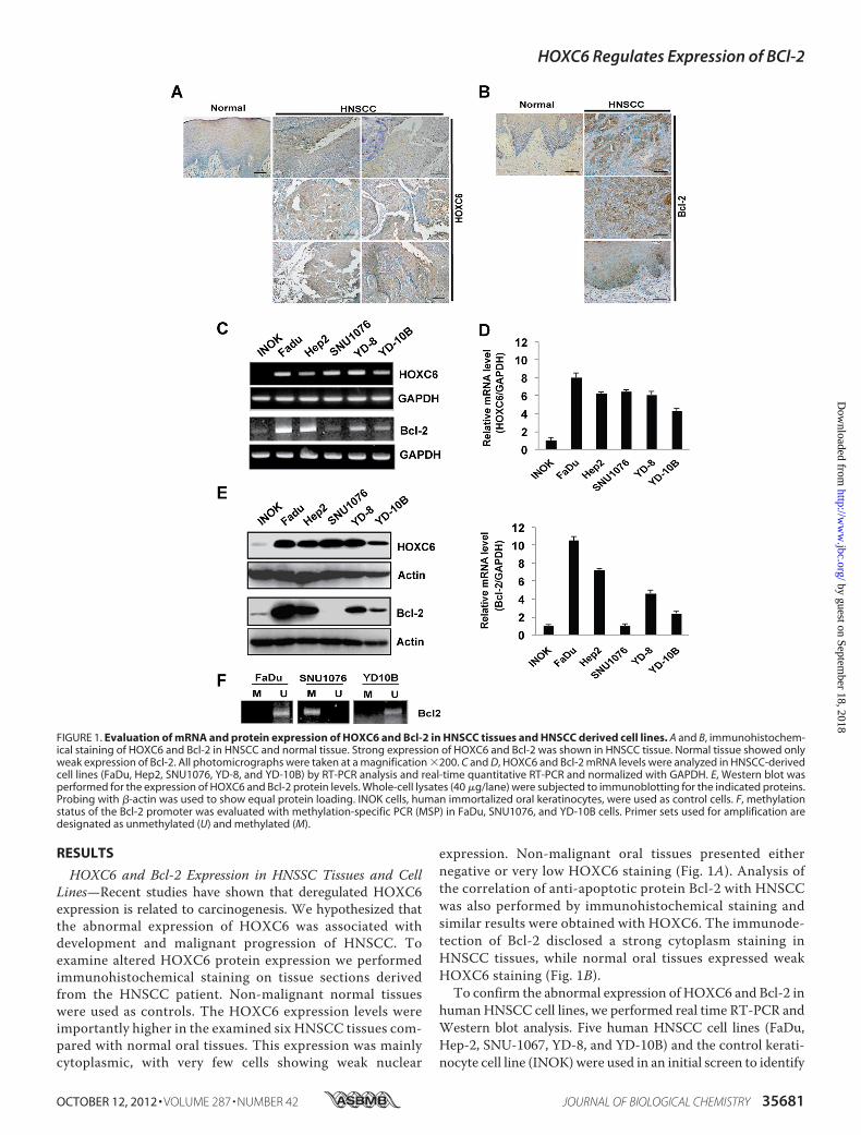
HOXC6 and Bcl-2 Expression in HNSSC Tissues and CellLines—Recent studies have shown that deregulated HOXC6expression is related to carcinogenesis. We hypothesized thatthe abnormal expression of HOXC6 was associated withdevelopment and malignant progression of HNSCC. Toexamine altered HOXC6 protein expression we performedimmunohistochemical staining on tissue sections derivedfrom the HNSCC patient. Non-malignant normal tissueswere used as controls. The HOXC6 expression levels wereimportantly higher in the examined six HNSCC tissues com-pared with normal oral tissues. This expression was mainlycytoplasmic, with very few cells showing weak nuclear
expression. Non-malignant oral tissues presented eithernegative or very low HOXC6 staining (Fig. 1A). Analysis ofthe correlation of anti-apoptotic protein Bcl-2 with HNSCCwas also performed by immunohistochemical staining andsimilar results were obtained with HOXC6. The immunode-tection of Bcl-2 disclosed a strong cytoplasm staining inHNSCC tissues, while normal oral tissues expressed weakHOXC6 staining (Fig. 1B).To confirm the abnormal expression of HOXC6 and Bcl-2 in
humanHNSCC cell lines, we performed real time RT-PCR andWestern blot analysis. Five human HNSCC cell lines (FaDu,Hep-2, SNU-1067, YD-8, and YD-10B) and the control kerati-nocyte cell line (INOK) were used in an initial screen to identify
FIGURE 1. Evaluation of mRNA and protein expression of HOXC6 and Bcl-2 in HNSCC tissues and HNSCC derived cell lines. A and B, immunohistochem-ical staining of HOXC6 and Bcl-2 in HNSCC and normal tissue. Strong expression of HOXC6 and Bcl-2 was shown in HNSCC tissue. Normal tissue showed onlyweak expression of Bcl-2. All photomicrographs were taken at a magnification �200. C and D, HOXC6 and Bcl-2 mRNA levels were analyzed in HNSCC-derivedcell lines (FaDu, Hep2, SNU1076, YD-8, and YD-10B) by RT-PCR analysis and real-time quantitative RT-PCR and normalized with GAPDH. E, Western blot wasperformed for the expression of HOXC6 and Bcl-2 protein levels. Whole-cell lysates (40 �g/lane) were subjected to immunoblotting for the indicated proteins.Probing with �-actin was used to show equal protein loading. INOK cells, human immortalized oral keratinocytes, were used as control cells. F, methylationstatus of the Bcl-2 promoter was evaluated with methylation-specific PCR (MSP) in FaDu, SNU1076, and YD-10B cells. Primer sets used for amplification aredesignated as unmethylated (U) and methylated (M).
HOXC6 Regulates Expression of BCl-2
OCTOBER 12, 2012 • VOLUME 287 • NUMBER 42 JOURNAL OF BIOLOGICAL CHEMISTRY 35681
by guest on September 18, 2018
http://ww
w.jbc.org/
Dow
nloaded from
potential associations between HOXC6 and Bcl-2. MostHNSCC cell lines had elevated HOXC6 mRNA and proteinlevels compared with the control INOK cells (Fig. 1, C and E).The real time RT-PCR analysis confirmed that HOXC6 andBcl-2 were significantly up-regulated in 5 HNSCC cell lines(Fig. 1D). The ratio of HOXC6 mRNA expression was �4-foldin all HNSCC cell lines, and the highest ratio was a 7-foldincrease, as determined using real time RT-PCR. Similarly,Bcl-2 expression was increased in these cell lines, with theexception of the SNU1076 cells.Aberrant methylation of DNA has been recognized as an
important mechanism for the regulation of the expression ofgenes (32). To investigate whether DNAmethylation is relatedto the suppression of Bcl-2 in SNU1076 cells, the methylationstatus of the Bcl-2 promoter was evaluated using MSP. Nomethylation of the Bcl-2 promoter was observed in FaDu andYD-10B cells, whereas methylation of the Bcl-2 promoter wasshowed in SNU1076 cells, regardless of HOXC6 expression;unmethylation was observed in FaDu and YD-10B, but was notobserved in SNU1076 (Fig. 1E). Our results showed that theincreased DNA methylation of this CpG region of the Bcl-2promoter is a possible mechanism for gene silencing in theSNU1076 cells. These results suggest that aberrant expressionof HOXC6 and Bcl-2 protein may be associated with the devel-opment of HNSCC. Hence, FaDu and YD-10B cells were cho-sen for this approach.HOXC6 Regulates the Expression of Bcl-2 and Cell Prolifera-
tion in HNSCC Cells—We then examined whether the overex-pression of HOXC6 in FaDu and YD-10B cells affects theexpression of Bcl-2. Correlations between HOXC6 levels andBcl-2 expression were assessed following the measurement oftranscripts using RT-PCR and Western blotting. Notably, theoverexpression of HOXC6 increased expression of Bcl-2mRNA and protein in the FaDu and YD-10B cells (Fig. 2,A andB). However, the mRNA and protein levels of Bax, a pro-apop-totic protein, in HOXC6-induced FaDu and YD-10B cells werenot affected. As expected, the overexpression of HOXC6caused increased activity of Bcl-2 promoters in FaDu andYD-10B cells (Fig. 2C). In addition, theMTT assay showed thatcells transfected with HOXC6 had significantly higher cell via-bility levels than their controls (Fig. 2D). These results indicatethat expression of HOXC6 induces expression of Bcl-2 andincrease the cell viability.We used siRNA transfection to knock downHOXC6 expres-
sion in FaDu and YD-10B cells to determine the impact onBcl-2 expression. RT-PCR and Western blots were performedto analyze mRNA and protein expression levels, respectively.The decrease in Bcl-2 mRNA levels occurred with knock downof HOXC6 by siRNA as shown in Fig. 2E. When HOXC6 pro-tein expression was decreased using siRNA against HOXC6,Bcl-2 protein expression was also repressed by 70% as shown inFig. 2F. Therefore, these results suggest that HOXC6 positivelyregulates Bcl-2 in both cell lines.HOXC6 Directly Regulates Bcl-2 Promoter Activity—To test
the effects ofHOXC6 expression onBcl-2 promoter activity, wegenerated a luciferase reporter construct driven by the 1.2 kbBcl-2 promoter. INOK, FaDu, and YD-10B cells were tran-siently transfected with the Bcl-2 promoter-luciferase reporter
plasmids. As shown in Fig. 3A, the ratio of Bcl-2 activity was�3-fold in FaDu and YD-10B cells compared with INOK cells(Fig. 3A).We next attempted to define the binding sites of HOXC6 in
the Bcl-2 promoter by serially deleting regions within the pro-moter. As depicted in Fig. 3B, the deletion constructs pGL3-Bcl-2-B1, -B2, -B3, and -B4 did not have drastic effects on theluciferase activity. However, the deletion construct pGL3-Bcl-2-B5 dramatically decreased the ability to be activated byHOXC6 (Fig. 3B). These data suggest that there may be criticalbinding sites for HOXC6 in the pGL3-Bcl-2-B4 promoterregion.It was demonstrated in the previous report that HOXC6
belongs to the HOX family of transcription factors that bind tothe TAAT sequences of target gene promoters to regulate geneexpression (14). According to the promoter sequence analysisfor pGL3-Bcl-2-B4, there is one tentative TAAT element bind-ing site (�420 bp) for HOXC6 (Fig. 3C).To confirm binding of HOXC6 to the Bcl-2 promoter, we
performed a chromatin immunoprecipitation (ChIP) assay.The crosslinked extracts in FaDu andYD-10B cells were immu-noprecipitated with either antibodies against HOXC6 or con-trol anti-IgG (Fig. 3D). The crosslinked DNA was analyzed byPCR using primers designed to amplify theHOXC6-responsiveregion that covers the TAATmotif (�420 bp) of the pGL3-Bcl-2-B4 promoter. Comparedwith the IgG control group,HOXC6was determined to be associated with the Bcl-2 promoterregion containing theTAATmotif. In addition, a PCR fragment(147 bp) corresponding to �609 to �463 (containing �480TAAT motif from start codon) of Bcl-2 promoter was used asHOXC6 binding negative control. This fragment was notdetected when HOXC6 antibody was used for the pull-downassay (Fig. 3D).To examine the binding of HOXC6 to the Bcl-2 promoter in
tumor, we performed a ChIP assay in HNSCC tissues. Wefound that Bcl-2 promoter-specific PCR primers amplified thispromoter region fromDNA that was immunoprecipitated withthe anti-HOXC6 antibody in tumor tissue but not in normaltissue, demonstrating that the interaction between HOXC6protein and the Bcl-2 promoter was specific in tumor. (supple-mental Fig. S1).To confirm whether the �420 bp TAATmotif was involved
in HOXC6-mediated regulation of Bcl-2 promoter activity, the�420 bp TAAT binding site within the Bcl-2 promoter regionwasmutated and the activity of themutated promoter was eval-uated in FaDu and YD-10B cells without HOXC6 overexpres-sion. As shown in Fig. 3E, the introduction of point mutation atHOXC6 (�420 bp TAAT) binding site of the Bcl-2 promoterattenuated Bcl-2 promoter activity compared with that of thewild-type control. These results indicate that HOXC6 directlybinds the �420 TAAT elements on Bcl-2 promoter region andregulates its gene expression.Knockdown of HOXC6 Induces Cell Death in HNSCC Cell
Lines—To determine whether depletion of HOXC6 by siRNAcan induce apoptosis followed by cell growth inhibition, weperformed MTT assay and flow cytometry analysis usingAnnexin V/PI double staining. The MTT assay indicated thatexpression of siHOXC6 significantly inhibited the growth of
HOXC6 Regulates Expression of BCl-2
35682 JOURNAL OF BIOLOGICAL CHEMISTRY VOLUME 287 • NUMBER 42 • OCTOBER 12, 2012
by guest on September 18, 2018
http://ww
w.jbc.org/
Dow
nloaded from

FaDu and YD-10B cells, whereas down-regulation of HOXC6did not affect the growth of INOK control cells (Fig. 4A). Asshown in Fig. 4B, siHOXC6 induced the cell death comparedwith the untreated control cells. In the vehicle-treated cells,3.0% were positive for Annexin V-FITC staining whilesiHOXC6 treatment resulted in increases of 13.7% and 13.2% inFaDu and YD-10B cells, respectively. We also found thatsiHOXC6 inhibited the expression level of Bcl-2 and increasedthe level of cleaved PARP and cytochrome c release in both celllines (Fig. 4C). In addition, activation of caspase-3was observedin siHOXC6-treated cells (Fig. 4D). These results suggest thatsiHOXC6 induces caspase-3-dependent apoptosis in FaDu andYD-10B cells.HOXC6 Inhibits Paclitaxel-induced Apoptosis in HNSCC
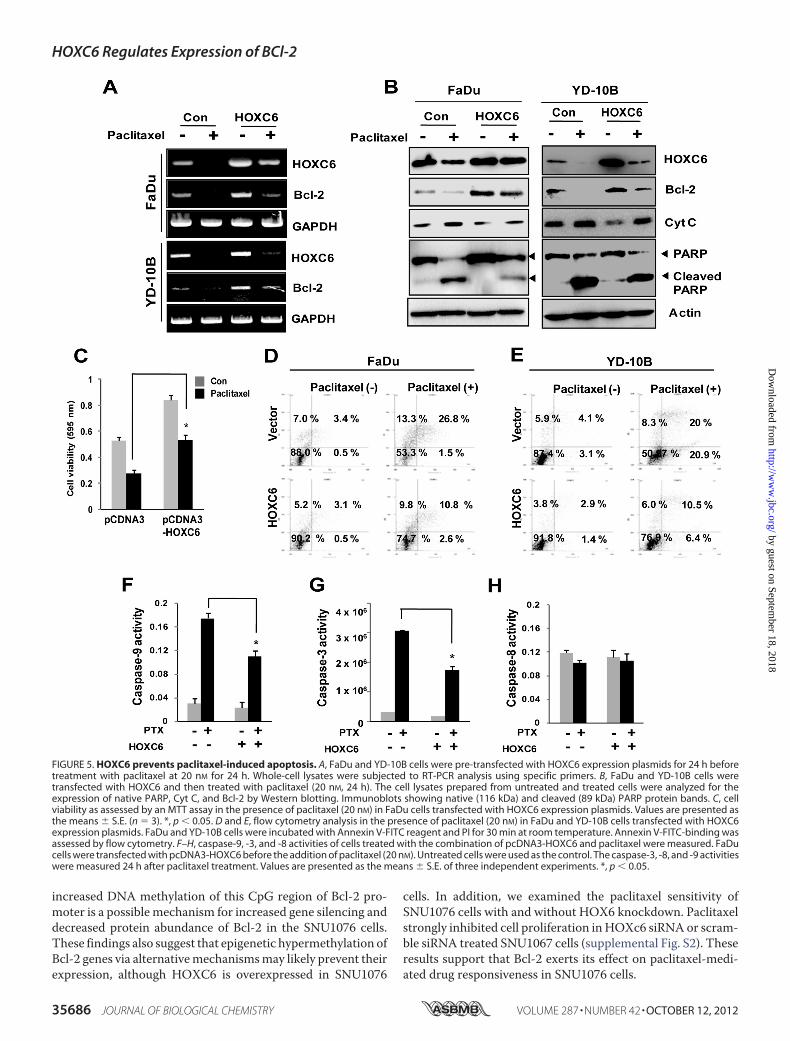
Cells—To examine whether the cytotoxic effect of paclitaxel onHNSCC cells would be affected by HOXC6 expression, we
compared the apoptosis related protein levels in FaDu andYD-10B cells cell treated with paclitaxel alone or with the com-bination of paclitaxel and HOXC6 transfection. RT-PCR andWestern blot analysis revealed that the expression of Bcl-2decreased when FaDu and YD-10B cells were exposed to pacli-taxel. Remarkably, overexpression of HOXC6 recovered thedecreased expression levels of Bcl-2 mRNA and protein withpaclitaxel treatment (Fig. 5,A and B). We further examined theeffect of HOXC6 on the level of cytosolic cytochrome c (Cyt C)and poly (ADP-ribose) polymerase (PARP), a component of thesignaling pathway of apoptosis. Western blot analysis revealedthat paclitaxel caused an increased Cyt C and cleaved PARPlevels in FaDu and YD-10B cells. HOXC6-transfected cellsstrongly inhibited the levels of cleaved PARP and Cyt C by pacli-taxel treatment, suggesting that they inhibit paclitaxel-inducedapoptosis (Fig. 5B). AnMTT assay was performed to evaluate the
FIGURE 2. HOXC6 regulates the expression of Bcl-2 at the transcriptional level as well as cell proliferation. FaDu and YD-10B cells were transfected withpcDNA3-HOXC6 plasmid as described under “Experimental Procedures.” A, after 48 h transfection, mRNA was determined by RT-PCR assays, and (B) HOXC6,Bcl-2, and Bax protein levels were determined by Western blot analysis. C, FaDu and YD-10B cells transiently transfected with pCDNA3-HOXC6. At 24 hpost-transfection, cells were co-transfected pGL3-Bcl-2 promoter region together with pRL-TK for 24 h (which carried the Renilla luciferase gene controlled byHSV-tk promoter as internal control). Cell lysates were subjected to luciferase activity assays using the dual luciferase reporter assay system, and the luciferaseactivity was normalized to pRL-TK activity (mean � S.D. of three independent experiments. *, p � 0.05; **, p � 0.01). D, FaDu and YD-10B cells were transfectedfor 48 h with either pcDNA3-HOXC6 or control vector. After transfection, the cell viability was analyzed using the MTT assay. **, p � 0.01 (E) RT-PCR quantitationof Bcl-2 mRNA in FaDu and YD-10B cells transiently transfected with scRNA or HOXC6 siRNA. After 48 h of transfection, mRNA level of Bcl-2 was determined byRT-PCR analysis. F, Western blots of HOXC6 and Bcl-2 expression in cells co-transfected with scRNA or HOXC6 siRNA. Whole cell lysates were obtainedpost-transfection and the samples were separated by 12% SDS/PAGE and probed for HOXC6, Bcl-2, Bax, and actin as a loading control.
HOXC6 Regulates Expression of BCl-2
OCTOBER 12, 2012 • VOLUME 287 • NUMBER 42 JOURNAL OF BIOLOGICAL CHEMISTRY 35683
by guest on September 18, 2018
http://ww
w.jbc.org/
Dow
nloaded from

effects of HOXC6 on paclitaxel sensitivity of the cells. The cellviability of the paclitaxel-treated FaDu cells was significantlydecreased compared with the untreated cells, while overexpres-sion ofHOXC6 increased cell viability in the FaDu cells comparedwith the vehicle-transfected cells (Fig. 5C). Interestingly, paclitaxeltreated HOXC6 overexpressing cells significantly experiencedincreased cell viability compared with paclitaxel alone. Theseresults strongly support thatHOXC6has the cell protection effecton paclitaxel-mediated cell sensitivity.We investigated the effect of HOXC6 on paclitaxel-induced
apoptosis inFaDuandYD-10Bcells using flowcytometry analysis.WefoundthatAnnexin-Vpositivecellswere inducedbypaclitaxeltreatment in both cells. In contrast, HOXC6-expressing cellstreated with paclitaxel displayed inhibited late apoptosis com-pared with the cells treated with paclitaxel alone (Fig. 5, E and F).
To support these results, we investigated caspase activityafter the cells were treated with paclitaxel and/or HOXC6. Thecolorimetric assay revealed that caspase-3 and -9 activities wereremarkably increased in the paclitaxel-treated FaDu cells,whereas the caspase-3 and -9 activities in HOXC6 overexpress-ing FaDu cells treated with paclitaxel were significantly lowerthan those in the FaDu/paclitaxel cells (Fig. 5, E and F).Caspase-8 activity was not affected by paclitaxel treatment andHOXC6 expression (Fig. 5G).
DISCUSSION
HOX genes constitute a family of transcription factors thatare key to developmental processes, embryonic morphogene-sis, and differentiation (33, 34). They are also expressed in var-ious human cancers including esophagus, prostate, and colon,
FIGURE 3. Identification of the HOXC6 binding site in the promoter of Bcl-2 gene. A, INOK, FaDu, and YD-10B cells were plated in 12-well cell culture dishes.Each group of cells was transiently transfected with pCDNA3-HOXC6. At 24 h post-transfection, cells were co-transfected pGL3-Bcl-2 promoter region togetherwith pRL-TK. Cell lysates were subjected to luciferase activity assays using the dual luciferase reporter assay system, and the luciferase activity was normalizedto pRL-TK activity. B, deletion analysis of the Bcl-2 promoter-reporter constructs (pGL3-Bcl-2-B1 to pGL3-Bcl-2-B7). Dual-reporter gene assay were performedwith expression constructs carrying various lengths of Bcl-2 promoter. FaDu cells were co-transfected with one of seven constructs with a pRL-TK. After 48 h oftransfection, cell extracts were prepared and analyzed for luciferase activity and normalized to Renilla luciferase activity. C, pGL3-Bcl-2-B4 promoter sequencecontaining one putative HOXC6 binding sites (TAAT motif, �420 bp). A diagram of the proximal 150 bp region of the pGL3-Bcl-2-B4 promoter. D, demonstra-tion of HOXC6-binding site (TAAT motif, �420 bp) on the Bcl-2 promoter by ChIP assay. Chromatin lysates from FaDu, and YD-10B cells were immunoprecipi-tated (IP) with antibody against HOXC6. Nonimmune IgG was used as negative control for immunoprecipitation. Samples were processed as described under“Experimental Procedures.” The left panel shows that PCR using chromatin (Input) pulled down by anti-HOXC6 antibody (HOXC6 Ab) as template yielded theBcl-2 promoter, which was verified the Bcl-2 fragment (�420 TAAT containing, positive primer) of Bcl-2 promoter. When negative control IgG or no antibody(No Ab) was used for pull-down assay, no PCR product was observed. The right panel shows that immunoprecipitated DNA was amplified using primersrepresenting a site upstream of the �420 TAAT HOXC6-binding site (�480 TAAT containing, negative primer) on the Bcl-2 promoter. No PCR product wasobserved pull-down of HOXC6 antibody. N-CAM promoter used as HOXC6 binding positive control using ChIP assay. E, pGL3-Bcl-2 promoter assay with TAATmotif (�420 bp) point mutation constructs in FaDu and YD-10B cells. FaDu and YD-10B cells were transfected with wild type pGL3-Bcl-2 promoter orpoint-mutated pGL3-MDR-1 (TAAT3TGGT). pRL(Renilla luciferase) plasmid was co-transfected as an internal control. The cells were harvested 48 h aftertransfection. The promoter activity of each preparation was normalized to the Renilla value. The relative promoter activity is averaged from at least threeindependent experiments. *, p � 0.05; **, p � 0.01.
HOXC6 Regulates Expression of BCl-2
35684 JOURNAL OF BIOLOGICAL CHEMISTRY VOLUME 287 • NUMBER 42 • OCTOBER 12, 2012
by guest on September 18, 2018
http://ww
w.jbc.org/
Dow
nloaded from

suggesting a potential role in carcinogenesis (13, 14). Recently,a study described the expression ofHOXgenes in oral dysplasiaand oral squamous cell carcinoma (OSCC), suggesting thatoverexpression of particular HOX genes is implicated in thedevelopment of oral dysplasia and oral SCC (19, 35). Moreover,another study using human salivary glands showed the possi-bility that HOX genes play a role in salivary gland carcinogen-esis (36). Among them, aberrant expression of HOXC6 geneswas observed in oral SCC and esophageal SCC (10, 19). Therewas a tendency for increased expression ofHOXC6 in SCCwithlymph node metastasis (20); this finding suggests that HOXC6may play a key role in the acquisition of metastatic phenotypesofOSSC.However, the current study is insufficient in establish-ing its function, the specific cellular signaling to which it con-tributes, or its relationships with the HNSCC phenotype.In this study, we identified that the levels of HOXC6 expres-
sion experience, importantly, an increase in expression byHNSCC cell lines compared with immortalized oral keratino-cyte (INOK) cells when assessed bymeasuringmRNA. The lev-els of Bcl-2mRNAwere also significantly higher inHNSCC celllines than in control INOK cells. Consistently, the analysis ofimmunoblotting demonstrated a statistically significant corre-
lation between expression of HOXC6 and Bcl-2 in HNSCC celllines. In SNU1076 cells, the level of Bcl-2 expression was lowerthan other HNSCC cell lines, although HOXC6 was increasedin SNU1076 cells compared with INOK cells. Recently, inHOXC6 expressed tumors cell, target genes of HOXC6, includ-ing FGFR2, CD44, andWIF, have been shown to be silenced byhypermethylation (37–39). It was still not clear whether thatthe expression of Bcl-2 ismediated throughmethylation of spe-cificCpG sites on theBcl-2 promoter in SNU1076 cells.Nguyenet al. provided evidence that a 417 bp CpG island of the Bcl-2gene was identified as hypermethylated in all co-twins exhibit-ing autistic traits (40). Some studies reported Bcl-2 exon 2methylation status in cancer (41). It was reported that Bcl-2promoter is hypermethylated in colorectal cancer tissue (42).They suggest that aberrant methylation of Bcl-2 promoter maybe involved in the expression of Bcl-2. This possibility wasexamined, for bisulfite-treated DNA from SNU601 cells, bynested methylation-specific PCR (MSP). We found that Bcl-2promoter from SNU1076 cells yielded a PCR product only withmethylation-specific primers, whereas DNA from FaDu andYD-10B cells yielded a PCR product only with unmethylation-specific primers (Fig. 1F). Our results suggested that the
FIGURE 4. Silencing of HOXC6 caused caspase 3-dependent apoptosis. A, FaDu and YD-10B cells were transfected for 48 h with either HOXC6 siRNA orscrambled siRNA (scRNA). After 48 h, cell viability was analyzed using the MTT assay. The results of three experiments (n 3) are summarized. *, p � 0.05; **, p �0.01. B, representative of cell apoptosis with flow cytometry analysis. After siHOXC6 transfection, cells were stained with Annexin V/PI to analyze apoptotic cellpopulations. C, Western blotting of Bcl-2, cytochrome c, and cleaved PARP. Whole cell lysates were obtained after 48 h of transfection, and the samples wereseparated by 12% SDS/PAGE and probed for cleaved PARP, Bcl-2, Cyt C, and actin as a loading control. D, FaDu and YD-10B cells were plated in 96-well platesand treated with scRNA or SiHOXC6 as described under “Experimental Procedures.” Caspase-Glo3 reagent was added to wells and luminescence was measuredafter 2 h of incubation. Data were collected from three independent experiments. *, p � 0.05.
HOXC6 Regulates Expression of BCl-2
OCTOBER 12, 2012 • VOLUME 287 • NUMBER 42 JOURNAL OF BIOLOGICAL CHEMISTRY 35685
by guest on September 18, 2018
http://ww
w.jbc.org/
Dow
nloaded from
increased DNA methylation of this CpG region of Bcl-2 pro-moter is a possible mechanism for increased gene silencing anddecreased protein abundance of Bcl-2 in the SNU1076 cells.These findings also suggest that epigenetic hypermethylation ofBcl-2 genes via alternativemechanismsmay likely prevent theirexpression, although HOXC6 is overexpressed in SNU1076
cells. In addition, we examined the paclitaxel sensitivity ofSNU1076 cells with and without HOX6 knockdown. Paclitaxelstrongly inhibited cell proliferation inHOXc6 siRNA or scram-ble siRNA treated SNU1067 cells (supplemental Fig. S2). Theseresults support that Bcl-2 exerts its effect on paclitaxel-medi-ated drug responsiveness in SNU1076 cells.
FIGURE 5. HOXC6 prevents paclitaxel-induced apoptosis. A, FaDu and YD-10B cells were pre-transfected with HOXC6 expression plasmids for 24 h beforetreatment with paclitaxel at 20 nM for 24 h. Whole-cell lysates were subjected to RT-PCR analysis using specific primers. B, FaDu and YD-10B cells weretransfected with HOXC6 and then treated with paclitaxel (20 nM, 24 h). The cell lysates prepared from untreated and treated cells were analyzed for theexpression of native PARP, Cyt C, and Bcl-2 by Western blotting. Immunoblots showing native (116 kDa) and cleaved (89 kDa) PARP protein bands. C, cellviability as assessed by an MTT assay in the presence of paclitaxel (20 nM) in FaDu cells transfected with HOXC6 expression plasmids. Values are presented asthe means � S.E. (n 3). *, p � 0.05. D and E, flow cytometry analysis in the presence of paclitaxel (20 nM) in FaDu and YD-10B cells transfected with HOXC6expression plasmids. FaDu and YD-10B cells were incubated with Annexin V-FITC reagent and PI for 30 min at room temperature. Annexin V-FITC-binding wasassessed by flow cytometry. F–H, caspase-9, -3, and -8 activities of cells treated with the combination of pcDNA3-HOXC6 and paclitaxel were measured. FaDucells were transfected with pcDNA3-HOXC6 before the addition of paclitaxel (20 nM). Untreated cells were used as the control. The caspase-3, -8, and -9 activitieswere measured 24 h after paclitaxel treatment. Values are presented as the means � S.E. of three independent experiments. *, p � 0.05.
HOXC6 Regulates Expression of BCl-2
35686 JOURNAL OF BIOLOGICAL CHEMISTRY VOLUME 287 • NUMBER 42 • OCTOBER 12, 2012
by guest on September 18, 2018
http://ww
w.jbc.org/
Dow
nloaded from

Although the exact action mechanism of HOXC6 inSNU1076 cells is unknown, many different molecules and sig-naling pathways have been demonstrated in various cancer celllines. Because different types of tumor cells have differentgenetic alterations and characteristics, the molecular targets ofHOXC6 could be different in SNU1076 cell lines.HOXC6 is a HOX family member that presumably functions
by binding directly to DNA promoter elements via its home-odomain, which is a DNA binding domain located within its Nterminus (43). The DNA core sequence of the homeodomainbinding sites (HBSs) frequently contains the sequence TAAT.Previous studies showed thatHOXC6binds to similar elementson the neural cell adhesion molecule (N-CAM) promoter (44).In our study, the results of the promoter deletion mutants con-firm that HOXC6 directly binds to the specific TAAT, asequence motif in the Bcl-2 gene promoter. In addition, ChIPapproaches clearly demonstrated that HOXC6 was associatedwith the TAATmotif (�420 bp from start codon) in Bcl-2 pro-moter. These studies support the newHOXC6mechanism thatinvolves direct binding of HOXC6 to specific promoter ele-ments to activate Bcl-2 expression in HNSCC cells. Moreover,siRNA directed against HOXC6 resulted in decreased Bcl-2mRNA levels and reduced luciferase reporter activity in FaDuand YD-10B cells. Therefore, we believe that HOXC6 inducesBcl-2 expression in HNSCC cells by directly increasing its pro-moter activity.Generally, cells harboring multiple pathphysiologic altera-
tions are normally eliminated by apoptosis. In normal cells,diminished apoptosis plays a critical role in tumor initiation,progression, and drug resistance (26). Several proteins thatinhibit apoptosis have been identified, including Bcl-2 familymembers Bcl-2 and Bcl-XL. Particularly, Bcl-2 acts as a keyregulator of cellular apoptosis and is an important determinantof cellular sensitivity or resistance to chemotherapy drugs (31).Overexpression of Bcl-2 is commonly observed in HNSCC andmay play an important role in overcoming apoptosis inHNSCC(45, 46).We found evidence that the presence of HOXC6 in HNSCC
is strongly associated with Bcl-2 expression. Small-interferingRNA knock-down of HOXC6 expression induces apoptosis,and overexpression of HOXC6 increases the growth/prolifera-tion of FaDu andYD-10B cells. In this study, the overexpressionofHOXC6was investigated to understand the effect ofHOXC6upon apoptosis induced by the chemotherapeutic agent pacli-taxel using theMTT test and a flow cytometry approach.One ofthe interesting findings of this study was that the expressionof HOXC6 was significantly associated with the inhibition ofpaclitaxel-induced apoptosis, thereby confirming the anti-apo-ptotic role of HOXC6. Paclitaxel decreased expression of theanti-apoptotic protein Bcl-2 and increased caspase-3-depen-dent cleaved PARP. The reduced mRNA and protein levels ofBcl-2 were recovered by HOXC6 expression in FaDu andYD-10B cells. In addition, overexpression of HOXC6 inhibitsthe activity of paclitaxel-induced caspase-3 and -9 and thearrest of cell growth, suggesting that HOXC6-mediated Bcl-2expression plays a protective role in enhanced paclitaxel cyto-toxicity. The overexpression of HOXC6 in HNSCC may block
this apoptotic pathway and allow aberrant cell growth throughmitosis.Previous studies suggested that the PI3K/Akt pro-prolifera-
tive and survival pathway are influenced by several HOXC6direct targets such as BMP7, IGFBP3, and PDGFRA (22–24). Itis possible that anothermechanism by whichHOXC6 exerts itspro-survival function may be the PI3K/Akt pathway.In conclusion, the evidence presented in this report indicates
that HOXC6 is highly expressed in HNSCC cell lines andinduces proliferative activity. Our results also provide novelinformation about the function of HOXC6 in the regulation ofBcl-2 gene expression and its potential anti-apoptotic role.
REFERENCES1. Jemal, A., Siegel, R., Xu, J., andWard, E. (2010) Cancer statistics, 2010.CA
Cancer J. Clin. 60, 277–3002. Parkin, D. M. (2001) Global cancer statistics in the year 2000. Lancet
Oncol. 2, 533–5433. Warnakulasuriya, S. (2009) Global epidemiology of oral and oropharyn-
geal cancer. Oral Oncol. 45, 309–3164. Warnakulasuriya, S. (2009) Significant oral cancer risk associatedwith low
socioeconomic status. Evid Based Dent. 10, 4–55. Argiris, A., and Eng, C. (2003) Epidemiology, staging, and screening of
head and neck cancer. Cancer Treat Res. 114, 15–606. Abate-Shen, C. (2002) Deregulated homeobox gene expression in cancer:
cause or consequence? Nat. Rev. Cancer 2, 777–7857. Veraksa, A., Del Campo, M., and McGinnis, W. (2000) Developmental
patterning genes and their conserved functions: frommodel organisms tohumans.Mol. Genet. Metab. 69, 85–100
8. Friedmann, Y., Daniel, C. A., Strickland, P., and Daniel, C. W. (1994) Hoxgenes in normal and neoplastic mouse mammary gland. Cancer Res. 54,5981–5985
9. Castronovo, V., Kusaka, M., Chariot, A., Gielen, J., and Sobel, M. (1994)Homeobox genes: potential candidates for the transcriptional control ofthe transformed and invasive phenotype. Biochem. Pharmacol. 47,137–143
10. Chen, K. N., Gu, Z. D., Ke, Y., Li, J. Y., Shi, X. T., and Xu, G. W. (2005)Expression of 11 HOX genes is deregulated in esophageal squamous cellcarcinoma. Clin. Cancer Res. 11, 1044–1049
11. Takahashi, Y., Hamada, J., Murakawa, K., Takada, M., Tada, M., Nogami,I., Hayashi, N., Nakamori, S., Monden, M., Miyamoto, M., Katoh, H., andMoriuchi, T. (2004) Expression profiles of 39 HOX genes in normal hu-man adult organs and anaplastic thyroid cancer cell lines by quantitativerealtime RT-PCR system. Exp. Cell Res. 293, 144–153
12. van Oostveen, J., Bijl, J., Raaphorst, F., Walboomers, J., and Meijer, C.(1999) The role of homeobox genes in normal hematopoiesis and hema-tological malignancies. Leukemia. 13, 1675–1690
13. Yahagi, N., Kosaki, R., Ito, T., Mitsuhashi, T., Shimada, H., Tomita, M.Takahashi, T., and Kosaki, K. (2004) Position-specific expression of HOXgenes along the gastrointestinal tract. Congenit Anom. 44, 18–26
14. Bodey, B., Bodey, B. Jr., Siegel, S. E., and Kaiser, H. E. (2000) Immunocy-tochemical detection of the homeobox B3, B4, and C6 gene products inbreast carcinomas. Anticancer Res. 20, 3281–3286
15. Fujiki, K., Duerr, E. M., Kikuchi, H., Ng, A., Xavier, R. J., Mizukami, Y.,Imamura, T., Kulke, M. H., and Chung, D. C. (2008) Hoxc6 is overex-pressed in gastrointestinal carcinoids and interacts with JunD to regulatetumor growth. Gastroenterology 135, 907–916
16. Bodey, B., Bodey, B. Jr., Siegel, S. E., Luck, J. V., and Kaiser, H. E. (2000)Homeobox B3, B4, and C6 gene product expression in osteosarcomas asdetected by immunocytochemistry. Anticancer Res. 20, 2717–2721
17. Miller, G. J., Miller, H. L., van Bokhoven, A., Lambert, J. R., Werahera,P. N., Schirripa, O., Lucia, M. S., and Nordeen, S. K. (2003) AberrantHOXC expression accompanies themalignant phenotype in human pros-tate. Cancer Res. 63, 5879–5888
18. Tucci, R., Campos, M. S., Matizonkas-Antonio, L. F., Durazzo, M., PintoJunior Ddos, S., and Nunes, F. D. (2011) HOXB5 expression in oral squa-
HOXC6 Regulates Expression of BCl-2
OCTOBER 12, 2012 • VOLUME 287 • NUMBER 42 JOURNAL OF BIOLOGICAL CHEMISTRY 35687
by guest on September 18, 2018
http://ww
w.jbc.org/
Dow
nloaded from

mous cell carcinoma. J. Appl. Oral Sci. 19, 125–12919. Hassan, N. M., Hamada, J., Murai, T., Seino, A., Takahashi, Y., Tada, M.,
Zhang, X., Kashiwazaki, H., Yamazaki, Y., Inoue, N., and Moriuchi, T.(2006) Aberrant expression of HOX genes in oral dysplasia and squamouscell carcinoma tissues. Oncol Res. 16, 217–224
20. Bijl, J., van Oostveen, J. W., Kreike, M., Rieger, E., van der Raaij-Helmer,L.M.,Walboomers, J.M., Corte, G., Boncinelli, E., van den Brule, A. J., andMeijer, C. J. (1996) Expression of HOXC4, HOXC5, and HOXC6 in hu-man lymphoid cell lines, leukemias, and benign and malignant lymphoidtissue. Blood 87, 1737–1745
21. McCabe, C. D., Spyropoulos, D. D., Martin, D., and Moreno, C. S. (2008)Genome-wide analysis of the homeobox C6 transcriptional network inprostate cancer. Cancer Res. 68, 1988–1996
22. Grishina, I. B., Kim, S. Y., Ferrara, C., Makarenkova, H. P., Walden, P. D.(2005) BMP7 inhibits branching morphogenesis in the prostate gland andinterferes with Notch signaling. Dev. Biol. 288, 334–347
23. Ricort, J. M., and Binoux, M. (2002) Insulin-like growth factor-bindingprotein-3 activates a phosphotyrosine phosphatase. Effects on the insulin-like growth factor signaling pathway. J. Biol. Chem. 277, 19448–19454
24. van der Geer, P., Hunter, T., and Lindberg, R. A. (1994) Receptor protein-tyrosine kinases and their signal transduction pathways. Annu. Rev. CellBiol. 10, 251–337
25. Lin, Y, Liu, G., Zhang, Y., Hu, Y. P., Yu, K., Lin, C., McKeehan, K., Xuan,J. W., Ornitz, D. M., Shen, M. M., Greenberg, N., McKeehan, W. L., andWang, F. (2007) Fibroblast growth factor receptor 2 tyrosine kinase isrequired for prostatic morphogenesis and the acquisition of strict andro-gen dependency for adult tissue homeostasis. Development 134, 723–734
26. Danial, N. N., and Korsmeyer, S. J. (2004) Cell death: critical controlpoints. Cell 116, 205–219
27. Trask, D. K., Wolf, G. T., Bradford, C. R., Fisher, S. G., Devaney, K., John-son, M., Singleton, T., and Wicha, M. (2002) Expression of Bcl-2 familyproteins in advanced laryngeal squamous cell carcinoma: correlation withresponse to chemotherapy and organ preservation. Laryngoscope 112,638–644
28. Sharma, H., Sen, S., Lo Muzio, L., Mariggiò, A., Singh, N. (2005) Anti-sense-mediated downregulation of antiapoptotic proteins induces apo-ptosis and sensitizes head and neck squamous cell carcinoma cells tochemotherapy. Cancer Biol. Ther. 4, 720–727
29. Wolter, K. G., Wang, S. J., Henson, B. S., Wang, S., Griffith, K. A., Kumar,B., Chen, J., Carey, T. E., Bradford, C. R., D’Silva, N. J. (2006) (�)-Gossypolinhibits growth and promotes apoptosis of human head and neck squa-mous cell carcinoma in vivo. Neoplasia 8, 163–172
30. Li, R., Boehm, A. L., Miranda, M. B., Shangary, S., Grandis, J. R., Johnson,D. E. (2007) Targeting antiapoptotic Bcl-2 family members with cell-per-meable BH3 peptides induces apoptosis signaling and death in head andneck squamous cell carcinoma cells. Neoplasia 9, 801–811
31. Cory, S., Adams, J. M. (2002) The Bcl2 family: regulators of the cellularlife-or-death switch. Nat. Rev. Cancer 2, 647–656
32. Jones, P. A., Baylin, S. B. (2002) The fundamental role of epigenetic events
in cancer. Nat. Rev. Genet. 3, 415–42833. Gehring, W. J., Hiromi, Y. (1986) Homeotic genes and the homeobox.
Annu. Rev. Genet. 20, 147–17334. Krumlauf, R. (1994) HOX genes in vertebrate development. Cell 78,
191–20135. De Souza Setubal Destro, M. F., Bitu, C. C., Zecchin, K. G., Graner, E.,
Lopes, M. A., Kowalski, L. P., Coletta, R. D. (2010) Overexpression ofHOXB7 homeobox gene in oral cancer induces cellular proliferation andis associated with poor prognosis. Int. J. Oncol. 36, 141–149
36. Cazal, C., Sobral, A. P., de Almeida, F. C., das Graças Silva-Valenzuela,M.,Durazzo,M. D., Nunes, F. D. (2006) The homeoboxHOXB13 is expressedin human minor salivary gland. Oral Dis. 12, 424–427
37. Wissmann, C., Wild, P. J., Kaiser, S., Roepcke, S., Stoehr, R., Woenckhaus,M., Kristiansen, G., Hsieh, J. C., Hofstaedter, F., Hartmann, A., Knuechel,R., Rosenthal, A., Pilarsky, C. (2003)WIF1, a component of theWnt path-way, is down-regulated in prostate, breast, lung, and bladder cancer.J. Pathol 201, 204–212
38. Park, S., Kim, J. H., Jang, J. H. (2007) Aberrant hypermethylation of theFGFR2 gene in human gastric cancer cell lines. Biochem. Biophys. Res.Commun. 357, 1011–1015
39. Zhu, X., Lee, K., Asa, S. L., Ezzat, S. (2007) Epigenetic silencing throughDNA and histone methylation of fibroblast growth factor receptor 2 inneoplastic pituitary cells. Am. J. Pathol. 170, 1618–1628
40. Nguyen, A., Rauch, T. A., Pfeifer, G. P., Hu, V. W. (2010) Global methyla-tion profiling of lymphoblastoid cell lines reveals epigenetic contributionsto autism spectrum disorders and a novel autism candidate gene, RORA,whose protein product is reduced in autistic brain. FASEB J. 24,3036–3051
41. Babidge, W. J., Butler, L. M., Burton, M. A., Cowled, P. A. (2001) Methy-lation of CpG sites in exon 2 of the bcl-2 gene occurs in colorectal carci-noma. Anticancer Res. 21, 2809–2814
42. Zhu, Q., Jin, Z., Yuan, Y., Lu, Q., Ge, D., Zong, M. (2011) Impact ofMTHFR gene C677T polymorphism on Bcl-2 gene methylation and pro-tein expression in colorectal cancer. Scand J. Gastroenterol. 46, 436–445
43. Svingen, T., Tonissen, K. F. (2006) Hox transcription factors and theirelusive mammalian gene targets. Heredity 97, 88–96
44. Jones, F. S., Holst, B. D., Minowa, O., De Robertis, E. M., Edelman, G. M.(1993) Binding and transcriptional activation of the promoter for the neu-ral cell adhesion molecule by HoxC6 (Hox-3.3). Proc. Natl. Acad. Sci.U.S.A. 90, 6557–6561
45. Drenning, S. D., Marcovitch, A. J., Johnson, D. E., Melhem, M. F.,Tweardy, D. J., Grandis, J. R. (1998) Bcl-2 but not Bax expression is asso-ciated with apoptosis in normal and transformed squamous epithelium.Clin. Cancer Res. 4, 2913–2921
46. Trask, D. K., Wolf, G. T., Bradford, C. R., Fisher, S. G., Devaney, K., John-son, M., Singleton, T., Wicha, M. (2002) Expression of Bcl-2 family pro-teins in advanced laryngeal squamous cell carcinoma: correlation withresponse to chemotherapy and organ preservation. Laryngoscope 112,638–644
HOXC6 Regulates Expression of BCl-2
35688 JOURNAL OF BIOLOGICAL CHEMISTRY VOLUME 287 • NUMBER 42 • OCTOBER 12, 2012
by guest on September 18, 2018
http://ww
w.jbc.org/
Dow
nloaded from

Sung-Min Moon, Soo-A Kim, Jung-Hoon Yoon and Sang-Gun AhnModulates Bcl-2 Expression
HOXC6 Is Deregulated in Human Head and Neck Squamous Cell Carcinoma and
doi: 10.1074/jbc.M112.361675 originally published online August 15, 20122012, 287:35678-35688.J. Biol. Chem.
10.1074/jbc.M112.361675Access the most updated version of this article at doi:
Alerts:
When a correction for this article is posted•
When this article is cited•
to choose from all of JBC's e-mail alertsClick here
Supplemental material:
http://www.jbc.org/content/suppl/2012/08/30/M112.361675.DC1
http://www.jbc.org/content/287/42/35678.full.html#ref-list-1
This article cites 46 references, 9 of which can be accessed free at
by guest on September 18, 2018
http://ww
w.jbc.org/
Dow
nloaded from