hugo alexandre mendes de oliveira - universidade do minho · thesis for phd degree in chemical and...
TRANSCRIPT

Hugo Alexandre Mendes de Oliveira
April, 2014UM
inho
|201
4
Molecular studies on bacteriophage endolysins and their potential to control Gram-negative bacteria
Mo
lecu
lar
stu
die
s o
n b
act
eri
op
ha
ge
en
do
lysi
ns
an
d
the
ir p
ote
nti
al t
o c
on
tro
l Gra
m-n
eg
ati
ve b
act
eri
aH
ugo
Alex
andr
e M
ende
s de
Oliv
eira
Universidade do Minho
Escola de Engenharia

Thesis for PhD degree in Chemical and Biological Engineering
Hugo Alexandre Mendes de Oliveira
April, 2014
Universidade do Minho
Escola de Engenharia
SUPERVISOR: Professor Joana Cecília Valente Rodrigues Azeredo
CO-SUPERVISOR: Doctor Leonardus Dorothea Kluskens
Molecular studies on bacteriophage endolysins and their potential to control Gram-negative bacteria

AUTHOR: Hugo Alexandre Mendes de Oliveira
E-MAIL: [email protected]
TITLE OF THE THESIS: Molecular studies on bacteriophage endolysins and their potential to control
Gram-negative bacteria
SUPERVISOR: Professor Joana Cecília Valente Rodrigues Azeredo
CO-SUPERVISOR: Doctor Leonardus Dorothea Kluskens
CONCLUSION YEAR: 2014
PhD in Chemical and Biological Engineering
THE INTEGRAL REPRODUCTION OF THIS THESIS IS ONLY AUTHORIZED FOR RESEARCH
PURPOSES, PROVIDED PROPER COMMITMENT AND WRITTEN DECLARATION OF THE
INTERESTED PART.
University of Minho, April 2014
__________________________________

“The true sign of intelligence is not knowledge but imagination”
Albert Einstein


v
Acknowledgments
I would like to acknowledge all persons that, made this work possible to
accomplish for their disposal and kindness provided.
First and foremost, I would like to thank my Supervisors, Doctor Joana Azeredo
and Doctor Leon Kluskens. Without their support, flexibility, and guidance, I would
never have extended myself so far or achieved so much.
To my mentors and co-supervisors abroad, Craig Billington (Institution of
Environmental Science and Research, Christchurch), Rob Lavigne (Katholieke
Universiteit, Leuven), Teresa Petersen (International Iberian Nanotechnology
Laboratory, Braga), and Francesco Secundo (Istituto di chimica del riconoscimento
molecolare, Milan) and colleagues that became friends, Maarten Walmagh,
Pieter-Jan Ceyssens and Thiagarajan Viruthachalam, for their teaching and
leadership related to this thesis.
Many thanks go to all my colleagues at the Centre of Biological Engineering, for
their collaboration, sympathy, availability and assistance.
Finally, I’d like to extend a special thanks to family and friends outside the
University for their love and friendship, making it possible to finish my doctoral
degree.
I would like to acknowledge “Fundação para a Ciência e Tecnologia” (FCT),
Portugal for supporting this thesis though the Grant SFRH/BD/63734/2009 and
the projects FCOMP-01-0124-FEDER-019446, FCOMP-01-0124-FEDER-027462 and
PEst-OE/EQB/LA0023/2013. I would also like to acknowledge the financial support
from the project “BioHealth - Biotechnology and Bioengineering approaches to
improve health quality", Ref. NORTE-07-0124-FEDER-000027, co-funded by the
Programa Operacional Regional do Norte (ON.2 – O Novo Norte), QREN, FEDER.

vii
Abstract
Bacteriophages are viruses that specifically infect bacterial hosts to reproduce. At
the end of the infection cycle, progeny virions are confronted with a rigid cell wall
that impedes their release into the environment. Consequently, bacteriophages
encode hydrolytic enzymes, called endolysins, to digest the peptidoglycan and
cause bacteriolysis.
In contrast to their extensively studied counterparts, active against Gram-
positives, endolysins from bacteriophages from a Gram-negative background
remain less explored. This knowledge gap is largely due to their limited potential
as an antimicrobial, which is related to the presence of an impermeable outer
membrane in Gram-negatives that blocks the exogenous endolysin action. The
experimental work developed in the scope of this thesis aimed at developing
efficient strategies to potentiate the endolysin action against these pathogens.
An extensive in silico analysis was performed to provide new insights about
endolysins structure and function and bacteriophage-endolysin-host ecology. It
was possible to identify and analyze 723 putative endolysins sequences from 5
distinct bacteriophages families, infecting 64 different bacterial genera. These
endolysins are tremendously diverse in terms of enzymatic function (24 different
enzymatic and 13 binding domains), architecture arrangements (89 different types
with either globular or modular design) and length (72 to 578 amino acid
residues).
Three different novel endolysins (Lys68, ABgp46 and PVP-SE1gp146) were studied
in detail. Biochemical characterization of Lys68 (from a Salmonella-infecting
bacteriophage) showed that it is highly thermostable, withstanding temperatures
up to 100°C, and able to refold to its original conformation upon thermal
denaturation. Lys68 was able to lyse a wide panel of Gram-negative bacteria in
combination with outer membrane permeabilizers. While the Lys68/EDTA
combination could only inactivate Pseudomonas strains, the use of citric or malic
acid as permeabilizer broadened and increased its antibacterial effect. Particularly
against Salmonella, the combinatory effect of malic or citric acid with Lys68 led to
approximately 3 to 5 log reductions after 2 hours, respectively. During an acid-
promoted effect, weak acids permeabilized the lipopolysaccharide of most
bacteria to Lys68, which retained a relative high activity under these acidic
conditions. In case of EDTA, its chelation effect was only observed against

viii
Pseudomonas membranes, where ionic interactions are crucial stabilizing forces.
The endolysin ABgp46 (from an Acinetobacter-infecting bacteriophage) was shown
to naturally inactivate 1 log of certain Acinetobacter strains. Tests in the presence
of a number of weak acids (citric, malic, lactic, benzoic and acetic acid) resulted in
a powerful antibacterial effect when combined with Abgp46. Higher bactericidal
activity was consistently obtained when ABgp46 was combined with citric and
malic acid, reducing all planktonic Cronobacter, Klebsiella and E. coli O157
(reductions of 1 to 3 logs) and Pseudomonas, Acinetobacter and Salmonella
(reduction of more than 4 logs) species tested. It can be speculated that the major
weak acid differences observed are related to their acid dissociated constant, that
seems to favor compounds (with lower pKa values) that tend be more ionized. The
same combinations did not have significant antibacterial activity when applied
against Pseudomonas and Acinetobacter biofilms.
To enhance the activity of endolysins against Gram-negative cells, modified
endolysins where constructed by fusing PVP-SE1gp146 (from a Salmonella-
infecting bacteriophage) with different LPS-destabilizing peptides of polycationic,
hydrophobic and amphipathic nature. This strategy resulted in an improvement of
the activity of the modified endolysin compared to the native one (1 log reduction
on Pseudomonas and Salmonella cells was obtained). The bactericidal activity of all
modified variants was increased further in the presence of EDTA. A polycationic
nonapeptide was the most efficient tag (maximum reduction of 5 logs). With a
different purpose, attempts of increasing the endolysin (in this case Lys68) action
against Listeria monocytogenes cells, by inserting species-specific peptidoglycan-
binding peptides, did not result in a higher activity.
From the moment their genetic identity became known, endolysins have sparked
the interest as alternatives for existing antibiotics. Here it was shown that
endolysins can be used to kill not only Gram-positive, but also Gram-negative
bacterial pathogens. These obtained results underline the great potential of using
an endolysin-based strategy for prevention and/or control of Gram-negative
pathogens in foodstuff, food processing surfaces, veterinary and medical
applications.

ix
Sumário
Os bacteriófagos são vírus que especificamente infectam bactérias para se
reproduzirem. No fim do ciclo de infecção, os vírus descendentes são confrontados
com uma parede celular rígida que impede a sua libertação para o meio ambiente.
Consequentemente, os bacteriófagos codificam enzimas hidrolíticas, chamados
endolisinas, para digerir o peptidoglicano e causar lise bacteriana.
Em oposição às suas homólogas extensivamente estudadas contra bactérias Gram-
positivas, as endolisinas de bacteriófagos que infectam bactérias Gram-negativas
permanecem menos exploradas. Este facto está relacionado com o seu potencial
antibacteriano limitado, devido à presença de uma membrana externa que
bloqueia a sua acção exógena. O trabalho desenvolvido nesta dissertação teve
como objectivo o desenvolvimento de estratégias eficientes para potenciar a
acção das endolisina contra estes agentes patogénicos.
A partir de bibliotecas genómicas públicas, foi realizada uma extensa análise
bioinformática sobre endolisinas e a ecologia estabelecida entre o bacteriófago-
endolisina-hospedeiro bacteriano. Foi possível analisar 723 sequências que
codificam endolisinas de bacteriófagos de 5 famílias distintas e que infectam 64
géneros bacterianos diferentes. Estas endolisinas são tremendamente diversas em
termos de sua função enzimática (encontrados 24 domínios enzimáticos e 13
domínios de ligação diferentes), estrutura molecular (89 tipos diferentes
identificadas) e do seu tamanho (72-578 resíduos de aminoácidos).
Foram estudados em detalhe três endolisinas diferentes (Lys68, ABgp46 e PVP-
SE1gp146). A Lys68 (isolada de um bacteriófago que infecta Salmonella) mostrou
ser altamente termoestável, resistindo temperaturas até 100°C, sendo capaz de
renaturar para a sua conformação original após um efeito térmico desnaturante. A
Lys68 foi capaz de lisar um painel alargado de bactérias Gram-negativas, em
combinação com permeabilizantes de membrana externa. Enquanto da
combinação Lys68/EDTA apenas resultou na inactivação de estirpes de
Pseudomonas, o uso de ácido cítrico ou ácido como permeabilizantes, aumentou e
alargou o espetro da sua acção antibacteriana. Particularmente contra Salmonella,
a combinação Lys68/cítrico e Lys68/málico levou a reduções aproximadamente de
3 a 5 logs ao fim de 2 horas, respectivamente. Os ácidos fracos permeabilizam os
lipopolissacáridos das bactérias, permitindo a entrada da Lys68 que permanece
com uma elevada actividade nestas condições ácidas. No caso do EDTA, o seu

x
efeito quelante apenas é eficaz contra as membranas da Pseudomonas que são
essencialmente estabilizadas por interacções iónicas.
A endolisina ABgp46 (isolada de um bacteriófago que infecta Acinetobacter)
mostrou ter uma capacidade natural de eliminar células de Acinetobacter. O seu
efeito bactericida foi testado na presença de uma lista extensa de ácidos fracos
(cítrico, málico, láctico, benzóico e ácido acético), mostrando ter um poderoso
efeito antibacteriano. A actividade bactericida foi sempre superior quando ABgp46
foi combinada com o ácido cítrico ou málico, reduzindo populações planctónicas
pertencentes as células de Cronobacter, Klebsiella e E. coli O157 (reduções de 1 a 3
logs) e de Pseudomonas, Acinetobacter e Salmonella (reduções de mais de 4 logs).
Pensa-se que as diferenças observadas entre os ácidos fracos usados estão
relacionados com a sua constante de dissociação, que parece favorecer os
compostos (com valores mais baixos de pKa), que tendem ficar mais ionizados. As
mesmas combinações não tiveram actividade antibacteriana significativa quando
aplicada contra biofilmes de Pseudomonas e Acinetobacter.
Para melhorar a actividade, a endolisina PVP-SE1gp146 (isolada de um
bacteriófago que infecta Salmonella) foi geneticamente alterada, adicionando
péptidos de natureza policatiónica, hidrofóbica e anfipática que destabilizam os
lipopolissacáridos da membrana de células Gram-negativas. Estas proteínas
quiméricas tiveram melhor actividade antibacteriana quando comparada com a
proteína nativa (reduções de 1 log em células de Pseudomonas e Salmonella). A
actividade bactericida foi ainda melhorada na presença de EDTA. O péptido
policatiónico (contendo nove aminoácidos) foi a fusão mais eficiente (reduções
máximas de 5 logs). Com um objectivo diferente, tentou-se melhorar a actividade
da endolisina (neste caso a Lys68) contra células de Listeria monocytogenes,
inserindo péptidos que especificamente reconhecem o peptidoglicano de células
de Listeria, contudo sem nenhum efeito positivo alcançado.
Desde o momento da sua descoberta, as endolisinas têm despertado o interesse
como agentes antibacterianos alternativos. Aqui mostrou-se que as endolisinas
podem não só ser usadas para matar bactérias Gram-positivas, mas também
patogénicos Gram-negativos. Os resultados obtidos sublinham o grande potencial
do uso de uma estratégia baseada em endolisinas para a prevenção e/ou controlo
de patogénicos Gram-negativos em alimentos, superfícies de processamento
alimentar, na veterinária e em aplicações médicas.

xi
Table of contents
Acknowledgments ................................................................................................. v
Abstract .................................................................................................................. vii
Sumário .................................................................................................................. ix
List of publications ................................................................................................. xv
List of abbreviations ............................................................................................... xvii
List of figures and tables ........................................................................................ xix
Chapter 1 - Introduction and background ................................................................. 1
1.1 General introduction ....................................................................................... 3
1.1.1 The problem - Gram-negative infections ................................................. 3
1.1.2 A “possible” solution - Bacteriophage lytic proteins ............................... 5
1.2 Endolysin as murein hydrolases ....................................................................... 11
1.2.1 Endolysins structure organization ........................................................... 11
1.2.2 Endolysin biotechnological characteristics .............................................. 16
1.3 Gram-negative outer membrane barrier for external endolysins ................... 23
1.3.1 LPS structure and diversity ...................................................................... 23
1.4 Strategies to cross the Gram-negative outer membrane ................................ 28
1.4.1 Outer membrane permeabilizer agents .................................................. 28
1.4.2 Covalent modification - inserting LPS-destabilizing peptides .................. 31
Chapter 2 - Roadmap of the Thesis ..................................................................... 33
Chapter 3 - In silico analysis of bacteriophage endolysins .................................... 37
3.1 Introduction ..................................................................................................... 39
3.2 Materials and methods .................................................................................... 40
3.2.1 Endolysin search and database................................................................ 40
3.2.2 Cladogram of bacteriophage-endolysin-host .......................................... 40
3.3 Results .............................................................................................................. 42
3.3.1 Outline - Endolysins structure diversity and distribution ........................ 42
3.3.2 Enzymatic catalytic domains .................................................................... 44
3.3.3 Cell binding domains ................................................................................ 48
3.3.4 Cladogram bacteriophage-host-endolysin relationship .......................... 53
3.4 Discussion ........................................................................................................ 55

xii
3.4.1 The enzymatic catalytic domains - Evolutionary claims .......................... 55
3.4.2 The cell binding domains - Evolutionary claims ....................................... 56
Chapter 4 - A highly thermostable Salmonella phage endolysin, Lys68, with broad anti-Gram-negative activity in presence of weak acids ................................................ 61
4.1 Introduction ..................................................................................................... 63
4.2 Materials and methods .................................................................................... 64
4.2.1 Bacterial strains, bacteriophage and chemicals....................................... 64
4.2.2 In silico analysis ........................................................................................ 66
4.2.3 Cloning, recombinant protein expression and purification ..................... 66
4.2.4 Biochemical characterization ................................................................... 70
4.2.5 Antibacterial assays.................................................................................. 74
4.3 Results .............................................................................................................. 76
4.3.1 In silico analysis ........................................................................................ 76
4.3.2 Recombinant purification and substrate specificity ................................ 76
4.3.3 Biochemical characterization ................................................................... 78
4.3.4 In vitro antibacterial activity .................................................................... 84
4.4 Discussion ......................................................................................................... 92
Chapter 5 - Activity of an Acinetobacter phage endolysin, ABgp46, against Gram-negative pathogens in planktonic and biofilm cultures ........................................ 97
5.1 Introduction ..................................................................................................... 99
5.2 Materials and methods .................................................................................... 100
5.2.1 Bacterial strains, bacteriophage and chemicals....................................... 100
5.2.2 In silico analysis ........................................................................................ 101
5.2.3 Cloning, recombinant protein expression and purification ..................... 101
5.2.4 Biochemical characterization ................................................................... 103
5.2.5 Antibacterial assays.................................................................................. 103
5.3 Results .............................................................................................................. 105
5.3.1 In silico analysis ........................................................................................ 105
5.3.2 Recombinant expression and purification ............................................... 105
5.3.3 Biochemical characterization ................................................................... 106
5.4.4 In vitro antibacterial activity .................................................................... 109
5.4 Discussion ......................................................................................................... 114

xiii
Chapter 6 - Chimeric endolysins for better antibacterial performance ................. 119
6.1 Introduction ..................................................................................................... 121
6.2 Materials and methods .................................................................................... 124
6.2.1 Bacterial strains, bacteriophage and chemicals ...................................... 124
6.2.2 Cloning, recombinant protein expression and purification ..................... 125
6.2.3 Antibacterial assays of endolysins with LPS-destabilizing peptides ........ 129
6.2.4 Antibacterial assays of endolysins with cell binding domains ................. 129
6.3 Results .............................................................................................................. 130
6.3.1 Modified endolysins recombinant expression......................................... 130
6.3.2 Antibacterial assays of endolysins with LPS-destabilizing peptides ........ 131
6.3.3 Antibacterial assays of the endolysins with cell binding domains .......... 133
6.4 Discussion ........................................................................................................ 135
Chapter 7 - General conclusions and future perspectives ..................................... 139
7.1 General conclusions ......................................................................................... 141
7.2 Future perspectives ......................................................................................... 144
References .................................................................................................................. 151
Supplemental Material .............................................................................................. 163

xii

xv
List of publications
Peer reviewed articles
Submitted
Oliveira H; Viruthachalam T; Walmagh M; Sillankorva S; Lavigne R; Petersen T;
Kluskens LD; Azeredo J (2014). A highly thermostable Salmonella phage endolysin,
Lys68, with broad anti-Gram-negative activity in the presence of weak acids.
Briers Y; Walmagh M; Puyenbroeck V; Cornelissen A; Oliveira H; Azeredo J;
Verween G; Pirnay JP; Miller S; Volckaert G; Lavigne R (2014). Engineered
endolysin-based “Artilysins” to combat multidrug resistant Gram-negative
pathogens.
Published
Oliveira H; Azeredo J; Lavigne R; Kluskens LD (2012). Bacteriophage endolysins as
a response to emerging foodborne pathogens. Trends in Food Science &
Technology 28: 103-115.
Oliveira H; Melo L; Santos S; Nóbrega F; Ferreira E; Cerca N; Azeredo J; Kluskens,
L (2013). Molecular aspects and comparative genomics of bacteriophage
endolysins. J Virol 87: 4558-4570.

xvi
Other scientific output
Oral presentations
Oliveira H; Melo L; Santos S; Nóbrega F; Ferreira E; Cerca N; Azeredo, J;
Kluskens L. Molecular and functional aspects of bacteriophage endolysins.
BioMicroWorld 2013 - V International Conferenec on Environmental, Industrial
and Applied Microbiology, Madrid, Spain, October 2-4th, 2013.
Invited oral presentations
Oliveira H. Development of Bacteriophage Endolysins to Control Gram-negative
Pathogens, at the Istituto di Chimica del Riconoscimento Molecolare CNR,
Milan, Italy, January 28th, 2014.
Posters in conferences
Oliveira H; Walmagh M; Kluskens LD; Sillankorva S; Lavigne R; Azeredo J.
Biochemical and antibacterial characterization of a novel phage endolysin against
the Gram-negative pathogen Pseudomonas aeruginosa. 19th Evergreen
International Phage Biology Meeting, August 7-12th, 2011.
Oliveira H; Walmagh M; Kluskens LD; Sillankorva S; Lavigne R; Azeredo J.
Antibacterial activity on opportunistic Pseudomonas aeruginosa pathogen by a
novel Salmonella phage endolysin. MicroBiotec'11 - Book of Abstracts, Braga,
Portugal, 1-3th December, 2011. - Best poster prize
Oliveira H; Melo, L; Nóbrega F; Santos SB; Cerca N; Ferreira EC; Azeredo J;
Kluskens LD. Unraveling the insights into phage endolysin association. Viruses of
Microbes - EMBO Conference, Brussels, Belgium, 16-20th July, 2012.
xvii
List of abbreviations
AMP Antimicrobial Peptide
α4 α-helix 4 of T4 lysozyme (amino acids 143-155)
bp Base Pair(s)
CBD Cell Binding Domain
CEB Centre of Biological Engineering
CD Circular Dichroism
CFU Colony Forming Unit
CHAP Cysteine-, Histidine-dependent Aminohydrolase/Peptidase
CPP Cell-Penetrating Peptide
dCTP Deoxycytidine Triphosphate
DNA Desoxyribonucleic Acid
ds Double-stranded
ECD Enzymatic Catalytic Domain
EDTA Ethylene Diamine Tetra Acetic acid
(E)GFP (Enhanced) Green Fluorescent Protein
G-/ G+ Gram-negative / Gram-positive
GHxx Glycoside Hydrolase xx family
HEWL Hen Egg White Lysozyme
HEPES 4-(2-HydroxyEthyl)-1-Piperazine-EthaneSulfonic acid
IPTG Isopropyl-β-D-1-thiogalactopyranoside
LB Lysogeny Broth
LIC Ligation Independent Cloning
LPS Lipopolysaccharide
kDa Kilodalton
NAG or GluNAc N-acetylglucosamine
NAM or MurNAc N-acetylmuramic acid
Ni2+-NTA Nickel-Nitrilotriacetic Acid
OD xxxnm Optical Density at xxx nm wavelength
OM Outer Membrane
OMP Outer Membrane Permeabilizer
ORF Open Reading Frame
PBS Phosphate Buffered Saline
PCR Polymerase Chain Reaction
PG Peptidoglycan
PGH Peptidoglycan Hydrolases
SDS-PAGE Sodium Dodecyl Sulfate Polyacrylamide Gel Electrophoresis
ss Single-Stranded
Tm Melting temperature
Tris Tris(hydroxymethyl)aminomethane
wt Wild-Type

xviii

xix
List of figures and tables
List of figures
Figure 1.1. Lytic life cycle found in most double-stranded nucleic acid phages ........ 6
Figure 1.2. Schematic representation of how phage endolysins gain access to the
peptidoglycan at the end of the phage lytic cycle through a holin dependent and
independent manner .................................................................................................. 9
Figure 1.3. Schematic representation of the main peptidoglycan differences (in bold)
between a Gram-positive and Gram-negative bacteria and endolysin mode of action
according to their murein activity ............................................................................... 13
Figure 1.4. Schematic representation of a typical Gram-negative cell envelope ....... 24
Figure 1.5. Detailed view of the inner core oligosaccharide and Lipid A components of S.
Typhimurium ............................................................................................................... 26
Figure 1.6. Simplified view of the Gram-negative bilayer with an overview of its
destabilization by using different membrane active agents (EDTA, organic acid and
antimicrobial peptide) ................................................................................................ 30
Figure 3.1. Diversity of enzymatic catalytic domains/cell binding domains found at the
N- and C-terminus of the four major endolysin classes centrally located (from all 723
putative endolysins) .................................................................................................... 49
Figure 3.2. Nature and repartition of peptidoglycan hydrolase domains encoded by
derived from 727 endolysins. ..................................................................................... 52
Figure 3.3. Radial cladogram of endolysins of all characterized phages. ................... 53
Figure 4.1. Visualization of the Lys68 lytic activity on chloroform S. Typhimurium LT2
permeabilized lawn. .................................................................................................... 78

xx
Figure 4.2. Muralytic activity of Lys68. ....................................................................... 79
Figure 4.3. Circular dichroism spectra of Lys68 as a function of pH using a universal
buffer with adjusted pH (3.0-10.0)... ........................................................................... 80
Figure 4.4. Lys68 muralytic activity after thermal stress.. .......................................... 81
Figure 4.5. Melting curves for Lys68, measured by monitoring the absorbance at 222 nm
against increasing (denaturation) and decreasing temperatures (renaturation) at pH
7.0... ............................................................................................................................. 82
Figure 4.6. Circular dichroism spectra of Lys68 after thermal stress... ...................... 84
Figure 4.7. Epifluorescence microscopy of the S. Typhimurium LT2 cells permeabilized
with citric acid... .......................................................................................................... 89
Figure 4.8. Muralytic activity and secondary structure comparisons between Lys68 wild-
type and mutant variants... ......................................................................................... 90
Figure 4.9. Proposed Lys68 mechanism of action... ................................................... 91
Figure 5.1. Far-UV Circular dichroism spectra of ABgp46 under different pH values (4.0-
8.0) .............................................................................................................................. 107
Figure 5.2. Correlation between fluorescence spectra and conformational changes
deduced by plotting Parameter A as a function of temperature ................................ 109
Figure 6.1. Turbidity measurements of Lys68 and its variants against L. monocytogenes
CECT 5725... ................................................................................................................. 134
Figure S4.1. Circular dichroism spectrum of HEWL at basic (7.0) and acid pH (4.0)... 163
Figure S5.1. BlastP multiple alignment of ABgp46 with top five close-related
sequences... ................................................................................................................. 164
Figure S5.2. Amino acid sequence of ABgp42... .......................................................... 165

xxi
List of tables
Table 1.1. Endolysins that have been tested in animal models and food samples since
2000 ............................................................................................................................ 22
Table 3.1. Identified putative endolysin catalytic and binding domains .................... 43
Table 4.1. Overview of bacterial pathogens strains, references and sources used in
chapter 4 ..................................................................................................................... 64
Table 4.2. Genotypes of E. coli cloning and expression strains used in chapter 4 ..... 65
Table 4.3. Lytic activity of Lys68 against Gram-negative or Gram-positive strains. ... 77
Table 4.4. Apparent melting temperatures (Tm) of Lys68 as a function of pH (3.0-10.0) as
determined by circular dichroism signal at 222 nm in thermal denaturation and
renaturation experiments. .......................................................................................... 83
Table 4.5. Combinatorial antibacterial activity of outer membrane permeabilizers with
(A and B) or without (c) HEWL/Lys68 on Gram-negative bacterial pathogens. ......... 85
Table 4.6. Influence of different S. Typhimurium LT2 physiological states
(planktonic/biofilm) on the combinatorial effect of Lys68 and outer membrane
permeabilizers (citric and malic acid). ........................................................................ 87
Table 4.7. S. Typhimurium LT2 log reduction units after incubation with enzymes and
acids. ........................................................................................................................... 88
Table 5.1. Overview of bacterial pathogens strains, references and sources used in
chapter 5.. ................................................................................................................... 100
Table 5.2. Genotypes of E. coli cloning and expression strains used in chapter 5.. ... 101
Table 5.3. Apparent melting temperatures (Tm) of ABgp46 as a function of pH (4.0-8.0)
and temperature monitored by Circular dichroism intensity at 222 nm. .................. 107
Table 5.4. Antibacterial activity of ABgp46 against several Gram-negative bacterial
pathogens. .................................................................................................................. 109

xxii
Table 5.5. Combinatorial antibacterial activity of ABgp46 with outer membrane
permeabilizers under different concentrations against E. coli O157:H7. ................... 111
Table 5.6. Combinatorial antibacterial activity of the best ABgp46/outer membrane
permeabilizers (EDTA, citric and malic acid) formula against broad range of planktonic
Gram-negative pathogens. .......................................................................................... 112
Table 5.7. Combinatorial antibacterial activity of the best outer membrane
permeabilizers in absence (A) and presence (B) of ABgp46 against A. baumannii 2 and P.
aeruginosa PAOI biofilms.. .......................................................................................... 113
Table 6.1. List of selected LPS-destabilizing/Cell binding domain fusion peptides. ... 122
Table 6.2. Overview of bacterial pathogens strains, references and sources used in
chapter 6. ................................................................................................................... 124
Table 6.3. Genotypes of E. coli cloning and expression strains used in chapter 6. .... 125
Table 6.4. Overview of cloning and expression vectors used in chapter 6. ................ 125
Table 6.5. Overview of all expression constructs used in chapter 6. .......................... 127
Table 6.6. Recombinant purification yields of Lys68 and PVP-SE1 wild-type proteins and
their C-or N-terminal modified variants. ..................................................................... 130
Table 6.7. Antibacterial activity of PVP-SE1gp146 (wild-type and variants) against P.
aeruginosa PAO1, S. Typhimurium LT2 and E. coli XL1-Blue MRF´ in absence (A) and
presence (B) of EDTA. .................................................................................................. 131
Table 6.8. Endolysins relative lysis activity. ................................................................ 133
Table S4.1. Combinatorial effect of Lys68 (2 µM) with EDTA (5 mM) against different
Enterobacteriaceae species. ........................................................................................ 163

Chapter 6
Chapter 1
This chapter is adapted from the following paper: Oliveira H; Azeredo J; Lavigne
R; Kluskens LD (2012). Bacteriophage endolysins as a response to emerging
foodborne pathogens. Trends in Food Science & Technology 28:103-115.
Introduction and Background

Chapter 1
2
Introduction and Background
3
1.1 General introduction
Bacterial infections have always been a threat to human health. Gram-negative (G-)
bacterial pathogens in particular prevail in various surroundings, from food to
clinical settings. A possible way to prevent or control such pathogens is by using
bacteriophage (phages) lytic enzymes, called endolysins, evolved to destroy the
peptidoglycan (PG), the structural component of the bacterial cell wall. However,
part of the G- bacterial resilience originates from the presence of a protective
outer membrane (OM) that prevents toxic substances, such as endolysins, from
entering the cell. This literature review starts by addressing the G- bacterial
infection problems, and gives a detailed overview of a possible solution involving
endolysins to control these pathogens. Subsequently, the natural diffusion barrier
of G- bacterial OM for external endolysins is discussed as well as possible
strategies to cross and overcome it.
1.1.1 The problem - Gram-negative bacterial infections
Foodborne contaminations that are dominated by G- bacteria, represent a global
incidence, causing 9.4 million acquired illnesses on a yearly basis in the United
States alone (Scallan et al, 2011). With an estimated cost of $6 billion dollars each
year in medical expenses and lost productivity, according to the United States
Department of Agriculture’s Economic Research Service (USDA-ERS, 2011), control
of foodborne outbreaks has become a vital issue in the last decade. The actual
figure is higher since this estimate reflects illnesses caused only by the major
foodborne pathogens like Salmonella, Escherichia coli O157:H7, Shigella sonnei
and Campylobacter. According to several food regulatory entities, there are other
less common G- foodborne infectious species like Cronobacter sakazakii, Pantoea
agglomerans and Pseudomonas fluorescens that contribute to yearly costs related
to foodborne outbreaks (EFSA, 2009; CDC, 2011) .

Chapter 1
4
In clinical settings, hospital-acquired infections represent one of the most
important leading causes of death worldwide, often associated with mechanical
ventilation, invasive medical devices, or surgical procedures. With total of 1.7
million episodes and almost 99,000 deaths annually (Klevens et al, 2007; Kung et
al, 2008), hospital-acquired infections lead to an estimated United States health
care budget of more than $5 billion every year (Chopra et al, 2008). G- bacteria
represent more than 30% of hospital-acquired infections, and these bacteria
predominate in cases of ventilator-associated pneumonia (47%) and urinary tract
infections (45%). Pseudomonas aeruginosa, Acinetobacter baumannii, Klebsiella
oxycota and Proteus mirabilis species are the most important G- nosocomial
infections reported.
Today, several antimicrobial agents are used to prevent food spoilage (chemical
preservatives such as benzoates, nitrites, sulphites) or to treat infectious diseases
(naturally produced antibiotics or synthetic chemotherapeutic agents). However,
the beneficial, efficient and safety aspects of many applied antimicrobials are one
of the hottest topics of debate among researchers specialized in food and medical
science. For instance, sodium nitrite and nitrate are synthetic food preservatives
that have been associated to cause chest tightness, elevated pulse rate and
cancer among other adverse health effects (US EPA, 2006). The alarming
emergence of bacteria resistant to antibiotics (P. aeruginosa, A. baumannii and E.
coli are prevalent examples), acquired from foodborne or nosocomial infections, is
considered one of the greatest threats to human health of the new millennium, as
considered by the Infectious Diseases Society of America and the European
Society of Clinical Microbiology and Infectious Diseases, hampering the
effectiveness of everyday applied antibiotics (El-Tahawy, 2004; Gandhi et al,
2010).
Thus, despite the vast number of preservation methodologies adopted by the
food industry and hospitals, contaminations by pathogens still occur, leading to
serious economic consequences and threats associated with outbreaks. One

Introduction and Background
5
promising approach to prevent or destroy pathogenic bacteria is the use of PG-
degrading enzymes of phage origin as alternative bacteriolytic agents to currently
applied antimicrobials. This topic will be discussed in detail in the next section.
1.1.2 A “possible” solution - Bacteriophage lytic proteins
Phages are viruses that specifically infect bacteria and are harmless to humans,
animals and plants. They are the most abundant living entities on Earth,
outnumbering bacteria by an estimated tenfold, and accounting for a total
population size of 1031 phage particles able to infect 108 bacterial species
(Brussow & Hendrix, 2002; Rohwer, 2003). Since their discovery, first by Hankin in
1896 and later rediscovered by Twort (1915) and d`Herelle (1917) (Summers,
2004), phage research has contributed to some of most important scientific
breakthroughs in history of biological science, from molecular biology to genetic
regulation.
The morphology of phage particles and the type of nucleic acids form the basis of
their classification, a responsibility attributed to the International Committee on
the Taxonomy of Viruses. The major order of phages is the Caudovirales, with 96%
of all reported phages (Ackermann, 2003). Caudovirales virions possess double-
stranded (ds) DNA genomes enclosed in heads with icosahedral symmetry, and
with tails that vary in length. These tailed phages are subdivided in three families:
Siphoviridae (61%) with long, flexible and noncontractile tails; Myoviridae (25%)
having long, rigid and contractile tails; and Podoviridae (14%) with short and
noncontractile tails. The remaining non-tailed phages belong to a small and highly
variable group, with single-stranded (ss) DNA, ssRNA, or dsRNA (Ackermann,
2007).
In terms of reproduction cycle, being obligatory parasites, upon bacterial
infection, phages can either cause cell lysis to release the newly formed virus

Chapter 1
6
particles (lytic pathway) or lead to integration of the genetic information into the
bacterial chromosome without cell death (lysogenic pathway). The lytic life cycle,
illustrated in Figure 1.1, is predominantly found in all double-stranded nucleic acid
(dsNA) phages.
Figure 1.1. Lytic life cycle found in most double-stranded nucleic acid phages. Phages attach to
cells (absorption), inject their DNA and hijack the host protein machinery (infection), to produce
new phage particles (replication), causing bacterial disruption and subsequent release of their
progeny (lysis) to initiate a new life cycle. The schematic drawing was kindly provided by Dr. Rob
Lavigne, head of the Laboratory of Gene Technology at Katholieke Universiteit Leuven.
To be able to enter the cell and replicate, phages rely on random motion to attach
to specific receptors (e.g. lipopolysaccharides, teichoic acids, proteins) on the
surface of bacteria that narrows the host range of bacterial infection. The
attachment can occur by a variety of mechanisms, depending on the morphology
of the virus, often involving tail contraction and enzymatic degradation of a small
portion of the cell membrane, to allow genetic material injection. Intracellularly,
the phages multiply by taking advantage of the host’s DNA replication and protein
synthesis machinery, manufacturing and assembling new virus particles. At the
end of the infection cycle, progeny virions are confronted with a rigid cell wall that

Introduction and Background
7
hinders their release into the environment and the opportunity to start a new
infection cycle.
In this last step, phage encodes lytic proteins (discussed in detail in the next
section) responsible for the bacterial lysis and subsequent release of new phage
particles. The regulation of the lysis event has evolved individually to optimize
phage fitness. Consequently, a diverse lysis cassette is found in dsNA phage
genomes, although all contain a common lytic enzyme to degrade the bacterial
cell wall - the endolysin.
1.1.2.1 Endolysin-mediated lysis
Phage replication demands a strategy for progeny release and dispersion to
enable infection of new hosts. Two general strategies (holin-dependent and holin-
independent export) to accomplish lysis have been described and are illustrated in
Figure 1.2. They are exclusively reported for dsNA Caudovirales phages, as not
much is known about lytic systems present in cassettes of phages outside this
order.
Holin-dependent export
The holin-endolysin two-component cell lysis system, known as the lambda
paradigm, is thought to be universal in almost all dsNA phages, with some
exceptions (Sao-Jose et al, 2000; Kakikawa et al, 2002). In this system, soluble and
active endolysins, lacking secretory signals, accumulate in the intracellular space
due to the bacterial inner membrane. To gain access to the PG, small hydrophobic
membrane-spanning proteins called holins are expressed at a genetically
predetermined time and accumulate in clusters, producing homo-oligomeric
pores in the inner membrane (340 nm - 1 µm diameter width) and thereby
exposing the PG layer to the endolysins (Young, 1992; Young & Blasi, 1995).

Chapter 1
8
Consequently, by degrading the PG, the endolysins compromise the mechanical
strength and resistance of the cell wall that is needed to withstand the internal
cytoplasmic turgor (osmotic) pressure, causing bacteriolysis and the subsequent
release of the phage progeny. The lysis is then solely determined by the holin, but
in some cases further fine-tuned by the presence of a holin inhibitor (Young &
Blasi, 1995). This complex system illustrates the evolutionary pressure to optimize
lysis timing, which is crucial for phage fitness. On the one hand, the vegetative
cycle should be extended to allow sufficient accumulation and maturation of new
phage particles. On the other hand, the vegetative cycle should be shortened,
partially sacrificing the phage maturation period, to release early progeny viruses
with better opportunity to infect more hosts and multiply exponentially.
Holin-independent export
Two holin-independent lytic systems are found in phages. In the first system,
endolysins present N-terminal signal peptides that enable them to pass the
cytoplasmic membrane and reach the PG, by making use of the host Sec system.
Examples are endolysins found in Bacillus cereus phage TP21-L, Oenococcus oeni
phage fOg44 and Lactobacillus plantarum phage Øg1e (Sao-Jose et al, 2000;
Kakikawa et al, 2002). Secreted endolysins are translocated in an inactive form,
after which the N-terminal signal is subjected to proteolytic cleavage. Already in
the periplasm, the endolysins are only activated after dissipation of the proton
motive force, triggered by holins that accumulate till an allele-specific time. So,
rather than permeabilizing the endolysins through the cytoplasmic membrane,
holins play here an endolysin activation role to control the lysis timing event.

Introduction and Background
9
Figure 1.2. Schematic representation of how phage endolysins gain access to the peptidoglycan
at the end of the phage lytic cycle through a holin dependent and independent manner. a) Holin-
dependent export containing canonical endolysins; b) Holin-independent export with endolysins
containing a SP, and c) Holin-independent export with endolysin containing SAR. Abbreviations: SP,
signal peptide; SAR, signal-arrest-release; PG, peptidoglycan; CM, cytoplasmic membrane; Cyt,
cytosol. Adapted from (Catalao et al, 2012).

Chapter 1
10
The signal-arrest-release sequence is the second system found that uses the host
Sec system for the secretion of endolysins. However in this case, the endolysin
possesses a noncleavable N-terminal type II signal anchor that remains part of the
mature endolysin and stays embedded in the inner cell membrane in an inactive
form (Xu et al, 2004; Briers et al, 2011a). Consequently, these endolysins require
pinholins to cause a membrane depolarization to release the SAR anchor,
triggering endolysin activation and its access to the cell wall (Park et al, 2007;
Pang et al, 2009). Pinholins are here distinguished from holins for their role of
only promoting lesions (small pores < 2 nm) large enough to depolarize de
cytoplasmic membrane in order to control the timing of lysis.
More recently, a new model of holin-independent endolysin export was found in
Mycobacteriophage Ms6 (Catalao et al, 2012). Although not yet completely
understood, evidence shows that Ms6 endolysins, lacking any predicted Sec-type
or signal-arrest-release sequence, are assisted in their export by a chaperone-like
protein (gp1). Endolysin regulation and activation is assumed to be a combined
effort between two genes (gp4 and gp5), suggested to encode a holin and holin-
inhibitor proteins.
Overall, lysis is accomplished by two steps: the holin permeabilization of the
cytoplasmic membrane or signal-arrest-release endolysin activation (first step),
followed by PG degradation by endolysins (second step). Additionally, G- infecting
dsNA phages have also acquired accessory proteins (spanins and LysB) in what can
be considered a third step of host lysis - degradation of the OM (last barrier for
progeny phage egress) (Catalao et al, 2012). Spanins (Rz/Rz1), first discovered in
lambda phage, are proteins involved in the fusion of both inner/outer
membranes. As for LysB, it has only been observed in Mycobacteriophages and
their role is to digest bonds between the mycolic acids and the arabinogalactan, a
particular content of Mycobacterium OM.

Introduction and Background
11
1.2 Endolysins as murein hydrolases
The rigid PG layer, also known as murein, is responsible for the physical integrity
and shape of bacteria. It is composed of several chains of alternating residues of
N-acetylmuramic acid (MurNAc) and N-acetylglucosamine (GlcNAc), connected by
β-1,4 glycosidic bonds, linked to a short stem tetrapeptide. Whereas the
carbohydrate backbone is conserved in all bacteria, the peptide moiety made of L-
and D-amino acids is only conserved in G- organisms (Vollmer & Born, 2009). In
Gram-positives (G+), it is considerably more diverse in terms of length and
composition.
The amino acid residue in position 3 defines two types of PG: a meso-2,6-
diaminopimelic acid type (mDAP-type) in G- and some G+ species (i.e. Bacillus and
Listeria spp.) and an L-lysine type (Lys-type) typical for G+ organisms. In the mDAP-
type the peptide stem is usually directly linked via an amide bond, while in the
Lys-type they are connected through an interpeptide bridge, such as in
pentaglycine (Staphylococcus aureus) or dialanine (Streptococcus pyogenes).
This complexity and variety of more than 100 different reported eubacterial PG
chemotypes has resulted in an evolutionary pressure on phages to refine their
lytic cassette in order to compromise the host cell wall (Schleifer & Kandler,
1972). Toward this end, phages have acquired a huge diversity endolysins, varying
in structure, type of domain and number, all discussed in detail in the next
sections.
1.2.1 Endolysin structure organization
Depending on their origin, endolysins can vary in their molecular structure.
Endolysins can either be globular proteins, with an enzymatic catalytic domain
(ECD) alone responsible for the PG bonds cleavage, or modular proteins with an
extra cell wall (or PG) binding domain (CBD) to help with substrate recognition.

Chapter 1
12
Endolysins from G+ infecting phages possess a typical, well-defined modular
architecture, with an N-terminal ECD and a C-terminal CBD, separated by a short
linker. In some cases, endolysins can also contain more than one ECD or CBDs. A
unique example is the streptococcal C1 phage endolysin (PlyC). While in general
endolysins have a molecular mass of 25 to 40 kDa (Fischetti, 2008), this peculiar
endolysin weighs 114 kDa. PlyC is a multimeric protein that consists of one heavy
chain representing the catalytic site (PlyCA), and eight PlyCB chains that bind to
the cell wall (Nelson et al, 2006). In contrast to their G+ counterparts, endolysins
active against G- generally display a globular structure (i.e. only contain the ECD)
and rarely show a modular organization. Few examples of modular structure are
reported to have inverted molecular orientations compared to G+ endolysins, with
the ECD at the C-terminal and the CBD at the N-terminal side (e.g. KZ144 of phiKZ)
(Briers et al, 2007b).
1.2.1.1 Enzymatic catalytic domain
Depending on their ECD and the bonds that they hydrolyse, endolysins can be
divided into different classes (Figure 1.3). Glycosidases cleave the glycan
component at the reducing end of GlcNAc (Fig. 1.3, target 1), as shown for the
streptococcal LambdaSa2 endolysin (Pritchard et al, 2007), or at the reducing end
of MurNAc (Fig. 1.3, target 2), as described for the streptococcal B30 endolysin
(Pritchard et al, 2004). The N-acetyl-β-D-muramidases (often called lysozymes or
muramidases) share the same glycan target as the lytic transglycosidases (Fig. 1.3,
target 3), however both deliver different end products: transglycosidases do not
work as genuine hydrolases since no water is involved in the cleavage of the
glycoside bond, and a 1,6-anhydro bond is formed instead in the muramic acid
residue (Vollmer et al, 2008). This is an uncommon class among the phage
endolysins with the P. aeruginosa phage phiKZ gp144 endolysin as an example

Introduction and Background
13
Figure 1.3. Schematic representation of the main peptidoglycan differences (in bold) between a
Gram-positive and Gram-negative bacteria and endolysin mode of action according to their
murein activity. Legend: 1) N-acetyl-β-D-glucosaminidase; 2) N-acetyl-β-D-muramidase; 3) lytic
transglycosylase; 4) N-acetylmuramoyl-L-alanine amidase; 5) L-alanoyl-D-glutamate
endopeptidase; 6) D-alanyl-glycyl endopeptidase. Abbreviations: GlcNAc, N-acetyl glucosamine;
MurNAc, N-acetyl muramic acid.
Chapter 1
14
(Fokine et al, 2008). Distinct classes of amidases catalyze the hydrolysis of the
critical amide bond between MurNAc and the L-alanine (Fig 1.3, target 4),
separating the glycan strand from the stem peptide. Therefore, amidases are
predicted to cause the strongest destabilization effect in the PG. In addition,
distinct classes of endopeptidases and carboxypeptidases attack the LD- and DD-
bonds in the stem peptides, as has been demonstrated for Listeria Ply500 and the
Ply118 L-alanyl-D-glutamate endopeptidases (Loessner et al, 1995) (Fig. 1.3,
target 5). Other endopeptidases, like the staphylococcal phi11 D-alanyl-glycyl
endopeptidase, also cleave within peptides that crosslink the cell wall (Navarre et
al, 1999) (Fig. 1.3, target 6).
Related with the possibility to cleave more than one PG bond, some endolysins
have even acquired more than one ECD, such as the endopeptidase/muramidase
from the Streptococcus agalactiae phage B30 (Pritchard et al, 2004), the
muramidase/endopeptidase of a pneumococcal phage Cpl-1 lytic enzyme
(Hermoso et al, 2003), the endopeptidase/amidase of the phage phi11 endolysin
(Navarre et al, 1999) and the endopeptidase/glucosaminidase of a streptococcal
LambdaSa2 endolysin (Donovan & Foster-Frey, 2008). However, the presence of
two ECDs does not necessarily mean that they are equally active. For instance, the
D-glutaminyl-L-lysine endopeptidase of the streptococcal lambdaSa2 was found to
be responsible for almost the entire hydrolytic activity of the protein (Donovan &
Foster-Frey, 2008).
1.2.1.2 Cell binding domain
Endolysin modularity is represented when ECDs are accompanied by a CBD to
target different receptors (e.g. PG subunits, saccharides, proteins, lipoteichoic
acid, choline, and PG itself). The pneumococcal phage endolysin Cpl-1 CBD
recognizes choline decorations on teichoic acids, resulting in a very narrow activity
spectrum (Fernandez-Tornero et al, 2005). The CBDs of the Listeria

Introduction and Background
15
monocytogenes phage endolysins Ply118 and Ply500 can bind to different Listeria
serovars, with an extremely high, nanomolar range affinity, comparable with
levels of affinity-matured antibodies (Loessner et al, 2002). Another example is
the action of an endolysin against Streptococcus group A that binds to
polyrhamnose, which is a molecule indispensable for the growth of these bacteria
(Fischetti, 2003). In the case of PlyC, the total CBD consists of 8 monomeric
subunits which are able to specifically bind certain group A (S. pyogenes) and C (S.
equi, S. dysgalactiae) streptococci with high affinity using an unknown cell wall
epitope (McGowan et al, 2012). Notably, CBDs are mostly present in endolysins
from G+-infecting phages and are often found in double motifs. While it is shown
that, generally, CBDs of endolysins targeting G+ are highly specific, those found in
G-, like the endolysins KZ144 and EL188, show a broad binding spectrum (Briers et
al, 2007b).
1.2.1.3 Domain swapping, truncation and addition
The urge to develop endolysins with desired or enhanced properties for several
biotechnological applications has led to the creation of tailor-made enzymes. In
this approach, the emphasis is laid on adding, truncating or swapping endolysin
domains to obtain enzymes with optimized applications. Domain swapping can be
done to enhance the endolysin`s lytic activity, overcome insolubility problems or
for labelling purposes. Exchanging catalytic domains of a Streptococcus
pneumoniae phage endolysins resulted in enzymes with the same binding
characteristics but with multiple PG targets (Garcia et al, 1990). In the case of the
staphyloccocal P16 endolysin, a similar modification was made with the intention
to overcome solubility problems (Manoharadas et al, 2009). Probably, the most
representative case of domain swapping studies lies in exchanging the ECD for a
fluorescent protein (e.g. GFP). By exploiting the high affinity and specificity of the
CBDs, these CBD-GFP proteins are used to label and capture different bacterial

Chapter 1
16
cells, e.g. Listeria and Bacillus cells (Low et al, 2005; Kretzer et al, 2007). In case of
endolysins from G- phages, their CBDs (that bind to G- PG unspecifically) fused
with GFPs are also applied, but now as biomarkers, to assess the G- OM
permeability (Briers et al, 2009).
Domain truncations of both the ECD and the CBD have been carried out. CBD
truncations have resulted in mixed successes. For instance, CBD truncation of the
Listeria phage Ply118 and Ply500 endolysins (Loessner et al, 2002) or the
Clostridium perfringens phage phi3626 endolysin (Zimmer et al, 2002) abolished
its antibacterial activity, while for the Mur endolysin lytic activity remained
unaltered (Vasala et al, 1995). In some cases, the truncation of the endolysin`s
CBD resulted in a decrease of muralytic activity (Walmagh et al, 2012). For other
studies, a higher bacteriolytic activity was achieved (Loessner et al, 1998; Loessner
et al, 1999; Gaeng et al, 2000; Low et al, 2005) and in the case of the CBD-
truncated LysK and PlyGBS endolysins, a 2-fold and approximately 25-fold increase
in muralytic activity was attained, respectively (Cheng & Fischetti, 2007; Horgan et
al, 2009). Interestingly, despite that CBDs are responsible for targeting the
bacterial cell wall, they do not always seem to be essential for endolysin
antibacterial activity.
Finally, constructs containing extra repetitive CBDs to increase affinity have also
been made. This was the case for Listeria endolysins where duplicating the
CBD500 increased the equilibrium cell wall binding affinity by approximately 50-
fold (Schmelcher et al, 2011).
1.2.2 Endolysin biotechnological characteristics
Before considering the application of endolysins as a means to control foodborne
and clinical diseases (see compilation of biotechnological applications in Table
1.1), an analysis of their physiochemical properties should be considered,

Introduction and Background
17
alongside other aspects concerning antimicrobial efficiency and safety. The
following section debates relevant endolysin biotechnology features, giving
examples of their positive impact in food and clinical in situ applications.
1.2.2.1 Host specificity
Endolysins only cleave PG linkages that are exclusively present in bacteria;
however by displaying dissimilar lytic spectra they can be exploited differently.
When a wide range of bacteria has to be controlled, endolysins with a broad host
range will be required. For instance, in agriculture, endolysins targeting different
bacterial species could play an important role as biopesticides, preventing tomato
scabs, wilts and spots caused by Streptomyces scabies, Clavibacter michiganensis
and Xanthomonas campestris, respectively. In health care units, endolysins could
act as sanitizers targeting etilological agents of hospital-acquired infections (e.g. S.
aureus or P. aeruginosa).
Endolysins with a narrower range of action, targeting a specific species or at least
closely related bacterial species or genus, could prove useful for the elimination or
control of specific pathogens or spoilage organisms. In livestock, endolysin -
prebiotic - products could be administered to control enteric diseases in the cattle
and poultry gastro-intestinal tract (e.g. with C. perfringens Ply2626 endolysin on
poultry), while leaving the gut microflora unaltered (Zimmer et al, 2002).
Alternatively, endolysins can control mastitis-causing bacteria in mammary glands
of dairy cattle (e.g. with Streptococcus uberis Ply700 and S. aureus LysH5
endolysins) (Celia et al, 2008; Obeso et al, 2008), preventing cross-contamination
of dairy products and animal carcasses during milking and slaughtering
procedures, respectively. In industrial fermentations, endolysins like Ply511 could
be secreted in situ in modified starter cultures, to control L. monocytogenes
spoilage in milk (Gaeng et al, 2000), or used as a food additive, like the clostridial-
Chapter 1
18
specific CTP1 endolysin preventing cheese spoilage and blowing (Mayer et al,
2010).
1.2.2.2 Temperature
The effect of thermal stress on endolysins can be variable. It is important to
determine their antimicrobial activity under different temperatures (e.g. chilled
food of approximately 4°C), to allow for efficient biocontrol. Surprisingly,
endolysins have been shown to be active at a wide temperature range. While the
T4 lysozyme retains only a minor fraction of its activity after a 5 min treatment at
65°C (Nakagawa et al, 1985), KZ144 and EL188 endolysin activities remain high
even when exposed for 10 min to 50°C (Briers et al, 2007b). Others are shown to
be stable for 30 min at 45°C (Loeffler et al, 2003), or exhibit only a 30% decrease
in activity when exposed for 30 min to 90°C (Schmelcher et al, 2012b). Even higher
thermostability has been described for the lysozyme domain of gp36 from a P.
aeruginosa phage phiKMV, which is resistant to temperatures up to 100°C
(Lavigne et al, 2004). However, it is likely that phages isolated from more
thermostable bacteria will contain endolysins that are even more thermostable.
Good examples of sources of potentially thermostable endolysins are Bacillus
phage W1 and Geobacillus phage E1, isolated from deep-sea thermophilic
bacteria, or the phage phiTMA isolated from the extreme thermophile Thermus
thermophilus, originating from hot springs (Liu et al, 2006; Tamakoshi et al, 2011).
Endorsing this hypothesis is the characterized Thermus aquaticus phage phiIN93
endolysin with an optimal activity between 60 and 120°C (Matsushita & Yanase,
2008).
Some bacteria, such as Listeria spp., are able to multiply even in refrigerated
conditions. However, little is known about endolysin activity on this side of the
temperature spectrum. Recently, an endolysin LysZ5 report showed the ability to
reduce Listeria contamination at 4°C on soya milk (Zhang et al, 2012). It is

Introduction and Background
19
therefore estimated that with the characterization of more phages, novel thermo-
and cryo-resistant endolysins will be identified and new potential biotechnology
applications that require cryo/thermal processing will be established.
1.2.2.3 pH tolerance
Phage endolysin studies have indicated that their optimum pH can vary, ranging
from acidic to basic. While some have an optimal pH within 4.0-6.0 (Borysowski et
al, 2006), other have a better antibacterial activity at pH 7.0 (Walmagh et al, 2012)
and even between pH 8.0-9.0 (Schmelcher et al, 2012b). In addition, the endolysin
PlyPH, with a specific activity against Bacillus cereus and B. anthracis, remains
active over an unusually broad pH range of 4.0 to 10.5 (Yoong et al, 2006).
1.2.2.4 Long-term storage (shelf life)
A good food antimicrobial extends the shelf-life of food products. Several
endolysins demonstrate an extraordinarily long-term stability, such as Cpl-1,
which showed no loss in activity over 6 months at 4°C and 3 weeks at 37°C
(Loeffler et al, 2003). Other examples of endolysins with a long-term stability are
KZ144 and EL188, which can be stored for 4 months at 4°C without loss of activity
(Briers et al, 2007b).
1.2.2.5 Antimicrobial efficiency
Nanogram quantities of endolysin are able to eliminate bacteria from suspensions
in seconds (Loeffler et al, 2001). To date, no other known biological compound
has been found to kill microorganisms so quickly. In addition, affinity detection
methods using nanomolar amounts of CBDs have been described (Loessner et al,
2002).

Chapter 1
20
The possibility to eradicate antibiotic-resistant bacteria makes endolysins
attractive antimicrobials. This has been shown, for example, on methicillin-
resistant and multidrug-resistant S. aureus (endolysins LysK and MV-L,
respectively) (O'Flaherty et al, 2005; Rashel et al, 2007), on vancomycin-resistant
E. faecalis and E. faecium (endolysin PlyV12) (Yoong et al, 2004) and against multi-
resistant P. aeruginosa strains (endolysin OBPgp279) (Walmagh et al, 2012). To
further improve the antimicrobial efficacy, an endolysin cocktail or a combination
with antibiotics could be employed to eliminate less accessible bacteria.
Consistent with this, antibiotic-resistant S. pneumoniae strains have been targeted
using Cpl-1 and Pal endolysins with the same target specificity, but different
catalytic activities in vitro (Loeffler & Fischetti, 2003) and in vivo (Jado et al, 2003).
Also, synergistic effects have also been demonstrated with the Cpl-1 endolysin
and penicillin or gentamicin antibiotics against S. pneumoniae strains with
different levels of susceptibilities to penicillin (Djurkovic et al, 2005).
Still regarding to antimicrobial efficiency, endolysins can also eliminate bacterial
biofilms, which are often associated with potential problems in food and clinical
processing units. Despite the higher resistance of biofilms to antimicrobials,
compared to planktonic cells, three staphylococcal (phi11, phi12 and SAL-2) and
one streptococcal phage endolysin (PlyC) were shown to successfully remove
biofilms (Sass & Bierbaum, 2007; Son et al, 2010; Shen et al, 2013). In addition,
endolysin activity has been shown in the presence of nonionic detergents (i.e.
hard water and organic compounds), as well as its enzymatic disinfectant
capacities on surfaces (Hoopes et al, 2009), potentiating even more the endolysins
antimicrobial efficiency.

Introduction and Background
21
1.2.2.6 Safety
Several successful preclinical treatments with endolysins have been carried out in
animal models and thus far no potential toxicity was observed (Loeffler et al,
2003; McCullers et al, 2007). Moreover, since phages are daily ingested via water
and food without any associated health problems, endolysins may be regarded as
safe to humans and animals. Additionally, in contrast to the several antiviral
mechanisms of bacteria described against phages (Labrie et al, 2010), to date,
resistance to endolysins has not yet been reported. Repetitive exposure of
bacteria grown on agar plates to low concentrations of endolysin did not lead to
the recovery of resistant strains; neither did bacterial resistance occur after
several cycles of exposure to low concentrations of enzyme in liquid conditions
(Loeffler et al, 2001; Rodriguez-Rubio et al, 2013). It has been postulated that the
lack of bacterial resistance towards endolysins is due to their unique mode of
action on targeting essential molecules that are difficult to be altered by bacteria
(Loeffler et al, 2001; Fischetti, 2005).
Since their discovery, phage-encoded endolysins have been exploited in many
fields. Table 1.1 shows, however, an overwhelming application of endolysins in
combating G+ pathogens. The absence of their use against G- bacteria is explained
by the presence of an impermeable OM that hinders the action of external
endolysins. Because G- bacteria are also an important group of foodborne and
hospital related infections, the next sections introduce the bacterial OM
composition and diversity, which acts as a natural barrier for external endolysins.
Furthermore, a set of different strategies are discussed to show how to
permeabilize the bacterial membrane to endolysins.

Chapter 1
22
Table 1.1. Endolysins that have been tested in animal models and food samples since 2000.
Gram -type
Bacterial pathogen
Endolysin or CBD
Animal model or Food Sample
Reference
G+ Listeria monocytogenes
Ply511 Food Sample (milk fermentation)
(Gaeng et al, 2000)
G+ Streptococcus pneumoniae
Cpl-1 Animal model (mouse model induced bacteremia)
(Loeffler et al, 2003)
G+ Streptococcus pyogenes
Pal Animal model (mouse model of pharyngitis)
(Nelson et al, 2001)
G+ Bacillus anthracis
PlyG Animal model (mouse model of nasopharyngeal carriage)
(Schuch et al, 2002)
G+ Clostridium perfringens
Phi3626 Food Sample (poultry intestines)
(Zimmer et al, 2002)
G+ Streptococcus pneumoniae
Pal Animal model (murine sepsis)
(Jado et al, 2003)
G- Erwinia amylovora
phiEalh Food Sample (Pears surface)
(Kim et al, 2004)
G+ Group B streptococci
PlyGBS Animal model (mouse model vagina and oropharynx)
(Cheng et al, 2005)
G+ Staphylococcus aureus
phi11 Food Sample (bovine milk)
(Donovan et al, 2006b)
G+ Bacillus anthracis
PlyPH Animal model (B. cereus RSVF1 model of peritonitis)
(Yoong et al, 2006)
G+ Staphylococcus aureus
MV-L Animal model (mouse of systemic MRSA disease)
(Rashel et al, 2007)
G+ Listeria monocytogenes
CBD118 or CBD500
Food Sample (meat, dairy and ready-to-eat products)
(Kretzer et al, 2007)
G+ Streptococcus uberis
Ply700 Food Sample (bovine milk)
(Celia et al, 2008)
G+ Staphylococcus aureus
LysH5 Food Sample (pasteurized milk)
(Obeso et al, 2008)
G+ Streptococcus equi
PlyC Food Sample (horse-related material surfaces)
(Hoopes et al, 2009)
G+ Clostridium tyrobutyricum
phiCTP1 Food Sample (semi-skim milk)
(Mayer et al, 2010)
G+ Staphylococcus aureus
LysH5 Food Sample (pasteurized milk)
(Garcia et al, 2010)
G+ Listeria monocytogenes
CBDP40 Food Sample (milk and cheese)
(Schmelcher et al, 2010)
G+ Staphylococcus aureus
P-27 or HP
Animal model (mouse model induced bacteremia)
(Gupta & Prasad, 2011)
G+ Listeria monocytogenes
Lys Food Sample (soya milk)
(Zhang et al, 2012)
G+ Staphylococcus aureus
λSA2 Animal model (mouse model of bovine mastitis)
(Schmelcher et al, 2012a)
G+ Staphylococcus aureus
SAL-1 Animal model (mouse model of MRSA infection)
(Jun et al, 2013)
G+ Streptococcus Pneumonia
Cpl-1 Animal model (mouse model)
(Doehn et al, 2013)

Introduction and Background
23
1.3 Gram-negative outer membrane barrier for external endolysins
Although G- bacteria class is represented by a variety of genera, the cell envelope
of these bacteria has a common general architecture, comprising of a cytoplasmic
membrane (symmetric membrane of mainly phospholipids in both leaflets)
separated by a periplasmic space (containing the PG and some transport-
proteins), from the OM. With a few exceptions, the OM consists of an asymmetric
membrane of phospholipids (inner leaflet), lipopolysaccharides (LPS) (outer
leaflet) and proteins, like porins, serving as defusing channels, and lipoproteins,
covalently bound to the PG and anchored to the inner leaflet (Figure 1.4 a).
The LPS is the major component of the OM and represents the major virulence
factor, providing the bacterium a permeability barrier for many external agents,
such as endolysins. An overview of the LPS structure typically seen in G- bacteria is
presented, giving specific examples within the Enterobacteriaceae (Salmonella, E.
coli) and Pseudomonadaceae (P. aeruginosa). Furthermore, OM permeabilization
strategies to potentiate the endolysins` antibacterial activity are discussed.
1.3.1 LPS structure and diversity
The LPS is an extremely diverse structure that can be modified in response to
prevailing environmental conditions (Nikaido, 2003). Three different regions can
be distinguished according to their chemical structure, biosynthesis, genetics and
function: i) the lipid A component, ii) the core region and iii) the O-antigen, also
known as O-specific polysaccharides or O-side chains, illustrated in Figure 1.4 b.
The LPS may be presented in a rough (R-type, without O-antigen), smooth (S-type,
with O-antigen) or a mixture (SR-type).

Chapter 1
24
Figure 1.4. Schematic representation of a typical Gram-negative cell envelope. a) Illustration of
the main cell envelope components (cytoplasmic membrane, the periplasmic space and the outer
membrane). b) A zoomed image of a typical E. coli and S. Typhimurium LPS chemical structure is
illustrated. Two different sub-regions (inner and outer) are found in core oligosaccharide based on
their chemical composition. Small black dots represents divalent cations (Mg2+
, Ca2+
) that bridge
LPS molecules via ionic linkages to form a network. Abbreviation: KDO, 2-keto-3-deoxy-octonic
acid. Adapted from (Vaara, 1999).

Introduction and Background
25
1.3.1.1 O-polysaccharide or O-antigen
The O-antigen, the major component of the LPS surface, is a highly antigenic
structure consisting of a linear chain of oligosaccharide units, attached to the lipid
A-core molecules by a ligase (Figure 1.4 b). The O-antigen is a highly diverse
structure that provides the bacterium with an extra shelter against external
factors. Contributing to this diversity are the chain length (up to 50 mono- or
oligosaccharides), the sugar composition (containing 2-8 monosaccharide residues
of which D-glucose and D-galactose are more common), their modifications (O-
acetylation, epimerization, glycosylation and amidation) and the glycosyl linkages
between the repeating units (i.e. by α-(1→2)-, α-(1→3)- or β-(1→3)). This is
reflected by the polymorphisms presented in the genes responsible for the
biosynthesis of these structures (Knirel, 2009).
1.3.1.2 Core oligosaccharide
The core oligosaccharide is a short chain of oligosaccharides of ≈15 sugars. Being
chemically diverse (although to a lesser extent than the O-antigen), the core
region can be divided into an inner and outer core. The basis of the inner core
consists of keto-deoxyoctanoate, also known as 3-deoxy-D-manno-2-octulosonic
acid (Kdo) and L-glycero-D-manno-heptose (Hep) residues. Their number, linkages
and substituent units present (e.g. other sugars or phosphate residues, or
sometimes by acetyl groups or amino acids) account for structural diversity of this
core region. Common to all is the Kdo residue that covalently links the inner core
to the Lipid A (Figure 1.5) (Holst & Molinaro, 2009). The outer core is made of
hexose residues attaching the O-antigen to the last heptose of the inner core.
Common sugar residues found here are D-glucose, D-mannose and D-galactose. In
particular, the P. aeruginosa inner core contains a higher amount of Kdo and
phosphate residues, which gives an average of 6.5 formal negative charges per
LPS molecule, compared to the 5.5 formal negative charges of the E. coli Lipid A.

Chapter 1
26
This indicates that ionic interactions in the P. aeruginosa Lipid A with surrounding
divalent cations have a stronger contribution for the stabilization of LPS molecules
(Nikaido, 2003; Pier, 2007).
Figure 1.5. Detailed view of the inner core oligosaccharide and Lipid A components of S.
Typhimurium. Dotted lines represent incomplete substitutions. Abbreviations: Glc, D-glucose; Gal,
D-galactose, GlcN, D-glucosamine; P, phosphate; Hep, L-glycero-D-mannoheptose; EtN, 2-
aminoethanol; Ara4N, 4-amino-4-deoxy-L-arabinose; KDO, 2-keto-3-deoxy-octonic acid; GlcN, D-
glucosamine; GlcNAc, N-acetyl-D-glucosamine. Retrieved from (Vaara, 1999).

Introduction and Background
27
1.3.1.3 Lipid A
Lipid A is the innermost of the three regions of the LPS that anchors the whole
molecule to the OM. It represents the endotoxic centre of the LPS, which is
responsible for the pyrogenicity of LPS. Lipid A is known to possess a rather
conservative structure which is characterized by a β (1’,6) linked disaccharide of
D-glucosamine backbone, phosphorylated (at C1 and C4’) and acetylated on
different positions (C2, C3, C2’ and C3’) by primary hydroxy and non-hydroxy acyl
groups (Figure 1.5). Heterogeneity found in lipid A can arise from the type of
sugar present, the degree of phosphorylation, the presence of phosphate
substitutes, ethanolamine or other groups, and nature, chain length, number, and
position of the acyl groups (Raetz & Whitfield, 2002; Holst & Molinaro, 2009)
(Figure 1.5). Particularly, lateral interaction between the neighbouring Lipid A
moieties is responsible for the LPS stability and subsequent permeabilizing
efficiency. First, the negatively charged phosphate groups presented throughout
the Lipid A (and also the core oligosaccharide) are important for strengthening the
LPS monolayer by linking molecules via ionic bridges with divalent cations (Vaara,
1999). However, Lipid A phosphate group substitutes can occur, lowering the
overall negative charge of the LPS. Second, the presence of a higher number and
length of acyl chains (fatty acids) in lipid A increases intermolecular hydrophobic
stacking, also contributing to a more stabilized OM (Nikaido, 2003). A higher
acylated lipid A is found in E. coli and Salmonella (composed of hexa- or hepta-
acylations, with a minimum of 14 carbon atoms long), compared to the less
acylated Lipid A of P. aeruginosa (composed of penta-acylations, with a 12 carbon
atoms long), favouring the E. coli and Salmonella for a more stabilized LPS
structure (Nikaido, 2003).

Chapter 1
28
1.4 Strategies to overcome the outer membrane
In view of the conserved G- bacterial PG structure, the application of endolysins
on these bacteria is considered to be the ”holy grail” in endolysin-based microbial
control (Schleifer & Kandler, 1972). As mentioned before, two main OM-stabilizing
forces, the LPS cation-binding sites (ionic interactions) and the dense hydrophobic
stacking of the Lipid A acyl chain groups (hydrophobic interactions), are present.
Possible strategies to weaken the OM are: i) the use of outer membrane
permeabilizing (OMP) compounds (such as EDTA and organic acids) or ii) covalent
modification of endolysins by inserting LPS-destabilizing peptides (like
antimicrobial peptides. Their OMP mechanisms are illustrated in Figure 1.6.
Liposome delivery, OM vesicle-delivery and pressure-promoted permeabilization
are other possible OMP techniques that are not discussed here, as they are
beyond the scope of this research.
1.4.1 Outer membrane permeabilizer agents
The G- bacteria OM functions as a natural barrier against external harmful agents.
Its integrity can be challenged by targeting the aforementioned stabilizing divalent
cations interactions with the ionic LPS structure. Polycations, chelators and
organic acids form three distinct groups of OMPs interacting with the OM in a
different way.
Polycations (mostly represented by polymyxin B and their derivatives, lysine
polymers and aminoglycosides) compete with the stabilizing cations to bind to the
ionic sites of the LPS. This results in displacement of the divalent cations and
destabilization and/or permeation of the OM (Vaara, 1992). Among polymyxin
groups, polymyxin B (four net positive charges) is the strongest OMP because it
not only weakens the OM by cation-binding competition in range of 0.3-1 µg/mL
(sublethal effect) but also can cause leakage of cytoplasmic components (lethal

Introduction and Background
29
effect) with a minimal inhibitory concentration of 1 µg/mL in E. coli and S.
Typhimurium (Vaara & Vaara, 1983). As for lysine polymers, lysine20 (20 lysine
residues) has the strongest permeabilizing effect in the range of 0.3 to 3 µL/mL
(Vaara & Vaara, 1983). In case of aminoglycosides (three to six net positive
charges), they are weaker permeabilizers, strongly dependent on the ionic
strength (Vaara, 1992).
Chelators (mostly represented by EDTA) remove the stabilizing Mg2+ and Ca2+
cations from the LPS (Figure 1.6). Consequently, LPS components are released to
the extracellular environment (Leive et al, 1968) and this results in an uptake of
phospholipids that migrate from the inner layers, creating patches that facilitate
transport of hydrophobic/lipophilic agents (Nikaido & Vaara, 1985). Like most
chelators, EDTA has a weak bactericidal effect, able to, for example, sensitize G-
bacteria to several hydrophobic antibiotics (e.g. erythromycin, rifampin,
cloxacillin) (Scudamore et al, 1979). To date, EDTA is the only OMP agent
described to act synergistically with endolysins, permeabilizing the OM of P.
aeruginosa to these enzymes (Briers et al, 2007b).
Recently, weak organic acids (e.g. citric, malic, benzoic and lactic acid) have been
considered as another group of OMPs. Although some agents, such as citric acid,
have, to a lesser extent, chelating properties, additional acidity can contribute to
OM disruption (Alakomi et al, 2000; Theron & Lues, 2011) (Figure 1.6). It has been
proposed that the LPS disintegration is accomplished by the ability of
undissociated acid groups to interact/pass through the negatively charged LPS
(where negatively charged acid forms are repulsed) and migrate inside the cells to
cause sublethal injuries. Intracellularly, the organic acid meets a higher internal
pH (7.2) and dissociates to produce protons (that can exit through specific proton
channels) and anions (Breidt et al, 2004). The persistent presence of the higher
concentration of protons increases the cytoplasmic acidity, compromising some
enzymatic functions (e.g. inactivation of acid-sensitive enzymes) (Roe et al, 2002;
Hirshfield et al, 2003). The presence of anion parts that cannot exit also

Chapter 1
30
contributes to the inhibition of several metabolic reactions (e.g. increase of turgor
pressure) (Roe et al, 1998; Kirkpatrick et al, 2001). Citric, malic and lactic acid are
examples of weak organic acids reported to possess OM-disrupting activity against
P. aeruginosa, S. Typhimurium and E. coli O157 (Alakomi et al, 2000; Kim & Rhee,
2013). They can become active antimicrobial agents against G- bacteria at higher
concentrations or when combined with fatty acids (In et al, 2013; Kim & Rhee,
2013).
Figure 1.6. Simplified view of the Gram-negative bilayer with an overview of its destabilization
by using different membrane active agents (EDTA, organic acid and antimicrobial peptide). EDTA
chelates outer membrane-bound stabilizing divalent cations, causing dispersion of LPS molecules
from the cells. Organic acids can diffuse freely into intracellular spaces in their undissosiated form
(oppositely, dissociated acids are limited to secondary transport). Increase of anion as well as
proton concentrations affects several metabolic processes (e.g. enzyme inhibitions, amino acid
decarboxylation, membrane disruption). Antimicrobial peptides have a binary mode of action.
They can either lyse the cell by disruption of cytoplasmic membrane (pore formation) or
translocate inside the cells (direct penetration) to lethally interfere with several cellular processes
(e.g. DNA, RNA synthesis). Abbreviations: RCOOH, undissociated acid; H+ RCOO
-, dissociated acid;
pHi, intracellular pH; pKa, acid dissociation constant; OM, outer membrane.

Introduction and Background
31
1.4.2 Covalent modification - inserting LPS-destabilizing peptides
Covalent modification by the addition of (LPS-destabilizing) peptides with a
polycationic, hydrophobic or amphipathic nature can allow the passage of
external proteins through the bacterial OM barrier. Antimicrobial peptides (AMPs)
are short peptides with such characteristics that could be fused to endolysins. The
OM-AMP interaction is not completely understood and several mechanisms have
been proposed. Generally, two different AMP modes of action have been
described: they can make OM pores and lyse the cells upon contact; or penetrate
through the OM, exerting its antimicrobial effect intracellularly by inhibition of
various vital cellular processes (Splith & Neundorf, 2011). Figure 1.6 illustrates all
membrane active agents (EDTA, organic acid and AMPs, representing sources of
LPS-destabilizing peptides) used in this study to permeabilize the G- OM for
endolysins. Polycatonic peptides (such as polymyxin B or poly-L-lysine)
compromise the OM integrity by displacement of the divalent cations responsible
for the normal packing between phosphate groups of phospholipids and LPS
(Vaara, 1992). A similar peptide with positively charged amino acids has been
found in the Bacillus amyloliquefaciens phage IAM1521 endolysin C-terminal side,
thought to be responsible for its spontaneous antibacterial activity against G- cells
(Morita et al, 2001). Hydrophobic peptides are able to intercalate and disorder the
lipid A acyl chain-phospholipid moieties of the OM. This leads to a reduced
hydrophobic stacking and subsequent OM destabilization, no longer acting as an
efficient diffusion barrier (Nikaido, 2003). In the past, several modifications of the
hen egg white lysozyme (HEWL) with increased hydrophobic content expanded its
activity to G- bacteria. The addition of a hydrophobic Phe-Phe-Val-Ala-Pro
pentapeptide, which was shown to adopt β-strand conformation and interact with
the bacterial OMs, is an example of enhanced HEWL bactericidal effect, reducing
85% of E. coli cells (Ibrahim et al, 1994). Amphipathic peptides are α-helical
structures that contain both cationic and hydrophobic amino acids on opposite
sides of the helical axis, thereby exerting a powerful permeabilization effect on

Chapter 1
32
the bacterial membranes (Oren & Shai, 1998). An example is found within the C-
terminal sequence of the T4 lysozyme. The 13-mer peptide located between the
amino acids 143-155, corresponds to a typical positively charged amphipathic α-
helix (named α4), enabling the enzyme to interact with the negatively charged LPS
groups of G- bacteria. Synthetic α4, is highly active on different biological
membranes, and its antibacterial properties is apparently more important that
the enzymatic PG degradation (During et al, 1999).
.

Chapter 6
Chapter 2
Roadmap of the Thesis

Chapter 2
34

Roadmap of the Thesis
35
G- pathogenic bacteria are a global human health concern as they are common
causes of foodborne, environmental and zoonotic infectious diseases. This
problem is exacerbated by their ability to display intrinsic (natural) or acquired
antibiotic resistant mechanisms. Therefore there is an urgent need to develop
efficient mechanisms to combat harmful G- bacteria. The work developed in the
scope of this thesis aimed at developing innovative methods of G- control, based
on phage endolysins. The scientific challenge behind this approach is to make
endolysins accessible to the bacterial OM that naturally acts as protective barrier
for external agents.
This thesis comprises 7 different chapters grouped in 5 main parts, each
presenting:
1- An extensive literature review on phage endolysin functions and their
biotechnological interest to fight the problematic G- bacterial infections. A
detailed discussion was given on the structure and composition of the
protective G- OM, introducing several strategies to permeabilize it for
endolysins and facilitate access to the PG (chapter 1).
2- A deep in silico analysis of all available endolysins genes covering the
endolysins distribution in phages/infecting hosts and their molecular
differences, particularly those from a G- background, aiming at indicating
the best endolysin candidates to prevent or destroy pathogenic bacteria
(chapter 3).
3- The characterization of two novel phage-encoded endolysins (Lys68 and
ABgp46) to expand the knowledge on the few endolysins described from
G- background. Their antibacterial effect in combination with different kind
of OMPs (chelators and organic acids) was evaluated against a broad range
of G- pathogens in planktonic and biofilm cultures (chapter 4 and 5).
4- A description of an engineered class of endolysins (PVP-SE1gp146 and
Lys68) fused with a set of different tags (LPS-destabilizing peptides and

Chapter 2
36
CBDs), to enhance the endolysin`s antibacterial properties. Modified
PVPSE1-gp146 with different LPS-destabilizing peptides was tested against
G- pathogens. With a different objective, Lys68 variants possessing
different PG-binding peptides, derived from listerial phage endolysin CBDs,
were used to direct and amplify their activity against L. monocytogenes,
illustrating an additional usage of G--like endolysins (chapter 6).
5- Major conclusions of the work, addressing the main advantages and
limitations of the developed endolysin strategies to kill G- bacterial
pathogens, suggesting important issues that should be clarified in future
works (chapter 7).
Chapter 6
Chapter 3
The work presented in this chapter was published in the following paper:
Oliveira H*; Melo L*; Santos S; Nóbrega F; Ferreira E; Cerca N; Azeredo J; Kluskens
L (2013). Molecular aspects and comparative genomics of bacteriophage
endolysins. J Virol 87:4558-4570.
*Oliveira, H. and Melo, L. contributed equally to this article
In silico analysis of bacteriophage endolysins

Chapter 3
38

Endolysins in silico analysis
39
3.1 Introduction
Phages are the most abundant living entities on Earth and can be found in every
conceivable habitat (Rohwer, 2003). Regarding their diversity, phages can exist
with different morphotype (e.g. Myoviridae, Siphoviridae and Podoviridae with
different tail types), nucleic acid (double- or single-stranded DNA), genome size
and type of replication mechanisms (lytic or lysogenic pathway). Tremendously
diversified, they constitute a reservoir of great genetic diversity, which is
supported by the high frequency of novel genes found in newly characterized
phage genomes. This high diversification in phage population can be attributed to
the high number of different bacterial hosts available for phage infection and the
ability of phages to rapidly evolve in order to circumvent bacterial resistance
mechanisms (Sturino & Klaenhammer, 2006; Labrie et al, 2010). Therefore, among
other proteins, phages potentially encoded a diverse group of PG hydrolases
(PGHs) - endolysins - worth of exploring.
This chapter presents a deep in silico study of all known endolysins encoded by
double-stranded nucleic acid (dsNA) completely sequenced phage genomes.
There domains (ECDs/CBDs), structure (globular and modular design), as well as
their distribution through phage families and bacterial hosts was assessed. With
this analysis it was intended not only to resolve previous misconception regarding
endolysins nomenclature (simply calling all endolysins lysozymes as a generic term
for PGHs) but also to better understand the phage-endolysin-host ecology. This
will allow in the future the study of novel uncharacterized endolysins and
selection of the best endolysin candidates to fight specific bacterial pathogens.

Chapter 3
40
3.2 Materials and methods
3.2.1 Endolysin search and database
To identify endolysin genes and create a database, a comprehensive list of all
endolysins from 890 retrieved dsNA phages with completed genome sequences
deposited in the NCBI database was gathered. An in silico analysis of all putative
endolysin sequences were screened by the HHpred webserver (URL
http://toolkit.tuebingen.mpg.de/hhpred) using Pfam, InterProScan and COG; with
an E-value cut-off of 1x10⁻⁵ and at least 80% of query coverage. The MSA
generation method used was HHblits (Remmert et al, 2012). To indicate a given
ORF as a probable endolysin, two criteria were followed: (i) the presence of a
putative ECD and (ii) the absence of additional domains with extraneous function.
The latter allowed distinguishing endolysins involved in programmed lysis from a
second phage-associated muralytic activity often found to be involved in DNA-
injection (structural lysins, also known as exolysins). For each putative encoded
endolysin found, the following information was gathered: (i) presence and
position of ECD/CBD; (ii) phage family type; (iii) Gram reaction corresponding
host; and (iv) host genus.
3.2.2 Cladogram of bacteriophage-endolysin-host
Phage-endolysin-host cladogram was constructed using putative endolysin
sequences of only completely sequenced dsNA phage genomes. All these protein
sequences were retrieved from our database in FASTA format, aligned using
Clustal Omega (beta version for proteins only, v1.1.0) with the -o output.aln -
MAC-RAM=30000 –v –v parameters. The conversion to the PHYLIP format was
done using ClustalX multiple alignment software (version 2.0). The phylogenetic
construction was obtained using the Phylogeny Inference Package (PHYLIP v3.68).
The phylogenic analysis was performed using the following algorithms: Maximum

Endolysins in silico analysis
41
Likelihood, Neighbor-Joining and Parsimony. These methods were applied to the
PHYLIP sequences base using PROML, PROTDIST+NEIGHBOR and PROTPARS, with
SEQBOOT programmed to 100 replicates to bootstrap and number seed variation
(the trees were compared to address similarity). Bootstrap values were calculated
using CONSENSE. In all methods, the jumble number used was 10 and the
bootstrap values were all above 80%. Although more than one method was used
to construct the trees, the Maximum Likelihood method was selected since it
chooses those trees that maximize the probability of observing the data.
Phylogenetic trees obtained from PHYLIP were represented and arranged, without
being altered, using the FigTree software (version 1.3.1).

Chapter 3
42
3.3 Results
3.3.1 Outline - Endolysins structure diversity and distribution
With an in silico analysis of a total of 890 complete phage sequences found in the
NCBI, 723 putative endolysins were identified, from phages infecting 64 different
bacterial genera. The endolysins were identified in 136 Myoviridae, 378
Siphoviridae, 97 Podoviridae, and nine Tectiviridae (all dsDNA), four Cystoviridae
(dsRNA) and 99 unclassified phages. Despite their conserved biological function,
phage endolysins are enzymatically and architecturally tremendously diverse and
vary hugely in length and size. With a remarkable number of 24 different ECDs
and 13 CBD types (Table 3.1), these specialized enzymes comprehend 89 different
types of architectural organizations (Figure 3.1) ranging from 72 to 578 amino
acids residues (on average around 177 and 390 amino acids for endolysins from a
G- and G+ background, respectively). All the information mentioned above was
gathered in a database available at http://dx.doi.org/10.1128/JVI.03277-12.
For a systematic analysis, predicted similar domains found in different databases
(CHW and LYSM from both Pfam and SMART databases) were referred to as the
same domain. It is important to note that, despite AMI-5 being a domain similar
to other amidases, this bioinformatic analysis showed a similarity with the
peptidase NLPC-P60 domain (E-values differ at most 5 log). Additionally, in vitro
studies with AMI-5-endolysin from streptococcal phage Dp-1 indicate a peptidase
activity (Sheehan et al, 1997). Since the 16 Ami-5 domains found were all from
Streptococcus spp., they were further considered as NLPC-P60.

Endolysins in silico analysis
43
Table 3.1. Identified putative endolysin catalytic and binding domains.
PREDICTED CATALYTIC DOMAIN
CONSERVED DOMAINS PHAGE EXAMPLE HHpred
probability/E-value
Phage_lysozyme (LYSO) PF00959 / IPR002196 Burkholderia phage Bcep176 100.0 / 3.1E-34
Muramidase (MURA) COG4678 Enterobacteria phage lambda 100.0 / 6.2E-52
Glyco_hydro_19 (GH19) PF00182 / IPR000726 Pseudomonas phage F10 100.0 / 6.8E-42
Glyco_hydro_25 (GH25) PF01183 / IPR002053 Clostridium phage phiCTP1 100.0 / 4.9E-48
Glyco_hydro_108 (GH108) PF05838 / IPR008565 Vibrio phage VP882 100.0 / 1.5E-37
SLT (SLT) PF01464 / IPR008258 Pseudomonas phage B3 99.9 / 2.9E-23
Transglycosylase (TRANG) PF06737 / IPR010618 Mycobacterium phage Blue7 100.0 / 8.3E-31
Glucosaminidase (GLUCO) PF01832 / IPR002901 Bacillus phage Bam35c 100.0 / 6.8E-32
Amidase02_C (AMI02-C) PF12123/ IPR021976 Clostridium phage phiCD38-2 98.2 / 1.2E-06
Amidase_5 (AMI-5) PF05382 / IPR008044 Streptococcus phage 858 99.9 / 3.0E-26
Amidase_3 (AMI-3) PF01520 / IPR002508 Listeria phage 2389 100.0 / 6.4E-44
Amidase_2 (AMI-2) PF01510 / IPR002502 Gordonia phage GTE7 100.0 / 2.4E-29
NlpD (NLPD) COG0739 Mycobacterium phage Timshel 97.8 / 2E-05
VanY (VANY) PF02557 / IPR003709 Yersinia phage PY100 99.9 / 1.2E-24
Peptidase_U40 (PET-U40) PF10464 / IPR19505 Pseudomonas phage phi-6 100.0 / 2E-138
Peptidase_M15_3 (PET-15-3) PF08291 / IPR013230 Bacteroides phage B40-8 100.0 / 1.1E-38
Peptidase_M15_4 (PET-15-4) PF13539 Salmonella phage ST64T 99.7 / 8.2E-17
Peptidase_M23 (PET-M23) PF01551 / IPR016047 Thermus phage P23-45 100.0 / 2E-28
YkuD (YKUD) PF03734 / IPR005490 Synechococcus phage S-CBS4 99.7 / 7.1E-18
NLPC_P60 (NLPC-P60) PF00877 / IPR000064 Mycobacterium phage Dori 99.8 / 2.9E-19
Peptidase_C39_2 (PET-C39-2) PF13529 Rhodococcus phage REQ3 99.8 / 1.8E-17
CHAP (CHAP) PF05257 / IPR007921 Staphylococcus phage phi13 99.8 / 9.4E-22
DUF3597 (DUF) PF12200 / IPR022016 Listeria phage A188 99.5 / 7.6E-15
PREDICTED BINDING DOMAIN
PG_binding_3 (PG-3) PF09374 / IPR018537 Vibrio phage VP882 99.9 / 7.9E-25
LysM (LYSM) PF01476/ IPR018392 Enterococcus phage phiEf11 99.0 / 1.4E-10
LysM (LYSM) smart00257 Lactobacillus phage Lb338-1 99.5 / 5.0E-14
SH3_3 (SH3-3) PF08239/ IPR013247 Lactobacillus phage Lv-1 97.6 / 5.3E-05
SH3_5 (SH3-5) PF08460 / IPR013667 Staphylococcus phage phi2958PVL 99.5 / 1.5E-14
PG_binding_1 (PG-1) PF01471 / IPR002477 Mycobacterium phage Hertubise 99.5 / 8.7E-14
ChW (CHW) smart00728 Lactobacillus phage A2 99.3 / 2.3E-12
ChW (CHW) PF07538 / IPR006637 Lactococcus phage 949 99.2 / 7.1E-12
Cpl-7 (CPL7) PF08230/ IPR013168 Streptococcus phage SMP 99.5 / 2.4E-14
LGFP (LGFP) PF08310/ IPR013207 Nocardia phage NBR1 99.7 / 1.6E-17
SH3-related (SH3-r) SUPFAM0051050 Listeria phage A500 100.0 / 4.4E-54
FOG (FOG) COG5263 Listeria phage B054 99.8 / 4.0E-19
SH3b (SH3b) smart00287 Lactococcus phage P087 97.5 / 4.4E-05
SPOR (SPOR) PF05036/ IPR007730 Bacillus phage AP50 99.1 / 1.8E-10
SLAP (SLAP) PF03217/ IPR004903 Bacillus phage 0305phi8-36 98.9 / 3.0E-08
The acronyms used in all ECDs and CBDs are illustrated in parentheses. Domain abbreviations: Glyco_hydro,
Glycoside hydrolase; SLT, Soluble Lytic Transglycosylase; CHAP, Cysteine, Histidine-dependent
Amidohydrolases/Peptidases; DUF, Domain of Unknown Function; LYSM, Lysin motif; CHW, Clostridial
hydrophobic with conserved W (Trp); SPOR, Sporulation related domain; SLAP, bacterial Surface LAyer
Protein. Databases: PF, Pfam; COG, Clusters of Orthologous Groups of proteins; IPR, InterProScan; Smart,
Simple Modular Architecture Research Tool. AMI-5 is further considered as NLPC-P60.
Chapter 3
44
3.3.2 Enzymatic catalytic domains
3.3.2.1 Phage glycosidase PGHs
This group of enzymes belongs to the lysozyme-like superfamily that catalyzes the
glycolytic cleavage of the O-glycosidic bond of the bacterial PG. The PGHs are very
diverse and can be defined by sequence similarity according to the Carbohydrate-
Active Enzymes database (Cantarel et al, 2009). GH24, GH25 and GH108 (EC
3.2.1.17) are a structurally diverse set of phage endolysin members with the same
lysozyme activity cleaving the MurNAc-N-GlcNAc bond of the carbohydrate
backbone. GH24 are the most predominant PGHs, generally containing globular
MURA or LYSO domains that are only present in lytic cassettes of phages infecting
G- bacteria. GH25 endolysins, on the other hand, are acquired by Siphoviridae
viruses targeting G+ organisms, mostly belonging to the Firmicutes family (e.g.
Bacillus spp., Staphylococcus spp., Streptococcus spp.) that can be linked to
several distinct C-terminal CBDs (SLAP, CPL-7, LYSM, SH3 and FOG). The GH108
class contains just nine endolysins with an enzymatic activity restricted to phages
infecting only seven G- genera and have never been characterized in vitro. Other
SLT and TRANG domains belong to the lytic transglycosylase group of enzymes.
Although degrading the same PG covalent bond as the previous group, they are
not hydrolases. SLT-encoding endolysins are globular and acquired by phages to
target G- PG (found in Burkholderia, Campylobacter, Escherichia and Pseudomonas
phage endolysins), while the TRANG-type enzymes are modular and specialized in
G+ PG degradation (only found in Mycobacteriophages Gladiator, Da Vinci,
Hammer, Trixie, Jeffabunny, Blue7 and Turbido). P. aeruginosa phage phiKZ
endolysin is an example of a (SLT) lytic transglycosylase for which activity has been
biochemically confirmed (Paradis-Bleau et al, 2007). GH19 (EC 3.2.1.14) is a
distinct class cleaving the glycosidic β-1,4 linkages of unbranched chains of N-
acetylglucosamine polymers. This structure is uncommon in bacterial cell walls;
therefore one can speculate that GH19-identified endolysins can have glycosidase
activity, acting either as N-acetylglucosaminidase or as N-acetylmuramidase. They

Endolysins in silico analysis
45
are mainly found in phages infecting Mycobacterium spp., Gordonia spp. and
Corynebacterium spp. and have been recently characterized in the P. fluorescens
OBP and S. Enteritidis phage PVP-SE1 (Lai et al, 2011; Walmagh et al, 2012).
Finally, glucosaminidases (GLUCO) (EC.3.2.1.52) are core-specific lysosomal
enzymes cleaving the GlcNAc-(β-1,4)-MurNAc bond. In phage endolysins, this
domain is repeatedly found in Siphoviridae phage lytic cassettes that mostly target
hosts within the Firmicutes family. Although this catalytic class is frequently
present in Staphylococcus infecting phages containing three domains in a
CHAP/AMI-2/GLUCO module, only the Streptococcus agalactiae prophage
LambdaSa2 was shown to display β-D-N-acetylglucosaminidase activity (Pritchard
et al, 2007).
3.3.2.2 Phage amidase PGHs
N-acetylmuramoyl-L-alanine amidases (EC 3.5.1.28) are endolysins composed of
domains of the AMI-2, AMI-3, AMI-5 and AMI02-C type, cleaving the same PG
bond between the N-acetylmuramoyl residues and L-amino acid residues. AMI-2,
the most predominant domain (representing 28.22%), found in Mycobacterium-
like and Staphylococcus-like endolysins, is usually located as a central domain.
Escherichia phage T7 and Staphylococcus phage Twort endolysins are well studied
enzymes (Cheng et al, 1994; Loessner et al, 1998). AMI-3 domains are
predominantly found in G+-like endolysins. It is important to highlight that all
staphylococcal phage endolysins bearing AMI-3 have a CHAP/AMI-3/SH3-5
structure. Finally, AMI02-C is located at the C-terminal side and is merely present
in some Bacillus and Clostridium-like endolysins. To our knowledge, no in vitro
studies have yet been performed to confirm lytic activity of this domain.

Chapter 3
46
3.3.2.3 Phage carboxy/endopeptidases
Phage carboxy/endopeptidase PGHs form the larger group of ECDs that cleave
short peptides that link the sugar polymers of the PG. A total of nine different
peptidases were found: endopeptidase domains PET-M23, NLPC-P60, NLPD, PET-
U40 and PET-C39-2; and the carboxypeptidase domains VANY, PET-M15-4, PET-
M15-3 and YKUD. PET-M23 is found in phage endolysins from several genera. It
has been reported that the Bacillus subtilis prophage Sp-β endolysin (named CwlP)
contains a related domain that has been shown to function as a DD-
endopeptidase cleaving 4R3 D-Ala–m-DAP interpeptide linkages (Sudiarta et al,
2010). The NLPC-P60 domain is described as a superfamily with a very different
range of activities. Only found in G+-like endolysins, they can cleave N-
acetylmuramate-L-alanine linkages, as was shown for the Streptococcus phage Dp-
1 and Lactococcus lactis phage BK5-T endolysins (Boyce et al, 1995; Sheehan et al,
1997). More recently, NLPC-P60, found in some Mycobacteriophage endolysins,
was described as a cysteine proteinase cleaving the 4→3 linkage between D-Glu
and m-DAP residues (Payne & Hatfull, 2012). PET-C39-2 was first identified in Lysin
A proteins (Mycobacteriophage cell wall hydrolytic enzymes), cleaving the D-Glu-
m-DAP linkages (Payne & Hatfull, 2012). This peptidase is also a cysteine
proteinase and is mainly present in Mycobacteriophages. VANY is described in the
Pfam database as a D-alanyl-D-alanine carboxypeptidase, possibly due to the
Enterococcus faecium vancomycin resistance protein VanY (Wright et al, 1992).
Concerning its phage origin, this domain was identified both in G+ and G--like
endolysins, particularly in phages infecting Listeria, Bacillus and Escherichia
organisms. In opposition to what is described by Pfam, the Listeria phage
endolysins Ply118 and Ply500 and the enterobacteriophage T5 endolysin classified
as VANY carboxypeptidases act as L-alanyl-D-glutamate endopeptidases (Loessner
et al, 1995; Mikoulinskaia et al, 2009). PET-M15-4 is a domain also described as D-
alanyl-D-alanine carboxypeptidases, however it has been predicted to display L-
Ala-D-Glu activity (Payne & Hatfull, 2012). This domain was found in several

Endolysins in silico analysis
47
Mycobacteriophages and in some phages infecting G- organisms, such as
Salmonella. PET-M15-3 peptidase domains are metallopeptidase domains
belonging to family M15A, found in only nine different endolysins, mainly from G-
infecting phages. The exact cleavage site has not yet been identified, due to the
lack of biochemical evidence. CHAP is a common domain found in phage
endolysins identified in 87 proteins. This CHAP domain is strictly encoded in
phages infecting G+ hosts, predominantly in phages infecting Streptococcus spp.
and Staphylococcus spp. Of particular interest is their association with several
other families of amidases, which suggests that they might act in a cooperative
manner to cleave multiple PG substrates. Reports have shown that CHAP can
serve as a peptidase or amidase, acting as D-alanyl-L-alanyl endopeptidase (in
Streptococcus phage B30), as a D-alanyl-glycyl endopeptidase (in Staphylococcus
phage phi11) or as an N-acetylmuramoyl-L-analine amidase (in Streptococcus
phage C1). This latter endolysin (PlyC) is a unique multimeric protein composed of
two separate gene products (PlyA and PlyB) (Navarre et al, 1999; Baker et al,
2006; Nelson et al, 2006). The remaining NLPD, PET-U40 and YKUD have only been
found in one phage endolysin each, Mycobacteriophage Timshel (PET-M15-
4/NLPD), Cystoviridae Pseudomonas phage phi-6 (PET-U40), and Synechococcus
phage S-CBS4 (GLUCO/YKUD) endolysins, respectively. NLPD and YKUD remain
experimentally uncharacterized.
Phage endolysins with multi-catalytic activities
Through evolution, endolysins appear to have acquired certain substrate
specificities by obtaining multiple ECDs found in phages infecting a range of 11
different genera, but predominantly present in Staphylococcus and
Mycobacterium-like phages. The latter have 26 different types of structures, 14 of
which combine two ECDs. The most prevalent ones contain an N-terminal
predicted peptidase, a centrally located AMI-2, MURA, and TRANG, and a C-

Chapter 3
48
terminal PG-1. Less frequently, staphylococcal-like endolysins with multiple ECDs
can contain up to three ECDs with alternating CHAP/AMI-3/SH3-5, CHAP/AMI-
2/SH3-5 or CHAP/AMI-2/GLUCO modules. An unusual double amidase activity was
found with an AMI-2/AMI-2C and an AMI-3/AMI-2C structure in six Bacillus
phages and two Clostridium phages, respectively. In G--like endolysins, the few
multi-ECDs found belong to the Prochlorococcus marinus phage PSS2 and 4
Synechococcus phages with an N-terminal GH activity (MURA or GLUCO) and an
additional peptidase activity (PET-C39-2, YKUD or PET-M15-3). Another notable
detail is that 150 (77%) of the 196 annotated multi-ECDs phage endolysins belong
to Siphoviridae viruses, mostly infecting G+ hosts. Studied examples including
plural active lytic domains are the staphylococcal phi11 (Navarre et al, 1999), the
Group B streptococcal endolysin B30 (Pritchard et al, 2004) and the streptococcal
lSa2 phage endolysin (Pritchard et al, 2007).
3.3.3 Cell binding domains
Endolysin modularity can also associate ECDs with a selected CBD (Table 3.1).
Notably, CBDs are predominantly present in Bacillus, Lactobacillus, Lactococcus,
Mycobacterium, Staphylococcus and Streptococcus phage endolysins (Figure 3.1).
Among the great variety of CBD motifs found, only PG-1, SH3, LYSM and CPL-7
have been characterized in vitro (Loessner et al, 2002; Hu et al, 2010; Walmagh et
al, 2012). PG-1 is almost only restricted to phages infecting G+ genera with few
exceptions detected in G--like endolysins (Salmonella phage PVP-SE1 and
Pseudomonas phages phiKZ, EL, 201phi21 and OBP), where they occur in a unique
inverse rearrangement (one or more PG-1 CBDs at the N-termini). PG-3 type has
been acquired from phages with a rare modular arrangement (GH108/PG-3),
infecting G- counterparts. SH3 domains commonly found in autolysins and phage
endolysins often belong to the SH3b, SH3-3 or SH3-5 type. This CBD, shared by
lysostaphin (a bacteriocin from Staphylococcus simulans bv. staphylolyticus) and

Endolysins in silico analysis
49
Figure 3.1. Diversity of enzymatic catalytic domains/cell binding domains found at the N- and C-
terminus of the four major endolysin classes centrally located (from all 723 putative endolysins):
N-acetylmuramidases (blue scale), N-acetylglucoaminidases (purple), N-acetylmuramoyl-L-alanine
and carboxy/endopeptidases (green scale). CHAP (orange) can act as an amidase or peptidase.
These four major classes have been centrally located to show all ECDs (with colours mentioned
above) and CBDs (gray scale) found attached to their N- and C-termini. For the correct
interpretation of the schematic representation: of all endolysins found with a CHAP domain, a
GLUCO at their N-terminus was observed. In some proteins a GLUCO, AMI-2 and AMI-3 domain at
the C-terminus was identified.

Chapter 3
50
its homologue, hydrolase ALE-1, has been uncovered to recognize pentaglycine
cross bridges in PG (Grundling & Schneewind, 2006; Lu et al, 2006). Therefore, the
fact that most of SH3 domains are found in Staphylococcus-like endolysins is not
surprising. The LysM domain is considered to be the CBD with the widest range of
receptors identified in more than 4000 proteins of both prokaryotes and
eukaryotes. It has been postulated that this domain binds to various types of PG
and most likely recognizes the N-acetylglucosamine moiety (Buist et al, 2008). This
is in agreement with the findings presented herein that show that LysM is always
correlated to MurNAc-N-GlcNAc glycosidase activity (LYSO or GH25), with the
exception of three endolysins, in which LysM is joined with an amidase domain
(AMI-2 or AMI-3 type). Interestingly, the LysM domain is often found in double
motifs, having a unique architecture of three motifs in the Lactobacillus phage
Lb338-1 endolysin (GH25/LYSM/LYSM/LYSM). Endolysin modules containing the
less abundant CPL-7 are GH25/Cpl-7/Cpl-7, NLPC-P60/GH25/Cpl-7 and NLPC-
P60/Cpl-7/GLUCO present in Clostridium phage (phiCTP1), Mycobacterium phages
(Optimus and Baka) and in Streptococcus phages (SMP and 315.3), respectively.
Although CBD location is usually found at the N- or C-terminus, the
aforementioned CBDs (LYSM, CPL-7 and PG-1) can also be located as central
domains (e.g. Mycobacteriophage Adjutor with NLPC-P60/PG-1/PG-1/GH19,
Streptococcus phage SMP with NLPC/CPL-7/CPL-7/GLUCO and Streptococcus
phage phi-SsUD.1 with AMI-3/LYSM/LYSM/NLPC-P60). Concerning CHW, it has
been stated that this protein family is almost exclusively limited to the Clostridium
acetobutylicum bacterial species (Sullivan et al, 2007). The present results reveal
only nine CHW domains detected in three Lactococcus and six Lactobacillus phage
endolysins. Curiously, this domain is always present in endolysins attached to an
AMI-2 type domain and bears multi-CHW motif copies. The Lactococcus phage A2
endolysin has an AMI-2/CHW/CHW/CHW arrangement while Lactococcus phages
949, asccphi28 and KSY1 contain the AMI-2/CHW/CHW modules. The
acquirement of other uncommon CBD motifs, like LGFP, SPOR, FOG and SLAP, can

Endolysins in silico analysis
51
represent alternative ways for tighter binding, by directing the catalytic cavity into
target muropeptides or changes in species specificity. Remarkably, FOG motifs are
characterized by having six CBD (PF01473) repeats found only in six streptococcal
phages (Cp-1, Dp-1, EJ-1, MM1, PH10 and SM1) and one Listeria phage (B054). To
our knowledge these CBDs have never been characterized in vitro in the big pool
of phage PGHs.
Altogether, the structural endolysin design (ECD and CBD), their distribution
through phage genomes (dsDNA or dsRNA genomes) and phage infecting hosts
(different bacterial genera) are summarized in Figure 3.2. The diverse nature and
repartition of endolysin domains observed, is a reflection of the phages’ different
strategies to infect various bacterial and assure the same biological function, i.e.
to disrupt the PG. As mentioned before, specific patterns could be identified. For
instance the glycoside hydrolases are heavily present in G- infecting phages.
Another example is the predominant domain AMI-2 that is mostly found in
Mycobacterium-like and Staphylococcus phages.

Chapter 3
52
Figure 3.2. Nature and repartition of peptidoglycan hydrolase domains encoded by derived from
727 endolysins.

Endolysins in silico analysis
53
3.3.4 Cladogram bacteriophage-host-endolysin relationship
To comprehend the possible evolutionary position of phage endolysins, a
maximum likelihood tree was constructed. By analyzing the cladogram (Figure
3.3) we were able to perceive the great variety of endolysin domain structures,
generally with conserved ECDs and little homology within the CBDs.
Figure 3.3. Radial cladogram of endolysins of all characterized phages. The cladogram was
constructed using PHYLIP v3.68, with Neighbor-Joining algorithm. The resulting cladogram was
represented and arranged, without being altered, using the FigTree software (version 1.3.1).
Highlighted areas are numbered and correspond to the more conserved clades. 1 -
Mycobacterium/Siphoviridae/AMI-2; 2 - G+/GH25; 3 - Lactococcus/Siphoviridae/AMI-2; 4 - G
-/LYSO;
5 - G-/MURA; 6 - Streptococcus/Siphoviridae/NLPC-P60; 7 - G
-/Podoviridae/AMI-2; 8 - G
-
/Myoviridae/VANY; 9 - Staphylococcus/Siphoviridae; 10 - G-/GH19; 11 - Escherichia/Tectiviridae
/SLT; 12 - Mycobacterium/Siphoviridae/NLPC-P60/GH25; 13 - Mycobacterium/Siphoviridae/PET-
M15-4/PG1; 14 - Mycobacterium/Siphoviridae/PETM-15-4/GH19; 15 - Mycobacterium/
Siphoviridae/PET-M23/AMI-2; 16 - Mycobacterium/Siphoviridae/PET-C39-2/GH19.

Chapter 3
54
Interestingly, there is no distinct clade organization, which shows that the tree is
not conceived by just one factor, such as phage family, host or ECD. For tailed
phages, the parameter that is apparently more important for the cladogram
organization is endolysins’ domain architecture, which is visible on the highlighted
clades - the more robust clades. Regarding non-tailed phage endolysins,
Escherichia-like endolysins from Tectiviridae contain an SLT catalytic domain and,
as expected, they are in the same evolutionary clade. However, it was reported
that, despite the amount of similar Tectiviridae genomes, one of the major
differences was observed when comparing their lytic enzymes (Saren et al, 2005).
Oppositely, Cystoviridae Pseudomonas-like endolysins appear not to be conserved
as they are dispersed over the tree.

Endolysins in silico analysis
55
3.4 Discussion
3.4.1 The enzymatic catalytic domains - Evolutionary claims
The acquirement of specific ECDs from a large pool of potential candidates may be
influenced by three factors:
(i) Specific ECD substrate may be restricted to a few bacteria
For instance, because Staphylococcus, Streptococcus and Lactococcus have unique
3→4 glycine pentaglycine (Gly)5, dialanine (L-Ala-L-Ala) or L-Lys-D-Asp
interpeptide bridges within the PG, their specific phages have acquired a CHAP
domain that exclusively cleave these links (Vollmer & Born, 2009). LYSO activity is
dominant in phages infecting G- species. This can be explained by the ability of
many G+ pathogens to modify their glycan strands by N-glycosylation, de-N-
acetylations and/or O-acetylations that contribute to high levels of resistance to
lysozyme (Vollmer & Born, 2009).
(ii) PG composition and length varies with growth phase and conditions
As a result of generation of the cellular shape, its maintenance and bacterial
adaptation, PG structure and composition is continuously changing (incorporation
of new and removal of old material) (Glauner et al, 1988). This would drive phages
to a careful selection of a capable endolysin that efficiently digests all PG variants
(always with available substrate to digest). E. coli, Mycobacteria and S.
pneumoniae are known microorganisms in which muropeptides undergo several
changes (Pisabarro et al, 1985; Glauner et al, 1988; Lavollay et al, 2008).
Interestingly, their infecting phages represent the niche with lytic cassettes that
spans the greatest ECD diversity and multiplicity. Especially, Mycobacteriophage
endolysins have an impressive number of 127 multi-functional ECD combinations

Chapter 3
56
with preferred combinations of PEP-M13/AMI-2/PG-1, NLPC-P60/GH25 and PET-
M15-4/GH19 modules. In some cases, ECDs within the same endolysin may act
synergistically, cooperatively or even alone.
(iii) Distinct ECDs guarantee a similar biological function
Cleavage of a specific bacterial PG is not restricted to a single ECD, but can be
ensured by several ECDs with different cleavage sites. For instance, the MurNAc-
N-GlcNAc glycosidase activity is performed exclusively by the LYSO-containing
endolysins in the Xanthomonas phages, whereas at least four distinct domains
(LYSO, MURA, GH108 and SLT) are responsible for this activity in Escherichia
phage endolysins. The amidase cleavage of the D-lactyl moieties of MurNAc and L-
alanine of the short stem peptide (NAM-amidase) is carried out by AMI-2 in Vibrio
phage endolysins, while in Bacillus-like endolysins it is performed by distinct
AMI02-C, AMI-2 and AMI-3 domains. In general, ECDs are not global, but are
rather found in a restrictive number of bacterial genera. ECDs variety among
endolysins resulted from the historical battle between phage and bacterium
attaining a wealth of possible PGHs sequences to reach the same final outcome:
bacteriolysis.
3.4.2 The cell binding domains - Evolutionary claims
An evolutionary relationship between S. pneumoniae autolysins and some
streptococcal phage endolysins was originally proposed, based on their significant
nucleotide sequence similarities (Garcia et al, 1988). It was suggested that
modular endolysins may have evolved by the interchange of phage and bacterial
genes encoding individual modules (Garcia et al, 1988), gaining strong support
after the creation of functional chimeric phage-bacterial enzymes, which
consisted of one phage- and one bacterial-derived exolysin domain (Diaz et al,

Endolysins in silico analysis
57
1990; Diaz et al, 1991). The recruitment and distribution of CBD through phages
genomes (Figure 3.2) can be correlated by different facts:
(i) Specific CBD receptors may be restricted to a few bacteria
As the major role of endolysins is to enable progeny virions to be released from
the host cell, to ensure the effective release of phages the CBDs may have evolved
to target a unique and essential component of the host cell wall (Fischetti, 2003).
This would explain the variability of this module and the highly selective lytic
activity of endolysins. Therefore, in some cases, CBDs can confer highly specific
ligand recognition to endolysins, resulting in a very narrow substrate spectrum of
the full-length endolysin, often limited to the host species or strain (Perez-Dorado
et al, 2007), or to a lesser extent, genus-specific (Loessner et al, 1997; Loessner et
al, 2002). Well-studied examples of ligand recognition are the pneumococcal
endolysins derived from phages Cp-1 (Cpl-1 endolysin), Dp-1 (PAL endolysin) and
Ej-1 (Ej-1 endolysin). Their corresponding CBDs specifically bind choline, which is
only present in teichoic acids of the pneumococcal cell wall and which is essential
for bacterial viability (Garcia et al, 1988; Sheehan et al, 1997; Saiz et al, 2002).
Also, the CBDs of the L. monocytogenes phage endolysins Ply118 and Ply500 can
bind to different Listeria serovar groups as visualized by fusions with green
fluorescent protein (GFP), showing that they are correlated with the occurrence of
somatic antigens related to these serovars (Loessner et al, 2002).
(ii) Minimal disruption of normal flora
The reason for the presence of a CBD typically seen in G+-like endolysins, lies in
the absence of an OM in G+ bacteria. After external bacterial lysis, CBD keeps the
endolysin tethered to the PG, making them unavailable for degrading any
adjacent G+ PG from forthcoming potential hosts (bacterial threshold), thereby

Chapter 3
58
not compromising phage survival (Fischetti, 2008). The strong substrate specificity
of L. monocytogenes phage endolysins Ply118 and Ply500 of nanomolar affinities
is in favour of this hypothesis, suggesting that they have evolved to bind
irreversibly to their cell wall ligand (Loessner et al, 2002). On the other hand, the
presence of an OM in G- bacteria that impairs direct external endolysin contact
eliminates the need for a CBD in endolysins from phages infecting these bacteria.
In this in silico analysis, a predominant CBD distribution among G+ infecting
phages was observed (Figure 3.2). Lactobacillus and Lactococcus together with
Bacillus genera represent the phage niche with the most diverse CBD collection
(PG-1, SH3-3, SH3-5, SH3b, CHW, LYSM and SPOR). Excellent examples are phage
endolysins with 3 CBDs, only observed in Lactobacillus phage A2 (AMI-
2/CHW/CHW/CHW) and Lactobacillus phage Lb338-1 (GH25/LYSM/LYSM/LYSM),
or endolysins containing different types of CBDs, as is the case for the
Lactobacillus phage phiAT3 endolysin (GH25/SH3-5/LYSM).
(iii) Catalytic activity regulator
Rather than binding to the cell wall, CBD-associated domains sometimes can play
determining roles in the enzymatic efficiency by allowing enough substrate to
reach the catalytic site. Other carbohydrate hydrolases, such as xylanases and
cellulases that cleave insoluble carbohydrate polymers, share a similar modular
architecture (Khosla & Harbury, 2001) and their CBDs act by increasing enzyme-
substrate proximity (Bolam et al, 1998). Equilibrium association constants of CBDs
of Listeria phage endolysins Ply118 and Ply500 place them in the same range as
affinity-matured antibodies against bacterial cell surface antigens. Interestingly,
the deletion of their CBDs rendered enzymes with no muralytic activity (Loessner
et al, 2002). Also, the activity of a C-terminally truncated Clostridium phage
phi3626 endolysin was entirely abolished, emphasizing the important role that
CBDs play in the enzyme´s activity (Zimmer et al, 2002). As opposed to previous
Endolysins in silico analysis
59
observations, structural analysis of PlyL indicates that the C-terminal CBD not only
targets the endolysin to a specific ligand, but also inhibits the catalytic activity in
the absence of the cognate substrate. It has been hypothesized that substrate
recognition by the CBD of PlyL disrupts interactions between the CBD and the
ECD, relieving the inhibitory effect on the latter (Low et al, 2005). These inter-
domain interactions are absent in the C-terminal truncated variant, converting the
catalytic domain into a constitutively active domain. Therefore, B. subtilis, which is
lacking the cognate target, is hydrolyzed more efficiently by the truncated form
compared to the full-length version. Consequently, the role of the CBD is variable
and therefore each case must be examined individually.
(iv) Is it possible to lose non-essential domains?
Though a multi-domain architecture is indicative of a modular enzyme, it does not
guarantee its functionality as a modular protein. An analogy comparison can be
made for phage endolysins, in which the presence of a CBD does not often
constitute a prerequisite for enzyme activity. Deletion of the CBD of L.
monocytogenes endolysin PlyPSA only reduces the lytic activity (Korndorfer et al,
2006). Also the C-terminal domain of Lactobacillus delbrueckii subsp. lactis phage
LL-H endolysin can be removed without destroying the lytic activity (Vasala et al,
1995). C-terminal deletions of S. aureus phage endolysins PlyTW and Ply187, L.
monocytogenes phage endolysin Ply511, B. amyloliquefaciens phage endolysin
Morita2001, B. anthracis prophage endolysin PlyL and Bacillus cereus phage
endolysin Ply21 even increase the muralytic activity (Loessner et al, 1998;
Loessner et al, 1999; Gaeng et al, 2000; Morita et al, 2001; Low et al, 2005). This
pattern is also present in endolysins with dual lytic domains, where in theory both
should be equally active. Indeed, silent ECDs (almost devoid of activity) have been
observed in several multi-ECDs endolysins studied (streptococcal phage
lambdaSa2 and B30 and staphylococcal phage phi11 endolysins) (Donovan et al,

Chapter 3
60
2006a; Donovan & Foster-Frey, 2008; Horgan et al, 2009). Interestingly, in all
these cases the N-terminal ECD encodes the active domain with the highest
activity.
In conclusion, the ubiquity and huge genetic diversity found in phages is
considered to be a result of both vertical and horizontal evolution. Through these
processes, phages have acquired different lytic systems employing PG-degrading
enzymes to fight a diversified and changeable bacterial cell wall. Although
modular enzymes are not common in nature, a number of multicomponent
endolysins have been found able to digest the bacterial PG. Therefore, endolysins
can be seen as an important result of phage evolution to permit rapid adaptation
to new environmental conditions. With the structural and predicted mechanistic
basis for phage endolysins now being elucidated, their utility can be further
examined exploring novel uncharacterized domains and by tailoring modular
endolysins to acquire new functions.

Chapter 6
Chapter 4
A highly thermostable Salmonella phage
endolysin, Lys68, with broad anti-Gram-negative
activity in presence of weak acids

Chapter 4
62

Salmonella phage endolysin
63
4.1 Introduction
G- bacterial pathogens are common causes of foodborne (e.g. Salmonella,
Escherichia coli O157:H7) and hospital-acquired (e.g. Pseudomonas,
Acinetobacter) infectious diseases (Chopra et al, 2008; Scallan et al, 2011). In an
era in which the threat of antibiotic and multi-resistant bacteria is increasing and
solutions are becoming scarce, it is important to search for alternative
antimicrobials. One promising alternative approach to prevent or destroy
pathogenic bacteria is the use of bacterial cell wall hydrolases. Among them,
increasing interest has been given to lysozymes (e.g. hen egg white lysozyme -
HEWL) and phage endolysins. HEWL is a marketed protein, largely studied and
widely used in food and pharmaceutical industry products. Endolysins are
specialized PG-degrading enzymes, part of a universal lytic cassette system
encoded by all dsNA phages. In contrast to the obvious potential of endolysins in
fighting G+ pathogens, strategies to overcome the OM need to be developed in
order to extend their use to attack G- bacteria.
This chapter describes a novel Salmonella phage endolysin (further abbreviated as
Lys68). After an in silico analysis, its lytic spectrum, muralytic activity and stability
under different pH values and temperatures were analyzed, enlarging the pool of
the few characterized G- phage endolysins described so far. Its antibacterial action
against G- pathogens was also studied and compared to HEWL, an enzyme
comparable in size and catalytic activity, in combination with selected OMP
agents: EDTA (at 0.5 mM), citric (at 2 mM) and malic acid (at 5 mM),
concentrations described in literature as adequate for permeabilization (Doores,
1993; Alakomi, 2007; Walmagh et al, 2012). Finally, the endolysin PG digestion
and the OM permeabilizing mechanisms were discussed. As far as we know, this is
the first study that exploits the possibilities of combining a phage-encoded
endolysin with several weak acids as potential OMP agents, for an efficient killing
of several G- pathogens.
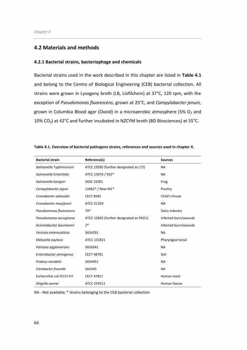
Chapter 4
64
4.2 Materials and methods
4.2.1 Bacterial strains, bacteriophage and chemicals
Bacterial strains used in the work described in this chapter are listed in Table 4.1
and belong to the Centre of Biological Engineering (CEB) bacterial collection. All
strains were grown in Lysogeny broth (LB, Liofilchem) at 37°C, 120 rpm, with the
exception of Pseudomonas fluorescens, grown at 25°C, and Campylobacter jenuni,
grown in Columbia Blood agar (Oxoid) in a microaerobic atmosphere (5% O2 and
10% CO2) at 42°C and further incubated in NZCYM broth (BD Biosciences) at 55°C.
Table 4.1. Overview of bacterial pathogens strains, references and sources used in chapter 4.
Bacterial strain Reference(s) Sources
Salmonella Typhimurium ATCC 19585 (further designated as LT2) NA
Salmonella Enteritidis ATCC 13076 / 932* NA
Salmonella bongori SGSC 31001 Frog
Campylobacter jejuni 12662* / New M1* Poultry
Cronobacter sakazakii CECT 8581 Child's throat
Cronobacter muytjensii ATCC 51329 NA
Pseudomonas fluorescens 7A* Dairy industry
Pseudomonas aeruginosa ATCC 15692 (further designated as PAO1) Infected burn/wounds
Acinetobacter baumannii 2* Infected burn/wounds
Yersinia enterocolitica SA54291 NA
Klebsiella oxytoca ATCC 131821 Pharyngeal tonsil
Pantoea agglomerans SA56341 NA
Enterobacter amnigenus CECT 48781 Soil
Proteus mirabilis SA54451 NA
Citrobacter freundii SA5345 NA
Escherichia coli O157:H7 CECT 47821 Human stool
Shigella sonnei ATCC 259311 Human faeces
NA - Not available; * strains belonging to the CEB bacterial collection

Salmonella phage endolysin
65
E. coli cloning and expression strains (Invitrogen) are given in Table 4.2.
Salmonella phage phi68 (encoding the Lys68 endolysin) was isolated from waste
effluents and belongs to the CEB phage collection. For other chemicals used, EDTA
was acquired from Pronalab while isopropyl-β-D-thiogalactopyranoside (IPTG),
HEPES, citric acid and malic acid were purchased from Sigma-Aldrich. HEWL was
obtained from Fisher Scientific.
Table 4.2. Gynotypes of E. coli cloning and expression strains and vector used in chapter 4.
Bacterial strains Genotype
TOP10 F
- mcrA Δ(mcrCB-hsdSMR-mrr) Ф80lacZΔM15 ΔlacX74 recA1 araD139 Δ(ara-
leu)7697 galU galK rpsL(Strr) endA1 λ
-
BL21(DE3) F– ompT hsdSB(rB
– mB
–) gal dcm (DE3)
For transformation of Lys68 constructs (described in the 4.2.3.1 Cloning
methodology section) chemically competent E. coli TOP10 and E. coli BL21 cells
were prepared for cloning and protein recombinant expression, respectively. The
procedure to make competent started to grow each colony overnight in 10 mL of
LB medium and diluted (1:100) to 50 mL fresh LB medium the following day. After
growing at 37°C and 120 rpm and reaching a OD of about 0.3, cell were pelleted
(10 min, 4°C, 5000 rpm) and resuspended in 25 mL ice-cold 0.1 M CaCl2 and
incubated for 30 min. Next, following a second centrifuge step, cells were
resuspended in 1.5 mL of 0.1 M CaCl2 in the presence of 10% glycerol. Competent
cells were stored at -80°C prior to use in 100 µL aliquots. Transformation of
foreign DNA was accomplished by the heat shock procedure. First, ≈ 200 ng of
plasmid was incubated with 100 µL of thawed competent cells on ice for 30 min,
after which a heat shock of 42°C for 45 s was carried out. For a proper selection,
cells were allowed to grow in liquid LB and plated onto LB agar plates containing
the proper antibiotic for each transformant.

Chapter 4
66
4.2.2 In silico analysis
Lys68 endolysin was screened by the HHpred webserver (URL
http://toolkit.tuebingen.mpg.de/hhpred) using Pfam, InterProScan and COG; with
an E-value cut-off of 1x10⁻⁵ and at least 80% of query coverage. The MSA
generation method used was HHblits (Remmert et al, 2012).
To identify putative catalytic residues within Lys68 sequence, BlastP searches
were conducted. Blasting the endolysin sequence, high homology residues,
indicated as probable catalytic residues can be found. To experimentally prove
their in silico prediction, point mutations (described in the next section) were
made using a QuickChange II XL Site-Directed Mutagenesis Kit (Agilent
Technologies). Mutagenic primers were designed using a web-based QuickChange
Primer Design Program (www.agilent.com/genomics/qcpd).
4.2.3 Cloning, recombinant protein expression and purification
Primers used to clone the Lys68 endolysin were purchased from Invitrogen. The
vector pET28a (Novagen) was used to clone and express Lys68, and has a resistant
marker for kanamycin (50 µg/mL). Restriction enzymes were purchased from NEB.
4.2.3.1 Cloning methodology
Standard PCR
The isolated Salmonella phage phi68 was partially sequenced showing
resemblance to the sequenced S. Enteritidis typing phage SETP3 (data not shown).
Based on phage SETP3 genomic sequence available at NCBI database (reference
sequence: NC_009232.2), primers were designed to amplify the putative
endolysin gene by PCR. Lys68 open reading frame was amplified using the
following PCR amplification mixture and end concentrations given: KAPA HiFi

Salmonella phage endolysin
67
Buffer Fidelity (1x); 10 mM dNTP Mix (0.3 mM); forward
(AGATATCATATGTCAAACCGAAACATTAGC) and revere primers
(GTGGTGCTCGAGCTACTTAG) (0.3 µM each), with introduced NdeI and XhoI
restriction sites are underlined; template Salmonella phage phi68 DNA (1 to 10
ng); KAPA HiFi DNA Polymerase from KaPaBioSystems (1 U) (Grisp) and PCR-grade
water up to 50 µL. The DNA thermal cycler (MJ Mini™ Gradient Thermal Cycler,
Biorad) was programmed for a standard PCR reaction of 35 cycles, including: an
initial DNA denaturation step (5 min at 98°C), DNA denaturation step (30 s at
98°C), primer annealing step (30 s at a 55°C), and extension step (2 min at 72°C),
followed by a final extension step (5 min at 72°C) to allow proper PCR product
elongation. After PCR reaction, samples were separated on a 1% agarose gel in
TAE electrophoresis buffer (40 mM Tris-HCl pH 7.2, 500 mM sodium acetate and
50 mM EDTA), stained with SYBR® Safe (Invitrogen), visualized by UV illumination
(BioRad) and cleaned (DNA Clean & ConcentratorTM-5k, Zymo Research). To clone
the amplified Lys68 gene (insert) in the pET28 expression vector selected, both
plasmid and insert were double-digested using the same NdeI/XhoI sticky end
endonucleases in the following reaction: 1 µL of DNA (approx. 300 ng), 1 µL of
both restriction enzymes (10 U), 1 µL of 10x CutSmart buffer (NEB), and PCR-grade
water up to 10 µL. After 2 h incubation, insert/vector digested products were
cleaned (DNA Clean & ConcentratorTM-5k, Zymo Research) and ligated
(overnight, 16°C) using a T4 ligase (New England Biolabs) and a 3:1 (insert/vector)
molar ratio. E. coli Top10 cells were transformed with the ligation mixture.
Colony PCR
Colony PCR was carried out to screen for the correct construct before being
confirmed by sequencing. E. coli TOP10 transformants were transferred by
toothpick to a 96-well microtiter plate. Colonies were grown individually in each
well containing 100 µL of LB supplemented with antibiotic (50 µg/mL kanamycin)

Chapter 4
68
for 1.5 h at 37°C and in static conditions. Next, the following PCR mixture was
made: forward and reverse primers (insert-specific combined with vector-specific
primers at 0.4 µM); dNTPs mix (0.2 mM each nucleotide); buffer Dream Taq (10x);
Dream Taq polymerase (2.5 U) (Fermentas); 2.5 µL of cell culture grown in each
well and PCR-grade water up to 25 µL total volume. Then, a PCR reaction was
conducted: initial denaturation step of 5 min at 95°C followed by 35 cycles
consisting of 30 s at 98°C/30 sec at 50°C/2 min at 72°C, ending with a final
extension period of 10 min at 72°C. PCR product was loaded onto agarose gels for
confirmation. Finally, plasmid of positive constructs was extracted and verified by
sequencing (Macrogen).
Mutagenesis
To substitute the putative catalytic residues within the Lys68 sequence, two active
site mutations (Glu18Ala and Thr35Ala) were introduced. Two sets of overlapping
mutagenic primers (5'-CGCGGCATTCGCGGGGTTCCGGG, forward, 5'-
CCCGGAACCCCGCGAATGCCGCG, reverse, for Glu18Ala, and 5'-
AGAATGAGAAGTACCTTGCTATTGGCTACGGCCAC, forward 5'-
GTGGCCGTAGCCAATAGCAAGGTACTTCTCATTCT, reverse, for Thr35Ala, with
mutation basepairs underlined), were applied using the QuickChange II XL Site-
Directed Mutagenesis Kit (Agilent Technologies), following the manufacturer’s
instructions. Briefly, 10 ng of pET28-Lys68 template, 125 ng of the two mutagenic
primers, a dNTP mix (1x), a reaction buffer, a Quick Solution reagent 2.5 U of
PfuUltra HF DNA polymerase and PCR-grade water up to 50 µL (total volume) was
added together. After the recommended mutagenic PCR reaction, the product
was digested with DpnI for 1 h at 37°C, to allow degradation of parental
methylated DNA. After transformation into XL10-Gold ultracompetent cells
provided, positive transformants were grown, their plasmid extracted and sent for
sequencing (Macrogen).

Salmonella phage endolysin
69
4.2.3.2 Protein overexpression and purification
After sequencing, all vectors were introduced and expressed in E. coli BL21(DE3).
Cells were grown in 200 mL LB supplemented with the specific antibiotic (50
µg/mL of kanamycin) to an OD600nm of 0.6 (4 h, 120 rpm at 37°C), and recombinant
protein expression was induced with IPTG to a final concentration of 0.5 mM.
After optimization, E. coli expression temperature (16°C) and temperatures (18 h)
were chosen to maximize soluble expression yields. The culture was then
centrifuged (9500xg, 30 min) and cells were disrupted by resuspending the pellet
in 1/25 volumes of lysis buffer (20 mM NaH2PO4, 0.5 M NaCl/NaOH, pH 7.4),
followed by three cycles of freeze-thawing (-80°C to room temperature).
Maintaining the sample on ice, cells were further disrupted by sonication (Cole-
Parmer, Ultrasonic Processors) for 8-10 cycles (30 s pulse, 30 s pause). Insoluble
cell debris were removed by centrifugation (9500xg, 30 min, 4°C). The
supernatant was collected and filtered (0.22 µm filters) and applied to Ni2+-NTA
resin stacked in HisTrapTM HP 1 ml columns (GE Healthcare) for purification, using
protein-dependent imidazole concentrations optimized for and evolving four
steps:
1. Equilibrium step - 10 mL of lysis buffer (65 mM imidazole concentration).
2. Loading step - Loading of total soluble expressed E. coli extract that was
resuspended in lysis buffer (65 mM imidazole concentration).
3. Washing step - 10 mL of lysis buffer (65 mM imidazole concentration).
4. Elution step - 2 mL of lysis buffer with 300 mM imidazole.
Eluted protein fractions were then visualized by standard denaturation SDS-PAGE
(sodium dodecyl sulfate polyacrylamide gel electrophoresis) gels using the Bio-Rad
system under denaturation conditions. To perform the electrophoresis, a 5% of
upper and a 12% lower stacking and separating acrylamide gels were assembled.
From the eluted fractions, 10 µL were added to 10 µL of Laemmli buffer (2x)

Chapter 4
70
(Sigma), boiled at 95°C for 5 min and loaded onto the gels together with a Low
Molecular Weight ladder (GE Healthcare). Protein bands were routinely separated
for 2 h, at 120 volts. Gels were rinsed with water and stained with 0.05% of
Commassie brilliant blue R-250 (Sigma) solution for 2 h. Background destaining
was performed with several washing steps with water. Only protein samples with
purities equal to or higher than 90% were considered pure and used to perform
additional studies. Proteins were dialyzed in 10 mM phosphate buffered saline
(PBS) at pH 7.2 (using Maxi GeBAflex-tube Dialysis Kit - Gene Bio-Application
L.T.D). The Protein concentrations were then determined by the BCATM Protein
Assay Kit with bovine serum albumin as standard (Thermo Scientific).
4.2.4 Biochemical characterization
4.2.4.1 Endolysin lytic spectrum
To determine lytic spectrum of Lys68, a range of different strains was tested
(Table 4.1). G- and G+ overnight grown cultures were overnight grown and diluted
1:100 diluted in the following day in fresh 5 mL LB and allowed grow until
reaching the mid-exponential phase (OD600nm of 0.6). Cells were then plated (100
μL) onto LB agar Petri dishes for lawn formations. After 8 h incubation, a 30-μL
drop of 2 µM of purified Lys68 was added to visualize lysis halos. In the case of G-
cells a step involving treatment with chloroform vapours was included to
permeabilize the OM (Raymond Schuch, 2009). After lawn formations, in a
chemical fume hood, 10 mL of chloroform were added to an inverted lid glass
plate. Bacterial lawns were inverted on top of the chloroform and incubated for 1
h to allow chloroform evaporation. Finally, a 30-μL drop of 2 µM of Lys68 protein
was added to visualize lysis halos. In both cases, halo spots commonly appeared
after 30 min up to 1 h after the start of the incubation period.

Salmonella phage endolysin
71
4.2.4.2 Muralytic activity
The PG lytic (or muralytic) activity was quantified on P. aeruginosa PAO1 cells with
the OM permeabilized (exposed PG), as a substrate (Lavigne et al, 2004). This
substrate was prepared by growing P. aeruginosa cells to an OD600nm (optical
density at 600 nm) of 0.6, centrifuging them (4000xg, 15 min, 4°C) and
resuspending them in a chloroform-saturated 0.05 M Tris buffer (pH 7.7). After
gently shaking for 45 min at room temperature, protoplasts were collected
(4000xg, 15 min, 4°C), washed and resuspended in 80 mM phosphate buffer (pH
7.2) at an OD600nm of 1.0. Substrate was then stored at -20°C prior to use. To
perform the muralytic activity, 30 μL of serial dilutions of enzyme were added to
270 μL of OM permeabilized cells (prepared substrate). The endolysin activity was
measured over a period of 30 s or up to 30 min, through the decrease in OD600nm
using a BIO-TEK®, Synergy HT Microplate Reader. Obtained values for the negative
control (30 μL of PBS pH 7.2) were subtracted from the sample values. The
muralytic activity was calculated according to a standardized calculation method
optimized and extensively described elsewhere (Briers et al, 2007a). Enzymatic
activity was defined as follows: Activity (units/µM) = ((slope
(OD600nm/min)/µM))/0.001), where the activity of 1 unit is defined as the
concentration of enzyme (in µM) necessary to create a drop in OD600nm of 0.001
per minute. Averages ± standard deviations are given for n = 3 repeats.
pH dependence
The pH dependence on the muralytic activity was assessed using the same 30 μL
of serial dilutions of enzyme on 270 μL of OM permeabilized cells, but now
resuspended in a universal pH buffer (150 mM KCl, 10 mM KH2PO4, 10 mM Na-
citrate and 10 mM H3BO4) adjusted to different pH values within the range of 3.0
and 10.0. Averages ± standard deviations are given for n = 3 repeats.

Chapter 4
72
Cryo/Thermal stability
For cryostability tests, the same muralytic activity described was performed with
Lys68 samples stored during 2 months at cold temperatures (4°C and -20°C).
Thermostability was evaluated by incubating the endolysin at several
temperatures 40°C, 60°C, 80°C and 100°C for 30 min and in addition at 100°C
during different time intervals (5, 10, 20, 25, 30 and 45 min) in a MJ MiniTM BIO-
RAD Thermocycler. Following a 30-min cooling step on ice, the (residual) activity
was measured using 30 µL of 2 μM Lys68 on 270 µL of OM permeabilized cells
resuspended in 80 mM phosphate buffer pH 7.2. The residual muralytic activity of
each sample relative to the activity of unheated reference sample at time 0 (=
100% activity) was determined. Averages ± standard deviations are given for n = 3
repeats.
4.2.4.3 Circular dichroism
To analyse the Lys68 endolysin secondary structure, circular dichroism (CD)
experiments were performed in the far- and near-UV region, using a Jasco J-815
CD spectrometer equipped with a water-cooled Peltier unit. These experiments
were carried out at the INL, the International Iberian Nanotechnology Laboratory
in Portugal. The spectra were recorded in a cell width of 0.1-mm path length
(110.QS, Hellma) from 185 to 360 nm for all proteins with 1 nm steps, scan speed
of 20 nm/min, high sensitivity and a 16 s response time. Three consecutive scans
from each sample and its respective buffer baseline were obtained. The averaged
baseline spectrum was subtracted from the averaged sample spectrum measured
under the same conditions. Secondary structure estimates were derived from the
spectra using the CDSSTR (Compton & Johnson, 1986) and CONTINLL (Provencher
& Glockner, 1981; van Stokkum et al, 1990) routine of the DICHROWEB (Whitmore
& Wallace, 2004; Whitmore & Wallace, 2008) server run on the Set 4 optimized
for a wavelength of 190-240 nm. Thermal denaturation/renaturation of the

Salmonella phage endolysin
73
proteins was measured by monitoring the change in ellipticity in a cell width of 0.1
cm path- length at 222 nm over the range of 20°C to 75°C, in increments of 1°C.
The experimental denaturation/renaturation profiles were analyzed by a
nonlinear least squares fit assuming a two-state transition and used to calculate
the melting temperature (Tm). For CD measurements, a concentration of 8 µM of
protein was prepared in the presence of a universal buffer (10 mM KH2PO4, 10
mM Na-citrate and 10 mM H3BO4) adjusted to different pH values within the
range of 3 and 10 (for pH dependence tests) and in 80 mM phosphate buffer pH
7.2 (for thermostability tests). The same conditions were used for the analysis of
the pH and temperature on the muralytic activity described above.
4.2.3.4 Fluorescence binding assay
To visualize the OMP effect, S. Typhimurium LT2 cells were incubated with 2 mM
citric acid and 2 µM KZ-EGFP protein. KZ-EGFP is a previously described PG binding
domain from a Pseudomonas phage endolysin KZ144 coupled with a green
fluorescent protein that has strong G- PG affinity (Briers et al, 2009). After 30 min
incubation period, the sample was washed twice in 10 mM HEPES/NaOH (pH 7.2).
Afterwards, a 30-µL drop was spotted onto a microscope slide and images were
recorded using a confocal laser scanning microscopy (FluoView FV1000
microscope, Olympus) in search for fluorescent Salmonella. Cells treated with
water (instead of citric acid) and EFGP were used as negative control.

Chapter 4
74
4.2.5 Antibacterial assays
4.2.5.1 In vitro assays on planktonic exponential cells
An overnight culture of G- cells was diluted (1:100) in fresh 5 mL LB and incubated
at 37°C at 120 rpm. When reaching the mid-exponential phase with an OD600nm of
0.6, cells were resuspended in 10 mM HEPES/NaOH (pH 7.2), and then 100-fold
diluted to a final density of 106 colony forming units (CFU)/mL. Each culture (50
μL) was incubated for up to 2 h at room temperature with 25 μL of Lys68 (2 μM
final concentration) together with 25 μL of water or 25 μL of OMPs (EDTA, citric
acid and malic acid) dissolved in water. The final concentrations used were 0.5
mM, 2 mM and 5 mM, respectively, concentrations retrieved from literature
(Alakomi, 2007; Briers et al, 2011b). Following the same protocol, HEWL was
assessed at a final concentration of 2 μM to compare with the Lys68 results. As a
negative control, 50 μL of cells were incubated with 25 μL water and 25 μL of PBS
(pH 7.2) (culture control), with 25 μL of OMP and 25 μL of PBS (pH 7.2) (OMP
control) or with 25 μL water and 25 μL of endolysin dialyzed in PBS (pH 7.2)
(endolysin control). After a 30 min incubation period, cell suspensions were
serially diluted and colonies were counted after overnight incubation at 37°C in LB
agar plates. The antibacterial activity was quantified as the relative inactivation in
logarithmic units (= log10 (N0/Ni) with N0 = number of untreated cells (in the
negative control) and Ni = number of treated cells counted after incubation).
Averages ± standard deviations are given for n = 4 repeats.
4.2.5.2 In vitro assays on different bacterial physiological states
To study the influence against different bacterial physiological states, Lys68/OMPs
mixture was tested against planktonic S. Typhimurium LT2 under exponential and
stationary phase and also against biofilms. S. Typhimurium LT2 cells, grown
overnight at 37°C, were diluted 1:100 in fresh LB and grown for 3 h (planktonic

Salmonella phage endolysin
75
exponential phase), 12 h (stationary phase) and 24 h (in this case, 200 µL volume
on 96-microplate wells - biofilm cells). In the planktonic and stationary assay, 50
µL cells resuspended in 10 mM HEPES/NaOH (pH 7.2) to a final 106 CFU/mL were
incubated with 25 µL Lys68 (2 µM final concentration) together with 25 µL of citric
and malic acid (2 mM and 5 mM final concentrations, respectively) for 2 h. For
biofilm assays, overnight cultures of S. Typhimurium cells were normalized (OD
adjusted to 1.0 in LB medium) after which 2 μL of the cell suspension was
transferred to 96-well polystyrene plates (Orange Scientific) containing 198 μL of
LB. To establish mature biofilms the plate was incubated for 24 h at 37°C and 120
rpms. Afterwards, the culture medium was removed and non-adherent cells were
also removed by washing the biofilms twice with 200 µL of 10 mM HEPES/NaOH
(pH 7.2). Cells were then incubated with 100 µL of HEPES together with 50 µL
Lys68 (2 µM final concentration) and 50 µL of citric and malic acid (2 mM and 5
mM final concentrations, respectively) for 2 h. For all experiments, cells incubated
with water (instead of citric/malic acid) or PBS (instead of Lys68) were used as
negative control. After incubation, the effect of the Lys68 and OMP mixtures on G-
cells (planktonic and biofilms) was assessed by quantification of the number of
CFUs. Again, the antibacterial activity was expressed as the relative inactivation in
logarithmic units described above. Averages ± standard deviations are given for n
= 4 repeats.

Chapter 4
76
4.3 Results
4.3.1 In silico analysis
The 489-bps endolysin gene from Salmonella phage phi68, encoding a 162-amino
acid protein with a deduced molecular mass of 19.6 kDa, was amplified and
sequenced (GenBank accession number for Lys68 nucleotide sequence,
KJ475444). Based on data from HHpred output (an E-value of 2.3x10-47 and 100%
of query coverage) Lys68 is predicted to be a globular protein with a conserved
domain between amino acids 1-151 and belongs to the glycoside hydrolase family
GH24 (CAZY, 2014). This catalytic domain is predicted to degrade cell walls by
catalyzing the hydrolysis of 1,4-linkages between N-acetylmuramic acid and N-
acetyl-D-glucosamine residues. BlastP comparison revealed high homology (>90%)
to six other endolysin proteins from Salmonella phages, namely SETP3, ST4, SE2,
vB_SenS-Ent1, SS3e and wksl3, that have not yet been characterized in vitro. A
BlastP analysis also indicates conservation in both presumed catalytic residues
Glutamic acid (Glu18) and Threonine (Thr35) for the glycosylase reaction.
4.3.2 Recombinant purification and substrate specificity
Overexpression of Lys68 in E. coli BL21(DE3) yielded a soluble protein of 14.3
mg/L. To confirm the predicted catalytic activity and to determine the bacterial
host spectrum, Lys68 endolysin (2 µM) drops was spotted on several OM-
permeabilized G- cell substrates and several intact G+ strain lawns (listed in Table
4.3 and Figure 4.1) with different structural types of PG. All OM permeabilized G-
cell substrates together with L. monocytogenes were efficiently lysed. Conversely,
the remaining tested G+ bacteria all survived the treatment of Lys68.

Salmonella phage endolysin
77
Table 4.3. Lytic activity of Lys68 against Gram-negative or Gram-positive strains.
Bacterial strain Relative lysis activity
G- 30 min 1 h
Escherichia coli BL21(DE3) Susceptible
Escherichia coli O157:H7 CECT 47821 Susceptible
Salmonella Typhimurium LT21 Intermediate susceptible
Salmonella Enteritidis ATCC 13076 Susceptible
932* Susceptible
Salmonella bongori SGSC 31001 Susceptible
Campylobacter jejuni 12662* Susceptible
New M1* Susceptible
Cronobacter sakazakii CECT 8581 Susceptible
Cronobacter muytjensii ATCC 51329 Susceptible
Pseudomonas fluorescens 7A1* Susceptible
Pseudomonas aeruginosa PAO11 Susceptible
Acinetobacter Baumannii 21* Susceptible
Yersinia enterocolitica SA54291 Susceptible
Klebsiella oxytoca ATCC 131821 Susceptible
Pantoea agglomerans SA56341 Intermediate susceptible
Enterobacter amnigenus CECT 48781 Susceptible susceptible
Proteus mirabilis SA54451 Intermediate Intermediate
Citrobacter freundii SA5345 Intermediate susceptible
Shigella sonne ATCC 259311 Susceptible
G+
Listeria monocytogenes CECT 5725 Intermediate Intermediate
Staphylococcus epidermidis 9142* Resistant Resistant
Staphylococcus aureus CECT 976 Resistant Resistant
Paenibacillus larvae H23* Resistant Resistant
1strains tested with enzymes/OMPs;
*strains from the CEB bacterial collection;
Susceptible (clear lysis halo); Intermediate (turbid lysis halo); Resistant (absence of lysis halo)

Chapter 4
78
Figure 4.1. Visualization of the Lys68 lytic activity on chloroform S. Typhimurium LT2
permeabilized lawn. Six 10 µL-radial endolysin (2 µM) drops resulted in lysis halos. PBS added to
the center served as negative control (no lysis halo observed).
4.3.3 Biochemical characterization
4.3.3.1 pH dependence and muralytic activity
Muralytic activity
Biochemical characterization of Lys68 started exploring the muralytic activity
under different conditions. Regarding pH dependence, Lys68 remains active over
a pH range from 4.0 to 10.0 (maintaining 41.17%±4.81 and 46.31%±0.96 of its
activity, respectively) (Figure 4.2 a), with a pH optimum around 7.0 (pH of the
bacterial cytoplasm). When kept at optimal pH, Lys68 has a muralytic activity of
400 Units/µM that decreases to 310 Units/µM (23.3% less) and 100 Units/μM
(74.9% less) when the protein is stored for two months at 4°C and -20°C,
respectively (Figure 4.2 b).

Salmonella phage endolysin
79
Figure 4.2. Muralytic activity of Lys68. a) pH optimum for enzymatic activity of Lys68. Relative
muralytic activity is measured as the slope of the OD600nm/min curve, given in percentage by
comparing to the activity at pH 7.0 (the highest measured value) (Y-axis) on OM-permeabilized P.
aeruginosa PAO1 substrate and is shown for a pH range between 3.0 and 10.0 (X-axis). b)
Saturation curves for Lys68 muralytic activity under optimal pH (7.2). The activity in OD600 nm/min
(Y-axis) for incremental amounts of Lys68 (0 months - squares; 2 months at 4°C - circles; 2 months
at -20°C - triangles) is depicted. Muralytic activity reaches saturation at 2 to 3 μM.
0,0 0,5 1,0 1,5 2,0 2,5 3,00,00
0,05
0,10
0,15
0,20
0,25
0,30 Lys68 (0 months)
Lys68 (2 montshs/4°C)
Lys68 (2 montshs/-20°C)
y = 0.10
R2 = 0.92
y = 0.31
R2 = 0.97
Slo
pe (O
D a
t 600 n
m/m
in)
[Lys68]/M
y = 0.40
R2 = 0.95
b
2 3 4 5 6 7 8 9 10 110
20
40
60
80
100
120
% o
f re
lative m
ura
lytic a
ctivity
pH
a

Chapter 4
80
Circular dichroism analysis
To elucidate the major structural features of Lys68, CD studies were employed
using a universal buffer at different pH values (Figure 4.3). The Lys68 CD profile
exhibited two negative dichroic minima at 222 nm and 208 nm and a positive
dichroic maximum at 192 nm, which is characteristic of a protein with a high α-
helix content.
Figure 4.3. Circular dichroism spectra of Lys68 as a function of pH using a universal buffer with
adjusted pH (3.0-10.0).
Deconvolution of the CD spectra, using a dataset as specified in materials and
methods, allowed us to determine that 37% of Lys68 folds as α-helices, 17% as β-
sheet, and 18% as turns, while 27% was unordered. Interestingly, when the pH
was between 4.0 and 10.0, the obtained spectra were almost identical and the
predicted secondary structures content remained essentially unchanged. These
data shows that the secondary structure content of Lys68, at various pH
conditions, is highly conserved, even at pH 4.0, similar to the trend observed for
HEWL after analysis its CD spectrum (Supplementary Figure S4.1). Conformational
changes occurred around pH 3.0 with loss of secondary structure.
pH 3
pH 4
pH 5
pH 6
pH 7
pH 8
pH 9
pH 10
200 210 220 230 240 250 260 270
-10
0
10
Elli
pticity
Wavelength/nm
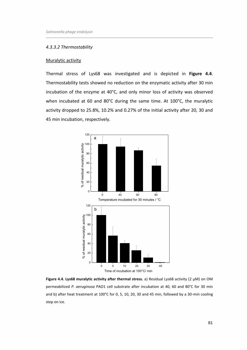
Salmonella phage endolysin
81
4.3.3.2 Thermostability
Muralytic activity
Thermal stress of Lys68 was investigated and is depicted in Figure 4.4.
Thermostability tests showed no reduction on the enzymatic activity after 30 min
incubation of the enzyme at 40°C, and only minor loss of activity was observed
when incubated at 60 and 80°C during the same time. At 100°C, the muralytic
activity dropped to 25.8%, 10.2% and 0.27% of the initial activity after 20, 30 and
45 min incubation, respectively.
Figure 4.4. Lys68 muralytic activity after thermal stress. a) Residual Lys68 activity (2 µM) on OM
permeabilized P. aeruginosa PAO1 cell substrate after incubation at 40, 60 and 80°C for 30 min
and b) after heat treatment at 100°C for 0, 5, 10, 20, 30 and 45 min, followed by a 30-min cooling
step on ice.
0 20 40 60
0
20
40
60
80
100
120
0 40 60 80
% o
f re
sid
ua
l m
ura
lytic a
ctivity
Temperature incubated for 30 minutes / °C
a
0 5 10 15 20 25
0
20
40
60
80
100
120
Time of incubation at 100°C/ min
0 5 10 20 30 45
b
% o
f re
sid
ua
l m
ura
lytic a
ctivity
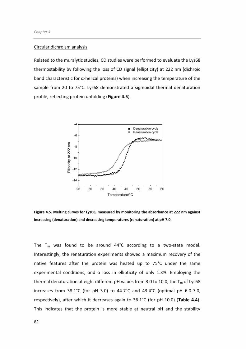
Chapter 4
82
Circular dichroism analysis
Related to the muralytic studies, CD studies were performed to evaluate the Lys68
thermostability by following the loss of CD signal (ellipticity) at 222 nm (dichroic
band characteristic for α-helical proteins) when increasing the temperature of the
sample from 20 to 75°C. Lys68 demonstrated a sigmoidal thermal denaturation
profile, reflecting protein unfolding (Figure 4.5).
Figure 4.5. Melting curves for Lys68, measured by monitoring the absorbance at 222 nm against
increasing (denaturation) and decreasing temperatures (renaturation) at pH 7.0.
The Tm was found to be around 44°C according to a two-state model.
Interestingly, the renaturation experiments showed a maximum recovery of the
native features after the protein was heated up to 75°C under the same
experimental conditions, and a loss in ellipticity of only 1.3%. Employing the
thermal denaturation at eight different pH values from 3.0 to 10.0, the Tm of Lys68
increases from 38.1°C (for pH 3.0) to 44.7°C and 43.4°C (optimal pH 6.0-7.0,
respectively), after which it decreases again to 36.1°C (for pH 10.0) (Table 4.4).
This indicates that the protein is more stable at neutral pH and the stability
25 30 35 40 45 50 55 60
-14
-12
-10
-8
-6
-4
Denaturation cycle
Renaturation cycle
Elli
pticity a
t 222 n
m
Temperature/°C

Salmonella phage endolysin
83
decreases at acidic or basic pH values, which is positively correlated with the
observed optimal pH from the Lys68 muralytic activity.
Table 4.4. Apparent melting temperatures (Tm) of Lys68 as a function of pH (3.0-10.0) as
determined by circular dichroism signal at 222 nm in thermal denaturation and renaturation
experiments.
pH Tm
(Denaturation cycle) Tm
(Renaturation cycle)
3 - -
4 38.1°C 35.0°C
5 43.0°C 40.7°C
6 44.7°C 41.8°C
7 43.4°C 40.8°C
8 41.5°C 38.5°C
9 39.4°C 34.6°C
10 36.1°C -
Afterwards, Lys68 was subjected to thermal stress under different temperatures
(40, 60, 80 and 100°C) and protein ellipticity was monitored (Figures 4.6 a, b). It
was observed that from 4°C to 40°C, Lys68 remained very stable and kept its
activity for longer periods. As the incubation temperature increased from 40°C to
60°C, 80°C and 100°C, evident changes in secondary structures occurred, with a
continuous decrease in the ellipticity of the 208 nm and 220 nm bands. In
accordance, CD thermal stability showed that the loss of muralytic activity upon
thermal stress is accompanied with gradual loss of endolysin secondary structure.

Chapter 4
84
Figure 4.6. Circular dichroism spectra of Lys68 after thermal stress. a) Lys68 (2 µM) incubated at
40, 60 and 80°C for 30 min followed by 30 min incubation on ice, and b) after heat treatment at
100°C for 0, 5, 10, 20, 25 and 30 followed by 30 min incubation on ice.
4.3.4 In vitro antibacterial activity
4.3.4.1 Enzymes/OMPs effect on Gram-negative cells
The in vitro antibacterial activity of Lys68/OMP and HEWL/OMP was investigated
on a wide panel of G- bacteria (Table 4.5). As is depicted, a stronger antibacterial
activity was observed against S. Typhimurium LT2, A. baumannii 2 and P.
aeruginosa PAO1, compared to the other tested species.
200 220 240 260 280-20
-10
0
10
20
30
40
Elli
pticity
Wavelength/nm
0 min
5 min
10 min
20 min
30 min
b
200 220 240 260 280-20
-10
0
10
20
30
40
Elli
pticity
Wavelength/nm
4°C
40°C
60°C
80°C
100°C
a

Salmonella phage endolysin
85
Table 4.5. Combinatorial antibacterial activity of outer membrane permeabilizers with (A and B)
or without (C) HEWL/Lys68 on Gram-negative bacterial pathogens. Marked in bold are significant
log reduction units observed (≥ 1 log).
A Bacterial Species HEWL/Water HEWL/EDTA HEWL/Citric HEWL/Malic
Salmonella Typhimurium LT2 0.03 ± 0.07 0.14 ± 0.23 0.08 ± 0.15 0.07 ± 0.09
Acinetobacter baumannii 2 0.01 ± 0.09 0.04 ± 0.06 0.10 ± 0.06 0.17 ± 0.15
Pseudomonas aeruginosa PAO1 0.02 ± 0.10 0.89 ± 0.17 0.24 ± 0.02 2.44 ± 0.52
Pseudomonas fluorescens 7A 0.04 ± 0.04 0.30 ± 0.38 0.28 ± 0.13 3.77 ± 0.45
Shigella sonnei ATCC 25931 0.22 ± 0.29 0.01 ± 0.11 0.02 ± 0.07 0.06 ± 0.12
E. coli O157:H7 CECT 4782 0.04 ± 0.15 0.11 ± 0.12 0.10 ± 0.05 0.09 ± 0.04
Cronobacter sakazakii CECT 858 0.11 ± 0.18 0.15 ± 0.20 0.30 ± 0.16 0.20 ± 0.13
Pantoea agglomerans SA5634 0.04 ± 0.10 0.08 ± 0.15 0.20 ± 0.19 0.22 ± 0.29
Enterobacter amnigenus CECT 4878 0.04 ± 0.07 0.07 ± 0.13 0.18 ± 0.17 0.26 ± 0.04
Proteus mirabilis SA5445 0.08 ± 0.03 0.09 ± 0.14 0.03 ± 0.25 0.17 ± 0.14
Salmonella bongori SGSC 3100 0.02 ± 0.10 0.10 ± 0.09 0.09 ± 0.13 0.47 ± 0.19
Klebsiella oxytoca ATCC 13182 0.01 ± 0.02 0.10 ± 0.05 0.13 ± 0.08 0.26 ± 0.06
Yersinia enterocolitica SA5429 0.06 ± 0.05 0.18 ± 0.11 0.11 ± 0.12 0.34 ± 0.11
B Bacterial Species Lys68/Water Lys68/EDTA Lys68/Citric Lys68/Malic
Salmonella Typhimurium LT2 0.14 ± 0.16 0.16 ± 0.12 2.93 ± 0.45 2.65 ± 0.40
Acinetobacter baumannii 2 0.04 ± 0.12 0.14 ± 0.22 1.01 ± 0.33 2.93 ± 0.59
Pseudomonas aeruginosa PAO1 0.04 ± 0.34 2.46 ± 0.48 1.48 ± 0.35 3.31 ± 0.21
Pseudomonas fluorescens 7A 0.02 ± 0.10 1.44 ± 0.67 1.40 ± 0.23 5.91 ± 0.43
Shigella sonnei ATCC 25931 0.13 ± 0.28 0.35 ± 0.44 1.40 ± 0.33 2.42 ± 0.43
E. coli O157:H7 CECT 4782 0.02 ± 0.02 0.21 ± 0.14 1.18 ± 0.12 2.37 ± 0.35
Cronobacter sakazakii CECT 858 0.16 ± 0.06 0.30 ± 0.09 1.09 ± 0.19 1.40 ± 0.30
Pantoea agglomerans SA5634 0.03 ± 0.18 0.13 ± 0.12 0.32 ± 0.18 1.26 ± 0.18
Enterobacter amnigenus CECT 4878 0.04 ± 0.11 0.28 ± 0.18 0.68 ± 0.36 1.21 ± 0.27
Proteus mirabilis SA5445 0.03 ± 0.12 0.09 ± 0.04 0.41 ± 0.29 0.97 ± 0.25
Salmonella bongori SGSC 3100 0.06 ± 0.10 0.08 ± 0.16 0.13 ± 0.09 0.94 ± 0.05
Klebsiella oxytoca ATCC 13182 0.07 ± 0.13 0.12 ± 0.17 0.24 ± 0.14 0.30 ± 0.22
Yersinia enterocolitica SA5429 0.11 ± 0.10 0.03 ± 0.06 0.18 ± 0.08 0.22 ± 0.07

Chapter 4
86
C Bacterial Species ------ Water/EDTA Water/Citric Water/Malic
Salmonella Typhimurium LT2 0.09 ± 0.10 0.07 ± 0.05 0.06 ± 0.09
Acinetobacter baumannii 2 0.04 ± 0.03 0.07 ± 0.05 0.07 ± 0.04
Pseudomonas aeruginosa PAO1 0.05 ± 0.06 0.60 ± 0.14 1.54 ± 0.47
Pseudomonas fluorescens 7A 0.42 ± 0.31 0.70 ± 0.14 2.70 ± 0.51
Shigella sonnei ATCC 25931 0.01 ± 0.14 0.02 ± 0.14 0.14 ± 0.03
E. coli O157:H7 CECT 4782 0.06 ± 0.04 0.01 ± 0.09 0.18 ± 0.18
Cronobacter sakazakii CECT 858 0.17 ± 0.12 0.06 ± 0.14 0.28 ± 0.10
Pantoea agglomerans SA5634 0.07 ± 0.10 0.03 ± 0.18 0.10 ± 0.18
Enterobacter amnigenus CECT 4878 0.08 ± 0.08 0.15 ± 0.15 0.16 ± 0.18
Proteus mirabilis SA5445 0.02 ± 0.10 0.01 ± 0.23 0.03 ± 0.12
Salmonella bongori SGSC 3100 0.07 ± 0.12 0.05 ± 0.14 0.16 ± 0.03
Klebsiella oxytoca ATCC 13182 0.12 ± 0.12 0.01 ± 0.09 0.18 ± 0.09
Yersinia enterocolitica SA5429 0.01 ± 0.06 0.30 ± 0.23 0.06 ± 0.07
The activity of both Lys68 and HEWL alone against exponentially growing cells was
insignificant. However, following permeabilization with OMPs a different outcome
was observed. Despite being both able to inactivate Pseudomonas cells,
Lys68/EDTA had a more pronounced killing effect than HEWL/EDTA. Both citric
and malic acid were significantly better than EDTA in enhancing the access of
Lys68 to the PG, however this advantageous effect was not observed in
combination with HEWL. Lys68/citric acid was not only able to kill Pseudomonas
cells, but also caused a significant reduction of S. Typhimurium LT2 (2.93±0.45 log
reduction) and A. baumannii (1.01±0.33 log reduction) as well of Shigella sonnei
(1.40±0.33 log reduction), E. coli O157:H7 (1.18±0.12 log reduction) and
Cronobacter sakazakii (1.09±0.19 log reduction).
Interestingly, the effect of Lys68/malic acid was even higher in all strains
compared to Lys68/EDTA and Lys68/citric acid. Reductions of 2-3 logs units were
observed on P. aeruginosa (3.31±0.21), S. Typhimurium LT2 (2.65±0.40) and A.
baumannii (2.93±0.59). Overall, the best log reduction values were consistently
obtained with malic acid in combination with Lys68 after 30 min of incubation,

Salmonella phage endolysin
87
killing 11 different G- bacterial species. In contrast to the other strains, none of
the OMPs could sensitize Lys68 to Klebsiella oxytoca and Yersinia enterocolitica
strains.
When incubated for longer periods (2 h), the combinatory effect of citric acid and
Lys68 on S. Typhimurium LT2 increased remarkably, resulting in a 5.05±0.48 log
reduction of viable cells and was also efficient against stationary cells (1.45±0.15
log units of viable cells) and biofilms (1.26±0.10 log units of viable cells) (Table
4.6).
Table 4.6. Influence of different S. Typhimurium LT2 physiological states (planktonic/biofilm) on
the combinatorial effect of Lys68 and outer membrane permeabilizers (citric and malic acid).
Marked in bold are significant log reduction units observed (≥ 1 log).
S. Tyhpimurium LT2 Planktonic Biofilm
Exponential phase Stationary phase
Lys68 + Water 0.11 ± 0.14 0.14 ± 0.07 0.15 ± 0.11
PBS + Citric acid 0.10 ± 0.11 0.08 ± 0.11 0.09 ± 0.19
PBS + Malic acid 0.08 ± 0.20 0.19 ± 0.18 0.15 ± 0.25
Lys68 + Citric acid 5.05 ± 0.48 1.45 ± 0.15 1.26 ± 0.10
Lys68 + Malic acid 3.19 ± 0.53 0.55 ± 0.28 1.31 ± 0.13
4.3.4.2 OM permeabilization mechanism
To further investigate whether the permeabilizing effect of citric and malic acid is
compound- or pH-related, S. Typhimurium LT2 cells were incubated with Lys68 in
the presence of HCl (to drop the pH to 4.2 and 3.8, similar to citric and malic acid,
respectively) for a 30 min period (Table 4.7).

Chapter 4
88
Table 4.7. S. Typhimurium LT2 log reduction units after incubation with enzymes and acids.
Marked at bold are significant log reduction units observed (≥ 1 log).
Water HEWL Lys68 Lys68 + MgCl2
HCl, pH 4.2 0.14 ± 0.18 0.12 ± 0.05 2.44 ± 0.56 0.10 ± 0.18
HCl, pH 3.8 0.01 ± 0.17 0.11 ± 0.10 2.66 ± 0.42 0.20 ± 0.13
Citric acid, pH 4.2 0.21 ± 0.14 0.09 ± 0.08 3.08 ± 035 0.24 ± 0.10
Malic acid, pH 3.8 0.11 ± 0.09 0.13 ± 0.06 2.63 ± 0.47 0.18 ± 0.16
The results summarized in Table 4.7 show that the log reduction units of viable
cells are mostly due to the acidity. In the presence of HCl, Lys68 is able to kill
2.44±0.56 log of viable cells at pH 4.2 (3.08±0.35 for citric acid) and a 2.66±0.42
log reduction of viable cells was obtained at pH 3.8 (2.63±0.47 for malic acid). In
the case of HEWL, both organic acids and HCl were inactive towards S.
Typhimurium LT2.
It has been described that MgCl2 can interact with the LPS layer via divalent
cations (Mg2+), contributing to the stability of the OM (Alakomi et al, 2000).
Subsequently, this results in a decrease in permeability of the OM to external
agents. Consequently, the addition of 5 mM of MgCl2 abolished the Lys68
antibacterial activity in all tested conditions (Table 4.7).
To visualise the effect of organic acids as G- OMPs, an experiment was conducted
incubating S. Typhimurium LT2 cells with citric acid in the presence of a
fluorescent probe. This fluorescent probe was constructed earlier and consists of
a Pseudomonas phage KZ144 endolysin binding domain fused with an enhanced
green fluorescent protein (KZ-EGPF) that has high affinity to G- PG (Briers et al,
2009). When incubating the cells for a 30-min period, green fluorescent
Salmonella cells could be observed by epifluorescence microscopy (Figure 4.7),
indicative of cell permeablization.

Salmonella phage endolysin
89
Figure 4.7. Epifluorescence microscopy of the S. Typhimurium LT2 cells permeabilized with citric
acid. a) Cells incubated with 2 mM citric acid and 2 µM of KZ-EGFP protein; b) Cells incubated with
2 mM citric acid and 2 µM of wild-type EGFP protein; and c) Cells incubated with water and 2 µM
of wild-type EGFP protein for 30 min. Cell pellets were then washed twice and visualized using
epifluorescence microscopy with a 1500 magnification. This image has been converted to black
and white.
4.3.4.3 Lys68 PG digestion mechanism
To assess the Lys68 PG digestion mechanism and explain the activity of the
endolysin at low pH, an analysis of its presumed essential catalytic residues was
made. Lys68 is classified as a member of the GH24 family (CAZY, 2014), which has
a glutamine (Glu) residue as the proton donor. The catalytic nucleophile however
is yet unknown. To identify a putative nucleophile, BlastP searches were
conducted. Results showed that, besides Glu-18, a second amino acid residue,
Thr-35, was highly conserved, which could indicate a role as a putative catalytic
residue of Lys68. Mutagenesis of both presumably essential catalytic residues
resulted in one inactive enzyme (Glu18Ala) and one enzyme with similar activity
(Thr35Ala) compared to the Lys68 wild-type (Figure 4.8 a). Analysis of mutated
proteins by CD did not provide any evidence for significant structural alterations
due to the amino acid replacements (Figure 4.8 b). Therefore, the absence of
activity was entirely attributed to the Glu18Ala substitution.
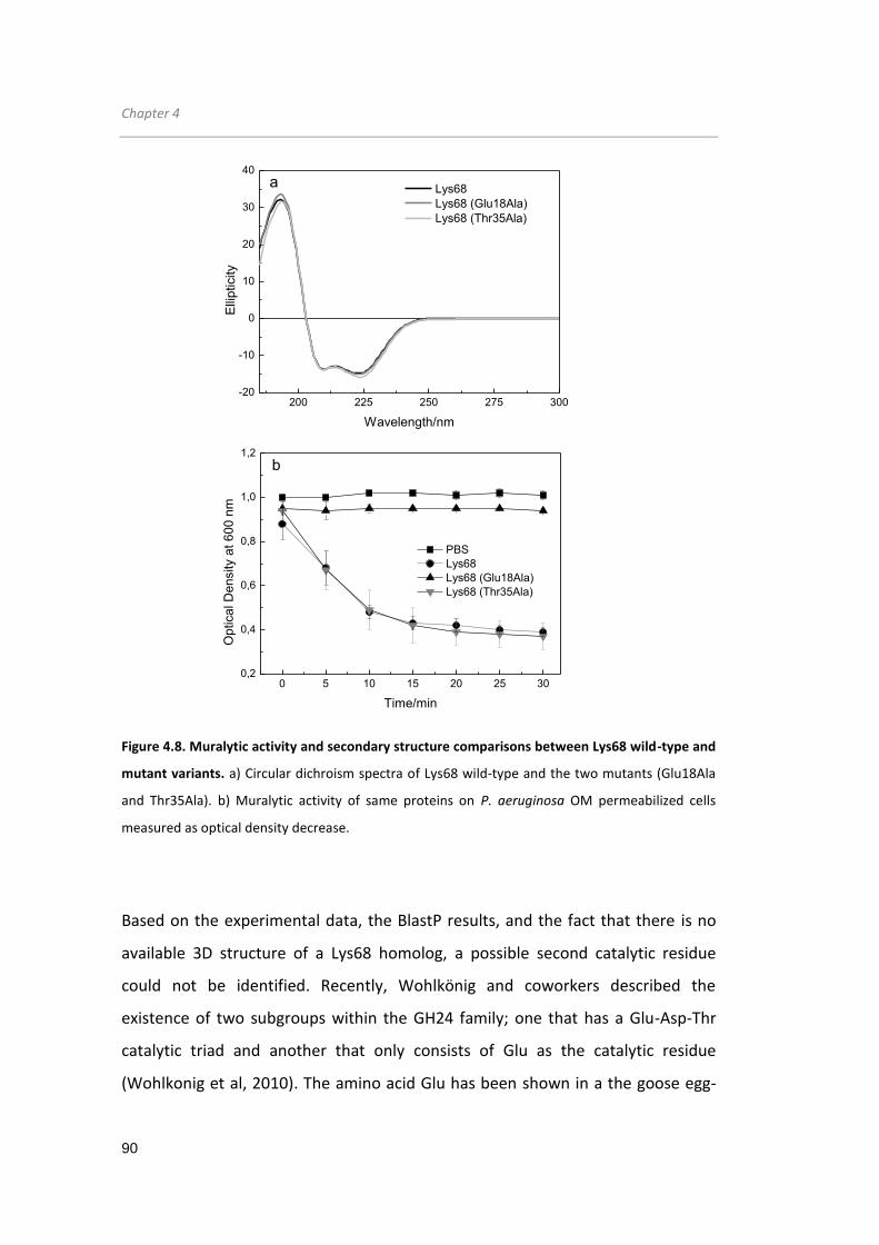
Chapter 4
90
Figure 4.8. Muralytic activity and secondary structure comparisons between Lys68 wild-type and
mutant variants. a) Circular dichroism spectra of Lys68 wild-type and the two mutants (Glu18Ala
and Thr35Ala). b) Muralytic activity of same proteins on P. aeruginosa OM permeabilized cells
measured as optical density decrease.
Based on the experimental data, the BlastP results, and the fact that there is no
available 3D structure of a Lys68 homolog, a possible second catalytic residue
could not be identified. Recently, Wohlkönig and coworkers described the
existence of two subgroups within the GH24 family; one that has a Glu-Asp-Thr
catalytic triad and another that only consists of Glu as the catalytic residue
(Wohlkonig et al, 2010). The amino acid Glu has been shown in a the goose egg-
PBS
Lys68
Lys68 (Glu18Ala)
Lys68 (Thr35Ala)
0 5 10 15 20 25 300,2
0,4
0,6
0,8
1,0
1,2
Op
tica
l D
en
sity a
t 6
00
nm
Time/min
b
200 225 250 275 300-20
-10
0
10
20
30
40
Elli
pticity
Wavelength/nm
Lys68
Lys68 (Glu18Ala)
Lys68 (Thr35Ala)
a

Salmonella phage endolysin
91
white lysozyme as the single responsible catalytic residue for the digestion of PG
bonds (Weaver et al, 1995). Therefore, it is proposed that Lys68 operates through
a two-step mechanism involving Glu-18 (Figure 4.9).
Figure 4.9. Proposed Lys68 mechanism of action. Lys68 hydrolyse the 1,4-linkages between N-
acetylmuramic acid and N-acetyl-D-glucosamine, through a two-step mechanism involving Glu-18
catalytic residue to cleave the peptidoglycan.
In the first step N-acetylhexosaminidases, which have an acetamido group, are
capable of neighboring group participation to form an intermediate oxazolinium
ion, with acidic assistance provided by the Glu-18 carboxylate. In the second step,
the now deprotonated acidic carboxylate acts as a base and assists a nucleophilic
water to hydrolyze the oxazolinium ion intermediate, giving the hydrolyzed
product (through individual inversions that lead to a net retention of
configuration).

Chapter 4
92
4.4 Discussion
In recent years, endolysins of phages infecting G+ bacteria have been extensively
studied (Fischetti, 2010). In contrast, G- phage endolysins have been poorly
explored, because the impermeable bacterial OM protects the underlying PG layer
from enzymatic hydrolysis by the exogenous endolysin. In this work, a novel
endolysin from a G- infecting phage is described. In silico characterization of Lys68
from the Salmonella phage phi68 shows elevated protein similarity (˃90%
identity) to other Salmonella phage endolysins belonging to the same lysozyme-
like superfamily. The observed globular structure (ECD alone) is also shared by
most other G- phage endolysins, contrary to modular archetypes (ECD and CBD)
typically seen in endolysins from a G+ background (Oliveira et al, 2013).
A biochemical characterization showed that Lys68 displays some interesting
characteristics. Regarding substrate specificity, Lys68 degrades the PG of all 20 G-
organisms tested, however it was ineffective against all included G+ strains, with
the exception of L. monocytogenes. Consequently, Lys68 displays a broader lytic
spectrum than the phage from which was derived from, whose spectrum is
limited to S. Enteritidis strains (Sillankorva et al, 2010). This broader spectrum is
explained by the common PG A1γ chemotype shared by the sensitive G- species
and also L. monocytogenes, whereas the remaining insensitive G+ strains like
Staphylococcus epidermidis 9142 and Staphylococcus aureus 976 presumably have
an A3α PG chemotype (Schleifer & Kandler, 1972). This chemotype was previously
shown to be resistant to other muralytic enzymes, like HEWL (Archibald AR, 1993).
The observed muralytic activity of the globular Lys68 (400 Units/µM) is lower
compared to other described modular G--like endolysins (e.g. KZ144 and EL188
with 2058 and 4735 U/µM, respectively, calculated using the same method)
(Walmagh et al, 2012). This can be explained by the fact that the latter two
enzymes contain cell wall binding domains that direct the enzymes to the PG
substrate, increasing the muralytic activity. Nevertheless, when compared to the

Salmonella phage endolysin
93
globular HEWL (35 Units/µM, reported elsewhere), Lys68 is 12 times more active
(Briers et al, 2007a). In addition to adequate profiles of activity and specificity,
antimicrobial enzymes require good structural stability. Therefore the pH effect
on the Lys68’s conformational stability was characterized. The endolysin’s
secondary structure showed to be highly conserved between pH values of 4.0 to
10.0. At pH 3.0, more turns and unordered/random coils are formed in Lys68 than
at higher pH values. Stability tests showed that Lys68 was more stable around pH
6.0-7.0 (with melting temperatures of 44.7°C and 43.4°C), matching the pH where
optimal muralytic activity is observed.
Once subjected to thermostability tests, some endolysins surprisingly maintain
their activity after long heating periods. Within endolysins from a G- background,
Pseudomonas phage endolysins KZ144 and EL188 activity still remains high after
being exposed at 50°C for 10 min (Briers et al, 2007b). In contrast, the phage T4
lysozyme only retains a minor fraction of its activity after a 5 min treatment at
65°C (Nakagawa et al, 1985). Recently, a highly thermostable modular endolysin
(PVPSE1gp146) with resistance to temperatures up to 100°C was described
(Walmagh et al, 2012). Lys68 is a globular protein where the muralytic activity is
only completely inactivated after 30 min at 100°C. Because the melting
temperature of the endolysin at pH 7.0 is 44°C, it seems that under the stress
caused by higher temperatures, the endolysin is able to gradually regain its
secondary structure to a point where, at high temperatures and prolonged
exposures, it cannot refold. Recently, a similar folding/refolding mechanism was
suggested by Schmelcher and coworkers to explain the high thermostability of the
G+ L. monocytogenes phage endolysins, HPL118 and HPL511 (Schmelcher et al,
2012b). In their study, HPL118 and HPL511 retained 35% of activity after 30 min at
90°C.
To allow a better passage of endolysins through the OM, a combinatorial
approach was used on G- bacteria, combining the bacterial cell wall-degrading

Chapter 4
94
enzyme Lys68 or HEWL with an OMP. Both proteins combined with EDTA could
reduce P. aeruginosa to some extent. A similar effect has been previously
described with endolysin EL188 from Pseudomonas infecting phage EL only on P.
aeruginosa strain PAOI (Briers et al, 2011b). The positive effect of EDTA on the
antibacterial activity is caused by the powerful OM permeabilization capacity of
EDTA through binding and withdrawal of the stabilizing divalent cations Mg2+ and
Ca2+, present in the LPS layer of the G- OM (Vaara, 1992). As a result, the bacterial
PG becomes more prone to the muralytic activity of the externally added
endolysins. However, the sensitivity of other bacterial species to the destabilizing
effect of EDTA was much lower, even when EDTA was increased to 5 mM (see
Supplementary Table S4.1). In seems that the type and structure of the OM plays
a key role for the antibacterial efficacy of the enzyme/EDTA combinatorial effect.
Possibly, due to the low number of phosphate groups per LPS molecule and the
corresponding amount of stabilizing divalent cations in the OM of
Enterobacteriaceae compared to the Pseudomonadaceae, EDTA turns out to be
ineffective to the former group. The observed synergetic effects of citric acid and
malic acid, has never been described for phage endolysins. In this respect, both
citric acid and malic acid had a better permeabilizing effect than EDTA, and were
able to permeabilize several G- strains tested enhancing the activity of Lys68. In
particular against exponential S. Typhimurium LT2 cells, an even more profound
effect was observed when extending the reaction from 30 minutes to 2 hours,
although proving to be less efficient against stationary cells and biofilms. The
lower efficiency in killing stationary cells can be a result of two aspects; i)
structural changes in the OM (e.g. LPS biosynthesis), compromising the endolysin
entry or LPS permeabilization through the organic acids, or ii) chemical
modifications in the PG (e.g. glycosylations or acetylations) known to contribute
to high levels of resistance to glycosyl hydrolases (Vollmer & Born, 2009; Rolfe et
al, 2012). As for biofilms, beside the presence of several bacterial physiological
states that can give rise to similar problems, additional diffusion limitations due to

Salmonella phage endolysin
95
the high density matrix content can hinder the efficacy of endolysins and
antimicrobials in general (Mah & O'Toole, 2001). Although the permeabilizing
capacity of citric acid by chelating divalent cations from the OM associated with
its protonated form has been reported, additional acidity can contribute to OM
damage. It has been proposed that the LPS disintegration is accomplished by the
ability of undissociated acid groups to interact/pass through the negatively
charged LPS (where negatively charged acids are repulsed) and migrate inside the
cell to cause sublethal damages (e.g. leading to OM membrane disruption)
(Alakomi et al, 2000; Theron & Lues, 2011). The 2 mM of citric acid (pKa 3.13 at
25°C) and 5 mM of malic acid (pKa 3.4 at 25°C) used, caused a drop of pH to 4.2
and 3.8, respectively. Because similar log reduction units were obtained using HCl
instead of citric or malic acid combined with Lys68, the acidity effect seems to
play a key role in the OM permeabilization. Because millimolar concentrations of
these organic acids by themselves did not have antibacterial effect on the cells
(with exception of Pseudomonas), they seem to be enough to compromise the cell
wall to a level that allows Lys68 to act on the PG, causing bacterial death.
Recently, a combined treatment with different organic acids to induce cell
permeability in E. coli O157:H7 was reported (Kim & Rhee, 2013). Transmission
electron microscopy images showed clear membrane disintegration that
potentiated the bactericidal action of medium-chain fatty acids. When bacterial
suspensions were supplemented with 5 mM of MgCl2, the effect of Lys68
combined with organic acids was completely abolished. MgCl2 has the ability to
protect the bacterial OM damaged from the acid challenge, an effect already
observed in the presence of lactic acid (Alakomi et al, 2000).
Based on the carbohydrate-active enzymes classification, several amino acids
have been identified within lysozyme-like proteins to play a key role in the
glycosidase reaction (CAZY, 2014). HEWL’s (belonging to the GH22 class) active
site, which cleaves polysaccharide chains in bacterial cell walls, is constituted
essentially by the two amino acids Glu-35 (pKa, 6.1-6.2) and Asp-52 (pKa, 3.4-4.5),

Chapter 4
96
which are only functional if the former is non-ionized and the latter is ionized
(Webb et al, 2010). Hence, the absence of enzymatic activity in the presence of
malic and citric acid is related to the deionization of Asp52 in an acidic pH. Lys68
has a different catalytic system. Since the Thr-35 mutation did not affect Lys68
antimicrobial activities, it can be concluded that Thr-35 is not involved in the
catalysis of PG. Therefore, it is tempting to speculate that Lys68 operates through
a two-step mechanism involving Glu-18, capable of digesting the 1,4-linkages
between N-acetylmuramic acid and N-acetyl-D-glucosamine residues of the PG in
a low pH environment (pH around 4.0) through a retaining mechanism. The
catalytic activity of amino acid Glu-73 in the goose egg-white lysozyme has been
proved by Weaver and co-workers to be enough to digest the PG bonds (Weaver
et al, 1995).
To summarize, G- bacterial pathogens prevail in various surroundings and their
resilience is aided by the presence of an OM that prevents toxic substances from
entering the cell. Lys68 demonstrates very good stability properties and its
efficient combination with EDTA, citric acid and malic acid could be greatly
explored in many fields to combat G- pathogens. Lys68/EDTA could be used as a
therapeutic product to control P. aeroginosa, typically present in burn wound
infections or as food preservative against P. fluorescents. The broad antimicrobial
effect of Lys68/citric acid and Lys68/malic acid could be explored in food and
clinical settings as an effective sanitizer to prevent food spoilage and nosocomial
bacterial infections. Additionally, the powerful effect of Lys68 with the organic
acids can be used therapeutically to treat for example topical infections as chronic
wounds, when applied in a cream or ointment. Altogether, this study proposes
the combination of a bacterial cell wall hydrolase from phage origin (Lys68) with
EDTA, citric and malic acid for biotechnological and medical applications to fight
G- pathogens in the agricultural, food and medical industry.
Chapter 6
Chapter 5
Activity of an Acinetobacter phage endolysin,
ABgp46, against Gram-negative pathogens in
planktonic and biofilm cultures

Chapter 5
98
Acinetobacter phage endolysin
99
5.1 Introduction
Although chapter 4 demonstrated the efficacy of endolysin/organic acid mixtures
to control G- pathogens, it is important to validate this technology for other
endolysins and prove their transversal application. Multidrug-resistant bacteria
and biofilms are also important factors that need to be addressed in depth, as
most G- bacteria, like Pseudomonas and Acinetobacter, are naturally resistant to
antibiotics (e.g. low outer membrane permeability) or able to quickly acquire new
resistance mechanisms (e.g. horizontal transfer of resistance genes) (Bonomo &
Szabo, 2006). Their ability to form biofilms is another important aspect that makes
them more tolerant to varied antimicrobial agents (e.g. related to the limited
diffusion transport and the presence of different bacterial physiological states)
(Mah & O'Toole, 2001).
This chapter describes the isolation of a novel phage-encoded endolysin (ABgp46)
and a depolymerase (ABgp42) (part of the tail-fiber), originating from an
Acinetobacter baumannii phage. Depolymerases are polysaccharide-degrading
enzymes encoded by phages that act against the extracellular polymeric
substances found in biofilms, increasing their anti-biofilm efficiency (Lu & Collins,
2007; Cornelissen et al, 2011). ABgp46 was first analyzed for its stability under
different pH values and then tested against planktonic G- (antibiotic resistant)
bacteria in the presence and absence of OMPs agents. In this chapter, several
organic acids were tested (citric, malic, lactic, benzoic and acetic acid) to widen
novel synergies with substantial biotechnological interest, and compared with
EDTA, as it is the only agent described so far able to act synergistically with
endolysins (Walmagh et al, 2012). In the end, it was intended to explore the anti-
biofilm endolysin-organic acid-depolymerase activity formula. Endolysin anti-
biofilm activity is a field that still remains scarcely explored, with only few
successes reported against G+ Staphyloccocus and Streptococcus biofilms (Sass &
Bierbaum, 2007; Shen et al, 2013).

Chapter 5
100
5.2 Materials and methods
5.2.1 Bacterial strains, bacteriophage and chemicals
Bacterial strains used are listed in Table 5.1. Without exceptions, all strains were
grown in LB (Liofilchem) at 37°C and 120 rpm. E. coli cloning and expression
strains (Agilent Technologies, Invitrogen and Merck4Biosciences) are shown in
Table 5.2. E. coli strains BL21-CodonPlus-(DE3)-RIL (30 µg/mL chloramphenicol),
BL21-CodonPlus-(DE3)-RP, (30 µg/mL chloramphenicol) and Origami™ 2(DE3)
(12.5 µg/mL tetracycline and 50 µg/mL streptomycin) were grown under the
specified conditions. E. coli Top10 was used for cloning purposes while the
remaining strains were used for recombinant expression. E. coli strains were
transformed as described in chapter 4. A. baumannii phage vb_AbaP_CEB1
(encoding the ABgp46 endolysin and the ABgp42 depolymerase part of the tail-
fiber) was isolated from waste effluents and belongs to the CEB phage collection.
Table 5.1. Overview of bacterial pathogens strains, references and sources used in chapter 5.
Bacterial strain Reference(s) Sources
Salmonella Typhimurium ATCC 19585 (further designated as LT2) NA
Pseudomonas aeruginosa ATCC 15692 (further designated as PAO1) Infected burn/wounds
Klebsiella oxytoca ATCC 131821 Pharyngeal tonsil
Acinetobacter baumannii 1*/ 2* / 3* / 4* / 5* / 6* / 7* / 8* / 10* Infected burn/wounds
Escherichia coli O157:H7 CECT 47821 Human stool
NA - Not available; * strains kindly provided by Hospital de Braga

Acinetobacter phage endolysin
101
Table 5.2. Genotypes of E. coli cloning and expression strains used in chapter 5.
Bacterial strains Genotype
TOP10 F
- mcrA Δ(mcrCB-hsdSMR-mrr) Ф80lacZΔM15 ΔlacX74 recA1 araD139 Δ(ara-
leu)7697 galU galK rpsL(Strr) endA1 λ
-
BL21(DE3) F– ompT hsdSB(rB
– mB
–) gal dcm (DE3)
BL21-CodonPlus-(DE3)-RIL F- ompT hsdS(rB
-mB
-) dcm + Tet
r gal λ(DE3) endA Hte (argU ileY leuW Cam
r)
BL21-CodonPlus-(DE3)-Rp F- ompT hsdS(rB
-mB
-) dcm + Tet
r gal λ(DE3) endA Hte ([argU proL Cam
r)
Origami™ 2(DE3) Δ (ara-leu)7697 Δ lacX74 Δ phoA PvuII phoR araD139 ahpC galE galK rpsL F’[lac
+ lacI
q pro] (DE3) gor522::Tn10 trxB (Str
R, Tet
R)
EDTA was acquired from Pronalab while IPTG, HEPES, citric, malic, lactic, benzoic
and acetic acid were purchased from Sigma-Aldrich.
5.2.2 In silico analysis of endolysins
The endolysin sequence used was screened by the HHpred webserver (URL
http://toolkit.tuebingen.mpg.de/hhpred) using Pfam, InterProScan and COG; with
an E-value cut-off of 1x10⁻⁵ and at least 80% of query coverage. The MSA
generation method used was HHblits (Remmert et al, 2012). Multiple sequence
alignments were generated through ClustalW online (URL
http://www.genome.jp/tools/clustalw/).
5.2.3 Cloning, recombinant protein expression and purification
All primers used to clone the ABgp46 endolysin and ABgp42 depolymerase were
purchased from Invitrogen. The vector pET15b (Novagen) was used to clone and
express both proteins, and has the resistant marker gene for ampicillin (100
µg/mL). Restriction enzymes were purchased from NEB and all DNA cleaning steps
were performed using DNA Clean & ConcentratorTM-5k (Zymo Research). All final
constructs were verified by sequencing (Macrogen).

Chapter 5
102
5.2.3.1 Cloning methodology
The standard PCR cloning procedure for the ABgp46 endolysin and the ABgp42
depolymerase ORFs into pET15b was carried out as explained in chapter 4. For
ABgp46, specific set of primer 5-GGCAGCCATATGATTCTGACTAAAGACGGGTTTAG
(forward) and 5- GCAGCCGGATCCCTATAAGCTCCGTAGAGCGC (reverse), with the
NdeI/BamHI restriction endonuclease sites underlined, and annealing
temperature of 55°C, were used in a PCR reaction. As for ABgp42, the primers 5-
GGCAGCCATATGATGCACGCTGACCCATGTTGG (forward) and 5-
CAGCTGTAAACGTTATCCGTTGCTAAGGATCCGGCTGC (reverse), with the
NdeI/BamHI restriction endonuclease sites underlined and an annealing
temperature of 56°C were chosen.
5.2.3.2 Protein overexpression and purification
Both proteins were cloned in pET15b, making use of the present N-terminal His6
for fused expression. The E. coli expression system, purification using Ni2+-NTA
chromatography, band purification visualization, dialysis in 10 mM PBS pH 7.2 and
protein quantification were carried out as explained previously in chapter 4. Only
proteins with a purity higher than 90%, as judged by SDS-PAGE analysis, were
considered for in vitro studies. E. coli BL21(DE3) expression strain and 25 mM
imidazole purification stringency were chosen for ABgp46 endolysin expression
and purification. For the ABgp42 depolymerase, a set of different optimization
parameters (E. coli expression strains listed in Table 5.2), inducer concentration
(from 0.5 to 1.5 mM), expression time (from 4 to 18 h) and temperatures (from
16°C to 37°C) were conducted, however without being able to express the
protein.
Acinetobacter phage endolysin
103
5.2.4 Biochemical characterization
5.2.4.1 Fluorescence measurements
Intrinsic fluorescence emission spectra of the ABgp46 endolysin were recorded
over the 300-400 nm range with a Jasco FP-750 spectrofluorimeter equipped with
a Peltier thermostat. An excitation wavelength of 295 nm was used to minimize
the mission arising from tyrosine residues. Two µM of protein was used in a
universal buffer with pH adjusted from 4.0 to 8.0 and thermal denaturation on
monitor heating the sample from 20 to 70°C. The variation of the tertiary
structure was analyzed based on parameter a defined as: (IF325/IF360) protein, IF is
intensity of fluorescence. These experiments were carried out at the ICRM, the
Istituto di chimica del riconoscimento molecolare, in Italy.
5.2.4.2 Circular dicrhoism
Endolysin circular dichroism (CD) spectra were recorded in the Far-UV region
(195-260 nm) with a Jasco 600. Thermal denaturation was recorded with 5°C
increment steps from 20 to 75°C, using a universal buffer (pH 4.0-8.0). All CD
signals were baseline corrected and smoothed with the Spectra Analysis JASCO
software. In all cases, a concentration of 8 µM (0.18 mg/mL) was used in an
optical path of 0.1 cm. These experiments were carried out at the ICRM, the
Istituto di chimica del riconoscimento molecolare, in Italy.
5.2.5 Antibacterial assays
5.2.5.1 In vitro assays on planktonic cells
In vitro assays on planktonic cells were performed as described in chapter 4 with
some minor modifications. Mid-exponential phase cells (OD600nm of 0.6),

Chapter 5
104
resuspended and 100-fold diluted in 10 mM HEPES/NaOH (pH 7.2), were
prepared. Each culture (50 μL) was incubated for 2 h at room temperature with 25
μL of ABgp46 endolysin (final concentration of 2 μM) together with 25 μL of water
or 25 μL or OMPs (EDTA, citric, malic, lactic, benzoic and acetic acid) dissolved in
water. The final concentrations used were optimized during this work (see
concentrations in Table 5.5). As a negative control, 50 μL of cells were incubated
with 25 μL of PBS (pH 7.2) (instead of ABgp46) or with 25 μL water (instead or
OMPs). Cell counting and antibacterial activity quantification was done as
previously described. Averages ± standard deviations are given for n = 4 repeats.
5.2.5.2 In vitro assays on biofilm cells
Overnight bacterial cells were normalized (OD adjusted to 1.0 in LB medium) after
which 2 μL of the cell suspension was transferred to 96-well polystyrene plates
(Orange Scientific) containing 198 μL of LB. To establish mature biofilms the plate
was incubated for 24 h at 37°C and 120 rpms. Afterwards, the culture medium
was removed and non-adherent cells were also removed by washing the biofilms
twice with 200 µL of 10 mM HEPES/NaOH (pH 7.2). Cells were then incubated for
2 h with 100 µL of HEPES together with 50 µL of ABgp46 endolysin (2 µM final
concentration) and 50 µL of the best OMP agent selected (EDTA, citric and malic
acid). Optimized final concentrations used were: 0.5 mM, 3.55 mM and 4.55 mM,
respectively. As negative controls, cells were incubated with water (instead of
OMPs) or incubated with PBS (instead of endolysin). After incubation CFUs were
counted in LB agar plates. Again, the antibacterial activity was quantified as the
relative inactivation in logarithmic units, as described previously. Averages ±
standard deviations are given for n = 4 repeats.

Acinetobacter phage endolysin
105
5.3 Results
5.3.1 In silico analysis
The A. baumannii phage vb_AbaP_CEB1 was previously isolated and sequenced
(unpublished data). ORF 46 (referred to as ABgp46), encoding a 185-amino acid
protein with a deduced molecular mass of 23.1 kDa, is predicted to act as a
peptidoglycan hydrolase. HHpred output shows that ABgp46 belongs to the GH19
of the glycosidase family, a class of enzymes that cleaves the O-glycosidic bond of
the bacterial PG. In silico analysis through BlastP showed close resemblance to
five other endolysins from Acinetobacter phages (phiAB2, phiAB3, ABP-01 and
ABP-04), with high homology (95%) (Supplementary Figure S5.1). ORF 42
encodes the tail-fiber gene and a depolymerase that is part of this gene (referred
to as ABgp42). Through HHpred, the depolymerase domain is predicted to consist
of 208 amino acids, resulting in a molecular mass of 22.6 KDa, (Supplementary
Figure S5.2) and resembles a pectate lyase 3 (PF12708) from the Pec lyase clan
(CLO268). BlastP analysis did not identify other close related phage proteins.
5.3.2 Recombinant expression and purification
Recombinant expression of ABgp46 endolysin in E. coli BL21(DE3) yielded a
soluble protein of 20.5 mg/L. The ABgp42 depolymerase encoding domain was
expressed under several conditions. However, despite numerous attempts to
express the protein in several E. coli expression strains (derived from both BL21
and K12 strains) and different induction temperatures (from 37 to 16°C), times (4
to 16 h) and concentrations (1.5 to 0.5 mM IPTG), no protein was expressed.
Therefore, the following sections only describe the biochemical characterization
and in vitro antibacterial assays of the endolysin protein.

Chapter 5
106
5.3.3 Biochemical characterization
An analysis of the ABgp46 secondary and tertiary structure was performed under
different pH values, using CD and protein intrinsic fluorescence, respectively. This
exploratory characterization was used to understand the prospect of a hurdle
approach using ABgpL46 and OMPs, especially at low pH. Finding a stable
endolysin under these conditions would enable possible combinations with
organic acids, as an antibacterial formula for clinical or food application against G-
bacterial pathogens.
5.3.3.1 Circular dicrhoism analysis
With CD measurements, the ABgp46 secondary structure was monitored. The CD
spectra of ABgp46 was analyzed as a function of pH with a universal buffer with a
pH adjusted from 4.0 to 8.0 (Figure 5.1).
190 200 210 220 230 240 250 260 270-50
-40
-30
-20
-10
0
pH 4
pH 5
pH 6
pH 7
pH 8
Elli
pticity
Wavelength/nm
Figure 5.1. Far-UV Circular dichroism spectra of ABgp46 under different pH values (4.0-8.0).

Acinetobacter phage endolysin
107
The signal minima around 208 and at 220 nm that prevail in the far-UV CD
spectra, is indicative of α-helices in this protein. Unfortunately, due to signal noise
generated in far-UV, CD spectra could only be recorded from 260 to 200 nm and,
subsequently, it was not possible to estimate the content of the α-helice, β-sheet,
turns and unordered structures and compare these results with the data obtained
for the Lys68 endolysin. Ellipticity overlapping signatures indicate an unchanged
protein secondary structure. To determine more in depth the enzyme’s
conformational stability, a thermal treatment was employed by increasing
temperature (5°C steps between 20 to 70°C) of the protein solution at different
pH values. Secondary structure changes were analyzed by monitoring CD intensity
at 222 nm where α-helix structures show a negative peak and the melting
temperatures (Tm) at which the enzyme underwent a secondary structure are
shown in Table 5.3.
Table 5.3. Apparent melting temperatures (Tm) of ABgp46 as a function of pH (4.0-8.0) and
temperature monitored by Circular dichroism intensity at 222 nm. Possible renaturation cycles
were not monitored due to protein aggregation above Tm.
pH Tm
(Denaturation cycle) Tm
Observations
4 40-45°C Not aggregated
5 45-50°C Aggregated
6 45-50°C Aggregated
7 45-50°C Aggregated
8 45-50°C Aggregated
No differences were observed in the pH range 5.0-8.0, where all Tm were recorded
as being between 45-50°C. Unfortunately, it was not possible to determine the
precise value of Tm because the spectropolarimeter employed was not equipped
with a thermostat that allows a variation of the temperature with a constant rate.
At temperatures above Tm secondary structure changes caused protein
aggregation. At pH 4.0, a clear shift of Tm, between 40-45°C, was observed

Chapter 5
108
indicating a less favourable condition to maintain the secondary structure
integrity. Interestingly, this was the only pH where protein aggregation did not
occur.
5.3.3.2 Fluorescence measurements
Similarly to CD, the fluorescence spectra of ABgp46 were obtained at different pH
values and temperatures in order to analyse changes in tertiary structure. Taking
advantage of the two Tryptophans (Trp-95 and Trp-135) present in the sequence,
an excitation wavelength of 295 nm was used, minimizing the contribution of
tyrosine fluorescence. As an indication of significant tertiary structure changes,
Trp fluorescence shifts were analyzed using the Parameter A defined as the
IF325/IF360 ratio (where IF equals to the intensity of fluorescence) (Figure 5.2).
10 20 30 40 50 60 70 80
0,8
0,9
1,0
1,1
1,2
1,3
Pa
ram
ete
r A
Temperature/°C
pH4
pH5
pH6
pH7
pH8
Figure 5.2. Correlation between fluorescence spectra and conformational changes deduced by
plotting Parameter A as a function of temperature.
Increasing the temperature from 20 to 70°C, parameter A showed an analogous
trend for all pH values, where a sudden signal shift was identified between 40 to
50°C. Such spectral variations are typically observed when the medium that

Acinetobacter phage endolysin
109
surrounds the fluorophore differs in polarity and/or as a consequence of
conformational changes. Based on CD and parameter A data, it is likely that
secondary and tertiary structure changes are happening when the protein is
heated from 40°C to higher temperatures, resulting in an unfolded and denatured
protein.
5.3.4 In vitro antibacterial activity
5.3.4.1 ABgp46 against planktonic cells
The in vitro antibacterial activity of the recombinant Abgp46 was investigated on a
wide set of G- strains (Table 5.4).
Table 5.4. Antibacterial activity of ABgp46 against several Gram-negative bacterial pathogens.
Marked in bold are significant log reduction units observed (≥ 1 log).
Bacterial Species ABgp46/Water Patterns of antibiotic resistance*
Pseudomonas aeruginosa PAOI 0.03 ± 0.02 NA
Salmonella Typhimurium LT2 0.14 ± 0.05 NA
E. coli O157:H7 CECT 4782 0.17 ± 0.12 NA
Cronobacter sakazakii CECT 858 0.23 ± 0.26 NA
Klebsiella oxytoca ATCC 13182 0.18 ± 0.13 NA
Acinetobacter baumannii 1 1.77 ± 0.06 Resistant to 13 out of 18 tested antibiotics
Acinetobacter baumannii 2 0.93 ± 0.25 NA
Acinetobacter baumannii 3 0.43 ± 0.61 Resistant to 14 out of 18 tested antibiotics
Acinetobacter baumannii 4 1.00 ± 0.32 NA
Acinetobacter baumannii 5 0.81 ± 0.21 NA
Acinetobacter baumannii 6 0.51 ± 0.68 NA
Acinetobacter baumannii 7 0.18 ± 0.11 Resistant to 14 out of 18 tested antibiotics
Acinetobacter baumannii 8 0.37 ± 0.39 Resistant to 11 out of 18 tested antibiotics
Acinetobacter baumannii 10 1.20 ± 0.46 Resistant to 11 out of 18 tested antibiotics
*Patterns of antibiotic resistance was provided by Hospital de Braga and were based on susceptibility tests
performed according to the NCCLS for antibiotic resistance; NA - not available

Chapter 5
110
The activity of 2 μM Abgp46 alone on tested cells was insignificant as expected,
with the exception of A. baumannii strains. Interestingly, some Acinetobacter
strains were sensitive to the endolysin, causing a log reduction units of viable
cells, from 0.18±0.11 (lowest effect) to 1.77±0.06 (highest effect), including strains
resistant to several antibiotics.
5.3.4.2 ABgp46/OMP formula against planktonic cells
To broaden and enhance the antimicrobial effect of the Abgp46 to other G-
pathogens, the previous unsensitized E. coli O157:H7 CECT 4782 was chosen as a
model strain, to test and optimize an ABgp46/OMP formula. In addition to the
previously characterized OMPs citric and malic acid, other organic agents were
used (lactic, benzoic and acetic acid) to sensitize the G- OM to the endolysin. Each
OMP was used in different concentrations (to achieve three distinct reaction pH
values of 7.0, 5.5 and pH 4.0) and combined with ABgp46 to assess the best
antibacterial condition after 2 h incubation (illustrated in Table 5.5). At pH 7.0,
only the ABgp46/citric acid was able to reduce E. coli cells (0.86±0.38 logs). At pH
5.5, the effect was broadened to ABgp46/citric acid, ABgp46/malic acid and
ABgp46/lactic acid mixtures reducing approx. 1 log of E. coli cells. Consistently, the
effect was more pronounced at pH of 4.0. The combinatorial effects of
ABgp46/citric acid, ABgp46/malic acid and ABgp46/lactic acid mixtures resulted in
2.98±0.77, 2.18±0.82 and 0.98±0.58 log reductions. In this condition, the
antibacterial effect was also extended to ABgp46/benzoic acid inactivating
approx. 1 log of E. coli cells. The ABgp46/acetic acid combination only had a
moderate effect (0.42±0.29).
Taken into account the most efficient conditions tested, ABgp46/OMPs mixtures
(OMPs concentrations used to achieve a pH of 4.0) were tested in the presence of
5mM MgCl2. MgCl2 provides an extra source of available divalent cations (Mg2+)
that can link to the LPS negatively charged phosphate groups, strengthening the

Acinetobacter phage endolysin
111
LPS monolayer. Therefore, as expected, the addition of 5 mM of MgCl2 abolished
the activity of all ABgp46/OMP combinations.
Table 5.5. Combinatorial antibacterial activity of ABgp46 with outer membrane permeabilizers
under different concentrations against E. coli O157:H7. For a specific pH, different concentrations
(given in mM) of EDTA, citric, malic, lactic, benzoic and acetic acid are used in combination with 2
µM of ABgp46 to reduce E. coli O157:H7 cells in presence or absence of 5 mM of MgCl2. Marked in
bold are significant log reduction units observed (≥ 1 log).
pH OMPs Molarity (mM) PBS/OMPs ABgp46/OMPs ABgp46/OMPs
+ 5mM MgCl2
7.0 ± 0.1 EDTA
Citric
Malic
Lactic
Benzoic
Acetic
0.50
0.36
0.60
1.20
1.20
1.20
0.27 ± 0.11
0.21 ± 0.14
0.24 ± 0.11
0.24 ± 0.14
0.26 ± 0.15
0.25 ± 0.14
0.32 ± 0.15
0.86 ± 0.38
0.53 ± 0.28
0.55 ± 0.31
0.49 ± 0.34
0.53 ± 0.30
-
5.5 ± 0.1 Citric
Malic
Lactic
Benzoic
Acetic
1.50
3.30
3.45
3.55
4.35
0.20 ± 0.13
0.17 ± 0.14
0.31 ± 0.07
0.27 ± 0.13
0.26 ± 0.12
1.55 ± 0.45
1.13 ± 0.39
0.95 ± 0.14
0.44 ± 0.33
0.21 ± 0.22
-
4.0 ± 0.1 Citric
Malic
Lactic
Benzoic
Acetic
3.65
4.55
8.00
10.00
20.00
0.28 ± 0.06
0.24 ± 0.18
0.27 ± 0.18
0.57 ± 0.58
0.22 ± 0.08
2.98 ± 0.77
2.18 ± 0.82
0.98 ± 0.58
1.08 ± 0.57
0.42 ± 0.29
0.15 ± 0.04
0.10 ± 0.03
0.15 ± 0.04
0.02 ± 0.03
0.11 ± 0.10
After optimizing, citric acid (at 3.65 mM) and malic acid (at 4.55 mM) were
selected as the best OMPs to be combined with ABgp46. Therefore, their
synergistic effect was further tested on the remaining unsensitized G- strains
tested earlier and compared with the gold standard EDTA (at 0.5 mM) (Table 5.6).
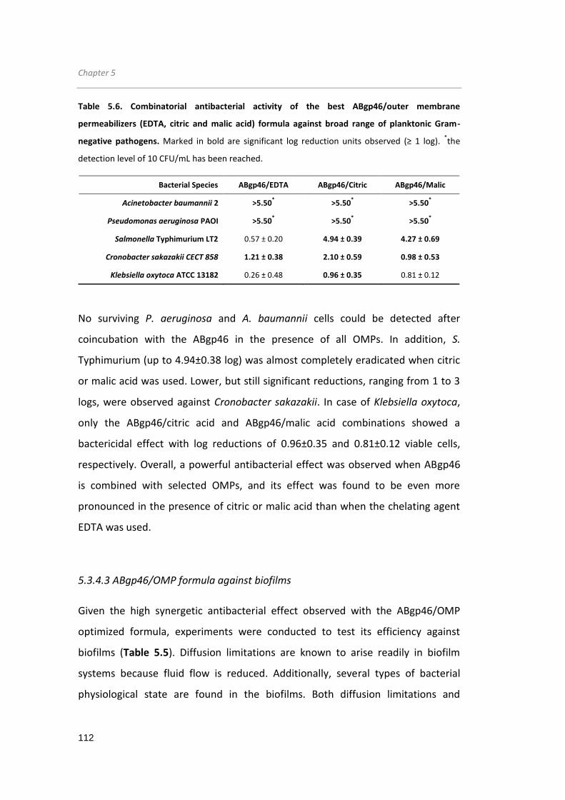
Chapter 5
112
Table 5.6. Combinatorial antibacterial activity of the best ABgp46/outer membrane
permeabilizers (EDTA, citric and malic acid) formula against broad range of planktonic Gram-
negative pathogens. Marked in bold are significant log reduction units observed (≥ 1 log). *the
detection level of 10 CFU/mL has been reached.
Bacterial Species ABgp46/EDTA ABgp46/Citric ABgp46/Malic
Acinetobacter baumannii 2 >5.50* >5.50
* >5.50
*
Pseudomonas aeruginosa PAOI >5.50* >5.50
* >5.50
*
Salmonella Typhimurium LT2 0.57 ± 0.20 4.94 ± 0.39 4.27 ± 0.69
Cronobacter sakazakii CECT 858 1.21 ± 0.38 2.10 ± 0.59 0.98 ± 0.53
Klebsiella oxytoca ATCC 13182 0.26 ± 0.48 0.96 ± 0.35 0.81 ± 0.12
No surviving P. aeruginosa and A. baumannii cells could be detected after
coincubation with the ABgp46 in the presence of all OMPs. In addition, S.
Typhimurium (up to 4.94±0.38 log) was almost completely eradicated when citric
or malic acid was used. Lower, but still significant reductions, ranging from 1 to 3
logs, were observed against Cronobacter sakazakii. In case of Klebsiella oxytoca,
only the ABgp46/citric acid and ABgp46/malic acid combinations showed a
bactericidal effect with log reductions of 0.96±0.35 and 0.81±0.12 viable cells,
respectively. Overall, a powerful antibacterial effect was observed when ABgp46
is combined with selected OMPs, and its effect was found to be even more
pronounced in the presence of citric or malic acid than when the chelating agent
EDTA was used.
5.3.4.3 ABgp46/OMP formula against biofilms
Given the high synergetic antibacterial effect observed with the ABgp46/OMP
optimized formula, experiments were conducted to test its efficiency against
biofilms (Table 5.5). Diffusion limitations are known to arise readily in biofilm
systems because fluid flow is reduced. Additionally, several types of bacterial
physiological state are found in the biofilms. Both diffusion limitations and

Acinetobacter phage endolysin
113
different physiological states are well-known aspects that reduce the
effectiveness of antimicrobials against biofilms. To anticipate these circumstances
and improve the antimicrobial efficiency, a depolymerase, encoded by the same
Acinetobacter phage vb_AbaP_CEB1, was intended to be used. As no protein was
expressed, as was mentioned before, biofilms antibacterial studies were
conducted by incubating ABgp46/OMP mixtures against A. baumannii 2 and P.
aeruginosa PAOI biofilms for 2 hours (Table 5.7).
Table 5.7. Combinatorial antibacterial activity of the best outer membrane permeabilizers in
absence (A) and presence (B) of ABgp46 against A. baumannii 2 and P. aeruginosa PAOI biofilms.
A Bacterial Species PBS/EDTA PBS/Citric PBS/Malic
Acinetobacter baumannii 2 0.05 ± 0.01 0.08 ± 0.11 0.24 ± 0.19
Pseudomonas aeruginosa PAOI 0.14 ± 0.01 0.20 ± 0.10 0.21 ± 0.13
B Bacterial Species ABgp46/EDTA ABgp46/Citric ABgp46/Malic
Acinetobacter baumannii 2 0.48 ± 0.28 0.20 ± 0.11 0.19 ± 0.05
Pseudomonas aeruginosa PAOI 0.32 ± 0.26 0.10 ± 0.14 0.54 ± 0.06
The results demonstrate an absence of killing effect when OMPs were added
alone to both bacteria. When ABgp46 was combined with OMPs, a slight
improvement was observed, although still with an insignificant effect. No major
differences were observed when ABgp46 was combined with citric or malic acid
that on average, were only able to reduce 0.3 to 0.5 log of both A. baumannii and
P. aeruginosa cells.

Chapter 5
114
5.4 Discussion
G- bacterial infections have always been a threat to human health. In particular,
infections caused by antibiotic resistant bacteria are problematic and their
incidence is constantly reported worldwide. Phage-encoded endolysins represent
one promising avenue of investigation to fight all these pathogens. After cloning
the ABgp46 endolysin, this protein was easily overexpressed in the E. coli
BL21(DE3) strain. For the heterologous production of the anti-biofilm-degrading
depolymerase, production efforts remained unsuccessful in several E. coli
expression strains. As no obvious solubility issues, hydrophobic regions or low E.
coli codon usage were detected, other expression systems rather than E. coli
should be considered in the future.
A biochemical characterization of ABgp46 was carried out in order to evaluate its
potential applications with organic acids to overcome the most obvious limitation
- the presence of a bacterial OM diffusion barrier. CD and fluorescent analysis
showed that pH changes from 4.0 to 8.0 do not have much influence on the
structure of ABgp46. As for the absence of aggregation only observed at pH 4.0, it
is possible that positively charged amino acids are repulsing protein interactions
avoiding aggregation.
Regarding the potential use of ABgp46 as an antimicrobial compound, the enzyme
was first tested without the help of any permeabilizer. Interestingly, despite the
present of the OM protecting the PG layer, externally added ABgp46 was active
against several Acinetobacter strains, including antibiotic-resistant phenotypes,
suggesting that the protein can intrinsically destabilize its OM. This is a rare event
reported for endolysins. The T4 lysozyme (from the T4 phage), was the first
endolysin described to naturally kill G- cells. It was proved that its C-terminal
positively charged amphipathic α-helix (named α-4) is responsible to cause OM
disruption, having a stronger bactericidal effect than the enzymatic PG hydrolysis
(During et al, 1999). Later, endolysins Lys1521 (from a B. amyloliquefaciens phage)
Acinetobacter phage endolysin
115
and LysAB2 (from A. baumanni phage) were also reported to spontaneously
inactivate G- cells. In these cases, it is assumed that their C-terminal side, rich in
positively charged amino acids surrounded with hydrophobic residues, interact
with the bacterial LPS. However, in opposite synthetic α-4 of the T4 lysozyme
bactericidal effect observed, studies conducted showed that Lys1521 and LysAB2
C-terminal region does not have an antibacterial effect themselves, but rather
mediate the N-terminal enzymatic domain to enter the cells, allowing them to
digest the PG and cause bacteriolysis (Morita et al, 2001; Lai et al, 2011). More
recently, the endolysin OBPgp279 (from A. baumanni and P. fluorescens phages,
respectively), was shown to lyse Pseudomonas cells (Walmagh et al, 2012),
although no predicted mechanism was identified. In all cases, it seems that when
endolysins are able to spontaneously kill G- bacteria (without any OMP), they only
have a moderate effect, reducing an approximately 1 log of viable cells. This effect
was also observed for ABgp46, able to reduce on average 1 log of Acinetobacter
strains. Because this enzyme has 96% identity to the previously characterized
LysAB2 (see the Supplemental Figure S5.1), the same C-terminal amphipathic
region (amino acid sequence between 112 and 145) identified can explain its
spontaneous activity against Acinetobacter cells.
To improve the antibacterial activity, ABgp46 was combined with different OMP
agents and concentrations (citric, malic, lactic, benzoic and acetic acid).
Consistently, an increasing antibacterial effect was observed using OMPs at higher
concentrations, reaching a maximum at low pH (4.0). Interestingly, besides CD and
fluorescence analyses predicting low structural stability at pH 4.0, the endolysin
muralytic activity is not compromised at the same pH, when combined to the
organic acids. As for the OM permeabilization mechanism, the relation between
low pH and an antibacterial effect suggests an acidity promoted killing effect.
However, different Abgp46/OMP outcomes, under the same condition (at pH 4.0),
were observed. Several aspects can individually potentiate the OM-destabilizing
effect: i) pKa of each acid, which will determine the percentage of deprotonated

Chapter 5
116
(undissociated) and protonated (dissociated) acid groups available outside de
cells. Only the undissociated forms are able to migrate inside the cells.
Intracellularly, acids dissociate (intracellular pH (≈7.2) is more alkaline than the
extracellular pH) and high anion and proton concentrations will
compromise/inhibit several cellular reactions, among which, the OM
stabilization/maintenance; ii) the level of hydrophobicity of the undissociated
groups, that will also influence their translocation through LPS hydrophobic
interactions to become toxic; iii) the different accumulation of the anions groups
that will inhibit specific types of cellular reactions, compromising the cells in a
different extent (Roe et al, 1998; Alakomi et al, 2000; Breidt et al, 2004). Because
the following bactericidal decreasing effect was observed: citric acid (pKa 3.13) >
malic acid (pKa 3.4) > lactic acid (pKa 3.86) ≈ benzoic acid (pKa 4.19) > acetic acid
(pKa 4.76) when combined with the endolysin, we speculate that this is a
reflection of acid dissociation constant (pKa) increase. This is somehow
unexpected, as increasing the pKa favours undissociated groups that could
penetrate into the cytoplasmic membrane potentiating cell damage. In opposite,
acids with low pKa values would produce more hydrogen ions (not able to
internalize) when exposed to an aqueous environment. Nevertheless, a similar
trend was observed when organic acids (with lower pKa values) were incubated
with medium-chain fatty acids (Kim & Rhee, 2013). These authors speculated that
the medium-chain fatty acid action, suggested to interact with the bacterial cell
membranes, results in sublethal injuries allowing hydrogen ions to pass into the
cell and achieve a marked bactericidal effect. Maybe a similar mechanism is
happening in the presence of ABgp46, explaining why ABgp46/citric acid and
ABgp46/malic acid are better combinations than when ABgp46 is combined with
lactic, benzoic or acetic acid. Additionally, the chelating properties only observed
in citric and malic acid will likely empower the LPS destabilization compared to the
remaining non-chelating acids. Indeed, the entire antibacterial effect was
abolished when the ABgp46/OMP formula was applied in presence of 5 mM of

Acinetobacter phage endolysin
117
MgCl2. Addition of an extra source of the divalent cation Mg2+ will strengthen the
electrostatic interactions between neighbouring LPS components, that seem to
avoid acid entry and sequential sublethal damage, and protects the LPS network
from the putative chelation effect.
After narrowing down an initial large list of OMP candidates, EDTA, citric acid and
malic acid were selected for further testing against a larger group of G- cells.
ABgp46/EDTA revealed to be only efficient to Pseudomonas and Acinetobacter
cells, although eradicating the cultures within 2 hours. Compared with EDTA, the
combinations of citric acid and malic acid with ABgp46 had a more powerful and
broaden effect, with an antibacterial effect on all tested strains. The same pattern
was previously observed with Lys68 in chapter 4. As mentioned before, while
EDTA acts as a chelating agent, sequestering LPS-stabilizing divalent cations,
organic acids are predicted to destabilize the bacterial OM by an acidic effect,
probably by lowering the bacterial internal pH and the accumulation of toxic
substances (sublethal injury) (Alakomi et al, 2000; Theron & Lues, 2011). P.
aeruginosa is known to have an LPS with a high phosphate content and
consequently a higher concentration of stabilizing divalent cations, increasing the
electrostatic interaction that contributes to LPS stability (Nikaido, 2003). Cation-
binding sites are therefore both the strength and the weakness of the P.
aeruginosa LPS layer. On the other hand, Enterobacteriaceae, such as E. coli and S.
Typhimurium, are known to have a lower phosphorylation degree in LPS,
compensated by increased hydrophobic packing of the lipid A moiety (Nikaido,
2003). Therefore one could speculate that this variation might explain the specific
EDTA action against Pseudomonas and Acinetobacter cell wall, which seems to be
more prone to a chelation effect.
Regarding biofilm control, diffusion limitations represent one of the major hurdles
to commonly applied antimicrobials. No antibacterial activity could be detected
against A. baumanni and P. aeruginosa biofilms when ABgp46 was combined with

Chapter 5
118
EDTA, citric acid or malic acid. As mentioned before, besides the obvious diffusion
problems, several types of bacterial physiological states are found in biofilms
(Mah & O'Toole, 2001). This physiological heterogeneity can contribute to higher
resistance to the endolysin/OMP mixtures. For instance, if a higher amount of
stationary cells is found, they will possess different OM composition (LPS and PG
chemical modifications), compromising the endolysin entry or LPS
permeabilization through the OMPs (Rolfe et al, 2012). Also the wide range of
physiological states found in biofilms can favour phenotypes known to activate
acid tolerance systems (e.g. consuming or removing excess of protons by
decarboxylation reactions and ion transporters, changing membrane
composition), protecting themselves from the organic acids OM-destabilization
(Warnecke & Gill, 2005). Altogether, these results indicate that ABgp46 is an
effective endolysin against several planktonic G- bacteria, including multiple drug-
resistant bacteria, in presence of weak acids. Achieving better performance
against biofilms, by completing the formulation with an efficient anti-biofilm
compound (using polysaccharides-degrading compounds such as a phage-
encoded-depolymerases or synthetic sodium periodate), will turn this technology
even more appealing as a therapeutic/disinfectant agent to control these
pathogens.

Chapter 6
Chapter 6
Chimeric endolysins for better antibacterial
performance

Chapter 6
120
Chimeric endolysins
121
6.1 Introduction
Endolysins, when externally administered to G- bacterial cells, are hampered in
their function to degrade the PG due to the presence of a protective OM. The LPS,
the major component of the OM, is the most important structure that provides
diffusion limitations for many external agents, like endolysins (Delcour, 2009).
Apart from the use of OMPs (described elsewhere in this thesis) or high
hydrostatic pressure treatments (Nakimbugwe et al, 2006; Briers et al, 2008) to
permeabilize the OM to endolysins, the covalent modification of endolysins with
LPS-destabilizing peptides could be an alternative approach. As mentioned before,
ionic and hydrophobic interactions represent the major LPS-stabilizing forces
(Vaara, 1999; Nikaido, 2003). Consequently, tagging peptides of polycationic,
hydrophobic or amphipathic nature, known to interfere with these stabilizing
forces, could assist endolysins to spontaneously penetrate this barrier and
become active antimicrobials.
In parallel, using G--like endolysins to attack G+ bacteria has received less
attention. In theory, endolysins are also able lyse G+ bacteria that share the same
A1γ PG chemotype as their G- counterparts. L. monocytogenes, an important
foodborne pathogen, is an example that contains this PG (Schleifer & Kandler,
1972). Listeria phage endolysins, known for their modular structure, have CBDs
described to be essential for their muralytic activity and specificity towards
Listeria PG (Loessner et al, 2002; Schmelcher et al, 2011; Eugster & Loessner,
2012). It is expected that G--like endolysins will also be active against Listeria cells
and their activity improved by inserting these specific CBDs.
This chapter describes an engineered-class of endolysins (PVP-SE1gp146 and
Lys68) fused to LPS-destabilizing peptides (chimeric PVP-SE1gp146) or to CBDs
(chimeric Lys68) to fight G- or G+ (L. monocytogenes), respectively. The list of LPS-
destabilizing and CBD peptides used in the work reported herein is listed in Table
6.1.

Chapter 6
122
Table 6.1. List of selected LPS-destabilizing/Cell binding domain fusion peptides. For each
peptide, their amino acid sequence and literature reference are depicted. LPS-destabilizing
peptides can either have a polycationic, hydrophobic, amphipathic nature. Cell binding domains
(CBDs) here described recognize specific Listeria serovars and come from different Listeria phage
endolysin sequences.
Peptide Physiochemical property Amino acid sequence Reference
LPS-destabilizing
α 4-helix of
T4 lysozyme Amphipathic PNRAKRVITTFRT (Matthews & Remington, 1974)
Lycotoxin1 Amphipathic IWLTALKFLGKHAAKKLAKQQLSKL (Yan & Adams, 1998)
Parasin1 Amphipathic KGRGKQGGKVRAKAKTRSS (Park et al, 1998)
PP Hydrophobic FFVAP (Ibrahim et al, 1994)
PK* Polycationic KRKKRKKRK Patent WO/2010/149792
ArtiMW1* Hydrophobic GFFIPAVILPSIAFLIVP Walmagh, unpubllshed
ArtiMW2* Amphipathic GKPGWLIKVALKFKKLIRRPLKRLA Walmagh, unpubllshed
Serogroups that
recognize From endolysin (protein ID) Reference
Cell binding domain
CBDP35 1/2, 3, 4, 5 and 6
YP_001468812.1 (Schmelcher et al, 2010)
CBDP40 1/2, 3, 4, 5 and 6 YP_002261442.1 (Schmelcher et al, 2010)
CBD500 4, 5 and 6 YP_001468411.1 (Schmelcher et al, 2010)
*randomly designed peptides at the Artilysins™ company;
Abbreviations: PP, pentapeptide; PK, polycationic peptide.
PVP-SE1gp146 is a previously characterized modular Salmonella phage endolysin
(Walmagh et al, 2012). This domain configuration is rare among G--like endolysins
and is associated with a high enzymatic activity. Addition of 0.5 mM EDTA turns
this enzyme active against multi-drug resistant G- bacteria. Due to these features,
PVP-SE1gp146 is an optimal candidate for covalent modification tests to further
improve its activity. To construct chimeric PVP-SE1gp146 proteins with enhanced
activity, several fusion patterns with (putative) LPS-destabilizing peptides were
chosen based on their: i) peptide length; ii) anti-G- activity and iii) cationic,
hydrophobic and amphipathic nature. Based on this rational, the cationic peptide

Chimeric endolysins
123
(PK) was designed to permeabilize G- OM by competing with/displacement of the
LPS stabilizing cation-binding sites. The hydrophobic peptides (PP and ArtiMW1)
are able to increase OM permeabilization by destabilizing the lipid A acyl chain-
phospholipid hydrophobic stacking (Nikaido, 2003). The PP peptide was previously
fused to HEWL, enhancing its activity towards G- cells (Ibrahim et al, 1994). Finally,
anti-G- active peptides (Lycotoxin1, Parasin1 and α4) and the newly designed
ArtiMW2, are small, linear amphipathic peptides that tend to promote the
formation of membrane pores. Lyxotoxin1 is a strong haemolytic, membrane pore
forming peptide, isolated from the venom of the Wolf Spider Lycosa carolinensis
(Yan & Adams, 1998). Parasin1 is a peptide with a wide antimicrobial spectrum
and low haemolytic activity, isolated from a catfish Parasilurus asotu (Park et al,
1998). The α4-helix of T4 lysozyme has a strong membrane disruption mechanism
that seems to be more active than the PG degradation bactericidal effect of the T4
lysozyme (During et al, 1999).
For chimeric Lys68 proteins, CBDs belonging to listerial phage full-length
endolysins (PlyP35, PlyP40 and Ply500) were selected as fusion patterns (Table
6.1) to direct and increase endolysin activity towards Listeria cells. These CBD are
known to confer species- and serovar-specificity to their corresponding
endolysins. While CBDP35 and CBDP40 are able to recognize a larger group of
Listeria cells, the CBD500 only confers ligands recognition of serovars belonging to
groups 4, 5 and 6 (Schmelcher et al, 2010). As mentioned before the presence of a
CBD can lower, maintain or increase the full-length endolysin activity and this
effect is protein-dependent (Oliveira et al, 2012). Therefore, besides conferring
specificity towards Listeria, it was intended to evaluate if the presence of an extra
CBD would increase the Lys68 muralytic activity (i.e. achieve a steeper slope of a
resulting lysis curve that measures optical density in function of time - translated
in a higher killing capacity), as observed with modular G--like endolysins (Walmagh
et al, 2012).

Chapter 6
124
6.2 Materials and methods
6.2.1 Bacterial strains, bacteriophage and chemicals
All bacterial strains used in the work described in this chapter are listed in Table
6.2. With no exceptions, all strains were grown in LB (Liofilchem) at 37°C and 120
rpm. E. coli cloning and expression strains (Invitrogen, Stratagene or Novagen) are
listed in Table 6.3. BL21-CodonPlus-(DE3)-RIL and XL1-Blue MRF' were both grown
in 30 µg/mL chloramphenicol. Salmonella phage phi68 (encoding the Lys68
endolysin) and the Salmonella phage PVP-SE1 (encoding the PVP-SE1gp146
endolysin) were isolated from waste effluents and belong to the CEB phage
collection. EDTA was acquired from Pronalab while IPTG and HEPES were
purchased from Sigma-Aldrich.
Table 6.2. Overview of bacterial pathogens strains, references and sources used in chapter 6.
Bacterial strain Reference(s) Sources
Salmonella Typhimurium ATCC 19585 (further designated as LT2) NA
Pseudomonas aeruginosa ATCC 15692 (further designated as PAO1) Infected burn/wounds
Escherichia coli XL1-Blue MRF’ Agilent commercial stain
Listeria inocua CECT 4030 NA
Listeria ivanovii CECT 5375 Goat
Listeria seeligeri CECT 5339 Animal faeces
Listeria welshimeri CECT 5371 NA
Listeria monocytogenes CECT 5725 / CECT 5873 / CECT 938 /
CECT 911 / CECT 933 / CECT 934 / CECT 937 7 CECT 936 / CECT 4031 / 923* / 994*
Chicken; 4c / NA / NA / Spinal fluid of man with cerebrospinal meningitis / Human / Brain of sheep with circling disease / 15 day old child / NA / Rabbit / Canning / Goat Milk
NA - Not available; * strains belonging to the CEB bacterial collection

Chimeric endolysins
125
Table 6.3. Genotypes of E. coli cloning and expression strains used in chapter 6.
Bacterial strains Genotype
TOP10 F
- mcrA Δ(mcrCB-hsdSMR-mrr) Ф80lacZΔM15 ΔlacX74 recA1 araD139 Δ(ara-
leu)7697 galU galK rpsL(Strr) endA1 λ
-
JM109 endA1 glnV44 thi-1 relA1 gyrA96 recA1 mcrB
+ Δ(lac-proAB) e14- [F' traD36
proAB+ lacI
q lacZΔM15] hsdR17(rK
-mK
+)
BL21(DE3) F– ompT hsdSB(rB
– mB
–) gal dcm (DE3)
BL21-CodonPlus-(DE3)-RIL F- ompT hsdS(rB
-mB
-) dcm + Tet
r gal λ(DE3) endA Hte (argU ileY leuW Cam
r)
XL1-Blue MRF' Δ(mcrA)183 Δ(mcrCB-hsdSMR-mrr)173 endA1 supE44 thi-1 recA1 gyrA96 relA1 lac [F′ proAB lacI
qZΔM15 Tn10 (Tet
r)]
E. coli Top10 and JM109 were used for cloning purposes. All strains, except E. coli
TOP10, were used for recombinant protein expression. E. coli strains were
transformed as described in chapter 4.
Table 6.4. Overview of cloning and expression vectors used in chapter 6. Vector name, resistance
marker and function are given. Ampr=ampicillin-resistant; kan
r=kanamycin resistant
Vector Resistance marker
Function
pEXP5CT/Topo® Ampr
Cloning and recombinant expression of PVP-SE1gp146 wt and PVP-SE1gp146 variants
pET28a Kanr Recombinant expression of Lys68
pQE-30 Ampr Cloning and recombinant expression of Lys68 variants
6.2.2 Cloning, recombinant protein expression and purification
A summary of all constructs, expression and purification methodology is given in
Table 6.5. All primers used were purchased from Invitrogen. The vectors pET28a
and pEXP5CT/Topo® were purchased from Novagen and Invitrogen, respectively.
E. coli strains harbouring pET28a (50 µg/mL kanamycin) or pEXP5CT/Topo® (100
µg/mL ampicillin) were grown in the presence of the indicated antibiotic
concentrations.

Chapter 6
126
6.2.2.1 Cloning methodology
Cloning procedures for Lys68 wt are described in chapter 4. Lys68 variants were
cloned in pQE-30 vector using similar standard PCR cloning procedure. DNA
templates, specific primers, restriction enzymes, and the CBD containing vectors
(pQE-30-P35, pQE-30-P40 and pQE-30-500) are listed in Table 6.5.
N-terminal peptide tags (α4 and PP) were fused to PVP-SE1gp146 using a TA
cloning technique (thymine(T)/adenine(A) complementary base pairs cloning
technique that avoids the use of restriction enzymes). A tail PCR was first carried
out using phage PVP-SE1 genomic purified DNA, a Pfu polymerase (1.25 U,
Thermo Fisher Scientific) and two primers, of which the forward primer is
harbouring the peptide-coding sequence (α4 and PP). After a PCR reaction and
product confirmation by agarose gel electroforesis, a second independent
extension (15 min at 72°C) was done by adding to the mixture 1 µL of Dream Taq
polymerase (5 U) (Thermo Fisher Scientific), compatible with the previous buffer
reaction used. This Taq polymerase has a non-template-dependent terminal
transferase activity and will add a single deoxyadenosine (A) to the 3′ ends of the
PCR. After this, the insert is cloned into the commercial pEXP5CT-TOPO®
expression vector (Life Technologies) following manufacturer’s procedures. This
vector is provided linearized with single 3′-thymidine (T) overhangs for TA cloning
and a Topoisomerase I covalently bound to the vector. Briefly, the amplified and
extended PCR product is added to the commercial vector and allowed to incubate
for 5 min at room temperature. To finalize the process competent E. coli cells
were transformed with the ligation mixture.
Table 6.5. Overview of all expression constructs used in chapter 6. Plasmid names, cloning
primers and amplification methodology used (“standard PCR”, T/A cloning or Ligation independent
cloning), as well as expression and purification conditions optimized for each construct
(temperature, inducer concentration, induction time, expression strain and purification stringency)
are depicted. For standard PCR cloning, restriction digestion sites are underlined in the primers.

Chimeric endolysins
127
Abbreviations: α4, PP, Lycotoxin1, Parasin1 are antibacterial peptides; PK, ArtMW1 and ArtMW2 are rationally design peptides; P35, P40 and P500, are different Listeria phage modular endolysins. Empty spaces detonate tasks not made.
1 Plasmids containing PVP-SE1gp146 and pEXP5CT/Ecl136II-PVP-SE1gp146 were provided by the Laboratory of Gene Technology from the katholieke universiteit of Leuven
2 PQ-30 containing three different Listeria phage endolysins were kindly provided by the ETH Zurich, Institute of Food, Nutrition and Health
CONSTRUCT NAME PRIMER SEQUENCES (5’ → 3’) CLONING METHODOLOGY (TEMPLATE AND VECTOR)
EXPRESSION CONDITIONS
PURIFICATION STRINGENCY
PVP-SE1gp146 PREPARATION CONSTRUCT PROVIDED 1 - - -
Ecl136II-PVP-SE1gp146 PREPARATION CONSTRUCT PROVIDED 1 - - -
α4-PVP-SE1gp146 TTGGAATGGGGAGCCCGAACCGTGCAAAACGTGTAATCA CATTGGAGGAGCCGGTACGGAAGGTGGTGATTACACGTT
Phage PVP-SE1 genomic DNA T/A cloning in pEXP5CT
16°C-18h BL21(DE3)-RIL
60 mM Imidazole
PP-PVP-SE1gp146 ATGGGATCCTTCTTCGTAGCACCGGGCTCCTCCAATGCTGCAAT
CGAGGTTAGAACAGATTTTGCCT Phage PVP-SE1 genomic DNA
T/A cloning in pEXP5CT 16°C-18h
BL21(DE3)-RIL 70 mM
Imidazole
Lycotoxin1-PVP-SE1gp146
GGAATGGGGAGCATCTGGCTGACCGCACTGAAATTCCTCGGCAAACACGCCGCAA CATTGGAGGAGCCCAGTTTGGATAATTGCTGTTTTGCCAGTTTCTTTGCGGCGTGTT
pEXP5CT/Ecl136II-PVP-SE1gp146 template Ligation independent cloning
16°C-18h BL21(DE3)-RIL
60 mM Imidazole
Parasin1-PVP-SE1gp146 TTGGAATGGGGAGCAAAGGCCGTGGCAAGCAGGGAGGCAAAGTACGTG
CATTGGAGGAGCCTGAGGAACGGGTCTTTGCTTTTGCACGTACTTTGC pEXP5CT/Ecl136II-PVP-SE1gp146 template
Ligation independent cloning 16°C-18h
BL21(DE3)-RIL 60 mM
Imidazole
ArtMW1-PVP-SE1gp146 TTATGGGCTTCTTCATCCCGGCAGTAATCCTGCCCTCCA
CATTGGAGGAGCCCGGTACGATCAGGAATGCGATGGAGGGCAGGATT pEXP5CT/Ecl136II-PVP-SE1gp146 template
Ligation independent cloning 16°C-18h
BL21(DE3)-RIL 65 mM
Imidazole
ArtMW2-PVP-SE1gp146 TTATGGGCAAACCGGGCTGGCTGATCAAAGTAGCACTGAAGTTCAAGA
CATTGGAGGAGCCTGCCAGTCTCTTCAGCGGACGACGGATCAGTTTCTTGAACTTCAG pEXP5CT/Ecl136II-PVP-SE1gp146 template
Ligation independent cloning 16°C-18h
BL21(DE3)-RIL 70 mM
Imidazole
Lys68 AGATATCATATGTCAAACCGAAACATTAGC
GTGGTGCTCGAGCTACTTAG phi68 genomic DNA
Standard PCR, pET28a NdeI/XhoI 16°C-18h BL21(DE3)
85 mM Imidazole
pQE-30-P35 PREPARATION CONSTRUCT PROVIDED IN JM109 2 - - -
pQE-30-P40 PREPARATION CONSTRUCT PROVIDED IN XL1-Blue MRF’ 2 - - -
pQE-30-500 PREPARATION CONSTRUCT PROVIDED IN JM109 - - -
Lys68-CBDP35 GGGGGGGATCCATGTCAAACCGAAACATTAGC
GGGGGGAGCTCGGAGGAGCCGGAGGTACCCTTAGCGCCACGGC pQE-30-P35 template
Standard PCR, pQE-30 BamHI/SacI 16°C-18h
JM109 25 mM
Imidazole
Lys68-CBDP40 GGGGGGGATCCATGTCAAACCGAAACATTAGC
GGGGGGAGCTCGGAGGAGCCGGAGGTACCCTTAGCGCCACGGC pQE-30-P40 template
Standard PCR, pQE-30 BamHI/SacI 16°C-18h
JM109 25 mM
Imidazole
Lys68-CBD500 GGGGGGGATCCATGTCAAACCGAAACATTAGC
GGGGGGAGCTCGGAGGAGCCGGAGGTACCCTTAGCGCCACGGC pQE-30-500 template
Standard PCR, pQE-30 BamHI/SacI 16°C-18h
JM109 25 mM
Imidazole

Chapter 6
128
For the remaining constructs, N-terminal peptide tags (ArtMW1, ArtMW2,
Lycotoxin1 and Parasin1) were fused to PVP-SE1gp146 endolysin by an adapted
version of the Ligation Independent Cloning (LIC) technique (Haun et al, 1992),
using pEXP5CT/Ecl136II-PVP-SE1gp146 as a template (having a C-terminal His6).
First, two complementary peptide-encoding cassettes (20 µM each, Table 6.5)
were hybridized (heated at 95°C and allowed to gradually cool down to 40°C). In
parallel, pEXP5CT/Ecl136II-PVP-SE1gp146 (≈200 ng) was linearized by the Ecl136II
digestion (introduced unique restriction endonuclease site present) for 2 h at
37°C. Both peptide-encoding cassette and linearized pEXP5CT/Ecl136II-PVP-
SE1gp146 vector were then incubated with a T4 DNA polymerase (Fermentas)
with a 3’→5’ exonuclease activity, in the presence of a dCTP mixture (Promega).
The T4 exonuclease activity stops at the first dCTP present in the sequence,
creating LIC-compatible sticky ends. A short 20 min incubation step at room
temperature of the LIC-compatible hybridized peptide-encoding cassettes and
vectors completes the cloning process, without the necessity of a ligase. As
described before, all constructs were first verified by colony PCR. Afterwards,
plasmid of positive transformants were extracted and verified by sequencing
(Macrogen or using Big Dye polymerase, ABI3130 Sequencer, Life Technologies).
6.2.2.2 Protein overexpression and purification
All expression vectors used contained an N-terminal (pET28a, pQE-30) or C-
terminal (pEXP5CT/Topo®) His6 tag for downstream expression. E. coli expression
system, expression conditions and the purification stringency for Ni2+-NTA affinity
chromatography, were individually optimized and are illustrated in Table 6.5.
Protein recombinant expression, purification and quantification procedures were
explained previously in chapter 4. Only proteins with a higher than 90% purity, as
judged by SDS-PAGE analysis, were considered for dialysis in 10 mM PBS pH 7.2
and to perform in vitro studies.

Chimera endolysins
129
6.2.3 Antibacterial assays of endolysins with LPS-destabilizing peptides
The antibacterial assay was executed in a similar way as described in chapter 4.
Briefly, mid-exponential phase cells (listed in Table 6.2) with an OD600nm of 0.6,
were resuspended in 10 mM HEPES/NaOH (pH 7.2) and then 100-fold diluted to a
final density of 106 CFU/mL. Each culture (50 μL) was incubated for 30 min at
room temperature with 25 μL of PVP-SE1gp146 wild-type (wt) and variants
dialyzed in PBS pH 7.2 (5 μM final concentration) together with 25 μL of water or
25 μL of EDTA. As a negative control, 50 μL of cells were incubated with 25 μL
water or 25 μL of PBS pH 7.2. After a 30 min incubation period, CFUs were
counted and the antibacterial activity assessed as previously described in chapter
4. Averages ± standard deviations are given for n = 3 repeats.
6.2.4 Antibacterial assays of endolysins with cell binding domains
Antibacterial assays of endolysins with CBDs were measured in terms of relative
activity. Listeria cells (listed in Table 6.2) were grown to mid-exponential phase
with an OD600nm of 0.6, resuspended in 10 mM HEPES/NaOH (pH 7.2) to achieve a
final OD600nm of 1.0. Turbidity measurements were then performed through the
decrease in OD by adding a 20-μL drop of each protein (2 μM) onto 180 μL of
different Listeria serovar cells (with an initial OD600nm of 1.0) for 2 h. As a negative
control 20 μL of PBS (pH 7.2) was used. Final OD values were categorized to
indicate the sensitivity of the reaction: -, non-sensitive (OD from 1.0 to 0.8); +,
sensitive (OD from 0.8 to 0.3) and ++, very sensitive (OD from 0.3 to 0.0).

Chapter 6
130
6.3 Results
6.3.1 Modified endolysins recombinant expression
A range of LPS-destabilizing peptides and CBDs were tagged to PVP-SE1gp146 and
Lys68, respectively. After optimization of different expression parameters, as
described in materials and methods, each construct was expressed differently, as
demonstrated in Table 6.6.
Table 6.6. Recombinant purification yields of Lys68 and PVP-SE1 wild-type proteins and their C-
or N-terminal modified variants. The total yield for each protein is indicated in amount of purified
protein (in mg) per liter of E. coli expression culture. For a given yield, a reduction percentage (%)
of soluble protein is given compared to the wild-type protein.
Fusion Fusion side PVP-SE1gp146 Lys68
Yield
(mg/L) Reduction percentage
Yield (mg/L)
Reduction percentage
Wild-type protein 38.2 -- 11.2 --
LPS-destabilizing peptide
α4 N-terminal 1.5 96 % NC
Lycotoxin1 N-terminal 4.3 89 % NC
Parasin1 N-terminal 5.1 87 % NC
PK N-terminal 65.2 (70%)*
ArtiMW1 N-terminal 3.2 92 % NC
ArtiMW2 N-terminal 2.4 94 % NC
Cell binding domain
CBDP35 C-terminal NC 6.5 42 %
CBDP40 C-terminal NC 9.1 19 %
CBD500 C-terminal NC 5.8 48 %
NC – Not constructed; * Increased (not reduction) of expression yield
N-terminal LPS-destabilizing peptides were inserted to give a final construct type
of peptide-PVP-SE1gp146-His6. In most of the cases, recombinant expressions
yields of modified proteins drastically decreased compared to the wt (reductions

Chimera endolysins
131
of more than 87%). The amount of soluble protein however, was still sufficient to
perform the antibacterial assays (results presented in the 6.3.2 Antibacterial
assays of endolysins with LPS-destabilizing peptides section). Interestingly, the PK-
SE1gp146-His6 construct was the only fusion, for which a higher expression yield
(70% more compared to the wt) was obtained.
Lys68 was C-terminally tagged with different CBDs to give the construct type of
His6-Lys68-CBD. While the introduction of LPS-destabilizing peptides (AMPs or
newly design) significantly reduced the expression yields, Lys68-CBD fusions
expression levels were influenced to a much lesser extent (with only 19% to 48%
reductions), albeit still lower than the protein wt. For all chimeras tested no
insoluble proteins were obtained (data not shown).
6.3.2 Antibacterial assays of endolysins with LPS-destabilizing peptides
After cloning and recombinant expression, PVP-SE1gp146 wt and its N-terminal
variants were incubated with mid-exponentially grown cells (P. aeruginosa PAO1,
S. Typhimurium LT2 and E. coli XL1-Blue MRF`) in the presence and absence of 0.5
mM of EDTA for 30 min. To determine the antibacterial activity in log reduction
units, cells suspensions were serially diluted and CFUs were counted (Table 6.7).
Table 6.7. Antibacterial activity of PVP-SE1gp146 (wt and variants) against P. aeruginosa PAO1,
S. Typhimurium LT2 and E. coli XL1-Blue MRF´ in absence (A) and presence (B) of EDTA. For each
protein, log reduction units are depicted and significant reductions (≥ 1 log) are marked in bold.
A Peptide P. aeroginosa PAO1 S. Typhimurium LT2 E.coli XL1-Blue MRF´
Wild-type 0.19 ± 0.01 0.13 ± 0.02 0.14 ± 0.04
PK 1.56 ± 0.10 0.30 ± 0.10 0.69 ± 0.09
α4 1.26 ± 0.12 0.14 ± 0.12 0.42 ± 0.21
PP 1.16 ± 0.17 0.24 ± 0.16 0.26 ± 0.09
ArtiMW1 1.26 ± 0.07 0.10 ± 0.07 0.20 ± 0.05
ArtiMW2 0.63 ± 0.11 0.12 ± 0.12 0.10 ± 0.09
Lycotoxin1 1.00 ± 0.24 0.20 ± 0.22 0.59 ± 0.39
Parasin1 0.52 ± 0.12 0.04 ± 0.03 0.49 ± 0.15

Chapter 6
132
In the absence of OMP, PVP-SE1gp146 displayed no antibacterial activity on the
three tested strains, as was expected. Once equipping the endolysin with different
LPS-destabilizing peptides, a bactericidal effect emerged against P. aeruginosa and
S. Typhimurium. The PK peptide proved to be the best fusion, not only able to
cause a log reduction of 1.56±0.10 of P. aeruginosa, but also inactivating
0.69±0.09 of E. coli cells in 30 min. As for α4, PP and ArtiMW2 peptides, they were
exclusively active against P. aeruginosa, reducing an approximate 1 log of viable
cells.
In the presence of EDTA (0.5 mM), all endolysin variants had a much stronger
effect. While the wt protein was able to reduce 3.68±0.13 logs of viable P.
aeruginosa cells, the PK, α4, ArtiMW1 and ArtiMW2 fusion peptides improved the
killing effect (log reductions of 4.92±0.23, 4.22±0.30, 3.95±0.09 and 3.99±0.17,
respectively). Regarding the antibacterial impact against S. Typhimurium and E.
coli, a much lower effect was observed. The Lycotoxin1 and α4 peptides were
shown to be active against two strains. Compared to the wt protein, these
peptides were similarly active towards P. aeruginosa, but could also reduce S.
Typhimurium or E. coli cells by almost 1 log after 30 min incubation. Remarkably,
the PK peptide not only gave the highest log reduction units of all proteins, but
was also the only peptide able to kill all three tested strains in combination with
EDTA (log reductions of 4.92±0.23, 0.73±0.30 and 0.85±0.17 for P. aeruginosa, S.
Typhimurium and E. coli, respectively).
B Peptide P. aeroginosa PAO1 S. Typhimurium LT2 E.coli XL1-Blue MRF´
EDTA 0.28 ± 0.02 0.05 ± 0.05 0.29 ± 0.11
Wild-type 3.68 ± 0.13 0.14 ± 0.21 0.34 ± 0.02
PK 4.92 ± 0.23 0.73 ± 0.30 0.85 ± 0.17
α4 4.22 ± 0.30 0.47 ± 0.24 0.85 ± 0.17
PP 3.69 ± 0.11 0.47 ± 0.07 0.54 ± 0.10
ArtiMW1 3.95 ± 0.09 0.27 ± 0.15 0.36 ± 0.11
ArtiMW2 3.99 ± 0.17 0.41 ± 0.25 0.16 ± 0.10
Lycotoxin1 3.79 ± 0.07 0.87 ± 0.28 0.34 ± 0.15
Parasin1 3.85 ± 0.08 0.59 ± 0.38 0.69 ± 0.24

Chimera endolysins
133
6.3.3 Antibacterial assays of the endolysins with cell binding domains
In order to improve the application potential of the G--like endolysin against L.
monocytogenes, Lys68 was equipped with three different Listeria CBDs. After
recombinant expression, all constructed endolysins were evaluated in terms of
relative lysis activity (Table 6.8, Figure 6.1).
Table 6.8. Endolysins relative lysis activity. Turbidity measurements were performed through the
decrease in OD by adding a 20-μL drop of each protein (2 μM) onto 180 μL of different Listeria
serovar cells (with an initial OD600nm of 1.0) for 2 h. As a negative control 20 μL of PBS (pH 7.2) was
used. Relative activity is given depending on the final OD value: -, non-sensitive (OD from 1.0 to
0.8); +, sensitive (OD from 0.8 to 0.3) and ++, very sensitive (OD from 0.3 to 0.0). The most
sensitive strain is highlighted in bold.
Relative lysis activity
Listeria species Serotype Lys68 Lys68-P35 Lys68-P40 Lys68-500
L. monocytogenes CECT 4031T 1a ++ + + +
CECT 5873 1/2a ++ + + +
CECT 936 1/2b ++ + + +
CECT 911 1/2c ++ + - +
CECT 933 3a ++ + + +
CECT 937 3b ++ + + +
CECT 938 3c ++ + - -
CECT 934 4a ++ + - -
923* 4b ++ + - -
CECT 5725 4c ++ ++ ++ ++
994* 4ab ++ + + +
Listeria welshimeri CECT 5371 6a ++ + - +
Listeria seeligeri CECT 5339 6b ++ + + +
Listeria inocua CECT 4030 Unknown + - - -
Listeria ivanovii CECT 5375 Unknown + - - -

Chapter 6
134
0 5 10 15 20 25 30
0,0
0,2
0,4
0,6
0,8
1,0
OD
60
0n
m R
ed
uc
tio
ns
Time (seconds)
PBS
Lys68
Lys68-P35
Lys68-P40
Lys68-500
Figure 6.1. Turbidity measurements of Lys68 and CBD variants against L. monocytogenes CECT
5725.
Results show that Lys68 was able to digest the cell wall of all Listeria cells. As
expected, being a globular endolysin displaying a single ECD, Lys68 activity
revealed to be unspecific towards Listeria cells. While the relative activity of Lys68
demonstrated the ability of killing all 15 strains tested, Lys68-P35, Lys68-P40 and
Lys68-500 (containing different Listeria-specific CBDs) could only reduce the OD of
13, 8 and 10 strains, respectively. Lys68 alone consistently remained more active
than its variants, and always reached lower OD values after incubation with the
different Listeria cells for 2 hours. Figure 6.1 shows the turbidity assay of Lys68
and its fusion variants against the most sensitive Listeria strain (CECT 5725). With
this strain, OD reductions from OD 1.0 to 0.2-0.3 were obtained within only 30
seconds. Kinetics curves demonstrate that the Lys68 wt displayed the steepest
lysis curve (i.e. highest muralytic activity). For all tested serovars a consistent
higher muralytic activity of Lys68 wt compared to the CBD variants was observed.
Chimera endolysins
135
6.4 Discussion
After a careful selection of peptides for the covalent modification of endolysins,
their recombinant expression revealed to be, in most cases, critical in obtaining
large amounts of soluble proteins to test. Several aspects can interfere with
protein E. coli recombinant expression (e.g. preferential codon usage,
physicochemical properties of its amino acid sequence, including hydrophobicity,
electrostatic charge, its toxicity) (Price et al, 2011; Gopal & Kumar, 2013).
Generally, it was possible to heterologously produce the endolysins fused to LPS-
destabilizing peptides, however the obtained production yield was very low (87%
to 96% reductions). Production levels decreased to a lower extent when fused to
CBD domains (47% of maximum reductions). This decrease in protein production
may indicate that inherent toxicity of the LPS-destabilizing peptides is
determinant for the lower recombinant expression levels of these proteins. A
unique event happened with the PK fusion, as it was the only protein expressed in
higher quantity (more 70% increased than the wt) and therefore seems to favour
protein synthesis. Expressed proteins were then tested in two separate parts;
PVP-SE1gp146 tagged with LPS-destabilizing peptides was tested against G-
bacteria and CBD-fused Lys68 was tested for enhanced antibacterial performance
against G+ Listeria cells.
When PVP-SE1gp146 was fused to different LPS-destabilizing peptides, an
increase of activity could be observed for most of the proteins. However, this
effect seems to be bacteria- and peptide-related. In general, when fused to PVP-
SE1gp146 the intrinsic LPS-destabilizing peptides were only able to inactivate
Pseudomonas cells. The polycationic peptide PK had the strongest effect
(1.56±0.10). When combined with EDTA, the effect could be increased and
extended to Salmonella and E. coli. Peptides such as the PK, α4, PP and
Lycotoxin1, could increase PVP-SE1gp146 wt activity towards Pseudomonas (max.
reductions of 4.92±0.23 logs), but also against Salmonella (max. reductions of

Chapter 6
136
0.87±0.28 logs) and E. coli (max. reductions of 0.85±0.17 logs). The structure and
composition of the different LPS can explain the higher sensitivity of
Pseudomonas over the Salmonella and E. coli. Pseudomonas LPS is strongly
dependent on high phosphate ionic interactions with surrounding divalent cations
(Nikaido, 2003). On the other hand, Salmonella and E. coli LPS is more stabilized
by hydrophobic interactions of the Lipid A moieties (and lower phosphorylation
degree) (Nikaido, 2003). The combined effect of the polycationic peptide PK and
EDTA had the strongest effect against Pseudomonas. They both act in a different
manner on the cations responsible for Pseudomonas LPS integrity, namely by
cation displacement (PK) and removal (EDTA). Peptides selected to increase the
cationic, hydrophobic and amphipatic strength of the PVP-SE1gp146 proved to be
effective against Salmonella and E. coli, although to a lesser extent. Nevertheless,
the results obtained should be taken in careful consideration. The addition of
extra amino acids may interfere differently with the chimeric protein folding and
influence its function. This could explain the different synergistic, additive and
antagonistic effects observed with the different peptides and EDTA combinations.
Overall, it was shown that this rational design of phage-encoded endolysins is able
to increase the antibacterial activity. However, unless in silico analysis could be
used to anticipate the chimeric endolysins expression, folding, function and
activity, only experimental validations will determine the best peptide fusion
partner.
A similar approach was pursued to clone CBD peptides into Lys68. CBDs were
tested to turn the Lys68 more specific and active against Listeria cells. Lys68 wt
demonstrated to have a broader lytic range compared to the Lys68-CBD variants.
These results were expected, as Lys68 is a globular protein (only containing an
ECD) that should behave unspecifically. For Lys68 tagged with CBPP35 or CBDP40
its lytic spectra still remained high as they also do not confer serovar-specificity.
However for Lys68-500, 10 different serovars were sensitive to this protein,
including serovars where CBD500 did not recognize in the Listeria phage endolysin

Chimera endolysins
137
Ply500 (e.g. type 1/2 and 3) (Schmelcher et al, 2010). Therefore, this Listeria CBD
is not displaying the same antibacterial specificity. In terms of activity, with no
exceptions, none of the CBDs assisted Lys68 in reducing the OD of the Listeria cells
worked in a faster way (i.e. higher muralytic activity). One can speculate, as
described earlier, that the presence of a CBD is hindering (instead of improving)
the muralytic activity of the catalytic domain (Cheng & Fischetti, 2007; Horgan et
al, 2009). Another aspect to take into account is that modified proteins might not
fold properly, thereby hindering ECD or CBD functionality. Either one of those
factors may hamper proper ECD muralytic activity and CBD recognition.
Additionally, a shift of ionic strength and pH condition for an optimal activity can
also be impairing the enzyme activity, as demonstrated before to different Listeria
phage modular endolysins (Schmelcher et al, 2012b). Therefore, to exclude
previous theories, future tests covering a range of different protein
concentrations, pH values and ionic strengths should be carried out to clarify the
role of the CBDs on the Lys68 activity.
Taking everything into consideration, (modified) G--like endolysins can become a
new generation of powerful antimicrobials to fight bacterial pathogens either
from food to veterinary and medical settings.

Chapter 6
138

Chapter 6
128
Chapter 7
General conclusions and future work

Chapter 7
140
General conclusions and future work
141
7.1 General conclusions
The main goal of this work was to expand the knowledge of phage-encoded
endolysins and explore their bactericidal properties against bacterial pathogens,
with particular emphasis on G- bacteria. Finding strategies to overcome the
presence of a highly protective OM that makes G- bacteria less accessible for
externally added antimicrobials, such as endolysins, represented the most
important scientific challenge of this study (described in chapter 1).
In silico analysis of all available endolysins sequences, showed an enormous
diversity of endolysins with different molecular structures and functions (chapter
3). This diversity is driven by the dynamic adaptation of phages that continuously
need to co-evolve with a diverse group of bacterial hosts. Therefore, endolysins
represent a varied group of antimicrobials that could be of use in fighting bacterial
infections. Additionally, through an ever increasing knowledge on protein design
and better available molecular biology tools, tailor-made enzymes can still be
developed to achieve the desired or enhanced properties for industrial and
medical use.
In vitro, the biochemical characterization of two different G- phage endolysins
(Lys68 and ABgp46) contributed to enlarge the knowledge of a group of enzymes
until recently scarcely explored (chapter 4 and 5). It was shown that an endolysin
from a G-- background can have a prolonged shelf-life (up to 2 months with no
significant activity lost), be stable and active under a broad range of pH values
(from acidic to basic conditions) and also be thermostable (withstanding
temperatures up to 100°C). The fact that the characterized Lys68 is able to refold
after thermal denaturation (>Tm) explains its high thermostability. A PG digestion
mechanism was also proposed for Lys68, explaining its muralytic activity in an
acidic environment. This biochemical characterization is of great importance to
discover new desirable traits that can be used for biotechnological applications.

Chapter 7
142
To overcome the bacterial OM diffusion limitation, both endolysins (Lys68 and
ABgp46) were combined with different OMPs against a vast panel of G- bacterial
pathogens (chapter 4 and 5). The observed specific antibacterial bacterial effect is
correlated with the bacterial LPS structure and composition. EDTA, which acts by
chelation, was able to permeabilize the OM for the penetration of endolysins, but
only in a few bacterial species, namely Pseudomonas and Acinetobacter species.
The organic acids citric and malic acids were able to promote a broader OM
permeabilization, acting synergistically with our endolysins to kill most of the G-
strains tested. The low pH present was recognized to be important for the OM
destabilization and subsequent permeabilization to endolysins. However, we
speculate that undissociated acid groups from organic acids tested, can penetrate
differently through the bacterial LPS, damaging or inhibiting several OM-
stabilizing enzymatic processes, explaining the differences in synergy observed
when combining endolysins with different weak acids at the same pH. We further
demonstrated that endolysins were not only efficient antimicrobials in the
presence of OMPs, but also that they were occasionally able to kill specific groups
of bacteria without the aid of any OM permeabilization, including antibiotic
resistant phenotypes (chapter 5). Overall, this endolysin-based antimicrobial
approach also has its limitations: some bacteria (K. oxytoca and Y. enterocolitica)
remain less sensitised by the endolysin/OMP formula; its efficiency decreased in
the presence of different bacterial physiological states and against biofilms; the
strategy of using weak acids (that mostly act at low pH) could limit future
biotechnological applications in sectors where these substances are prohibited or
allowed in very low concentrations. A certain concentration of these weak acids
must be present, in order to provide acidity needed to permeabilize the OM to
endolysins. Nevertheless, other OMPs, enzymes or combinations can still be
searched in order optimize and expand their use.
To boost the previous endolysin/OMP antibacterial efficiency, a rational design of
introducing specific LPS-destabilizing peptides (with different cationic,

General conclusions and future work
143
hydrophobic and amphipathic nature) was performed, to allow a spontaneous
endolysin penetration of the OM (chapter 6). This strategy revealed to be in most
cases efficient, however proved to be peptide- and bacteria- dependent. In the
absence of EDTA, peptide fusions could increase the PVP-SE1gp146 wt
antibacterial activity mostly against P. aeruginosa, from a minimum of 0.33 (for
Parasin1 peptide) to a maximum of 1.37 logs reductions (for PK peptide). When
combined with EDTA, the effect was in general more pronounced and broader
against all Pseudomonas, Salmonella and E. coli strains. The variation of the
primary LPS-stabilizing forces between Pseudomonas (mostly based on ionic
interactions) and Salmonella and E. coli (mostly based on hydrophobic packing)
appears to be determinant for the sensitivity observed to the different LPS-
destabilizing peptides tested (Raetz & Whitfield, 2002). However, this approach
implicates covalent modifications of the native protein. This is not only limiting
their recombinant expression, but also only small increased bactericidal effects
are observed in a few cases, that might limit their future biotechnology
application. Since it is likely that conformational changes of the fusion protein by
steric hindrance of the tagged peptides are hampering the endolysin`s action, it is
desirable to perform a preliminary bioinformatics analysis that might predict
upfront the protein folding, before advancing with the construction of the
envisaged fusions. Another option is to randomly select OM-destabilizing peptides
followed by a high-throughput screening procedure to speed up the process, as
rational design is a time-consuming process. This would allow to generate and
screen several LSP-destabilizing peptides with good potential before expressing
and purifying them at large scale. Despite all the covalent endolysin modifications,
this strategy still proved to be efficient in enhancing the endolysins`s antibacterial
performance, effect further improved in the presence of an OMP agent.
With a different purpose, endolysin covalent modifications with CBDs intended to
improve the activity of a G--like endolysin against L. monocytogenes. Several
examples are known in which the CBD is reported to be crucial for the endolysin’s

Chapter 7
144
specificity/activity (Loessner et al, 2002; Schmelcher et al, 2011; Eugster &
Loessner, 2012). However, fusing a CBD to Lys68 did not make the endolysin
specific or enhance its muralytic activity against Listeria cells. Similar folding issues
predicted to be happening with the endolysin tagged with different LPS-
destabilizing peptides, might explain the results obtained. To be sure of this,
changing the experimental conditions should be first considered, like modifying
the ionic strength or the pH and buffer composition. In theory, modifying these
enzymes can cause a shift in the optical physiological conditions in which they
perform the best, compared to the Lys68 wt conditions. Also, other antibacterial
specificity tests, such as spotting an endolysin drop into bacterial lawns, could be
performed to assess its lytic spectrum and to help establish the CBD functionality
in Lys68. Alternatively, CBD fusions can be tagged to other interesting endolysins
of G- origin (e.g. ABgp46 or PVP-SE1gp146) to accomplish the desired objective of
efficiently kill Listeria cells.
7.2 Future perspectives
With the expertise built up by previous studies that involved phage-encoded
endolysins to target G- pathogens, it is proposed some strategies to further
optimize the endolysin-based technology developed so far.
Widen the screening efforts to select better OMPs and endolysins
We have previously seen G--like endolysins with extraordinary characteristics. For
instance, the T4 lysozyme has a dual antibacterial activity; i) a bactericidal effect
owned by its amphipathic nature in disrupting the bacteria cellular membranes
and ii) lethal PG enzymatic digestion (During et al, 1999). Other enzymes have also
impressed us for their intrinsic ability to enter the OM, becoming active
antimicrobials against G- cells without the aid of OMPs (e.g. LysAB2, OBPgp279
General conclusions and future work
145
and ABgp46) or for their high thermostability (e.g. Lys68) (Lai et al, 2011;
Walmagh et al, 2012). Additionally, novel domains here described in silico (within
dsNA phages and even identified in Tectiviridae and Cystoviridae phages) can
represent valuable properties worth exploring. Therefore, a continuous search for
new phage-encoded endolysins in the enormous reservoir of phages is a
recommended strategy, combined with the exploitation of a more “friendly and
broad use” of OMPs. Although most of the OMPs here described are naturally
occurring compounds, their versatility can be compromised depending on their
use, type of bacteria and infection. This endolysin/OMP technology is more
efficient to certain groups of bacteria and, in case of organic acids, their use is
restricted to situations where low pH can be applied.
Improving anti-biofilm activity
Biofilms are microbial communities responsible for several infectious diseases
related to their ubiquitous nature. They are hard-to-treat cell populations that
have become highly tolerant to varied antimicrobial agents (Lewis, 2008). In order
to efficiently kill biofilms, a cocktail of different polysaccharides-degrading
compounds (e.g. cocktail phage-encoded depolymerases, synthethic sodium
periodate and dispersin B), DNases (like the Dornase from Pulmozyme®) and
proteases can be combined with endolysins to decompose the polysaccharides,
extracellular DNA and proteins typically present in biofilms, enhancing the anti-
biofilm activity. Dispersin B, an enzyme isolated from Aggregatibacter
actinomycetemcomitans (Kaplan et al, 2003) that possesses an anti-biofilm
activity, seems to be a good candidate. While many phage depolymerases are only
able to degrade specific biofilm matrix components, limiting the type of bacterial
biofilm (Hughes et al, 1998; Cornelissen et al, 2011), the poly-N-
acetylglucosamine-degrading activity of dispersin B is able to digest structural
poly-acetyl glucosamines present in both G+ (e.g. S. epidermidis) and G- bacteria

Chapter 7
146
biofilms (e.g. E. coli, P. fluorescens, Yersinia pestis,) (Kaplan et al, 2004; Itoh et al,
2005; Lu & Collins, 2007). Therefore, dispersin B is considered to be a broad
spectrum anti-biofilm agent and could be combined with the endolysin/OMP
formula to overcome the biofilm diffusion limitation problems. Additionally,
dispersin B detaches biofilms in a few minutes (<10 min) and has an optimal pH
value for activity around 4.0-5.0 (Kaplan et al, 2004). This is the range of pH where
the endolysin/OMP mixtures are more efficient.
Tailoring endolysins with LPS-destabilizing peptides
Three alternatives are proposed to the rational LPS-destabilising peptide design
addressed here. Rather than using AMPs, endolysins tagged with cell-penetrating
peptides (CPPs) could be constructed. CPPs, also known as protein transduction
domains, are short peptides (generally not exceeding 30 amino acids) found
within proteins that are able to cross cellular membranes (Bechara & Sagan,
2013). TAT (12 residues between the 48-60 region) and Penetratin (16 residues
between the 53-58 region) from the Trans-Activator of Transcription (from HIV
virus) and Antennapedia homeodomain (from the Drosophilia melanogaster
insect) proteins, respectively, were the first CPPs found to be responsible for
spontaneous translocation over the cell membrane (Derossi et al, 1994; Vives et
al, 1997). Although CPPs, like AMPs, have similar characteristics (physiochemical
properties, secondary structure), they are considered to have low toxicity and
haemolytic effect (manifested only at higher concentrations) (Zhu & Shin, 2009;
Splith & Neundorf, 2011). CPPs (like TAT and penetratin) are well-known peptides
able to interact with G- bacterial membranes (Bourre et al, 2010; Bahnsen et al,
2012; Garro et al, 2013). Their inherent low toxicity should improve the protein
recombinant expression (showed to be generally limited for AMPs containing
proteins). CPP-endolysin proteins could then be tested in the similar way as was
done with the PVP-SE1gp146 chimeras.

General conclusions and future work
147
A randomized strategy could be used to quickly generate and find novel AMPs or
CPPs. The rational design developed here is a time-consuming process that
sometimes creates low expression yields and results in enzymes with a moderate
antibacterial effect. For this, PVP-SE1gp146 gene for example, could be amplified
using a circular PCR approach, in which a forward primer (carrying the putative
LPS-destabilizing signal inserted in the protein N-terminal side) contains an N-
randomized domain. By linearizing the vector, each PCR cycle would generate a
protein with a different peptide. A high-throughput screening process could then
be used to identify the protein with the best LPS-destabilizing property.
One of the major hurdles of using LPS-destabilizing peptides in endolysins to
target G- cells is their lack of specificity, which is a crucial aspect when only certain
groups of bacteria need to be targeted. To narrow the action of endolysins,
understanding and exploring the bacteriocins` mode of action could provide
important information. For example, endolysins could be tagged with microcin
uptake domains. Microcins are gene-encoded peptides produced by
Enterobacteriaceae. After synthesis, they occasionally undergo post-translation
modifications, thus converting to mature molecules, with the aim to kill closely
related competitor strains that threat their survival (Morin et al, 2011). Their
narrow activity spectrum is exerted intracellularly, after translocation across the
OM through specific TonB-dependent receptors, involved in the energy-
dependent iron uptake. Once inside the cell, they can behave as bactericidal (e.g.
targeting the mannose permease) or bacteriostatic agents (e.g. inhibiting the
bacterial RNA polymerase) (Vincent et al, 2004; Bieler et al, 2006). Some of the
microcin uptake domains (e.g. Colicin V), that are responsible for their
internalization, have been identified (Azpiroz & Lavina, 2007). These domains
could be tagged to endolysins in order to target them to a specific group of
bacteria. An excellent example in which this strategy was applied is the creation of
a hybrid T4 lysozyme, coupled to a pesticin domain (Lukacik et al, 2012). This
engineered endolysin was able to reduce 1 log in 24 hours of TonB dependent

Chapter 7
148
Yersinia and E. coli species. This “slow mode of action” however, may lead to the
appearance of resistant phenotypes. Nevertheless, other endolysin/microcin
combinations must be constructed in order to evaluate their efficiency and
resistance development.
Tailoring endolysins with cell binding domains
Once modifying the molecular structure of a certain protein, its function can be
altered or shifted to different performance conditions. Lys68 was engineered into
a modular protein with three different listerial CBDs, however no improvement in
activity was observed. Some listeria endolysins possessing different CBDs are
known to acquire specific pH, ionic strength conditions (Schmelcher et al, 2012b).
To evaluate this possibility, photometric lysis assays could be performed on the
Lys68 mutants to calculate the range of concentration needed to perform
additional studies. Afterwards, all proteins could be tested to determine the pH
range (e.g. 3.0-10.0) and ionic strength (e.g. 10-1000 mM of NaCl) requirements
for optical muralytic activity of each protein. Additionally, the same modifications
can be applied to other endolysins (such ABgp46 or PVP-SE1gp146) or even by
inserting other CBDs, specific to other G+ bacteria that harbours similar A1γ PG
chemotype (such as Bacillus species).
Currently, G+ phage endolysins are recognized as useful antimicrobial or bacterial
specific recognition tools to fight bacterial pathogens, soon expected to make the
first R&D transitions into real commercial products. In fact, Micreos already has a
commercial endolysin-based product named Staphefekt™ to prevented or control
skin infections. In recent years, efforts have been made to fully explore the
biotechnological potential of endolysins by also tackling G- bacteria. This work
proves the enormous potential of G--like endolysins, once selecting proper
enzymes, OMPs and covalent modification. With an ever increasing knowledge, it
is expected that this endolysin-based technology can soon also make transitions

General conclusions and future work
149
from the proof-of-principle stage to food, agricultural and medicinal use. Such
antimicrobials are gaining particular and intensified interest in an era where
policies involving non-synthetic food preservatives and the threat of antibiotic and
multi-resistant bacteria are increasing and solutions are becoming scarce.

Chapter 7
150

151
References
Web references
European Food Safety Authority (EFSA):
www.efsa.europa.eu/EFSA/efsa_locale-1178620753812_1211902031795.htm
Centers for Disease Control and Prevention (CDC):
www.cdc.gov/ncidod/eid/vol5no5/mead.htm
United States Economic Research Service (USDA-ERS):
www.ers.usda.gov/amber-waves/2013-november/recent-estimates-of-the-cost-of-foodborne-
illness-are-in-general-agreement
United States Environmental Protection Agency (US EPA):
www.epa.gov/teach/chem_summ/Nitrates_summary.pdf
Carbohydrate-Active enZYmes (CAZY) Database:
www.cazy.org/Glycoside-Hydrolases
Other references Ackermann HW (2003). Bacteriophage observations and evolution. Res Microbiol 154: 245-51.
Ackermann HW (2007). 5500 Phages examined in the electron microscope. Arch Virol 152: 227-43.
Alakomi H-L (2007). Weakening of the Gram-negative bacterial outer membrane PhD thesis, University of Helsinki
Alakomi HL, Skytta E, Saarela M, Mattila-Sandholm T, Latva-Kala K, Helander IM (2000). Lactic acid permeabilizes gram-negative bacteria by disrupting the outer membrane. Appl Environ Microbiol 66: 2001-5.
Archibald AR HI, Harwood CR (1993). Bacillus subtilis and other Gram-positive bacteria, biochemistry, physiology and molecular genetics. In: Sonenshein A. L. HJA, Losick R. (eds.) Cell wall structure, synthesis and turnover, American Society for Microbiology. pp 381–410.
Azpiroz MF, Lavina M (2007). Modular structure of microcin H47 and colicin V. Antimicrob Agents Chemother 51: 2412-9.
Bahnsen JS, Franzyk H, Sandberg-Schaal A, Nielsen HM (2012). Antimicrobial and cell-penetrating properties of penetratin analogs: effect of sequence and secondary structure. Biochim Biophys Acta 1828: 223-32.
Baker JR, Liu C, Dong S, Pritchard DG (2006). Endopeptidase and glycosidase activities of the bacteriophage B30 lysin. Appl Environ Microbiol 72: 6825-8.
Bechara C, Sagan S (2013). Cell-penetrating peptides: 20 years later, where do we stand? FEBS Lett 587: 1693-702.

References
152
Bieler S, Silva F, Soto C, Belin D (2006). Bactericidal activity of both secreted and nonsecreted microcin E492 requires the mannose permease. J Bacteriol 188: 7049-61.
Bolam DN, Ciruela A, McQueen-Mason S, Simpson P, Williamson MP, Rixon JE, Boraston A, Hazlewood GP, Gilbert HJ (1998). Pseudomonas cellulose-binding domains mediate their effects by increasing enzyme substrate proximity. Biochem J 331 ( Pt 3): 775-81.
Bonomo RA, Szabo D (2006). Mechanisms of multidrug resistance in Acinetobacter species and Pseudomonas aeruginosa. Clin Infect Dis 43 Suppl 2: S49-56.
Borysowski J, Weber-Dabrowska B, Gorski A (2006). Bacteriophage endolysins as a novel class of antibacterial agents. Exp Biol Med (Maywood) 231: 366-77.
Bourre L, Giuntini F, Eggleston IM, Mosse CA, Macrobert AJ, Wilson M (2010). Effective photoinactivation of Gram-positive and Gram-negative bacterial strains using an HIV-1 Tat peptide-porphyrin conjugate. Photochem Photobiol Sci 9: 1613-20.
Boyce JD, Davidson BE, Hillier AJ (1995). Identification of prophage genes expressed in lysogens of the Lactococcus lactis bacteriophage BK5-T. Appl Environ Microbiol 61: 4099-104.
Breidt F, Jr., Hayes JS, McFeeters RF (2004). Independent effects of acetic acid and pH on survival of Escherichia coli in simulated acidified pickle products. J Food Prot 67: 12-8.
Briers Y, Cornelissen A, Aertsen A, Hertveldt K, Michiels CW, Volckaert G, Lavigne R (2008). Analysis of outer membrane permeability of Pseudomonas aeruginosa and bactericidal activity of endolysins KZ144 and EL188 under high hydrostatic pressure. FEMS Microbiol Lett 280: 113-9.
Briers Y, Lavigne R, Volckaert G, Hertveldt K (2007a). A standardized approach for accurate quantification of murein hydrolase activity in high-throughput assays. J Biochem Biophys Methods 70: 531-3.
Briers Y, Peeters LM, Volckaert G, Lavigne R (2011a). The lysis cassette of bacteriophage varphiKMV encodes a signal-arrest-release endolysin and a pinholin. Bacteriophage 1: 25-30.
Briers Y, Schmelcher M, Loessner MJ, Hendrix J, Engelborghs Y, Volckaert G, Lavigne R (2009). The high-affinity peptidoglycan binding domain of Pseudomonas phage endolysin KZ144. Biochem Biophys Res Commun 383: 187-91.
Briers Y, Volckaert G, Cornelissen A, Lagaert S, Michiels CW, Hertveldt K, Lavigne R (2007b). Muralytic activity and modular structure of the endolysins of Pseudomonas aeruginosa bacteriophages phiKZ and EL. Mol Microbiol 65: 1334-44.
Briers Y, Walmagh M, Lavigne R (2011b). Use of bacteriophage endolysin EL188 and outer membrane permeabilizers against Pseudomonas aeruginosa. J Appl Microbiol 110: 778-85.
Brussow H, Hendrix RW (2002). Phage genomics: small is beautiful. Cell 108: 13-6.
Buist G, Steen A, Kok J, Kuipers OP (2008). LysM, a widely distributed protein motif for binding to (peptido)glycans. Mol Microbiol 68: 838-47.
Cantarel BL, Coutinho PM, Rancurel C, Bernard T, Lombard V, Henrissat B (2009). The Carbohydrate-Active EnZymes database (CAZy): an expert resource for Glycogenomics. Nucleic Acids Res 37: D233-8.
Catalao MJ, Gil F, Moniz-Pereira J, Sao-Jose C, Pimentel M (2012). Diversity in bacterial lysis systems: bacteriophages show the way. FEMS Microbiol Rev 37: 554-71.
Celia LK, Nelson D, Kerr DE (2008). Characterization of a bacteriophage lysin (Ply700) from

References
153
Streptococcus uberis. Vet Microbiol 130: 107-17.
Cheng Q, Fischetti VA (2007). Mutagenesis of a bacteriophage lytic enzyme PlyGBS significantly increases its antibacterial activity against group B streptococci. Appl Microbiol Biotechnol 74: 1284-91.
Cheng Q, Nelson D, Zhu S, Fischetti VA (2005). Removal of group B streptococci colonizing the vagina and oropharynx of mice with a bacteriophage lytic enzyme. Antimicrob Agents Chemother 49: 111-7.
Cheng X, Zhang X, Pflugrath JW, Studier FW (1994). The structure of bacteriophage T7 lysozyme, a zinc amidase and an inhibitor of T7 RNA polymerase. Proc Natl Acad Sci U S A 91: 4034-8.
Chopra I, Schofield C, Everett M, O'Neill A, Miller K, Wilcox M, Frere JM, Dawson M, Czaplewski L, Urleb U, Courvalin P (2008). Treatment of health-care-associated infections caused by Gram-negative bacteria: a consensus statement. Lancet Infect Dis 8: 133-9.
Compton LA, Johnson WC, Jr. (1986). Analysis of protein circular dichroism spectra for secondary structure using a simple matrix multiplication. Anal Biochem 155: 155-67.
Cornelissen A, Ceyssens PJ, T'Syen J, Van Praet H, Noben JP, Shaburova OV, Krylov VN, Volckaert G, Lavigne R (2011). The T7-related Pseudomonas putida phage phi15 displays virion-associated biofilm degradation properties. PLoS One 6: e18597.
Delcour AH (2009). Outer membrane permeability and antibiotic resistance. Biochim Biophys Acta 1794: 808-16.
Derossi D, Joliot AH, Chassaing G, Prochiantz A (1994). The third helix of the Antennapedia homeodomain translocates through biological membranes. J Biol Chem 269: 10444-50.
Diaz E, Lopez R, Garcia JL (1990). Chimeric phage-bacterial enzymes: a clue to the modular evolution of genes. Proc Natl Acad Sci U S A 87: 8125-9.
Diaz E, Lopez R, Garcia JL (1991). Chimeric pneumococcal cell wall lytic enzymes reveal important physiological and evolutionary traits. J Biol Chem 266: 5464-71.
Djurkovic S, Loeffler JM, Fischetti VA (2005). Synergistic killing of Streptococcus pneumoniae with the bacteriophage lytic enzyme Cpl-1 and penicillin or gentamicin depends on the level of penicillin resistance. Antimicrob Agents Chemother 49: 1225-8.
Doehn JM, Fischer K, Reppe K, Gutbier B, Tschernig T, Hocke AC, Fischetti VA, Loffler J, Suttorp N, Hippenstiel S, Witzenrath M (2013). Delivery of the endolysin Cpl-1 by inhalation rescues mice with fatal pneumococcal pneumonia. J Antimicrob Chemother 68: 2111-7.
Donovan DM, Foster-Frey J (2008). LambdaSa2 prophage endolysin requires Cpl-7-binding domains and amidase-5 domain for antimicrobial lysis of streptococci. FEMS Microbiol Lett 287: 22-33.
Donovan DM, Foster-Frey J, Dong S, Rousseau GM, Moineau S, Pritchard DG (2006a). The cell lysis activity of the Streptococcus agalactiae bacteriophage B30 endolysin relies on the cysteine, histidine-dependent amidohydrolase/peptidase domain. Appl Environ Microbiol 72: 5108-12.
Donovan DM, Lardeo M, Foster-Frey J (2006b). Lysis of staphylococcal mastitis pathogens by bacteriophage phi11 endolysin. FEMS Microbiol Lett 265: 133-9.
Doores (1993). Organic acids. In: Davidson PMB, A.L. (eds.) Antimicrobials in Food., Marcel Dekker Ltd. pp 95-136.
During K, Porsch P, Mahn A, Brinkmann O, Gieffers W (1999). The non-enzymatic microbicidal

References
154
activity of lysozymes. FEBS Lett 449: 93-100.
El-Tahawy AT (2004). The crisis of antibiotic-resistance in bacteria. Saudi Med J 25: 837-42.
Eugster MR, Loessner MJ (2012). Wall teichoic acids restrict access of bacteriophage endolysin Ply118, Ply511, and PlyP40 cell wall binding domains to the Listeria monocytogenes peptidoglycan. J Bacteriol 194: 6498-506.
Fernandez-Tornero C, Garcia E, de Pascual-Teresa B, Lopez R, Gimenez-Gallego G, Romero A (2005). Ofloxacin-like antibiotics inhibit pneumococcal cell wall-degrading virulence factors. J Biol Chem 280: 19948-57.
Fischetti VA (2003). Novel method to control pathogenic bacteria on human mucous membranes. Ann N Y Acad Sci 987: 207-14.
Fischetti VA (2005). Bacteriophage lytic enzymes: novel anti-infectives. Trends Microbiol 13: 491-6.
Fischetti VA (2008). Bacteriophage endolysins: a novel anti-infective to control Gram-positive pathogens. Int J Med Microbiol 300: 357-62.
Fischetti VA (2010). Bacteriophage endolysins: a novel anti-infective to control Gram-positive pathogens. Int J Med Microbiol 300: 357-62.
Gaeng S, Scherer S, Neve H, Loessner MJ (2000). Gene cloning and expression and secretion of Listeria monocytogenes bacteriophage-lytic enzymes in Lactococcus lactis. Appl Environ Microbiol 66: 2951-8.
Gandhi TN, DePestel DD, Collins CD, Nagel J, Washer LL (2010). Managing antimicrobial resistance in intensive care units. Crit Care Med 38: S315-23.
Garcia E, Garcia JL, Garcia P, Arraras A, Sanchez-Puelles JM, Lopez R (1988). Molecular evolution of lytic enzymes of Streptococcus pneumoniae and its bacteriophages. Proc Natl Acad Sci U S A 85: 914-8.
Garcia P, Garcia JL, Garcia E, Sanchez-Puelles JM, Lopez R (1990). Modular organization of the lytic enzymes of Streptococcus pneumoniae and its bacteriophages. Gene 86: 81-8.
Garcia P, Martinez B, Rodriguez L, Rodriguez A (2010). Synergy between the phage endolysin LysH5 and nisin to kill Staphylococcus aureus in pasteurized milk. Int J Food Microbiol 141: 151-5.
Garro AD, Olivella MS, Bombasaro JA, Lima B, Tapia A, Feresin G, Perczel A, Somlai C, Penke B, Lopez Cascales J, Rodriguez AM, Enriz RD (2013). Penetratin and derivatives acting as antibacterial agents. Chem Biol Drug Des 82: 167-77.
Glauner B, Holtje JV, Schwarz U (1988). The composition of the murein of Escherichia coli. J Biol Chem 263: 10088-95.
Gopal GJ, Kumar A (2013). Strategies for the production of recombinant protein in Escherichia coli. Protein J 32: 419-25.
Grundling A, Schneewind O (2006). Cross-linked peptidoglycan mediates lysostaphin binding to the cell wall envelope of Staphylococcus aureus. J Bacteriol 188: 2463-72.
Gupta R, Prasad Y (2011). P-27/HP endolysin as antibacterial agent for antibiotic resistant Staphylococcus aureus of human infections. Curr Microbiol 63: 39-45.
Haun RS, Serventi IM, Moss J (1992). Rapid, reliable ligation-independent cloning of PCR products using modified plasmid vectors. Biotechniques 13: 515-8.
Hermoso JA, Monterroso B, Albert A, Galan B, Ahrazem O, Garcia P, Martinez-Ripoll M, Garcia JL,

References
155
Menendez M (2003). Structural basis for selective recognition of pneumococcal cell wall by modular endolysin from phage Cp-1. Structure 11: 1239-49.
Hirshfield IN, Terzulli S, O'Byrne C (2003). Weak organic acids: a panoply of effects on bacteria. Sci Prog 86: 245-69.
Holst O, Molinaro A (2009). Core region and lipid A components of lipopolysaccharides. In: Moran AP, Holst O, Brennan P, Itzstein Mv (eds.) Microbial glycobiology - structures, relevance and applications, Elsevier Inc. pp 29–56.
Hoopes JT, Stark CJ, Kim HA, Sussman DJ, Donovan DM, Nelson DC (2009). Use of a bacteriophage lysin, PlyC, as an enzyme disinfectant against Streptococcus equi. Appl Environ Microbiol 75: 1388-94.
Horgan M, O'Flynn G, Garry J, Cooney J, Coffey A, Fitzgerald GF, Ross RP, McAuliffe O (2009). Phage lysin LysK can be truncated to its CHAP domain and retain lytic activity against live antibiotic-resistant staphylococci. Appl Environ Microbiol 75: 872-4.
Hu S, Kong J, Kong W, Guo T, Ji M (2010). Characterization of a novel LysM domain from Lactobacillus fermentum bacteriophage endolysin and its use as an anchor to display heterologous proteins on the surfaces of lactic acid bacteria. Appl Environ Microbiol 76: 2410-8.
Hughes KA, Sutherland IW, Jones MV (1998). Biofilm susceptibility to bacteriophage attack: the role of phage-borne polysaccharide depolymerase. Microbiology 144 ( Pt 11): 3039-47.
Ibrahim HR, Yamada M, Matsushita K, Kobayashi K, Kato A (1994). Enhanced bactericidal action of lysozyme to Escherichia coli by inserting a hydrophobic pentapeptide into its C terminus. J Biol Chem 269: 5059-63.
In YW, Kim JJ, Kim HJ, Oh SW (2013). Antimicrobial Activities of Acetic Acid, Citric Acid and Lactic Acid against Shigella Species. Journal of Food Safety 33: 79-85.
Itoh Y, Wang X, Hinnebusch BJ, Preston JF, 3rd, Romeo T (2005). Depolymerization of beta-1,6-N-acetyl-D-glucosamine disrupts the integrity of diverse bacterial biofilms. J Bacteriol 187: 382-7.
Jado I, Lopez R, Garcia E, Fenoll A, Casal J, Garcia P (2003). Phage lytic enzymes as therapy for antibiotic-resistant Streptococcus pneumoniae infection in a murine sepsis model. J Antimicrob Chemother 52: 967-73.
Jun SY, Jung GM, Yoon SJ, Oh MD, Choi YJ, Lee WJ, Kong JC, Seol JG, Kang SH (2013). Antibacterial properties of a pre-formulated recombinant phage endolysin, SAL-1. Int J Antimicrob Agents 41: 156-61.
Kakikawa M, Yokoi KJ, Kimoto H, Nakano M, Kawasaki K, Taketo A, Kodaira K (2002). Molecular analysis of the lysis protein Lys encoded by Lactobacillus plantarum phage phig1e. Gene 299: 227-34.
Kaplan JB, Ragunath C, Ramasubbu N, Fine DH (2003). Detachment of Actinobacillus actinomycetemcomitans biofilm cells by an endogenous beta-hexosaminidase activity. J Bacteriol 185: 4693-8.
Kaplan JB, Ragunath C, Velliyagounder K, Fine DH, Ramasubbu N (2004). Enzymatic detachment of Staphylococcus epidermidis biofilms. Antimicrob Agents Chemother 48: 2633-6.
Khosla C, Harbury PB (2001). Modular enzymes. Nature 409: 247-52.
Kim SA, Rhee MS (2013). Marked Synergistic Bactericidal Effects and Mode of Action of Medium-Chain Fatty Acids in Combination with Organic Acids against Escherichia coli O157:H7. Applied and

References
156
Environmental Microbiology 79: 6552-6560.
Kim WS, Salm H, Geider K (2004). Expression of bacteriophage phiEa1h lysozyme in Escherichia coli and its activity in growth inhibition of Erwinia amylovora. Microbiology 150: 2707-14.
Kirkpatrick C, Maurer LM, Oyelakin NE, Yoncheva YN, Maurer R, Slonczewski JL (2001). Acetate and formate stress: opposite responses in the proteome of Escherichia coli. J Bacteriol 183: 6466-77.
Klevens RM, Edwards JR, Richards CL, Jr., Horan TC, Gaynes RP, Pollock DA, Cardo DM (2007). Estimating health care-associated infections and deaths in U.S. hospitals, 2002. Public Health Rep 122: 160-6.
Knirel YA (2009). O-Specific polysaccharides of Gram-negative bacteria. In: Moran AP, Holst O, Brennan P, Itzstein Mv (eds.) Microbial glycobiology - structures, relevance and applications, Elsevier Inc. pp 57–74.
Korndorfer IP, Danzer J, Schmelcher M, Zimmer M, Skerra A, Loessner MJ (2006). The crystal structure of the bacteriophage PSA endolysin reveals a unique fold responsible for specific recognition of Listeria cell walls. J Mol Biol 364: 678-89.
Kretzer JW, Lehmann R, Schmelcher M, Banz M, Kim KP, Korn C, Loessner MJ (2007). Use of high-affinity cell wall-binding domains of bacteriophage endolysins for immobilization and separation of bacterial cells. Appl Environ Microbiol 73: 1992-2000.
Kung HC, Hoyert DL, Xu J, Murphy SL (2008). Deaths: final data for 2005. Natl Vital Stat Rep 56: 1-120.
Labrie SJ, Samson JE, Moineau S (2010). Bacteriophage resistance mechanisms. Nat Rev Microbiol 8: 317-27.
Lai MJ, Lin NT, Hu A, Soo PC, Chen LK, Chen LH, Chang KC (2011). Antibacterial activity of Acinetobacter baumannii phage varphiAB2 endolysin (LysAB2) against both gram-positive and gram-negative bacteria. Appl Microbiol Biotechnol 90: 529-39.
Lavigne R, Briers Y, Hertveldt K, Robben J, Volckaert G (2004). Identification and characterization of a highly thermostable bacteriophage lysozyme. Cell Mol Life Sci 61: 2753-9.
Lavollay M, Arthur M, Fourgeaud M, Dubost L, Marie A, Veziris N, Blanot D, Gutmann L, Mainardi JL (2008). The peptidoglycan of stationary-phase Mycobacterium tuberculosis predominantly contains cross-links generated by L,D-transpeptidation. J Bacteriol 190: 4360-6.
Leive L, Shovlin VK, Mergenhagen SE (1968). Physical, chemical, and immunological properties of lipopolysaccharide released from Escherichia coli by ethylenediaminetetraacetate. J Biol Chem 243: 6384-91.
Lewis K (2008). Multidrug tolerance of biofilms and persister cells. Curr Top Microbiol Immunol 322: 107-31.
Liu B, Wu S, Song Q, Zhang X, Xie L (2006). Two novel bacteriophages of thermophilic bacteria isolated from deep-sea hydrothermal fields. Curr Microbiol 53: 163-6.
Loeffler JM, Djurkovic S, Fischetti VA (2003). Phage lytic enzyme Cpl-1 as a novel antimicrobial for pneumococcal bacteremia. Infect Immun 71: 6199-204.
Loeffler JM, Fischetti VA (2003). Synergistic lethal effect of a combination of phage lytic enzymes with different activities on penicillin-sensitive and -resistant Streptococcus pneumoniae strains. Antimicrob Agents Chemother 47: 375-7.
Loeffler JM, Nelson D, Fischetti VA (2001). Rapid killing of Streptococcus pneumoniae with a

References
157
bacteriophage cell wall hydrolase. Science 294: 2170-2.
Loessner MJ, Gaeng S, Scherer S (1999). Evidence for a holin-like protein gene fully embedded out of frame in the endolysin gene of Staphylococcus aureus bacteriophage 187. J Bacteriol 181: 4452-60.
Loessner MJ, Gaeng S, Wendlinger G, Maier SK, Scherer S (1998). The two-component lysis system of Staphylococcus aureus bacteriophage Twort: a large TTG-start holin and an associated amidase endolysin. FEMS Microbiol Lett 162: 265-74.
Loessner MJ, Kramer K, Ebel F, Scherer S (2002). C-terminal domains of Listeria monocytogenes bacteriophage murein hydrolases determine specific recognition and high-affinity binding to bacterial cell wall carbohydrates. Mol Microbiol 44: 335-49.
Loessner MJ, Maier SK, Daubek-Puza H, Wendlinger G, Scherer S (1997). Three Bacillus cereus bacteriophage endolysins are unrelated but reveal high homology to cell wall hydrolases from different bacilli. J Bacteriol 179: 2845-51.
Loessner MJ, Wendlinger G, Scherer S (1995). Heterogeneous endolysins in Listeria monocytogenes bacteriophages: a new class of enzymes and evidence for conserved holin genes within the siphoviral lysis cassettes. Mol Microbiol 16: 1231-41.
Low LY, Yang C, Perego M, Osterman A, Liddington RC (2005). Structure and lytic activity of a Bacillus anthracis prophage endolysin. J Biol Chem 280: 35433-9.
Lu JZ, Fujiwara T, Komatsuzawa H, Sugai M, Sakon J (2006). Cell wall-targeting domain of glycylglycine endopeptidase distinguishes among peptidoglycan cross-bridges. J Biol Chem 281: 549-58.
Lu TK, Collins JJ (2007). Dispersing biofilms with engineered enzymatic bacteriophage. Proc Natl Acad Sci U S A 104: 11197-202.
Lukacik P, Barnard TJ, Keller PW, Chaturvedi KS, Seddiki N, Fairman JW, Noinaj N, Kirby TL, Henderson JP, Steven AC, Hinnebusch BJ, Buchanan SK (2012). Structural engineering of a phage lysin that targets gram-negative pathogens. Proc Natl Acad Sci U S A 109: 9857-62.
Mah TF, O'Toole GA (2001). Mechanisms of biofilm resistance to antimicrobial agents. Trends Microbiol 9: 34-9.
Manoharadas S, Witte A, Blasi U (2009). Antimicrobial activity of a chimeric enzybiotic towards Staphylococcus aureus. J Biotechnol 139: 118-23.
Matsushita I, Yanase H (2008). A novel thermophilic lysozyme from bacteriophage phiIN93. Biochem Biophys Res Commun 377: 89-92.
Matthews BW, Remington SJ (1974). The three dimensional structure of the lysozyme from bacteriophage T4. Proc Natl Acad Sci U S A 71: 4178-82.
Mayer MJ, Payne J, Gasson MJ, Narbad A (2010). Genomic sequence and characterization of the virulent bacteriophage phiCTP1 from Clostridium tyrobutyricum and heterologous expression of its endolysin. Appl Environ Microbiol 76: 5415-22.
McCullers JA, Karlstrom A, Iverson AR, Loeffler JM, Fischetti VA (2007). Novel strategy to prevent otitis media caused by colonizing Streptococcus pneumoniae. PLoS Pathog 3: e28.
Mikoulinskaia GV, Odinokova IV, Zimin AA, Lysanskaya VY, Feofanov SA, Stepnaya OA (2009). Identification and characterization of the metal ion-dependent L-alanoyl-D-glutamate peptidase encoded by bacteriophage T5. FEBS J 276: 7329-42.

References
158
Morin N, Lanneluc I, Connil N, Cottenceau M, Pons AM, Sable S (2011). Mechanism of bactericidal activity of microcin L in Escherichia coli and Salmonella enterica. Antimicrob Agents Chemother 55: 997-1007.
Morita M, Tanji Y, Orito Y, Mizoguchi K, Soejima A, Unno H (2001). Functional analysis of antibacterial activity of Bacillus amyloliquefaciens phage endolysin against Gram-negative bacteria. FEBS Lett 500: 56-9.
Nakagawa H, Arisaka F, Ishii S (1985). Isolation and characterization of the bacteriophage T4 tail-associated lysozyme. J Virol 54: 460-6.
Nakimbugwe D, Masschalck B, Atanassova M, Zewdie-Bosuner A, Michiels CW (2006). Comparison of bactericidal activity of six lysozymes at atmospheric pressure and under high hydrostatic pressure. Int J Food Microbiol 108: 355-63.
Navarre WW, Ton-That H, Faull KF, Schneewind O (1999). Multiple enzymatic activities of the murein hydrolase from staphylococcal phage phi11. Identification of a D-alanyl-glycine endopeptidase activity. J Biol Chem 274: 15847-56.
Nelson D, Loomis L, Fischetti VA (2001). Prevention and elimination of upper respiratory colonization of mice by group A streptococci by using a bacteriophage lytic enzyme. Proc Natl Acad Sci U S A 98: 4107-12.
Nelson D, Schuch R, Chahales P, Zhu S, Fischetti VA (2006). PlyC: a multimeric bacteriophage lysin. Proc Natl Acad Sci U S A 103: 10765-70.
Nikaido H (2003). Molecular basis of bacterial outer membrane permeability revisited. Microbiol Mol Biol Rev 67: 593-656.
Nikaido H, Vaara M (1985). Molecular basis of bacterial outer membrane permeability. Microbiol Rev 49: 1-32.
O'Flaherty S, Coffey A, Meaney W, Fitzgerald GF, Ross RP (2005). The recombinant phage lysin LysK has a broad spectrum of lytic activity against clinically relevant staphylococci, including methicillin-resistant Staphylococcus aureus. J Bacteriol 187: 7161-4.
Obeso JM, Martinez B, Rodriguez A, Garcia P (2008). Lytic activity of the recombinant staphylococcal bacteriophage PhiH5 endolysin active against Staphylococcus aureus in milk. Int J Food Microbiol 128: 212-8.
Oliveira H, Azeredo J, Lavigne R, Kluskens LD (2012). Bacteriophage endolysins as a response to emerging foodborne pathogens. Trends in Food Science & Technology 28: 103-115.
Oliveira H, Melo LD, Santos SB, Nobrega FL, Ferreira EC, Cerca N, Azeredo J, Kluskens LD (2013). Molecular aspects and comparative genomics of bacteriophage endolysins. J Virol 87: 4558-70.
Oren Z, Shai Y (1998). Mode of action of linear amphipathic alpha-helical antimicrobial peptides. Biopolymers 47: 451-63.
Pang T, Savva CG, Fleming KG, Struck DK, Young R (2009). Structure of the lethal phage pinhole. Proc Natl Acad Sci U S A 106: 18966-71.
Paradis-Bleau C, Cloutier I, Lemieux L, Sanschagrin F, Laroche J, Auger M, Garnier A, Levesque RC (2007). Peptidoglycan lytic activity of the Pseudomonas aeruginosa phage phiKZ gp144 lytic transglycosylase. FEMS Microbiol Lett 266: 201-9.
Park IY, Park CB, Kim MS, Kim SC (1998). Parasin I, an antimicrobial peptide derived from histone H2A in the catfish, Parasilurus asotus. FEBS Lett 437: 258-62.

References
159
Park T, Struck DK, Dankenbring CA, Young R (2007). The pinholin of lambdoid phage 21: control of lysis by membrane depolarization. J Bacteriol 189: 9135-9.
Payne KM, Hatfull GF (2012). Mycobacteriophage endolysins: diverse and modular enzymes with multiple catalytic activities. PLoS One 7: e34052.
Perez-Dorado I, Campillo NE, Monterroso B, Hesek D, Lee M, Paez JA, Garcia P, Martinez-Ripoll M, Garcia JL, Mobashery S, Menendez M, Hermoso JA (2007). Elucidation of the molecular recognition of bacterial cell wall by modular pneumococcal phage endolysin CPL-1. J Biol Chem 282: 24990-9.
Pier GB (2007). Pseudomonas aeruginosa lipopolysaccharide: a major virulence factor, initiator of inflammation and target for effective immunity. Int J Med Microbiol 297: 277-95.
Pisabarro AG, de Pedro MA, Vazquez D (1985). Structural modifications in the peptidoglycan of Escherichia coli associated with changes in the state of growth of the culture. J Bacteriol 161: 238-42.
Price WN, 2nd, Handelman SK, Everett JK, Tong SN, Bracic A, Luff JD, Naumov V, Acton T, Manor P, Xiao R, Rost B, Montelione GT, Hunt JF (2011). Large-scale experimental studies show unexpected amino acid effects on protein expression and solubility in vivo in E. coli. Microb Inform Exp 1: 6.
Pritchard DG, Dong S, Baker JR, Engler JA (2004). The bifunctional peptidoglycan lysin of Streptococcus agalactiae bacteriophage B30. Microbiology 150: 2079-87.
Pritchard DG, Dong S, Kirk MC, Cartee RT, Baker JR (2007). LambdaSa1 and LambdaSa2 prophage lysins of Streptococcus agalactiae. Appl Environ Microbiol 73: 7150-4.
Provencher SW, Glockner J (1981). Estimation of globular protein secondary structure from circular dichroism. Biochemistry 20: 33-7.
Raetz CR, Whitfield C (2002). Lipopolysaccharide endotoxins. Annu Rev Biochem 71: 635-700.
Rashel M, Uchiyama J, Ujihara T, Uehara Y, Kuramoto S, Sugihara S, Yagyu K, Muraoka A, Sugai M, Hiramatsu K, Honke K, Matsuzaki S (2007). Efficient elimination of multidrug-resistant Staphylococcus aureus by cloned lysin derived from bacteriophage phi MR11. J Infect Dis 196: 1237-47.
Raymond Schuch VAF, Daniel C. Nelson (2009). A Genetic Screen to Identify Bacteriophage Lysins. In: Clokie AMKMRJ (eds.) Bacteriophages - Methods in Molecular Biology, pp 307-319.
Remmert M, Biegert A, Hauser A, Soding J (2012). HHblits: lightning-fast iterative protein sequence searching by HMM-HMM alignment. Nat Methods 9: 173-5.
Rodriguez-Rubio L, Martinez B, Rodriguez A, Donovan DM, Gotz F, Garcia P (2013). The phage lytic proteins from the Staphylococcus aureus bacteriophage vB_SauS-phiIPLA88 display multiple active catalytic domains and do not trigger staphylococcal resistance. PLoS One 8: e64671.
Roe AJ, McLaggan D, Davidson I, O'Byrne C, Booth IR (1998). Perturbation of anion balance during inhibition of growth of Escherichia coli by weak acids. J Bacteriol 180: 767-72.
Roe AJ, O'Byrne C, McLaggan D, Booth IR (2002). Inhibition of Escherichia coli growth by acetic acid: a problem with methionine biosynthesis and homocysteine toxicity. Microbiology 148: 2215-22.
Rohwer F (2003). Global phage diversity. Cell 113: 141.
Rolfe MD, Rice CJ, Lucchini S, Pin C, Thompson A, Cameron AD, Alston M, Stringer MF, Betts RP, Baranyi J, Peck MW, Hinton JC (2012). Lag phase is a distinct growth phase that prepares bacteria

References
160
for exponential growth and involves transient metal accumulation. J Bacteriol 194: 686-701.
Saiz JL, Lopez-Zumel C, Monterroso B, Varea J, Arrondo JL, Iloro I, Garcia JL, Laynez J, Menendez M (2002). Characterization of Ejl, the cell-wall amidase coded by the pneumococcal bacteriophage Ej-1. Protein Sci 11: 1788-99.
Sao-Jose C, Parreira R, Vieira G, Santos MA (2000). The N-terminal region of the Oenococcus oeni bacteriophage fOg44 lysin behaves as a bona fide signal peptide in Escherichia coli and as a cis-inhibitory element, preventing lytic activity on oenococcal cells. J Bacteriol 182: 5823-31.
Saren AM, Ravantti JJ, Benson SD, Burnett RM, Paulin L, Bamford DH, Bamford JK (2005). A snapshot of viral evolution from genome analysis of the Tectiviridae family. J Mol Biol 350: 427-40.
Sass P, Bierbaum G (2007). Lytic activity of recombinant bacteriophage phi11 and phi12 endolysins on whole cells and biofilms of Staphylococcus aureus. Appl Environ Microbiol 73: 347-52.
Scallan E, Hoekstra RM, Angulo FJ, Tauxe RV, Widdowson MA, Roy SL, Jones JL, Griffin PM (2011). Foodborne illness acquired in the United States - major pathogens. Emerg Infect Dis 17: 7-15.
Schleifer KH, Kandler O (1972). Peptidoglycan types of bacterial cell walls and their taxonomic implications. Bacteriol Rev 36: 407-77.
Schmelcher M, Powell AM, Becker SC, Camp MJ, Donovan DM (2012a). Chimeric phage lysins act synergistically with lysostaphin to kill mastitis-causing Staphylococcus aureus in murine mammary glands. Appl Environ Microbiol 78: 2297-305.
Schmelcher M, Shabarova T, Eugster MR, Eichenseher F, Tchang VS, Banz M, Loessner MJ (2010). Rapid multiplex detection and differentiation of Listeria cells by use of fluorescent phage endolysin cell wall binding domains. Appl Environ Microbiol 76: 5745-56.
Schmelcher M, Tchang VS, Loessner MJ (2011). Domain shuffling and module engineering of Listeria phage endolysins for enhanced lytic activity and binding affinity. Microb Biotechnol 4: 651-62.
Schmelcher M, Waldherr F, Loessner MJ (2012b). Listeria bacteriophage peptidoglycan hydrolases feature high thermoresistance and reveal increased activity after divalent metal cation substitution. Appl Microbiol Biotechnol 93: 633-43.
Schuch R, Nelson D, Fischetti VA (2002). A bacteriolytic agent that detects and kills Bacillus anthracis. Nature 418: 884-9.
Scudamore RA, Beveridge TJ, Goldner M (1979). Outer-membrane penetration barriers as components of intrinsic resistance to beta-lactam and other antibiotics in Escherichia coli K-12. Antimicrob Agents Chemother 15: 182-9.
Sheehan MM, Garcia JL, Lopez R, Garcia P (1997). The lytic enzyme of the pneumococcal phage Dp-1: a chimeric lysin of intergeneric origin. Mol Microbiol 25: 717-25.
Shen Y, Koller T, Kreikemeyer B, Nelson DC (2013). Rapid degradation of Streptococcus pyogenes biofilms by PlyC, a bacteriophage-encoded endolysin. J Antimicrob Chemother 68: 1818-24.
Sillankorva S, Pleteneva E, Shaburova O, Santos S, Carvalho C, Azeredo J, Krylov V (2010). Salmonella Enteritidis bacteriophage candidates for phage therapy of poultry. J Appl Microbiol 108: 1175-86.
Son JS, Lee SJ, Jun SY, Yoon SJ, Kang SH, Paik HR, Kang JO, Choi YJ (2010). Antibacterial and biofilm removal activity of a podoviridae Staphylococcus aureus bacteriophage SAP-2 and a derived recombinant cell-wall-degrading enzyme. Appl Microbiol Biotechnol 86: 1439-49.

References
161
Splith K, Neundorf I (2011). Antimicrobial peptides with cell-penetrating peptide properties and vice versa. Eur Biophys J 40: 387-97.
Sturino JM, Klaenhammer TR (2006). Engineered bacteriophage-defence systems in bioprocessing. Nat Rev Microbiol 4: 395-404.
Sudiarta IP, Fukushima T, Sekiguchi J (2010). Bacillus subtilis CwlP of the SP-{beta} prophage has two novel peptidoglycan hydrolase domains, muramidase and cross-linkage digesting DD-endopeptidase. J Biol Chem 285: 41232-43.
Sullivan L, Paredes CJ, Papoutsakis ET, Bennett GN (2007). Analysis of the clostridial hydrophobic with a conserved tryptophan family (ChW) of proteins in Clostridium acetobutylicum with emphasis on ChW14 and ChW16/17. Enzyme and Microbial Technology 42: 29-43.
Summers WC (2004). Bacteriophage research early history. In: Kutter E, Sulakvelidze A (eds.) Bacteriophages - Biology and Applications, CRC Press. pp 5-27.
Tamakoshi M, Murakami A, Sugisawa M, Tsuneizumi K, Takeda S, Saheki T, Izumi T, Akiba T, Mitsuoka K, Toh H, Yamashita A, Arisaka F, Hattori M, Oshima T, Yamagishi A (2011). Genomic and proteomic characterization of the large Myoviridae bacteriophage varphiTMA of the extreme thermophile Thermus thermophilus. Bacteriophage 1: 152-164.
Theron MM, Lues JFR (2011). Mechanisms of microbial inhibition. In: Theron MM, Lues JFR (eds.) Organic Acids and Food Preservation, CRC Press. pp 117-150.
Vaara M (1992). Agents that increase the permeability of the outer membrane. Microbiol Rev 56: 395-411.
Vaara M (1999). Lipopolysaccharide and the Permeability of the Bacterial Outer Membrane. In: Brade H, Opal SM, Vagel SN, Morrison DC (eds.) Endotoxin in Health and Disease, CRC Press. pp 31-39.
Vaara M, Vaara T (1983). Polycations sensitize enteric bacteria to antibiotics. Antimicrob Agents Chemother 24: 107-13.
van Stokkum IH, Spoelder HJ, Bloemendal M, van Grondelle R, Groen FC (1990). Estimation of protein secondary structure and error analysis from circular dichroism spectra. Anal Biochem 191: 110-8.
Vasala A, Valkkila M, Caldentey J, Alatossava T (1995). Genetic and biochemical characterization of the Lactobacillus delbrueckii subsp. lactis bacteriophage LL-H lysin. Appl Environ Microbiol 61: 4004-11.
Vincent PA, Delgado MA, Farias RN, Salomon RA (2004). Inhibition of Salmonella enterica serovars by microcin J25. FEMS Microbiol Lett 236: 103-7.
Vives E, Brodin P, Lebleu B (1997). A truncated HIV-1 Tat protein basic domain rapidly translocates through the plasma membrane and accumulates in the cell nucleus. J Biol Chem 272: 16010-7.
Vollmer W, Born P (2009). Bacterial cell envelope peptidoglycan. In: Moran AP, Holst O, Brennan P, Itzstein Mv (eds.) Microbial glycobiology - structures, relevance and applications, Elsevier Inc. pp 15–28.
Walmagh M, Briers Y, dos Santos SB, Azeredo J, Lavigne R (2012). Characterization of modular bacteriophage endolysins from Myoviridae phages OBP, 201phi2-1 and PVP-SE1. PLoS One 7: e36991.
Warnecke T, Gill RT (2005). Organic acid toxicity, tolerance, and production in Escherichia coli

References
162
biorefining applications. Microb Cell Fact 4: 25.
Weaver LH, Grutter MG, Matthews BW (1995). The refined structures of goose lysozyme and its complex with a bound trisaccharide show that the "goose-type" lysozymes lack a catalytic aspartate residue. J Mol Biol 245: 54-68.
Webb H, Tynan-Connolly BM, Lee GM, Farrell D, O'Meara F, Sondergaard CR, Teilum K, Hewage C, McIntosh LP, Nielsen JE (2010). Remeasuring HEWL pK(a) values by NMR spectroscopy: methods, analysis, accuracy, and implications for theoretical pK(a) calculations. Proteins 79: 685-702.
Whitmore L, Wallace BA (2004). DICHROWEB, an online server for protein secondary structure analyses from circular dichroism spectroscopic data. Nucleic Acids Res 32: W668-73.
Whitmore L, Wallace BA (2008). Protein secondary structure analyses from circular dichroism spectroscopy: methods and reference databases. Biopolymers 89: 392-400.
Wohlkonig A, Huet J, Looze Y, Wintjens R (2010). Structural relationships in the lysozyme superfamily: significant evidence for glycoside hydrolase signature motifs. PLoS One 5: e15388.
Wright GD, Molinas C, Arthur M, Courvalin P, Walsh CT (1992). Characterization of vanY, a DD-carboxypeptidase from vancomycin-resistant Enterococcus faecium BM4147. Antimicrob Agents Chemother 36: 1514-8.
Xu M, Struck DK, Deaton J, Wang IN, Young R (2004). A signal-arrest-release sequence mediates export and control of the phage P1 endolysin. Proc Natl Acad Sci U S A 101: 6415-20.
Yan L, Adams ME (1998). Lycotoxins, antimicrobial peptides from venom of the wolf spider Lycosa carolinensis. J Biol Chem 273: 2059-66.
Yoong P, Schuch R, Nelson D, Fischetti VA (2004). Identification of a broadly active phage lytic enzyme with lethal activity against antibiotic-resistant Enterococcus faecalis and Enterococcus faecium. J Bacteriol 186: 4808-12.
Yoong P, Schuch R, Nelson D, Fischetti VA (2006). PlyPH, a bacteriolytic enzyme with a broad pH range of activity and lytic action against Bacillus anthracis. J Bacteriol 188: 2711-4.
Young R (1992). Bacteriophage lysis: mechanism and regulation. Microbiol Rev 56: 430-81.
Young R, Blasi U (1995). Holins: form and function in bacteriophage lysis. FEMS Microbiol Rev 17: 191-205.
Zhang H, Bao H, Billington C, Hudson JA, Wang R (2012). Isolation and lytic activity of the Listeria bacteriophage endolysin LysZ5 against Listeria monocytogenes in soya milk. Food Microbiol 31: 133-6.
Zhu WL, Shin SY (2009). Antimicrobial and cytolytic activities and plausible mode of bactericidal action of the cell penetrating peptide penetratin and its lys-linked two-stranded peptide. Chem Biol Drug Des 73: 209-15.
Zimmer M, Vukov N, Scherer S, Loessner MJ (2002). The murein hydrolase of the bacteriophage phi3626 dual lysis system is active against all tested Clostridium perfringens strains. Appl Environ Microbiol 68: 5311-7.

163
Supplementary material
Figure S4.1. Circular dichroism spectrum of HEWL at pH (7.0) and acidic pH (4.0).
Table S4.1. Combinatorial effect of Lys68 with EDTA against different
Enterobacteriaceae species. Overnight bacterial cells were 1:100 diluted in fresh
LB, grown for approx. 3 h to reach exponential phase (OD600nm of 0.6). Cells
resuspended in 10 mM HEPES/NaOH (pH 7.2) were then 100-fold diluted to a final
density of 106 CFU/mL. Each cell (50 µL) was incubated with 25 µL Lys68 (2 µM
final concentration) together with 25 µL of EDTA (5 mM final concentration) for 30
min. Cells incubated with PBS (instead of Lys68) or with water (instead of EDTA)
were used as a negative control. After incubation CFUs were counted. Averages ±
standard deviations are given for n = 3 repeats.
Bacterial Species Lys68/Water PBS/EDTA Lys68/EDTA
S. Tyhpimurium LT2 0.08 ± 0.07 0.07 ± 0.08 0.18 ± 0.10
E. coli O157:H7 CECT 4782 0.14 ± 0.05 0.11 ± 0.12 0.10 ± 0.00
Cronobacter sakazakii CECT 858 0.09 ± 0.05 0.14 ± 0.05 0.13 ± 0.12

164
Figure S5.1. ClustalW multiple sequence alignment of ABgp46 with top five
close-related sequences. The query ABgp46 (boxed) is aligned to five endolysin
sequences belonging to Acinetobacter phages: phiAB2 (HM755898), phage AB3
(YP_008060133.1 and YP_008060134.1), phage ABP-01 (AHG30899.1) and phage
ABP-04 (AHG30900.1). For each protein the accession numbers are given.
Comparison shows conserved (*) and positive (:/.) amino acid matches.
Highlighted at grey is the putative amphipathic helices region found in the C-
terminus of the LysAB2 that matches 100% with ABgp46. Positively charged amino
acids are shown are shown in bold.

References
165
Figure S5.2. Amino acid sequence of ABgp42. A depolymerase-related domain
(Pectate_lyase_3), from the Pec lyase clan (CLO268), was found using HHpred
webserver (URL: http://toolkit.tuebingen.mpg.de/hhpred.html) using Pfam,
InterProScan and COG; with an E-value of 0.041 and 96.6% of query coverage
(amino acids 4-207 underlined).