human anatomy and physiology i chapter 3 - part 1 the cell instructor: mary holman
TRANSCRIPT

Human Anatomy and Physiology I
Chapter 3 - Part 1The Cell
Instructor: Mary Holman
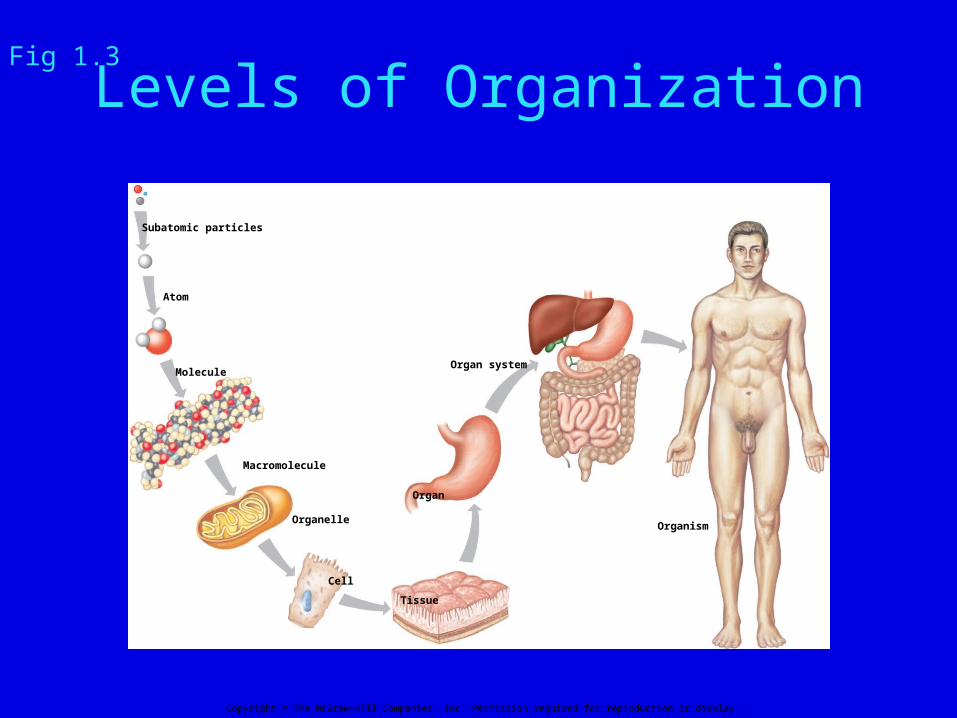
Levels of Organization
Subatomic particles
Atom
Molecule
Macromolecule
Organelle
Cell
Tissue
Organ
Organ system
Organism
Copyright © The McGraw-Hill Companies, Inc. Permission required for reproduction or display.
Fig 1.3

Cells
• Basic units of all living things• Differentiate into many different types• All cell types have some basic
characteristics in common
CytologyThe study of cellular structure
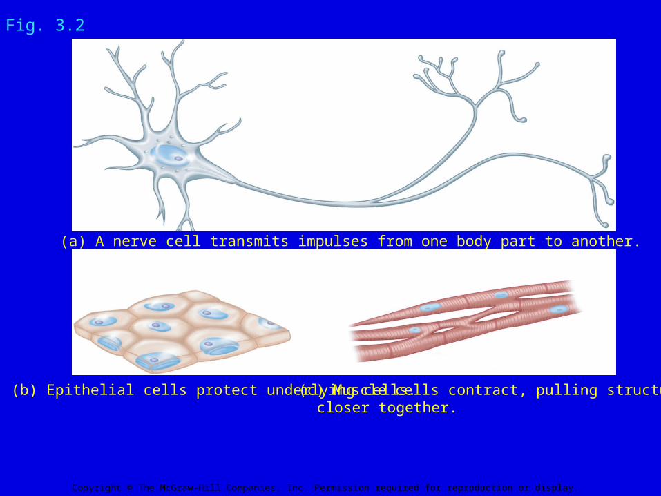
Fig. 3.2
(b) Epithelial cells protect underlying cells. (c) Muscle cells contract, pulling structures closer together.
(a) A nerve cell transmits impulses from one body part to another.
Copyright © The McGraw-Hill Companies, Inc. Permission required for reproduction or display.
Fig. 3.2
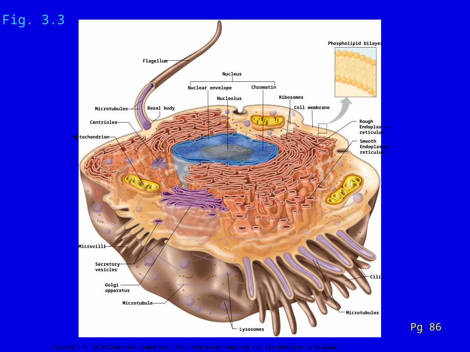
Fig. 3.3
Microtubules
Flagellum
Nuclear envelope
Basal body
Chromatin
Ribosomes
Cell membrane
Mitochondrion
Cilia
Microtubules
Microtubule
Golgiapparatus
Secretoryvesicles
Centrioles
Microvilli
Lysosomes
SmoothEndoplasmicreticulum
RoughEndoplasmicreticulum
Nucleolus
Nucleus
Phospholipid bilayer
Copyright © The McGraw-Hill Companies, Inc. Permission required for reproduction or display.
Pg 86
Fig. 3.3

Cell Membrane also called Plasma Membrane
• Outer surface of the cell
• Composed of phospholipid bi-layer
• Selectively permeable
• Protein molecules embedded in the membrane control entrance and exit of molecules

C
H H
HH
C
H
H
N
O
O
Fatty acid
Fatty acid
O
POCH
O–
Phosphate portion
(the unshaded portion may vary)
H
CH
C
H
H
O
Copyright © The McGraw-Hill Companies, Inc. Permission required for reproduction or display.
Fig. 2.15bA Phospholipid Molecule
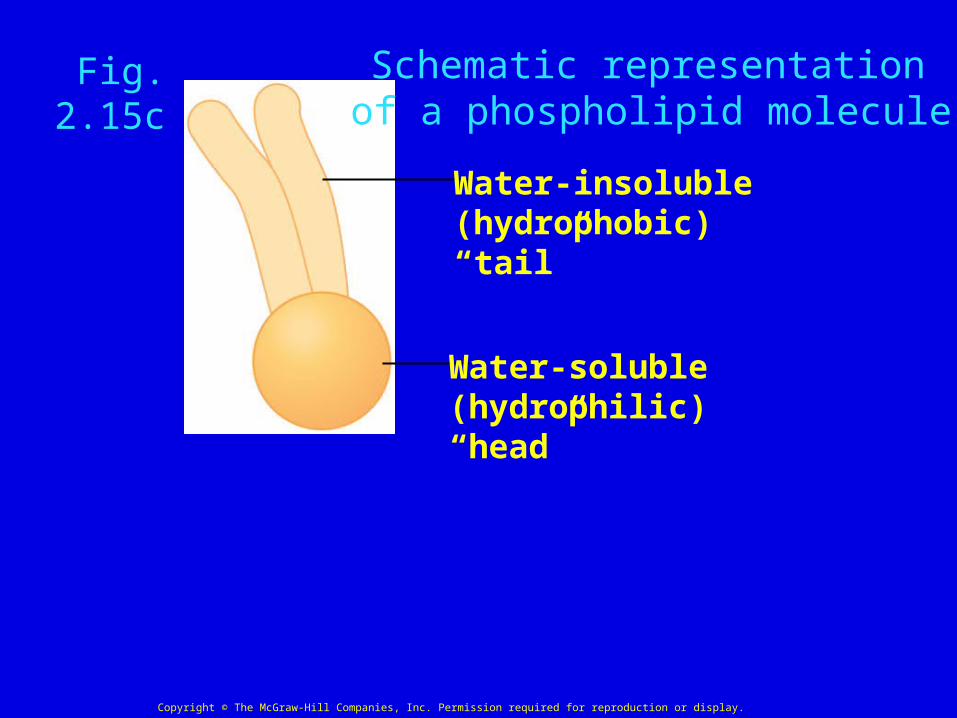
Fig. 2.15c
Schematic representationof a phospholipid molecule
Water-insoluble(hydrophobic) “tail”
Water-soluble(hydrophilic) “head”
Copyright © The McGraw-Hill Companies, Inc. Permission required for reproduction or display.

Cell membrane
“Heads” ofphospholipid
“Tails” ofphospholipid
Copyright © The McGraw-Hill Companies, Inc. Permission required for reproduction or display.
Hydrophilic
Middle of membrane
Hydrophobic
Fig. 3.6b
cytoplasm
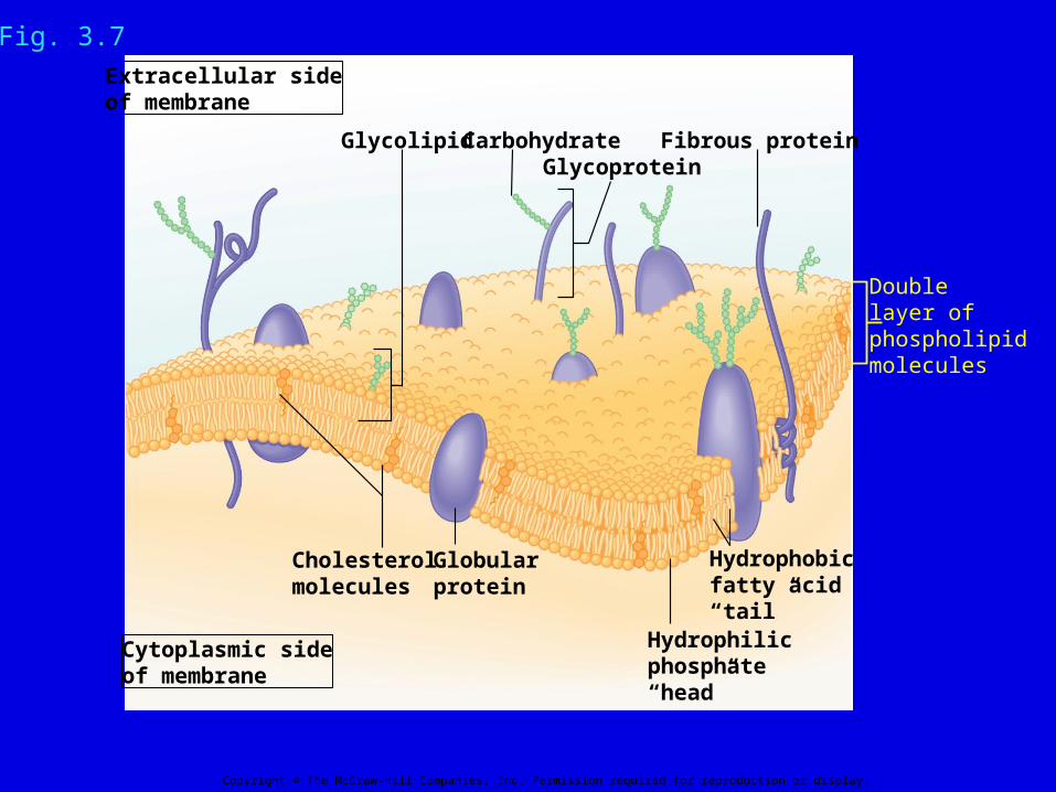
Fig. 3.7
Doublelayer ofphospholipidmolecules
Fibrous protein
Extracellular sideof membrane
Cytoplasmic sideof membrane
Carbohydrate
Hydrophobicfatty acid“tail”
Hydrophilicphosphate“head”
Cholesterolmolecules
Globularprotein
GlycolipidGlycoprotein
Copyright © The McGraw-Hill Companies, Inc. Permission required for reproduction or display.
Fig. 3.7
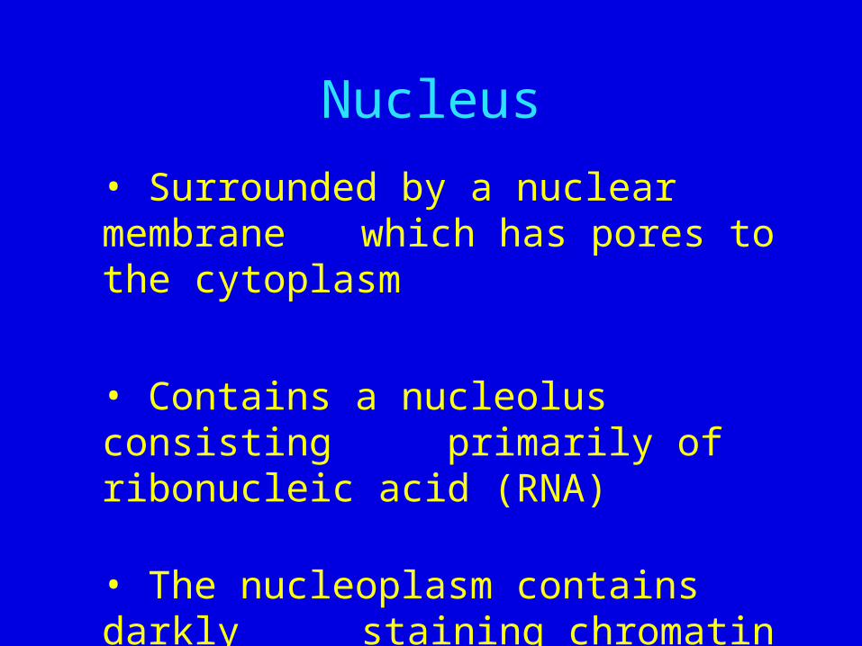
Nucleus
• Surrounded by a nuclear membrane which has pores to the cytoplasm
• Contains a nucleolus consisting primarily of ribonucleic acid (RNA)
• The nucleoplasm contains darkly staining chromatin

Fig. 3.19a
Nuclearpores
Nucleus
Nucleolus
Chromatin
Nuclearenvelope
Fig. 3.19The Cell Nucleus
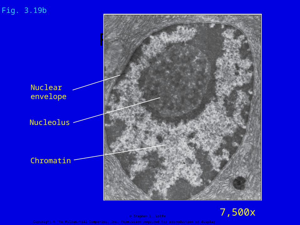
Fig. 3.19b
Nucleolus
Chromatin
Nuclearenvelope
Copyright © The McGraw-Hill Companies, Inc. Permission required for reproduction or display.
© Stephen L. Wolfe 7,500x
Fig. 3.19b

Cytoplasm
• Cellular material outside the nucleus but inside the cellular membrane
• Consists of a fluid portion called cytosol, a cytoskeleton, organelles, and inclusions

Microfilaments
• Slender fibers of the protein actin
• Bundles of microfilaments along with microtubules provide the framework of the cytoskeleton
• Connect to cellular components providing a matrix to the cytoplasm and lending strength

Microtubules
• Long slender hollow tubes made up of the protein tubulin
•Two to three times larger in diameter than microfilaments
• Provide the framework for movement of organelles within the cell
• Basic component of centrioles, cilia, and flagella

Fig. 3.18a
MitochondrionNucleus
Roughendoplasmicreticulum
Cell membrane
Microfilaments Ribosome Microtubules
Vesicle
Copyright © The McGraw-Hill Companies, Inc. Permission required for reproduction or display.
Fig. 3 18a
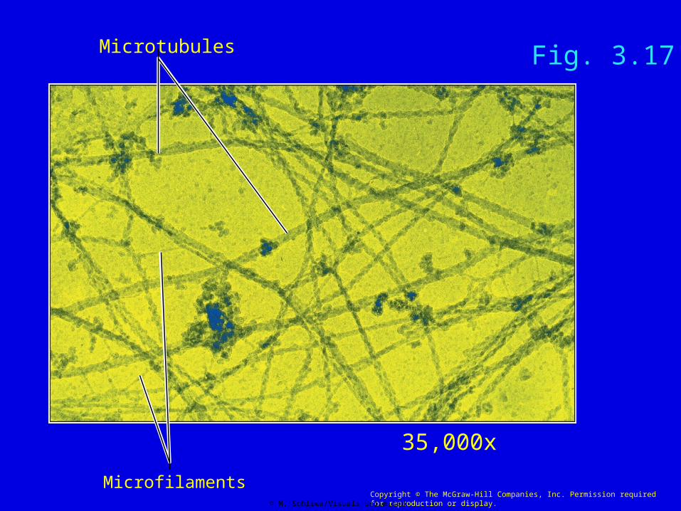
Microtubules
MicrofilamentsCopyright © The McGraw-Hill Companies, Inc. Permission required for reproduction or display.© M. Schliwa/Visuals Unlimited
Fig. 3.17
35,000x

Kirschner MW, Suter MM, Weingarten MD, Littman DR: The role of in the assembly of microtubules in vitro. Annals NY Acad Sci, 253:90-106, 1975.
Weingarten MD, Suter MM, Littman DR, Kirschner MW: Properties of the depolymerization products of microtubules from mammalian brain. Biochemistry,
13:5529-5537, 1974.
Organelles
Small structures within cells with specialized functions
The number and type of organelles within a specific cell are related to the specific structure and function of that cell

Ribosomes
• Site of protein synthesis
• Non-membranous
• Scattered throughout the cytoplasm and attached to endoplasmic reticulum (ER)
• Composed of two sub-units
• Subunits are manufactured
separately in the nucleolus

ER membrane
Ribosomes
(a)
Copyright © The McGraw-Hill Companies, Inc. Permission required for reproduction or display.
© Don W. Fawcett/Photo Researchers, Inc. 28,500x
Fig. 3.9a
Ribosomes on the Endoplasmic Reticulum
Endoplasmic Reticulum (ER)• Most extensive structure within the cytoplasm
• Composed of broad, flattened, interconnected sacs and tubules
• Interior spaces called cisternae
• Provides pathway for intracellular transport of molecules
• Called Rough ER (RER) when covered with ribosomes

ER Membranes
Ribosomes
Copyright © The McGraw-Hill Companies, Inc. Permission required for reproduction or display.
Endoplasmic ReticulumFig. 3.9

Smooth ER
Copyright © The McGraw-Hill Companies, Inc. Permission required for reproduction or display.
Fig. 3.9c
Golgi Apparatus• Connected to Rough ER (RER)
• Composed of stacks of flattened
membranous sacs called cisternae
• Modifies, packages, and distributes
proteins and lipids made on rough and
smooth ER
• Receives and transports molecules via
vesicles

Fig. 3.10b
Golgiapparatus
Nucleus
Cytosol
Transportvesicle
Secretion
Cell membrane
Nuclearenvelope
Copyright © The McGraw-Hill Companies, Inc. Permission required for reproduction or display.
RoughEndoplasmicreticulum
Fig. 3.10b Golgi Apparatus
Nucleus
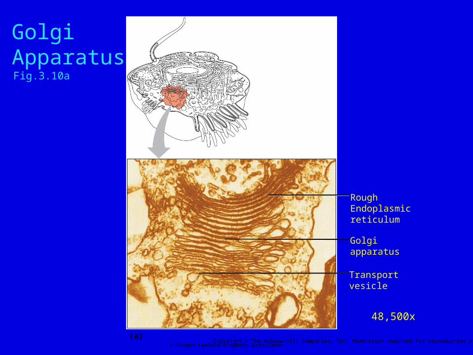
Fig. 3.10a
Golgiapparatus
RoughEndoplasmicreticulum
Transportvesicle
(a)Copyright © The McGraw-Hill Companies, Inc. Permission required for reproduction or display.
© Gordon Leedale/Biophoto Associates
48,500x
Golgi ApparatusFig.3.10a
Vesicles
• Small membranous sacs that vary in size and contents
• Form by pinching off of small pieces of membrane that surround a tiny amount of liquid or solid
• Method of distributing molecules within a cell and/or into and out of cell
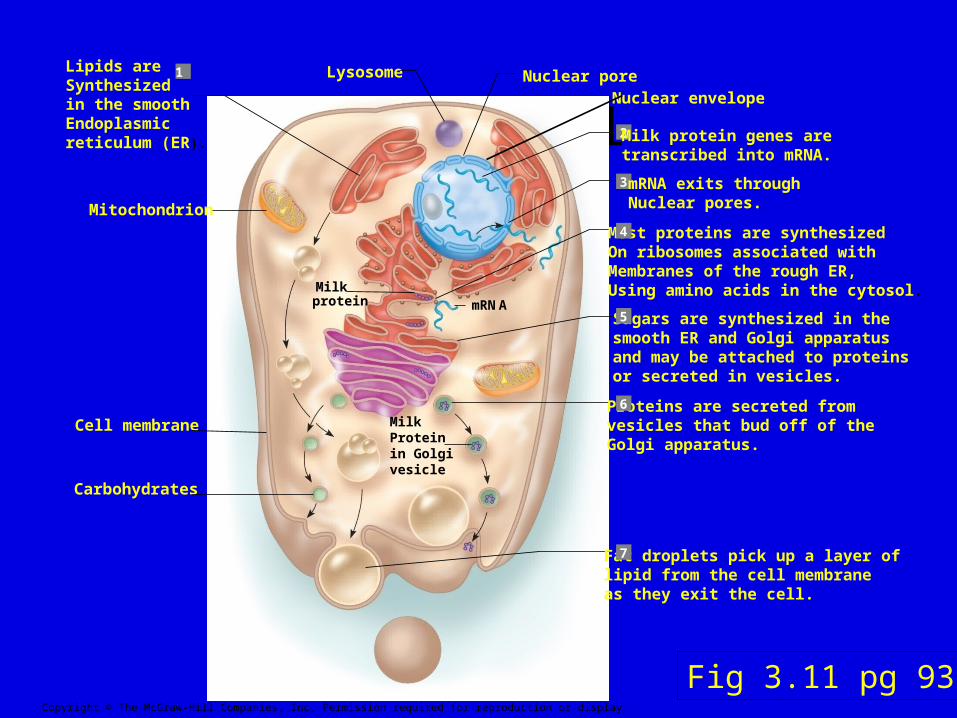
Fig. 3.11
Carbohydrates
Cell membrane
Mitochondrion
Lysosome Nuclear poreNuclear envelope
mRNAMilkprotein
Lipids areSynthesizedin the smoothEndoplasmicreticulum (ER).
Most proteins are synthesizedOn ribosomes associated withMembranes of the rough ER,Using amino acids in the cytosol.
Sugars are synthesized in thesmooth ER and Golgi apparatusand may be attached to proteinsor secreted in vesicles.
Proteins are secreted fromvesicles that bud off of theGolgi apparatus.
Fat droplets pick up a layer oflipid from the cell membraneas they exit the cell.
2
3
4
5
6
7
Milk protein genes aretranscribed into mRNA.
1
mRNA exits throughNuclear pores.
Milk Proteinin Golgivesicle
Copyright © The McGraw-Hill Companies, Inc. Permission required for reproduction or display.
Fig 3.11 pg 93
Mitochondrion
• Rod-shaped bodies with double- layered walls
• Inner layer has many infoldings called cristae
• Major site of energy (ATP) production
• Have their own DNA and ribosomes which are inherited solely from the mother
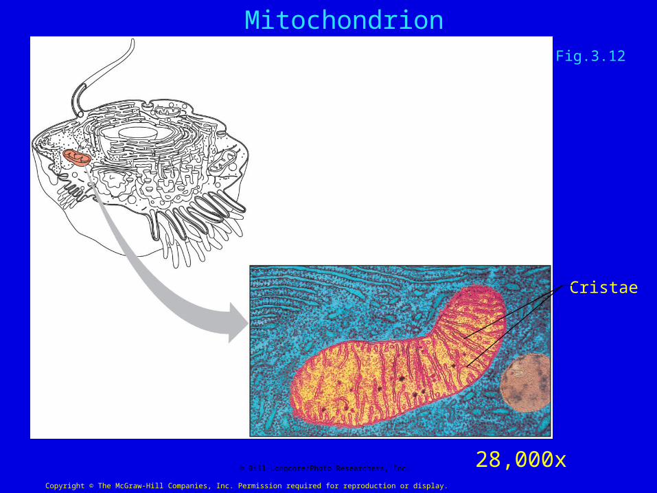
Fig. 3.12a
Cristae
Copyright © The McGraw-Hill Companies, Inc. Permission required for reproduction or display.
© Bill Longcore/Photo Researchers, Inc.
Mitochondrion
28,000x
Fig.3.12
Lysosomes
• Membrane-enclosed vesicles of varying shape
• Contain a variety of enzymes
• Clean up the cell by destroying debris, unnecessary molecules, aging organelles and foreign particles
• Can play a role in facilitating cell death
Peroxisomes
• Similar to lysosomes in structure but usually smaller
• Contain enzymes for breaking down various often toxic substances
peroxidases and catalase
•Most abundant in liver and kidney cells which are active in detoxification

Centrosome
• Located near the nucleus
• Consists of 2 non-membranous
cylinders - centrioles, at right angles to each other
• Made up of microtubules
• Distribute chromosomes during cell division by forming spindle fibers
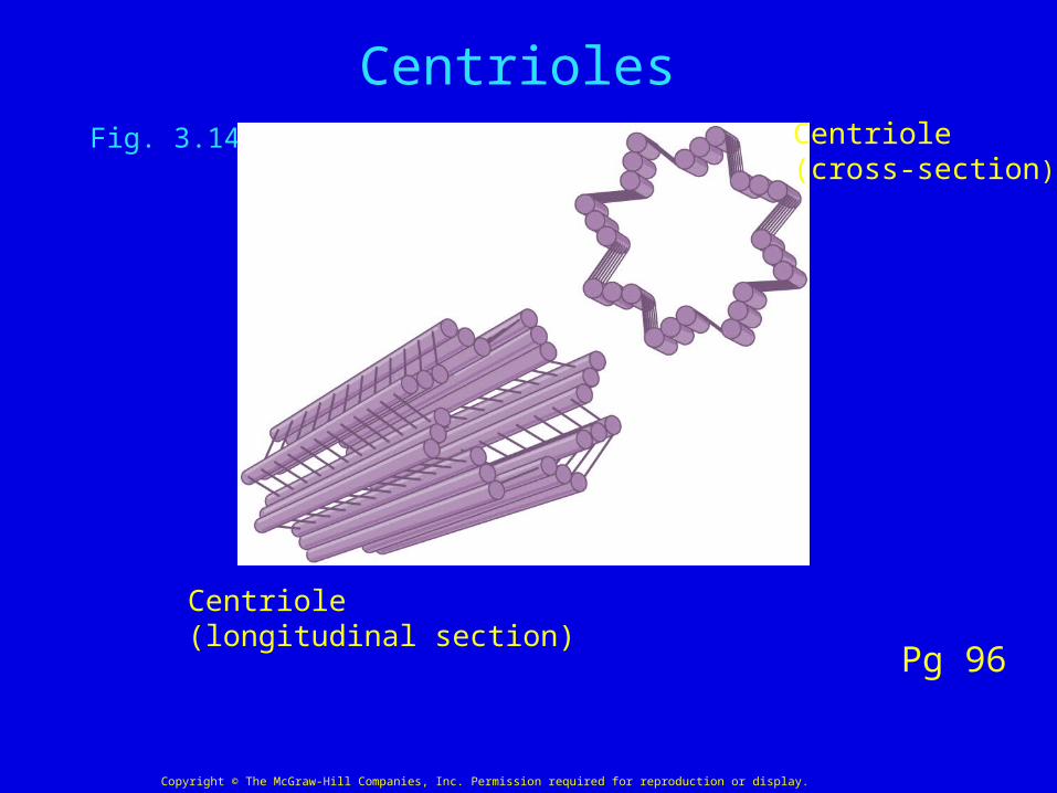
Fig. 3.14b Centriole(cross-section)
Centriole(longitudinal section)
Copyright © The McGraw-Hill Companies, Inc. Permission required for reproduction or display.
Centrioles
Pg 96
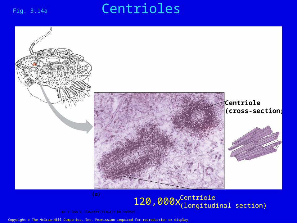
Fig. 3.14a
(a)
Centriole(cross-section)
Centriole(longitudinal section)
Copyright © The McGraw-Hill Companies, Inc. Permission required for reproduction or display.
a: © Don W. Fawcett/Visuals Unlimited
120,000x
Fig. 3.14a Centrioles

Cilia and Flagella• Hair-like appendages on some cells
• Function in providing movement
• Both originate from centrioles and are made up of microtubules
• Cilia are usually <20 um long
• Flagella can be thousands of um in length
• The only human cell with a flagellum is the sperm cell
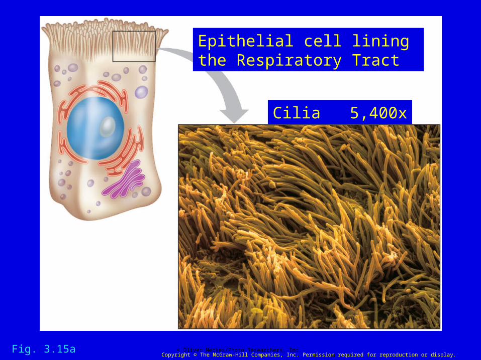
Fig. 3.15a
Copyright © The McGraw-Hill Companies, Inc. Permission required for reproduction or display.© Oliver Meckes/Photo Researchers, Inc.
Epithelial cell lining the Respiratory Tract
Cilia 5,400x
Fig. 3.15a

Fig. 3.16
Copyright © The McGraw-Hill Companies, Inc. Permission required for reproduction or display.
© Colin Anderson/Brand X/CORBIS 1,400x
Fig. 3.16

Fig. 3.3
Microtubules
Flagellum
Nuclear envelope
Basal body
Chromatin
Ribosomes
Cell membrane
Mitochondrion
Cilia
Microtubules
Microtubule
Golgiapparatus
Secretoryvesicles
Centrioles
Microvilli
Lysosomes
SmoothEndoplasmicreticulum
RoughEndoplasmicreticulum
Nucleolus
Nucleus
Phospholipid bilayer
Copyright © The McGraw-Hill Companies, Inc. Permission required for reproduction or display.
Pg 78

Copyright © The McGraw-Hill Companies, Inc. Permission required for reproduction or display.
Microvilli
Cell membrane
Mitochondrion
Golgi apparatus
Nucleolus
Nucleus
(a)
Lumen Microvilli
(b)
Roughendoplasmicreticulum
Cytoplasm ofepithelial cell
b: © The McGraw-Hill Companies, Inc./Al Telser, photographer 16,000x
Intestinal Epithelial Cell
Chap 17 pg 678