human bartonellosis
TRANSCRIPT

Human Human Bartonellosis Bartonellosis
caused bycaused byBartonella Bartonella
bacilliformisbacilliformis

César HenríquezCésar Henríquez11
Paul PachasPaul Pachas22
Phillip LawyerPhillip Lawyer33
Larry LaughlinLarry Laughlin33
Ciro MaguiñaCiro Maguiña11
1. Instituto de Medicina Tropical Alexander von Humboldt-1. Instituto de Medicina Tropical Alexander von Humboldt-Universidad Peruana Cayetano HerediaUniversidad Peruana Cayetano Heredia
2. Oficina General de Epidemiologia2. Oficina General de Epidemiologia
3. Uniformed Services University of the Health Sciences3. Uniformed Services University of the Health Sciences
20022002

Introduction
• Human bartonellosis is the clinical term to define the bacterial infections by the genus Bartonella.
• There are five important species that produce human diseases.

History and ArcheologyBartonellosis has been known since Pre-Inca times. Numerous artistic representations in clay “huacos” depict the chronic phase of the disease.
Historians and chronists described a disease with warts in Spanish troops when they arrived for the first time in Coaque-Ecuador.
For a long time it was thought that the disease was endemic only in Peru and that it had only one phase: “Peruvian wart” or “Verruga peruana”

Historical Figures
Dr. Alberto L. Barton
(1871-1950)
Daniel A. Carrión(1858-1885)
In 1875 an outbreak, characterized by fever and anemia (Oroya fever) occurred in the region of construction of the railroad line between Lima and Oroya. In 1885, Daniel A. Carrion, a Peruvian medical student, inoculated himself with material taken from a patient with Peruvian wart. He subsequently acquired Oroya fever and died a month later. Later, Alberto Barton discovered the etiologic agent of “Carrion’s disease”

Epidemiology• Bartonellosis is endemic
in Perú, Ecuador and Colombia.
• Geography and weather conditions vary depending of the region.
• Emergence or re-emergence of several infectious diseases, including bartonellosis, seem to coincide with “el Niño” weather phenomena.

Carrion’s disease cases (1945-2001)
YEAR

Ancash department was the most important endemic area of bartonellosis since 1945 until 1994.

AMAZONAS
CAJAMARCA
ANCASH
LIMA
AMAZONASPIURA
CAJAMARCASAN MARTIN
HUANUCOANCASH
LIMA
AYACUCHO
AMAZONASPIURA
CAJAMARCA
SAN MARTIN
LA LIBERTAD
ANCASH HUANUCO
LIMAJUNIN
CUSCO
LORETO
AM
AZ
ON
AS
PIURA
CA
JAM
AR
CA
AN
CA
SH
HUANUCO
LIM
A
CUSCO
1995 1997 1999 2001
Reported cases of Carrion’s disease
(1995-2001)


Incidence of Carrion’s disease by regions (1996-2002)

SAN IGNACIO
UTCUBAMBA
MOYOBAMBA
RODRIGUEZ DE MENDOZA
CASMA
CHANCHAMAYO
LA CONVENCION
QUISPICANCHI
ALTO AMAZONAS
RIOJA
BARRANCA
HUAURA
OYON
LEONCIO PRADOHUAMALIES
PACHITEAYAROWILCA
PAUCARTAMBO
CALCA
URUBAMBA
CANCHIS
ANTA
CUSCO
New foci of Carrion’s disease
February 2002
New epidemic areas identified.
Mortality during the outbreaks is high.
No cases of chronic phase (Peruvian wart)in epidemic areas.
No animal reservoir identified.
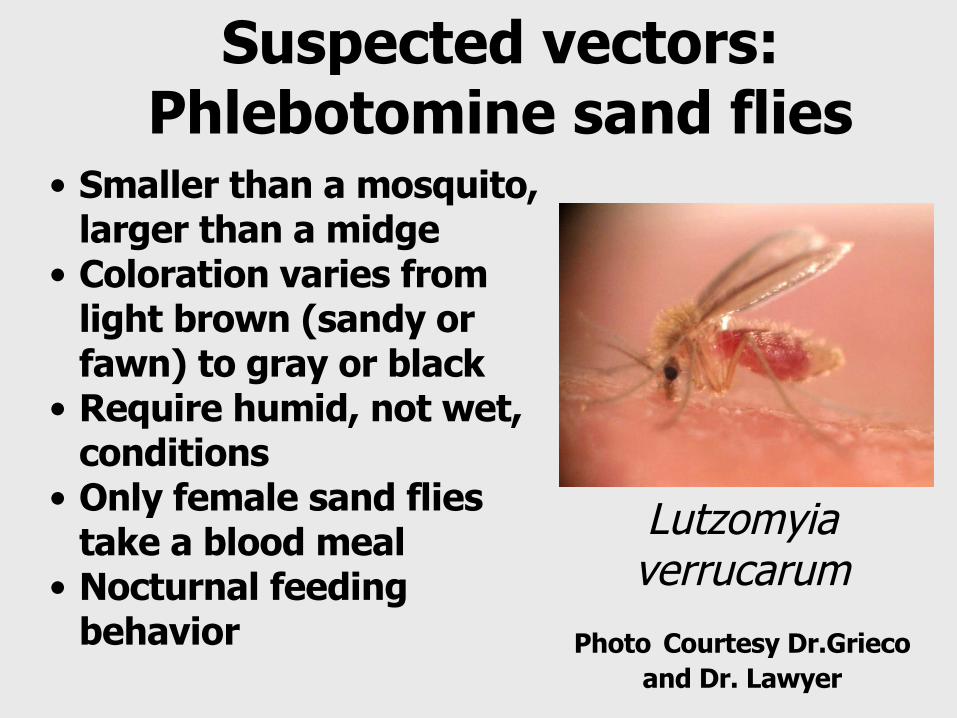
Suspected vectors:Phlebotomine sand flies
Lutzomyia verrucarum
Photo Courtesy Dr.Grieco and Dr. Lawyer
• Smaller than a mosquito, larger than a midge
• Coloration varies from light brown (sandy or fawn) to gray or black
• Require humid, not wet, conditions
• Only female sand flies take a blood meal
• Nocturnal feeding behavior

Suspected Vectors:Phlebotomine sand flies
Lutzomyia peruensis
Courtesy Dr.Grieco and Dr. Lawyer
• Sand fies are weak fliers
• Fly only at night unless disturbed in their daytime resting site
• Sand flies transmit Bartonella bacilliformis from infected to uninfected hosts by bite
• At least two species suspected in Peru: Lu. verrucarum and Lu. peruensis

Provinces with
Lutzomya verrucarum
Distribution of Carrion’s disease cases and Lutzomyia verrucarum
Provinces with Carrion’s disease cases

Etiologic agent:Bartonella bacilliformis
Gram negative aerobic, pleomorphic, flagellated, motile, coccobacillary, 2-3 m large and 0,2 - 0,5 m wide and facultative intracellular bacterium.
For its isolation, special cultures are required containing complemental soy agar, proteases, peptones, some essential amino acids and blood. The optimum growing temperature is 19-29 ºC.

Pathogenesis• Bartonella bacilliformis is
transmitted by the bite of the suspected vector Lutzomyia spp
• Following transmission, the bacterium infects red blood cells and endothelial cells
• The physical damage and introduction of antigens in the membranes of the red cells stimulate the Reticuloendothelial System to produce an intense erythrophagocytosis by macrophages and histiocytic cells resulting in severe extra vascular hemolytic anemia
•

Endothelial cells: the last target?
• The invasion of endothelial cells is an active process dependent on the activation of Rho, which is an intracellular signal implicated in the rearrangement of the host cell actin cytoskeletal network

The disease
• The clinical symptoms of bartonellosis are pleomorphic and some patients may be asymptomatic
• The two classical clinical presentations are the acute phase and the chronic phase, corresponding to the two different host cell types invaded by the bacterium

Acute phase: Oroya fever or Carrion’s disease
• The mean incubation time is 21 days (range 10 to 270 days)
• The diagnostic tests in this phase are:
Diagnostic test Sensitivity Specificity ReferenceBlood smear 36-73 91-96 1
Immunoblot 70 94 2
PCR(16S-23S) 47 98 3
Values in porcentaje

The diagnosis
The diagnosis in the acute phase can be done using the thin blood film with Giemsa stain.
It is possible to observe the bacillus inside the red blood cells.

M: DNA ladder (100 bp).1: B. bacilliformis DNA from culture extracted by thermal lysis (100°C, 10 min.) using 16S 23S primers (positive control).2: Whole blood extraction from an acute phase patient, using 16S 23S primers.3: Whole blood extraction from an acute phase patient, using primers for Citrate Synthetase gene.4: B. bacilliformis DNA from a culture extraction using primers for Citrate Synthetase gene.
Base pairs
1500 bp
600 bp
Molecular technics
M 1 2 3 4

Lane A: Positive control poolLane Band C: Bartonella bacilliformis-positive serum taken from a patient in acute phaseLane D: Negative control pool
Immunologic technics: Sonicated immunoblot
A B C D
20 kDa18 kDa17 kDa14 kDa

Chronic Phase: Peruvian wart (Verruga Peruana)
Mularlesions

Chronic Phase: Peruvian wart (Verruga Peruana)
Miliary lesions

Chronic Phase: Peruvian wart (Verruga Peruana)
Miliary lesions with overwhelming infection

Chronic phase: some numbers
• The diagnostic tests in this phase are blood culture (13% of patients with verruga have bacteriemia), culture of the verrugous warts and Immunoblot with a sensitivity of 70% and specificity of 100%
• The IFA has a sensitivity of 82% and specificity of 92%

Immunity and infection
• One factor that complicates the clearance of the bacterium is that intra-erythrocytic Bartonella are protected from both humoral and cellular immune responses due to a lack of major histocompatibility complex (MHC) molecules on the surface of the mature erythrocytes
• They are unable to present antigens of their invaders to the immune system

Conclusion• Human bartonellosis is a bacterial infection by
the genus bartonella• Bartonellosis caused by B. bacilliformis
(Oroya’s fever or Carrion’s disease) is endemic in Peru, Ecuador and Colombia
• No animal reservoir identified• Suspected vectors: Phlebotomine sand flies• About the disease, there are two classical
clinical presentations: acute and chronic phase
• New endemic areas identified: Emergent infectious disease