human cancer cell negative transcriptional regulator ofthe ...labs.biology.ucsd.edu/david/reprint...
TRANSCRIPT

MOLECULAR AND CELLULAR BIOLOGY, Feb. 1994, p. 1477-14860270-7306/94/$04.00+0Copyright © 1994, American Society for Microbiology
Human Cancer Cell Lines Express a Negative TranscriptionalRegulator of the Interferon Regulatory Factor Family
of DNA Binding ProteinsEMANUEL PETRICOIN III,1 MICHAEL DAVID,1'2 HUI FANG,2 PHILIP GRIMLEY,2
ANDREW C. LARNER,l12* AND SCOTIr VANDE POL3*
Division of Cytokine Biology, Centerfor Biologics Evaluation and Research,' Department ofPathology,Uniformed Services University of the Health Sciences,2 and Laboratory of Tumor
Virus Biology, National Cancer Institute,3 Bethesda, Maryland 20892
Received 2 August 1993/Returned for modification 18 September 1993/Accepted 15 November 1993
Members of the interferon regulatory factor (IRF) family ofDNA binding transcription factors have roles ingrowth regulation, antiviral responses, and transcriptional induction of interferon (IFN)-activated earlyresponse genes. The IRF family member ISGF3'y is the DNA binding component of IFN-stimulated gene factor3 (ISGF3), a multicomponent complex responsible for the stimulation of IFN-a-responsive genes. IFN-a-stimulated formation ofISGF3 and subsequent gene expression can be inhibited by phorbol esters or expressionof the adenovirus ElA protein. We have investigated IFN signaling in human malignant tumor cell lines of thelung, colon, ovary, cervix, and hematopoietic organs and found some of these cells to be defective forIFN-a-induced formation of ISGF3. In many cases, an inhibitory activity termed transcriptional knockout(TKO) correlated with nonresponsiveness. TKO purified from a human papillomavirus-negative cervicalcarcinoma cell line has a molecular size of 19 kDa. The purified protein interacted with the ISGF3y componentof ISGF3, preventing binding of ISGF3 to DNA. Purified TKO displaced ISGF3 from its DNA binding site invitro and prevented ISGF3'y, IRF-1, and IRF-2 from interacting with the IFN-stimulated response element.Partially purified TKO can also directly interact with ISGF3-y in the absence of DNA. This protein may beinvolved with the development of malignancies and the inability of IFN to exert its antiproliferative andantiviral effects.
The many biological activities of interferons (IFNs) ap-pear to be controlled at least in part by a set of cellular geneswhich are rapidly induced upon IFN-a or IFN--y binding tospecific cell surface receptors. IFN-stimulated gene factors(ISGFs) mediate gene induction by binding to enhancerswithin the promoters of both IFN-a- and IFN--y-inducedgenes. ISGFs have been characterized, and many of theseproteins have been purified and cloned (5, 12, 13, 25, 39).Assembly and DNA binding of the ISGF3 complex followsIFN-a activation of a membrane-associated tyrosine phos-phatase(s) and tyrosine kinase(s). A subset of the ISGF3proteins, called ISGF3a, becomes tyrosine phosphorylated(7, 11, 15, 39) and associates with the 48-kDa DNA bindingsubunit, ISGF3-y (referred to as 48kDay to avoid confusionwith ISGF3). The complex of ISGF3a and 48kDa-y com-
pletes the assembly of ISGF3, which then translocates to thenucleus and participates in the induction of IFN-activatedearly response genes.
Several studies indicated that growth promoters such as
phorbol esters and interleukin 4, as well as certain proteinsfrom DNA tumor viruses such as adenovirus ElA and thehepatitis B virus polymerase protein, also inhibit IFN-induced gene expression and induction of ISGF3 by IFN-a(1, 10, 14, 22-24, 32). In the case of phorbol esters and ElA,
* Corresponding authors. Mailing address for Andrew C. Lamer:Division of Cytokine Biology, Center for Biologics Evaluation andResearch, 8800 Rockville Pike, Bethesda, MD 20892. Phone: (301)402-0361. Present address for Scott Vande Pol: Department ofPathology, Case Western Reserve University School of Medicine,Cleveland, OH 44106. Phone: (216) 368-1679.
decreased formation of ISGF3 does not appear to reflect anyinability of IFN-at to modify the ISGF3a proteins (1, 14, 22,32). This conclusion is based upon evidence that ISGF3 can
be reconstituted by addition of excess 48kDay in nuclearextracts prepared from cells treated with IFN-a and phorbolester or extracts from cells which express ElA (1, 14, 22,32). Deletion of the conserved region 1 domain of ElAabrogated ElA's inhibitory effect on ISGF3 formation.Thus, in both phorbol ester-treated and ElA-expressingcells, the 48kDay subunit of ISGF3 either is not present or isincompetent to permit assembly of ISGF3. Alternatively, an
inhibitor which acts upon the 48kDay protein such that itinteracts less effectively with the activated ISGF3a proteinsmay be present in phorbol ester-treated cells or cells ex-
pressing ElA.The E6 and E7 proteins encoded by human papillomavirus
(HPV) have many biological and structural similarities toadenovirus early proteins (28, 33, 37). Studies were thusinitiated to determine whether expression of the E6 and E7proteins might modulate IFN signaling. During the course ofthese investigations, it became apparent that although ex-
pression of the E6 and E7 proteins in cervical cancer celllines correlated with inhibition of IFN-induced gene expres-sion in a manner analogous to that observed with ElA,HPV-negative cervical cancer cell lines and other cancer celllines also showed defects in IFN-a-induced gene expression.In both virally transformed cells and cells from spontaneoushuman malignancies, IFN-a signaling is inhibited by mech-anisms that target the interferon regulatory factor (IRF)family member 48kDay. Using one such HPV-negative cer-vical cancer cell line, we have purified a protein termedtranscriptional knockout (TKO) which specifically inhibited
1477
Vol. 14, No. 2
at UN
IV O
F C
ALIF
SA
N D
IEG
O on A
ugust 2, 2007 m
cb.asm.org
Dow
nloaded from
1478 PETRICOIN ET AL.
the binding to the IFN-stimulated response element (ISRE)of not only the 48kDa-y protein but also IRF-1 and IRF-2.
MATERIALS AND METHODS
Cells and culture. Human foreskin diploid fibroblasts(GM00468) from the National Institute of General MedicalSciences, HeLa S3 (CCL 2.2), CaSki (CRL 1550), SiHa(HTB 35), C-33A (HTB 31), and HT-3 (HTB 32) cells; aWI-38VA13 subline (CCL 75.1); and FaDu (HTB 43), K562(CCL 243), NCI-H498 (CCL 254), NCI-H146 (HTB 173),SK-OV3, 293, and MS 751 cells were obtained from theAmerican Type Culture Collection and grown and passagedaccording to the accompanying recommendations. N592cells were kindly grown and provided by Herbert Oie(National Cancer Institute).IFNs and reagents. Recombinant human IFN-a2a was a
generous gift from Hoffmann-LaRoche. Recombinant hu-man IFN-y was provided by Genentech Corp.
Preparation of nuclear and cytoplasmic cell extracts. Nu-clear and cytoplasmic cell extracts were prepared essentiallyas described elsewhere (32). Selected extracts were alky-lated with 10 mM N-ethylmaleimide for 20 min at roomtemperature followed by quenching with the addition of 15mM dithiothreitol at 4°C.
Electrophoretic mobility shift assays (EMSAs). Gel shiftassays were performed with a 32P-end-labeled double-stranded synthetic oligonucleotide (1.0 ng) with the se-quence 5'GATCCATGCCTCGGGAAAGGGAAACCGAAACTGAAGCC3' from the ISG15 (ISG with a 15-kDa prod-uct) promoter (32). The IFN-y response region (GRR) probewas labeled in the same manner and contained the sequence5'AGCATGTTTCAAGGATTTGAGATGTATTTCCCAGAAAAG3' (47). The AP1 competitor oligonucleotide waspurchased from Promega.
In vitro transcription and translation of 48kDa'y, IRF-1,and IRF-2 and formation of the TKO-ISGF3'y complex. The48kDa-y protein (clone 38-1) (42), the 113-kDa ISGF3ot pro-tein (13), and the full-length IRF-1 (pET-IRF1) and IRF-2(pET-IRF2) cDNAs (17) were in vitro transcribed and trans-lated with or without [ 5S]methionine in rabbit reticulocytelysate (Promega) according to the manufacturer's recom-mendation. Two microliters of translated or mock-translatedprotein was incubated with or without 5 ng of unlabeledISG15 double-stranded oligonucleotide probe in standardEMSA conditions on ice for 15 min. Five microliters ofbuffer A (20 mM KCl; 10 mM HEPES [N-2-hydroxyeth-ylpiperazine-N'-2-ethanesulfonic acid], pH 7; 5 mM dithio-threitol; 20% glycerol) or 5 ,ul of a column fraction enrichedfor TKO activity was added on ice for an additional 15 minand applied to a 6% native polyacrylamide gel as describedelsewhere (32). Gels were fixed exhaustively to removeunincorporated [315]methionine, dried, and exposed onX-ray film.
Purification of TKO from C-33A cells. C-33A cells wereharvested as described above, and extracts were chromato-graphically separated at 4°C. Active fractions were assayedby their ability to inhibit the binding of ISGF3 to the ISRE.The following resins were used to purify TKO: (i) heparin-Sepharose (Pharmacia) with a 0.05 to 0.3 M NaCl gradientand (ii) hydroxylapatite (Bio-Rad) with a 0 to 0.3 M Na2PO4(pH 7.0) elution gradient. The active fractions were dialyzedagainst 20 mM HEPES (pH 7.0)-20 mM KCl-1 mM dithio-threitol-20% glycerol (buffer A), and (NH4)2SO4 up to 25%was added to the dialyzed material. This solution wasapplied to phenyl-Sepharose (Pharmacia), and the flow-
through and wash were collected, dialyzed against buffer A,and concentrated on hydroxylapatite. Purified TKO wasrenatured from sodium dodecyl sulfate (SDS)-13% poly-acrylamide gels as described elsewhere (16).
RESULTS
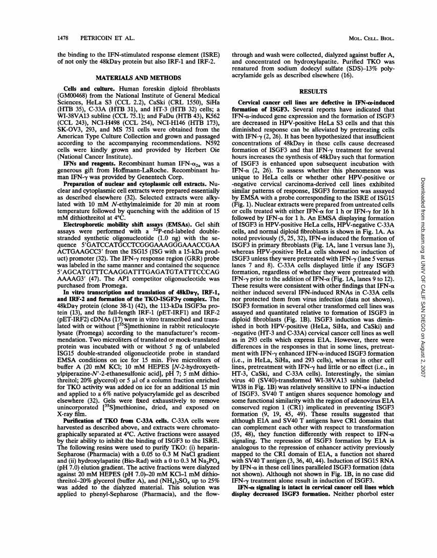
Cervical cancer cell lines are defective in IFN-cK-inducedformation of ISGF3. Several reports have indicated thatIFN-ot-induced gene expression and the formation of ISGF3are decreased in HPV-positive HeLa S3 cells and that thisdiminished response can be alleviated by pretreating cellswith IFN-y (2, 26). It has been hypothesized that insufficientconcentrations of 48kDa-y in these cells cause decreasedformation of ISGF3 and that IFN--y treatment for severalhours increases the synthesis of 48kDay such that formationof ISGF3 is enhanced upon subsequent incubation withIFN-a (2, 26). To assess whether this phenomenon wasunique to HeLa cells or whether other HPV-positive or-negative cervical carcinoma-derived cell lines exhibitedsimilar patterns of response, ISGF3 formation was assayedby EMSA with a probe corresponding to the ISRE of ISG15(Fig. 1). Nuclear extracts were prepared from untreated cellsor cells treated with either IFN-a for 1 h or IFN--y for 16 hfollowed by IFN-a for 1 h. An EMSA displaying formationof ISGF3 in HPV-positive HeLa cells, HPV-negative C-33Acells, and normal diploid fibroblasts is shown in Fig. 1A. Asnoted previously (5, 25, 32), IFN-a induced the formation ofISGF3 in primary fibroblasts (Fig. 1A, lane 1 versus lane 3),whereas HPV-positive HeLa cells showed no induction ofISGF3 unless they were pretreated with IFN--y (lane 5 versuslanes 7 and 8). C-33A cells displayed little if any ISGF3formation, regardless of whether they were pretreated withIFN-y prior to the addition of IFN-a (Fig. 1A, lanes 9 to 12).These results were consistent with other findings that IFN-aneither induced several IFN-induced RNAs in C-33A cellsnor protected them from virus infection (data not shown).ISGF3 formation in several other transformed cell lines wasassayed and quantitated relative to formation of ISGF3 indiploid fibroblasts (Fig. 1B). ISGF3 induction was dimin-ished in both HPV-positive (HeLa, SiHa, and CaSki) and-negative (HT-3 and C-33A) cervical cancer cell lines as wellas in 293 cells which express ElA. However, there weredifferences in the responses in that in some lines, pretreat-ment with IFN-y enhanced IFN-a-induced ISGF3 formation(i.e., in HeLa, SiHa, and 293 cells), whereas in other celllines, pretreatment with IFN--y had little or no effect (i.e., inHT-3, CaSki, and C-33A cells). Interestingly, the simianvirus 40 (SV40)-transformed WI-38VA13 subline (labeledWI38 in Fig. 1B) was relatively sensitive to IFN-a inductionof ISGF3. SV40 T antigen shares sequence homology andsome functional similarity with the region of adenovirus ElAconserved region 1 (CR1) implicated in preventing ISGF3formation (9, 19, 45, 49). These results suggested thatalthough ElA and SV40 T antigens have CR1 domains thatcan complement each other with respect to transformation(35, 48), they function differently with respect to IFN-asignaling. The repression of ISGF3 formation by ElA isanalogous to the repression of enhancer activity previouslymapped to the CR1 domain of EMA, a function not sharedwith SV40 T antigen (3, 36, 40, 44). Induction of ISG15 RNAby IFN-a in these cell lines paralleled ISGF3 formation (datanot shown). Although not shown in Fig. 1B, in no case didIFN-y treatment alone result in induction of ISGF3.IFN-a signaling is intact in cervical cancer cell lines which
display decreased ISGF3 formation. Neither phorbol ester
MOL. CELL. BIOL.
at UN
IV O
F C
ALIF
SA
N D
IEG
O on A
ugust 2, 2007 m
cb.asm.org
Dow
nloaded from

TKO INHIBITS ISGF3 BINDING TO DNA 1479
r HF - - eLa-a r- C-33A
IFN - 7 a y7a - 7 a 7/U - 7 a 'lu
_
....2 3 4 5 6 7 8 9
A COMPNEM
TRANSLATION
EXTRACT
_ + - _ + +
- - , 113 ^ 113
HF HF - - HF HF_
op_1W
ISRE -
+ -+HF C33A
qpm
l1
ISGF3 -*
B
48 kDa ^t
1 2 3 4 5 6 7 8
H--==v A IFNY->IFNa
CASKI
HT3 C33A
FIG. 1. Cervical cancer cell lines are defective in IFN-a-inducedISGF3 formation. (A) Cells were incubated with IFN-a (500 U/ml)for 1 h or with IFN-y (5 ng/ml) for 16 h followed by IFN-a for 1 h.Nuclear extracts were then prepared as described in Materials andMethods. ISGF3 activity was assayed by gel shift using a 32P-end-labeled ISRE probe corresponding to the ISG15 gene. Equalamounts of protein were assayed from each cell line. (B) BasalISGF3 or ISGF3 levels in response to IFN-a or the combination ofIFN-a and IFN--y were quantitated from EMSA gels such as thoseshown in panel A with an Ambis scanner. Equal amounts of nuclearextract protein were analyzed in each lane. Levels of ISGF3 relativeto that in human fibroblasts are shown, with the levels of IFN-a-induced ISGF3 from human fibroblasts arbitrarily set at 100%.HPV-positive SiHa, HeLa, and CaSki cervical carcinoma cell linesand HPV-negative HT-3 and C-33A cervical carcinoma cell lineswere used. HF, human diploid fibroblasts; 293, adenovirus E1A-transformed human epithelial cells; W138, WI-38VA13 subline.
nor adenovirus ElA appears to cause a defect in IFN-a-induced modification of the ISGF3a proteins (1, 14, 22, 32).In fact, in vitro addition of extracts which contained excess48kDay protein to extracts from IFN-a-treated cells whichexpress ElA or which have been treated with phorbolmyristate acetate restored formation of ISGF3 (1, 14, 22, 32).In order to see whether the virus-negative cell line C-33Awas similar to phorbol ester-treated or ElA-expressing cells,experiments were done to determine whether IFN-a-in-duced modification of the a peptides was intact. Similar tothe results observed with phorbol ester-treated cells andthose expressing ElA, addition of excess in vitro-translated48kDa-y protein also reconstituted ISGF3 formation in nu-clear extracts prepared from IFN-a-treated C-33A cells (Fig.2A, lane 8) to levels comparable to those in diploid fibro-blasts (lane 5). The effects of the addition of the 48kDa-yprotein were specific in that the addition of a nonspecificcontrol translation protein (the 113-kDa protein of ISGF3)did not reconstitute the complex (compare lanes 5 and 6 inFig. 2A). Similar results were seen with extracts of HeLa,SiHa, CaSki, and HT-3 cells after addition of in vitro-
B HF C33A
C aX C a
FCRFyl
FcRF-*2
..
t
FIG. 2. IFN-a signaling is intact, despite diminished ISGF3formation, in HPV-positive and -negative cervical carcinoma celllines. (A) The 48kDay protein reconstitutes ISGF3 in IFN-a-stimulated C-33A cervical cancer cells. ISGF3 was assayed byEMSA by using a 32P-end-labeled ISRE probe after the addition ofin vitro-translated 48kDa-y peptide (-y) (lanes 3, 5, 7, and 8) or113-kDa alpha peptide (113) (lanes 4 and 6). The 113-kDa protein,which is a component of ISGF3, was used as a negative control.Competitor (COMP) (50-fold molar excess) ISG15 oligonucleotidewas added to lane 7 to demonstrate specificity. N-ethylmaleimide(NEM) was used to inactivate endogenous 48kDay in nuclearextracts as indicated in the figure. HF, human diploid fibroblasts.(B) C-33A cells respond to IFN-a, as measured by the formation ofthe FcRFy DNA binding complex. IFN-ao signaling was measuredby the capacity to activate the FcRF-y complex in an EMSA utilizinga 32P-end-labeled GRR probe from the FcRy promoter. Cells weretreated with IFN-a for 1 h prior to harvest and preparation ofnuclear extracts. HF, human diploid fibroblasts; C, untreated cells;a, cells treated with IFN-a.
A
ISGF3 --
VOL. 14, 1994
at UN
IV O
F C
ALIF
SA
N D
IEG
O on A
ugust 2, 2007 m
cb.asm.org
Dow
nloaded from

1480 PETRICOIN ET AL.
translated 48kDay (data not shown). C-33A cell extracts donot contain the 48kDa-y protein by EMSA analysis (data notshown). An independent confirmation of intact IFN-a sig-naling in C-33A and HeLa cells was obtained by analyzingIFN-a-induced formation of the protein complex IFN--y-induced Fc receptor factor (FcRF-y), which interacts withthe GRR of the human high-affinity FcyRI gene promoter(21, 31, 47). Although the GRR was originally defined as aregion required for IFN--y-induced expression of this gene, ithas been determined that both IFN--y and IFN-a can activatethe protein(s) that binds to the GRR (21, 31, 47). Thetyrosine-phosphorylated 91-kDa protein found in ISGF3 isalso a component of FcRF-y, while the DNA binding 48kDa-yprotein is not present in the complex (21). Since induction ofISGF3 was completely absent in C-33A cells but could bereconstituted by addition of in vitro-translated 48kDay, weexamined IFN-a-induced formation of the FcRF-y complexin C-33A cells (Fig. 2B). A similar degree of induction of theFcRF,y complex was observed in IFN-a-treated C-33A cellsand diploid fibroblasts (Fig. 2B, compare lanes 2 and 4).IFN-a also induced approximately equivalent levels of theFcRF-y complex in the other cervical cancer cell linesanalyzed in Fig. 1 (data not shown). These data indicatedthat IFN-a binding to its receptor and activation of thetyrosine kinase which phosphorylates the 91-kDa ISGF3aprotein are intact in these cervical cancer cell lines, despitethe relative absence of ISGF3. Thus, in ElA-expressing,HPV-expressing, and HPV-negative cervical cancer celllines, ISGF3 formation is defective because of a functionaldecrease of the 48kDa-y protein. However, the ability ofIFN--y pretreatment of cells to improve IFN-a responsive-ness varied between cell lines (Fig. 1), indicating that morethan one mechanism might cause the decrease in the amountof ISGF3. We decided to focus our attention on the virus-negative cell line C-33A.An activity is present in cytoplasmic extracts of C-33A and
HT-3 cells which displaces ISGF3 from the ISRE. One mech-anism to account for the absence of ISGF3 despite intactIFN-a signaling in cervical cancer cell lines might be thepresence of a specific inhibitor for ISGF3. In order toexamine this possibility, unfractionated cytoplasmic extractsprepared from untreated cervical cancer cell lines wereadded to an ISGF3-containing nuclear extract prepared fromhuman fibroblasts treated with IFN-a (Fig. 3A). ISGF3 wasthen analyzed by EMSA. While addition of cytoplasmicextracts prepared from the HPV-positive cell lines (HeLa,SiHa, and CaSki) or human fibroblasts had little effect onISGF3, equal amounts of protein from HT-3 or C-33Acytoplasmic extracts caused a dramatic decrease (60% togreater than 90%) in the amount of detectable ISGF3 (Fig.3A, compare lanes 2 and 3 with lanes 4 to 7). Initialcharacterization of this TKO indicated that it completelydisplaced ISGF3 that had been prebound to DNA in lessthan 1 min at 4°C. Further characterization of TKO hasshown it to be heat labile and protease sensitive, but it wasnot inhibited by N-ethylmaleimide, RNases, or protease orphosphatase inhibitors (vanadate, NaF, okadaic acid, caly-culin A, or levamisol), suggesting that it is unlikely to be aphosphatase, kinase, or protease. TKO also displaced affin-ity-purified ISGF3 interaction with the ISRE (data notshown). Interestingly, extracts from 293 cells which consti-tutively express the adenovirus ElA protein showed nodisruption of ISGF3 from the DNA probe (data not shown).These results not only reinforced phenotypic differencesbetween these cell lines with regard to their inhibition ofIFN-a-induced ISGF3 formation but also provided direct
A Cx.Ca.Extr. - C33A
HF-ti. + +
HT-3 SiHa HeLa HF Caski
+ + + + +qs i 0
ISGF3 -*P
1 2 3 4 5 6 7
B EXTRACT - - C33A HT3 HF
HF-EXTR C ( 1 tX ti
FcRF-;:l
FcRFy.2-* ..
1 2 3 4 5
FIG. 3. An activity which prevents ISGF3, but not FcRFiy,binding to DNA is present in cytoplasmic extracts of C-33A andHT-3 cells. (A) Cytoplasmnic extracts (Cx.Ca.Extr.) (10 ptg) preparedfrom C-33A (lane 2), HT-3 (lane 3), SiHa (lane 4), and HeLa (lane 5)cells; human fibroblasts (HF) (lane 6); and CaSki cells (lane 7) wereincubated (4'C) with a nuclear extract containing ISGF3 preparedfrom human fibroblasts treated with IFN-at for 1 h (HF-at) EMSAswith an ISRE probe were used to measure ISGF3 formation. (B) Theextracts used in the experiment described in the legend to panel Awere analyzed for the formation of FcRF-y. Lane 1 contains nuclearextract prepared from untreated human fibroblasts (C), and lanes 2through 5 contain nuclear extracts prepared from human fibroblastsincubated with IFN-ot (oa) for 1 h. Cytoplasmic extracts from C-33Acells, HT-3 cells, and human fibroblasts were added in lanes 3, 4,and 5, respectively. HF, human fibroblasts; EXTR, extract.
evidence of the presence of a cellular factor which candisplace prebound ISGF3 from the ISRE.
In order to characterize the specificity of TKO in C-33Aand HT-3 cells, we took advantage of the fact that theIFN-induced FcRF-y complex contains the 91-kDa proteinbut not 48kDa-y or the 113-kDa protein (data not shown).C-33A or HT-3 cytoplasmic extracts were added to human
MOL. CELL. BIOL.
at UN
IV O
F C
ALIF
SA
N D
IEG
O on A
ugust 2, 2007 m
cb.asm.org
Dow
nloaded from

TKO INHIBITS ISGF3 BINDING TO DNA 1481
fibroblast nuclear extracts prepared from IFN-a-treatedcells, and FcRFy complexes instead of ISGF3 were analyzedwith the GRR probe. It can be seen (Fig. 3B) that C-33A andHT-3 cytoplasmic extracts that decreased ISGF3 levels inthe experiment described in Fig. 3A did not affect FcRF-y inthat described in Fig. 3B. This result indicated both thatTKO present in C-33A and HT-3 cells was specific for theISGF3 transcription complex and that TKO did not exert itsactivity by interacting with the 91-kDa protein of ISGF3.TKO disrupts binding of the 48kDa'y protein, IRF-1, and
IRF-2 to the ISRE. The data presented in Fig. 3 suggestedthat TKO might inhibit the 48kDay component of ISGF3from binding to the ISRE or associating with the ISGF3aproteins. To examine the former possibility, 48kDa-y wastested for its ability to interact with the ISRE in the presenceor absence of TKO (Fig. 4A). As described previously (41),48kDa-y translated in reticulocyte lysates specifically bindsto the ISRE and was displaced by the unlabeled ISREoligonucleotide (Fig. 4A, lane 1 versus lane 2) but not by anunlabeled oligonucleotide corresponding to an AP1 bindingsite (lane 1 versus lane 3). Addition of partially purified TKOprepared from C-33A cells to the binding reaction mixtureinhibited 48kDay binding to the ISRE in a dose-dependentmanner (Fig. 4A, lanes 5 to 9), while an inactive fractionfrom the heparin-Sepharose column, which contained anequal amount of protein, was without effect (compare lanes4 and 9).The 48kDay protein is a member of a family of DNA
binding proteins which recognize the core sequence of theISRE (42). Other members of this family include IRF-1,IRF-2, and ICSBP (8, 17, 27, 34). IRF-1 (ISGF2) and IRF-2bind specifically to the ISRE of IFN-stimulated genes or tothe PRD1 regulatory domain of the IFN-P gene, where theyfunction as either positive (IRF-1) or negative (IRF-2) regu-lators of IFN-j transcription (17). IRF-1 has negative growtheffects and has been found to be deleted in some humanleukemias (18, 46). IRF-2 antagonizes the actions of IRF-1and acts as an oncogene when overexpressed in NIH 3T3cells (18). Although ICSBP has not been shown to directlyinteract with the ISRE by EMSA, expression of the proteininhibited IFN-a-induced gene transcription (29). EMSAswere done to determine whether partially purified TKOinfluenced the binding of in vitro-translated IRF-1 or IRF-2to the ISRE (Fig. 4B and C). In a manner analogous to itsinhibitory effects on 48kDay binding to the ISRE, TKOclearly inhibited binding of both IRF-1 (Fig. 4B) and IRF-2(Fig. 4C) to the ISRE. C-33A extracts also abrogated bindingof IRF-1 and IRF-2 to the PRD1 oligonucleotide and theenhancer element within the IFN-1 promoter that bindsIRF-1 and IRF-2 (17) (data not shown). As shown in Fig. 4,there were no significant differences in the potency of theinhibitory effects of crude or partially purified TKO onbinding of IRF-1, IRF-2, and the 48kDay protein to theISRE.
Purified TKO consists of a single protein of 19 kDa. TKOactivity was purified approximately 5,000-fold from extractsof C-33A cells by a combination of heparin-Sepharose,hydroxylapatite, and phenyl-Sepharose column chromatog-raphy. To identify the polypeptide associated with TKOactivity, the most highly purified preparations were resolvedby SDS-polyacrylamide gel electrophoresis (PAGE) andsilver staining was performed (Fig. SA). A lane adjacent tothat which was silver stained was cut into slices, andproteins were eluted, renatured, and tested for TKO activityupon ISGF3 (Fig. 5B). TKO was recovered only fromfraction 6, which contained a single species of 19 kDa. (This
L u)+
< CL° TKO
A
* *. *4.
ISGF3- _*
IRF-1 -
IRF-2
1.. .2 3£4F .............5 6i 7 8 9
FIG. 4. TKO prevents the binding of the 48kDa-y protein, IRF-1,and IRF-2 to the ISRE. The 48kDa-y protein (A), IRF-1 (B), andIRF-2 (C) were in vitro transcribed and translated as described in thelegend to Fig. 2. The binding of these proteins to the ISRE wasassayed by EMSA with the ISRE probe (lanes 1). Specificity of thecomplexes was demonstrated by the fact that unlabeled ISRE (25molar excess) displaced the binding (lanes 2) while an oligonucleo-tide corresponding to APi did not affect binding (lanes 3). Increasingconcentrations of TKO activity, which was partially purified onheparin-Sepharose, were added to lanes 5 to 9, while an amount ofprotein equivalent to that used in lanes 9 from a column fractionwithout TKO activity was added to lanes 4. The identity of the moreslowly migrating complexes which migrate above 48kDay, IRF-1,and IRF-2 is unknown, but specificity is shown by the fact that thecomplexes are competed for by the unlabeled ISRE but not by AP1.Interestingly, TKO displaced the complexes seen in panels A and B.n.s., nonspecific heparin-Sepharose fraction without TKO activity.
particular protein stained a light tan color and thereforeappears to be white when photographed.) Other slices fromthe gel contained no TKO activity. Fraction 6, which con-tained TKO, and an adjacent slice from the gel were alsotested for activity with regard to inhibition of binding of invitro-translated 48kDay, IRF-1, and IRF-2 to the ISRE andFcRFy to the GRR (Fig.sB). While the TKO activityassociated with fraction 6 inhibited binding of 48kDa-y,IRF-1, and IRF-2 to the ISRE, it had no effect on the bindingof FcRFe y to the GRR (Fig. SB, compare lanes 11 and 12 with
VOL. 14, 1994
at UN
IV O
F C
ALIF
SA
N D
IEG
O on A
ugust 2, 2007 m
cb.asm.org
Dow
nloaded from

1482 PETRICOIN ET AL.
69
46 -
'30
21-
*14 -
B.U,(_
}2}3
}4- 5
}6}71}8
+1 +2 +3 -t4 +5 -t6 +7 -8i- ~ SGF3
1 2 5 t 7 3 C
W;:
IRF-1 FIP. iI'~. .Z2 3 4 5 6 7 8 9
I v:F91.:--t ? - .....
.G 1 1 7
FIG. 5. TKO purified from C-33A cells renatured from SDS-PAGE has a molecular size of 19 kDa and selectively inhibits thebinding of ISGF3, 48kDa-y, IRF-1, and IRF-2 to the ISRE. (A) TKOwas purified as described in Materials and Methods, and the proteinswere resolved on an SDS-13% PAGE gel. One lane of the gel wassubjected to silver staining (left panel), and an adjacent lane wassliced into fractions. The protein(s) was eluted from the gel, rena-tured, and assayed for TKO activity by using ISGF3 prepared fromIFN-a-treated human fibroblasts (right panel) by using a 32P-end-labeled ISRE probe. The fraction numbers are indicated on the rightand above the EMSA gel in the left and right panels, respectively.Molecular weight (MW) (left panel) is given in thousands. (B)Fraction 6, which inhibited ISGF3 binding to the ISRE, and fraction5, which was inactive, were tested for TKO activity by using invitro-translated 48kDa-y (lanes 1 to 3), IRF-1 (lanes 4 to 6), or IRF-2(lanes 7 to 9). As a negative control, fractions 5 and 6 were assayedfor their ability to inhibit the formation of the IFN-a-induced FcRFyto the GRR (lanes 10 to 12). The arrows indicate the specificcomplexes formed by binding of the 48 kDa-y, IRF-1, or IRF-2protein with the ISRE probe.
lanes 2, 3, 5, 6, 8, and 9). This pattern of TKO activity wasthe same as that of the crude C-33A cytoplasmic extractshown in Fig. 3 and the partially purified TKO activityshown in Fig. 4.TKO binds to the 48kDa'y protein. Because TKO did not
appear to bind to the ISRE when assayed by EMSA (datanot shown), experiments were performed to determinewhether it could interact directly with the 48kDa-y protein.The 48kDa-y protein was in vitro translated with [35S]methio-nine and incubated with either human fibroblast extracts orpartially purified C-33A extracts enriched for TKO activity(Fig. 6A). Formation of 48kDa-y-TKO complexes was thenassessed by using nondenaturing acrylamide gels underconditions identical to those used in the EMSA. The 48kDa-y
protein did not enter the gel in the absence of the unlabeledISRE oligonucleotide (Fig. 6A, lane 1). (The reason[s] that48kDay does not enter the gel is not apparent.) Addition ofthe ISRE permitted the formation of a complex (Fig. 6A,lane 2) which was not altered in the presence of humanfibroblast extract (lane 3). However, in the presence ofTKO, the retarded 48kDay protein-ISRE complex disap-peared and a new more slowly migrating and diffuse bandformed (Fig. 6A, lane 4). The formation of this complex didnot require the presence of the ISRE oligonucleotide, sincethe complex was seen with the addition of TKO in theabsence of DNA (Fig. 6A, compare lanes 4 and 7). Analysisof the 35S-labeled 48kDa-y protein on SDS-polyacrylamidegels after extended incubation with TKO showed that the48kDay protein was not degraded (data not shown). Toensure that the 48kDa-y-TKO complex which was present inthe gel shown in Fig. 6A (lane 7) correlated with the abilityto inhibit binding of the 48kDay protein to the ISRE, crudecytoplasmic C-33A extracts were fractionated on a heparin-Sepharose column. Fractions which contained TKO activityand side fractions which were devoid of activity wereassayed in parallel both for the ability to disrupt the 48kDa-y-ISRE complex (Fig. 6B, upper panel) and for the ability toform a TKO-48kDa-y complex in the absence ofDNA (lowerpanel). There was a complete correlation between fractionswhich inhibited binding of the 48kDa-y protein to the ISREand the formation of the 48kDay-TKO complex. This resultemphasized that TKO binding to the 48kDay protein is atleast one mechanism by which TKO disrupts formation ofthe ISGF3 transcription complex.
Several human cancer cell lines contain a TKO activitywhich disrupts binding of the 48kDa'y protein to the ISRE.Although our initial studies focused on characterization andpurification ofTKO from the C-33A cervical cancer cell line,many other IFN-resistant cell lines derived from humancancers have been reported (4). To determine whetherextracts from any other neoplastic cell lines might alsocontain analogous TKO activity, cytoplasmic extracts wereprepared from a variety of cell lines derived from humancancers. Cytoplasmic extracts prepared from these cell lineswere then added to 48kDay and assayed for their ability toinhibit binding of 48kDa-y to the ISRE, and quantitation ofTKO activity from the extracts prepared from the variouscell lines was performed (Fig. 7). Although some of these celllines clearly contained no TKO activity (Fig. 7, columns 5and 6), others significantly inhibited binding of 48kDay to theISRE (columns 7 to 12). Like C-33A cells, the cell lineswhich contained TKO activity also showed both decreasedendogenous ISGF3 formation and normal FcRF-y formationafter IFN-a treatment (Fig. 1 and 2) (data not shown).Whether the TKO activity observed in these human cancercell lines is identical to that purified from C-33A cellsremains to be determined.
DISCUSSION
IFN-induced signal transduction has been recently clari-fied by a number of different experimental approaches (5-7,11, 15, 25, 38). Although IFNs are well-described inhibitorsof cell growth, the specific mechanisms by which tumorpromoters, the expression of viral oncogenes, and celltransformation modulate IFN signaling remain poorly de-fined. Previous reports have indicated that phorbol esters,interleukin 4, expression of adenovirus ElA, or hepatitis Bvirus polymerase protein can all inhibit IFN-a-inducedISGF3 formation (1, 10, 14, 22-24, 32). The inhibitory effects
A
MOL. CELL. BIOL.
at UN
IV O
F C
ALIF
SA
N D
IEG
O on A
ugust 2, 2007 m
cb.asm.org
Dow
nloaded from

TKO INHIBITS ISGF3 BINDING TO DNA 1483
A
EXTRACT
unlabeled ISRE
B
48 kDa y I- - HF TKO - HF TKO
48 kDa y --
Fraction
mm mwU.--oqwimpp-
48 kDa y + 32P-labeled ISRE oligo
- 1 2 3 4 5 6 7
35S-48 kDa y 1 no ISRE oligo
48 kDa ,iTKOComplex
1 2 3 4 5 6 7
FIG. 6. Evidence of a direct interaction between TKO and the 48kDay protein. (A) A new complex is detected when a fraction containingTKO is incubated with 35S-labeled 48kDay protein. In vitro-translated 35S-labeled 48kDay protein was incubated at 4°C with (lanes 2 to 4) orwithout (lanes 1 and 5 to 7) unlabeled ISRE oligonucleotide. The incubation mixtures were then subjected to nondenaturing PAGE asdescribed in Materials and Methods. Lanes: 1, 35S-labeled 48kDay protein; 2; same as lane 1 with unlabeled ISRE; 3, same as lane 2 with 10,g of cytoplasmic extract prepared from human diploid fibroblasts (HF); 4, same as lane 2 with partially purified TKO, afterheparin-Sepharose chromatography. Lanes 5, 6, and 7 are the same as lanes 2, 3, and 4 except that no ISRE oligonucleotide was includedin the incubations. (B) TKO activity as assayed by disruption of binding of 35S-labeled 48kDay protein to the ISRE cofractionates with the48kDay-TKO complex as assayed in the experiment shown in Fig. 5A. Fractions (6 of 25) from heparin-Sepharose chromatography whichcontained TKO as assayed by disruption of binding of 35S-labeled 48kDay protein to the ISRE and adjacent side fractions (upper panel) weresimultaneously assayed for formation of the 48kDay-TKO complex (lower panel).
of ElA and phorbol esters on ISGF3 activation were re-versed in vitro by the addition of excess amounts of the48kDa-y DNA binding component of the ISGF3 transcriptioncomplex (1, 10, 14, 22, 32). This implied that both ElA andphorbol esters target the IRF family member 48kDay or thatabsent or modified 48kDa-y is a marker for these effectors. Inthis study, we found that HPV expression in HeLa, SiHa,and CaSki cells correlated with decreased IFN-a responsive-ness, and, as shown earlier with HeLa cells (2, 25), IFN-aunresponsiveness could be overcome in vitro by the additionof excess 48kDa'y (Fig. 1) or in vivo by treatment of cellswith IFN--y prior to the addition of IFN-a. Surprisingly,HPV-negative cervical cancer cell lines as well as otherhuman cancer cell lines were also found to be defective forISGF3 formation (Fig. 2A and 7).The defects in ISGF3 formation seen in E1A- or HPV-
expressing cells and the human malignancies examined inthis study were all due to defective and/or deficient 48kDa'y.Three experimental approaches suggested this to be thecase. (i) Addition of in vitro-translated 48kDa-y to nuclearextracts prepared from the cervical cancer cell lines afterIFN-a treatment reconstituted ISGF3 formation to a muchhigher level (e.g., compare Fig. 1A, lane 11, with Fig. 2A,
lane 8). (ii) IFN-a-induced formation of the FcRFy com-plexes (which do not contain 48kDay protein) was the samein all cervical cancer and transformed cell lines as it was indiploid fibroblasts (Fig. 2B). This result suggested intacttyrosine phosphorylation of the 91-kDa subunit of ISGF3, asIFN-induced tyrosine phosphorylation of the 91-kDa proteinis also required to form FcRFy (20, 30, 31). (iii) The TKOactivity present in C-33A and HT-3 cells had no effect on theassembly of the FcRFy complexes (Fig. 3A).
It should be noted that the defects of ISGF3 formationcharacterized in the malignant cell lines here are clearlydifferent from the mutation in the cell line described byVelazquez et al. (43), which lacks the tyrosine kinase Tyk2required to activate the ISGF3a proteins. While it is evidentfrom this study that a cellular competitor(s) exists in manycancer cell lines derived from cervical and other malignan-cies, at the moment it is uncertain whether mutations in theIFN-a receptor, Tyk2, or any of the ISGF3 components maybe also directly or indirectly related to transformation.Although all these cell lines are functionally deficient in
48kDa-y protein, they do demonstrate phenotypic differ-ences. Whereas in some HPV- or ElA-expressing cells,treatment with IFN--y prior to addition of IFN-a enhanced
48 kDaa;y4- 48 kDa ylTKO
Complex
VOL. 14, 1994
;;Z-i M.
N",
2dffQWL-,.Ir- -1
at UN
IV O
F C
ALIF
SA
N D
IEG
O on A
ugust 2, 2007 m
cb.asm.org
Dow
nloaded from

1484 PETRICOIN ET AL.
C33A HF LYM KER HTB9 FADU K562 H498 H146 OVC3 MS N592
ca
x
00
0
'i
FIG. 7. TKO activity is expressed in transformed cell linesderived from various tissues and disrupts binding of the 48kDayprotein to the ISRE. The 48kDay protein was prepared as describedin the legend to Fig. 2 and incubated with cytoplasmic extracts (10,ug) at 4'C as described in the legend to Fig. 3. Binding of the 48kDayprotein to an ISRE probe was analyzed by gel shift. The amount of48kDa-y protein bound to the ISRE was quantitated by counting ofthe 48kDay band. The amount of binding of 48kDay in the presenceof the HF extract was given an arbitrary value of 100. Cytoplasmicextracts added to the 48kDay protein included C33-A (C33A)(column 1), human fibroblasts (HF) (column 2), human peripheralblood lymphocytes (LYM) (column 3), human keratinocytes (KER)(column 4), HTB9 transitional-cell carcinoma cells (column 5),pharyngeal squamous-cell carcinoma cells (FADU) (column 6),myeloid leukemia cells (K562) (column 7), colon adenocarcinomacells (H498) (column 8), small-cell lung cancer cells (H146) (column9), OVCAR-3 ovarian adenocarcinoma cells (OVC3) (column 10),HPV-negative cervical cancer cells MS751 (MS) (column 11), andsmall-cell lung cancer cells (N592) (column 12).
ISGF3 formation, the effects of IFN--y were not significant inthe HPV-negative cervical cancer cell lines. The reason whytreatment of C33-A, HT-3, or CaSki cells with IFN-ot afterprior exposure to IFN--y did not lead to formation of ISGF3(Fig. 2B) is not understood. The fact that cytoplasmicextracts from C-33A and HT-3 cells, but not from HPV- orElA-positive cells, disrupted ISGF3 activity reinforced theidea that distinct phenotypes can be defined when IFN-ot-induced gene expression is depressed (Fig. 3A). The mech-anism(s) by which expression of ElA (or possibly E6 andE7) renders 48kDa-y protein less capable of interacting withthe ISGF3at proteins is not clear. However, it is clear thatseveral HPV-negative cervical cancer cell lines as well astransformed cell lines derived from a wide variety of neo-plasms contain an activity (TKO) which disrupts ISGF3.This activity is not present in normal cells such as fibro-blasts, lymphocytes, and keratinocytes (Fig. 3A and 7).Because the addition of excess 48kDa-y restored ISGF3 in
C-33A cell extracts (Fig. 2A), it seemed likely that TKOacted upon the 48kDay subunit of ISGF3 and thus inhibitedIFN-a-induced gene expression. To investigate this possibil-ity, we purified TKO from C-33A cellular extracts. Usingeither highly purified preparations of TKO or the 19-kDaprotein renatured from SDS-PAGE, we demonstrated thatTKO prevented binding not only of 48kDay but also of IRF-1and IRF-2 to the ISRE. Given the high degree of sequencehomology among the DNA binding domains of the IRFfamily members, this result is not surprising (42). Thebiological consequences of TKO expression with regard tothe functional effects of IRF-1 and IRF-2 are under investi-gation. In addition, by using highly enriched C-33A-derivedTKO, it was possible to demonstrate a direct interactionwith in vitro-translated 48kDa-y (Fig. 6). Other cancer celllines which contained TKO activity as assayed by disruption
of 48kDa-y binding to the ISRE also inhibited binding ofIRF-1 and IRF-2 to the same probe (data not shown).Although there is no proof that the TKO protein purifiedfrom C-33A cells is the same activity which is present inother human cancer cell lines, these results suggest that acommon mechanism which can inhibit IFN-a-induced geneexpression may be present in several types of transformedcells.
Thus, TKO appears to be a specific inhibitor of transcrip-tion factors involved in IFN signaling and induction ofIFN-P gene expression. It is interesting to note that the IRFproteins have also been implicated as promoters and/orrepressors of cell growth (18, 46). Attempts to obtain cDNAclones corresponding to the 19-kDa protein are being made.Experiments that permit us to manipulate the expression ofTKO will allow us to address some questions concerning therole of E6 and E7, ElA, and phorbol esters in modulation ofIFN-a-induced gene expression and how these proteins andTKO might alter the effects of IFN-a on cell growth andtransformation.
ACKNOWLEDGMENTS
Richard Pine generously provided the vector for in vitro transla-tion of IRF-1, T. Taniguchi provided both IRF-1 and IRF-2 vectors,J. E. Darnell provided the 113-kDa cDNA, and D. Levy providedthe vector for in vitro translation of the 48kDa-y protein. We thankD. Finbloom, P. Howley, and K. Munger for their critical reading ofthe manuscript.M.D. was supported by a Schrodinger fellowship from the Fonds
zur Forderung der wissenschaftlichen Forschung from Austria.
REFERENCES1. Ackrill, A. M., G. R. Foster, C. D. Laxton, D. M. Flavell, G. R.
Stark, and I. M. Kerr. 1991. Inhibition of the cellular responseto interferons by the products of the adenovirus type 5 ElAoncogene. Nucleic Acids Res. 19:4387-4393.
2. Bandyopadhyay, S. K., D. V. R. Kalvakolanu, and G. C. Sen.1990. Gene induction by interferons: functional complementa-tion between trans-acting factors induced by alpha interferonand gamma interferon. Mol. Cell. Biol. 10:5055-5063.
3. Borrelli, E., R. Hen, and P. Chambon. 1984. Adenovirus 2 ElAproducts repress enhancer-induced stimulation of transcription.Nature (London) 312:608-612.
4. Colamonici, 0. R., P. Domanski, L. C. Plantanias, and M. 0.Diaz. 1992. Correlation between interferon (IFN) a resistanceand deletion of the IFN a/1 genes in acute leukemia cell linessuggests selection against the IFN system. Blood 80:744-749.
5. Dale, T. C., A. M. Ali Imam, I. M. Kerr, and G. R. Stark. 1989.Rapid activation by interferon a of a latent DNA-binding proteinpresent in the cytoplasm of untreated cells. Proc. Natl. Acad.Sci. USA 86:1203-1207.
6. David, M., and A. C. Lamer. 1992. Activation of transcriptionfactors by interferon alpha in a cell free system. Science257:813-815.
7. David, M., G. Romero, Z. Zhang, J. E. Dixon, and A. C. Lamer.1993. In vitro activation of the transcription factor ISGF3 byIFNa involves a membrane associated tyrosine phosphataseand kinase. J. Biol. Chem. 268:6593-6599.
8. Driggers, P. H., D. L. Ennist, S. L. Gleason, M. Wai-Han, M. S.Marks, B.-Z. Levi, J. R. Flanagan, E. Appella, and K. Ozato.1990. An interferon y-regulated protein that binds the interfer-on-inducible enhancer element of major histocompatibility class1 genes. Proc. Natl. Acad. Sci. USA 87:3743-3747.
9. Figge, J., T. Webster, T. F. Smith, and E. Paucha. 1988.Prediction of similar transforming regions in simian virus 40large T, adenovirus ElA, and myc oncoproteins. J. Virol.62:1814-1818.
10. Foster, G. R., A. M. Ackrill, R. D. Goldin, I. M. Kerr, H. C.Thomas, and G. R. Stark. 1991. Expression of the terminalprotein region of hepatitis B virus inhibits cellular responses to
MOL. CELL. BIOL.
at UN
IV O
F C
ALIF
SA
N D
IEG
O on A
ugust 2, 2007 m
cb.asm.org
Dow
nloaded from

TKO INHIBITS ISGF3 BINDING TO DNA 1485
interferons a and -y and double-stranded RNA. Proc. Natl.Acad. Sci. USA 88:2888-2892.
11. Fu, X.-Y. 1992. A transcription factor with SH2 and SH3domains is directly activated by an interferon-a induced cyto-plasmic protein tyrosine kinase(s). Cell 70:323-335.
12. Fu, X.-Y., D. S. Kessler, S. A. Veals, D. E. Levy, and J. E.Darnell, Jr. 1990. ISGF-3, the transcriptional activator inducedby IFN-a, consists of multiple interacting polypeptide chains.Proc. Natl. Acad. Sci. USA 87:8555-8559.
13. Fu, X.-Y., C. Schindler, T. Improta, R. Aebersold, and J. E.Darnell, Jr. 1992. The proteins of ISGF-3, the interferon a-in-duced transcriptional activator, define a gene family involved insignal transduction. Proc. Natl. Acad. Sci. USA 89:7840-7843.
14. Gutch, M., and N. C. Reich. 1991. Response of the interferonsignal transduction pathway by the adenovirus ElA oncogene.Proc. Natl. Acad. Sci. USA 88:7913-7917.
15. Gutch, M. G., C. Daly, and N. C. Reich. 1992. Tyrosinephosphorylation is required for activation of an a interferon-stimulated transcription factor. Proc. Natl. Acad. Sci. USA89:11411-11415.
16. Hager, D. A., and R. R. Burgess. 1980. Elution of proteins fromsodium dodecyl sulfate-polyacrylamide gels, removal of sodiumdodecyl sulfate, and renaturation of enzymatic activity: resultswith sigma subunit of Escherichia coli RNA polymerase, wheatgerm DNA topoisomerase and other enzymes. Anal. Biochem.109:76-86.
17. Harada, H., T. Fujita, M. Miyamoto, Y. Kimura, M. Maruyama,A. Furia, T. Miyata, and T. Taniguchi. 1989. Structurally similarbut functionally distinct factors, IRF-1 and IRF-2, bind to thesame regulatory elements of IFN and IFN-inducible genes. Cell58:729-739.
18. Harada, H., M. Kitagawa, N. Tanaka, H. Yamamoto, K.Harada, M. Ishihara, and T. Taniguchi. 1993. Anti-oncogenicand oncogenic potentials of interferon regulatory factors-1 and-2. Science 259:971-974.
19. Harlow, E., P. Whyte, B. R. Franza, and C. Schley. 1986.Association of adenovirus early region 1A proteins with cellularpolypeptides. Mol. Cell. Biol. 6:1579-1589.
20. Igarashi, K., M. David, D. S. Finbloom, and A. C. Larner. 1993.In vitro activation of the transcription factor gamma interferonactivation factor by gamma interferon: evidence for a tyrosinephosphatase/kinase signaling cascade. Mol. Cell. Biol. 13:1634-1640.
21. Igarashi, K., M. David, A. C. Larner, and D. S. Finbloom. 1993.In vitro activation of a transcription factor by gamma interferonrequires a membrane-associated tyrosine kinase and is mim-icked by vanadate. Mol. Cell. Biol. 13:3984-3989.
22. Kalvakolanu, D. V. R., S. K. Bandyopadhyay, M. L. Harter, andG. C. Sen. 1991. Inhibition of interferon-inducible gene expres-sion by adenovirus ElA proteins: block in transcriptional com-plex formation. Proc. Natl. Acad. Sci. USA 88:7459-7463.
23. Kanda, T., et al. 1992. Independent association of antibodiesagainst human papillomavirus type 16 E1/E4 and E7 proteinswith cervical cancer. Virology 190:724-732.
24. Larner, A. C., E. F. Petricoin, Y. Nakagawa, and D. S. Fin-bloom. 1993. IL-4 attenuates the transcriptional activation ofboth IFNa and IFN-y induced cellular gene expression inmonocytes and monocytic cell lines. J. Immunol. 150:1944-1950.
25. Levy, D. E., D. S. Kessler, R. Pine, N. Reich, and J. E. Darnell.1988. Interferon-induced nuclear factors that bind a sharedpromoter element correlate with positive and negative transcrip-tional control. Genes Dev. 2:383-393.
26. Levy, D. E., D. J. Lew, D. S. Kessler, and J. E. Darnell. 1990.Synergistic interaction between interferon-a and interferon-ythrough induced synthesis of one subunit of the transcriptionfactor ISGF3. EMBO J. 9:1105-1111.
27. Miyamoto, M., T. Fujita, Y. Kimura, M. Maruyama, H. Harada,Y. Sudo, T. Miyata, and T. Taniguchi. 1988. Regulated expres-sion of a gene encoding a nuclear factor, IRF-1, that specificallybinds to IFN-1 gene regulatory elements. Cell 54:903-913.
28. Munger, K., M. Scheffner, J. M. Huibregtse, and P. M. Howley.1992. Interactions of HPV E6 and E7 with tumour suppressor
gene products. Cancer Surv. 12:197-217.29. Nelson, N., M. S. Marks, P. H. Driggers, and K. Ozato. 1993.
Interferon consensus sequence-binding protein, a member ofthe interferon regulatory factor family, suppresses interferon-induced gene transcription. Mol. Cell. Biol. 13:588-599.
30. Pearse, R. N., R. Feinman, K. Shuai, J. E. Darnell, Jr., and J. V.Ravetch. 1993. Interferon -y-induced transcription of the high-affinity Fc receptor for IgG requires assembly of a complex thatincludes the 91-kDa subunit of transcription factor ISGF3. Proc.Natl. Acad. Sci. USA 90:4314-4318.
31. Perez, C., J. Wietzerbin, and P. D. Benech. 1993. Two cis-DNAelements involved in myeloid-cell-specific expression andgamma interferon (IFN-y) activation of the human high-affinityFcy receptor gene: a novel IFN regulatory mechanism. Mol.Cell. Biol. 13:2182-2192.
32. Petricoin, E. F., R. H. Hackett, H. Akai, K. Igarashi, D. S.Finbloom, and A. C. Lamer. 1992. Modulation of interferonsignaling in human fibroblasts by phorbol esters. Mol. Cell.Biol. 12:4486-4495.
33. Phelps, W. C., C. L. Yee, K. Munger, and P. M. Howley. 1988.The human papillomavirus type 16 E7 gene encodes transacti-vation and transformation functions similar to adenovirus Ela.Cell 53:539-547.
34. Pine, R., T. Decker, D. S. Kessler, D. E. Levy, and J. E. Darnell,Jr. 1990. Purification and cloning of interferon-stimulated genefactor 2 (ISGF2): ISGF2 (IRF-1) can bind to the promoters ofboth beta interferon- and interferon-stimulated genes but is nota primary transcriptional activator of either. Mol. Cell. Biol.10:2448-2457.
35. Riley, T. E., A. Follin, N. C. Jones, and P. S. Jat. 1990.Maintenance of cellular proliferation by adenovirus early region1A in fibroblasts conditionally immortalized by using simianvirus 40 large T antigen requires conserved region 1. Mol. Cell.Biol. 10:6664-6673.
36. Rochette-Egly, C., C. Fromental, and P. Chambon. 1990. Gen-eral repression of enhanson activity by adenovirus-2 ElA pro-teins. Genes Dev. 4:137-150.
37. Scheffner, M., B. A. Werness, J. M. Huibregtse, A. J. Levine,and P. M. Howley. 1990. The E6 oncoprotein encoded by humanpapillomavirus types 16 and 18 promotes the degradation of p53.Cell 63:1129-1136.
38. Schindler, C., X.-Y. Fu, T. Improta, R. Aebersold, and J. E.Darnell, Jr. 1992. Proteins of transcription factor ISGF-3: onegene encodes the 91- and 84-kDa ISGF-3 proteins that areactivated by interferon a. Proc. Natl. Acad. Sci. USA 89:7836-7839.
39. Schindler, C., K. Shuai, V. R. Prezioso, and J. E. Darnell, Jr.1992. Interferon-dependent tyrosine phosphorylation of a latenttranscription factor. Science 257:809-813.
40. Stein, R. W., and E. B. Ziff. 1987. Repression of insulin geneexpression by adenovirus type 5 Ela proteins. Mol. Cell. Biol.7:1164-1170.
41. Veals, S. A., T. Santa Maria, and D. E. Levy. 1993. Twodomains of ISGF3-y that mediate protein-DNA and protein-protein interactions during transcription factor assembly con-tribute to DNA-binding specificity. Mol. Cell. Biol. 13:196-206.
42. Veals, S. A., C. Schindler, D. Leonard, X.-Y. Fu, R. Aebersold,J. E. Darnell, Jr., and D. E. Levy. 1992. Subunit of an alpha-interferon-responsive transcription factor is related to interferonregulatory factor and myb families of DNA-binding proteins.Mol. Cell. Biol. 12:3315-3324.
43. Velazquez, L., M. Fellous, G. R. Stark, and S. Pellegrini. 1992.A protein tyrosine kinase in the interferon a/P signaling path-way. Cell 70:313-322.
44. Velchich, A., and E. Ziff. 1985. Adenovirus ElA proteinsrepress transcription from the SV40 early promoter. Cell 40:705-716.
45. Wang, H.-G., Y. Rikitake, Y. Corrigan, P. Yaciuk, S. E.Abraham, B. Zerler, and E. Moran. 1993. Identification ofspecific adenovirus ElA N-terminal residues critical to thebinding of cellular proteins and to the control of cell growth. J.Virol. 67:476-488.
VOL. 14, 1994
at UN
IV O
F C
ALIF
SA
N D
IEG
O on A
ugust 2, 2007 m
cb.asm.org
Dow
nloaded from

1486 PETRICOIN ET AL. MOL. CELL. BIOL.
46. WIliman, C. L., et al. 1993. Deletion of IRF-1, mapping tochromosome 5q31.1, in human leukemia and preleukemia my-elodysplasia. Science 259:968-971.
47. Wilson, K. C., and D. S. Finbloom. 1992. Interferon -y rapidlyinduces in human monocytes a DNA-binding factor that recog-nizes the y response region within the promoter of the gene forthe high-affinity Fc-y receptor. Proc. Natl. Acad. Sci. USA89:11964-11968.
48. Yaciuk, P., M. C. Carter, J. M. Pipas, and E. Moran. 1991.Simian virus 40 large-T antigen expresses a biological activitycomplementary to the p300-associated transforming function ofthe adenovirus ElA gene products. Mol. Cell. Biol. 11:2116-2124.
49. Yee, S., and P. E. Branton. 1985. Detection of cellular proteinsassociated with human adenovirus type 5 early region ElApolypeptides. Virology 147:142-153.
at UN
IV O
F C
ALIF
SA
N D
IEG
O on A
ugust 2, 2007 m
cb.asm.org
Dow
nloaded from