human cytokine assays: tissue culture kit (384-well)/media/files/product inserts/human 384 well...
TRANSCRIPT
17056-v4-2017Aug | 1
Human Cytokine Assays:
Tissue Culture Kit (384-well)

17056-v4-2017Aug | 2
MSD Cytokine Assays
Human Cytokine Assays: Tissue Culture Kit (384-well)
This package insert must be read in its entirety before using this product.
FOR RESEARCH USE ONLY.
NOT FOR USE IN DIAGNOSTIC PROCEDURES.
MESO SCALE DISCOVERY® A division of Meso Scale Diagnostics, LLC. 1601 Research Boulevard Rockville, MD 20850-3173 USA www.mesoscale.com
MESO SCALE DISCOVERY, MESO SCALE DIAGNOSTICS, MSD, MSD GOLD, DISCOVERY WORKBENCH, MULTI-ARRAY, MULTI-SPOT, QUICKPLEX, SECTOR, SECTOR PR, SECTOR HTS, SULFO-TAG, R-PLEX, S-PLEX, U-PLEX, V-PLEX, STREPTAVIDIN GOLD, MESO, www.mesoscale.com, SMALL SPOT (design), 96 WELL 1, 4, 7, 9, & 10-SPOT (designs), 384 WELL 1 & 4-SPOT (designs), MSD (design), R-PLEX (design), S-PLEX (design), U-PLEX (design), V-PLEX (design), It’s All About U, and SPOT THE DIFFERENCE are trademarks and/or service marks of Meso Scale Diagnostics, LLC. ©2009-2017 Meso Scale Diagnostics, LLC. All rights reserved.

17056-v4-2017Aug | 3
Table of Contents
Introduction .................................................................................................................................................................. 4 Principle of the Assay ................................................................................................................................................... 4 Reagents Supplied ........................................................................................................................................................ 5 Additional Materials and Equipment ............................................................................................................................... 6 Safety ........................................................................................................................................................................... 6 Sample Preparation ....................................................................................................................................................... 6 Reagent Preparation ...................................................................................................................................................... 8 Assay Protocol ............................................................................................................................................................ 11 Assay Notes ................................................................................................................................................................ 12 Specificity .................................................................................................................................................................. 13 Appendix .................................................................................................................................................................... 14 Summary Protocol ...................................................................................................................................................... 15
Contact Information MSD Customer Service Phone: 1-240-314-2795 Fax: 1-301-990-2776 Email: [email protected]
MSD Scientific Support Phone: 1-240-314-2798 Fax: 1-240-632-2219 attn: Scientific Support Email: [email protected]

17056-v4-2017Aug | 4
Introduction MSD offers a broad range of human cytokine assays in 1-Spot MULTI-ARRAY® and 4-Spot MULTI-SPOT® 384-well plate formats. This product insert outlines an assay protocol recommended for tissue culture samples. This insert also describes ways the user can modify these protocols to meet specific work flow or performance requirements.
Principle of the Assay MSD Cytokine Assays measure one to four cytokines in an MSD 384-well MULTI-ARRAY or MULTI-SPOT plate. The assays employ a sandwich immunoassay (Figure 1). MSD provides a plate pre-coated with capture antibodies. The user adds the sample and a solution containing detection antibodies conjugated with electrochemiluminescent MSD SULFO-TAG™ labels over the course of one or more incubation periods. Analytes in the sample bind to capture antibodies immobilized on the working electrode surface; recruitment of the detection antibodies by the bound analytes completes the sandwich. The user adds an MSD buffer that provides the appropriate chemical environment for electrochemiluminescence and loads the plate into an MSD instrument where a voltage applied to the plate electrodes causes the captured labels to emit light. The instrument measures the intensity of emitted light to provide a quantitative measure of analytes in the sample.
Figure 1. Cytokine capture antibody is pre-coated on specific spots of a 4-Spot MSD MULTI-SPOT plate. Calibrator solutions or samples are incubated in the MULTI-SPOT plate, and each cytokine binds to its corresponding capture antibody spot. Cytokine levels are quantitated using a cytokine-specific detection antibody conjugated with MSD SULFO-TAG reagent.
17056-v4-2017Aug | 5
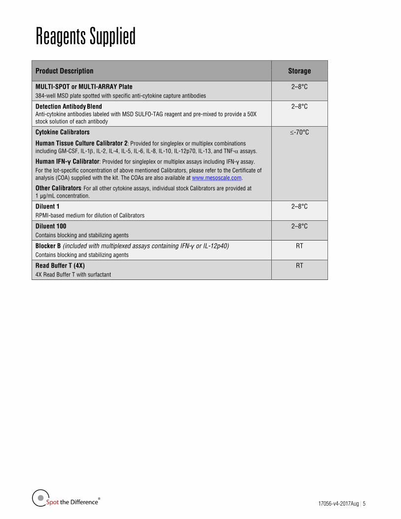
Reagents Supplied Product Description Storage
MULTI-SPOT or MULTI-ARRAY Plate 384-well MSD plate spotted with specific anti-cytokine capture antibodies
2–8°C
Detection Antibody Blend Anti-cytokine antibodies labeled with MSD SULFO-TAG reagent and pre-mixed to provide a 50X stock solution of each antibody
2–8°C
Cytokine Calibrators
Human Tissue Culture Calibrator 2: Provided for singleplex or multiplex combinations including GM-CSF, IL-1β, IL-2, IL-4, IL-5, IL-6, IL-8, IL-10, IL-12p70, IL-13, and TNF-α assays.
Human IFN-γ Calibrator: Provided for singleplex or multiplex assays including IFN-γ assay. For the lot-specific concentration of above mentioned Calibrators, please refer to the Certificate of analysis (COA) supplied with the kit. The COAs are also available at www.mesoscale.com.
Other Calibrators: For all other cytokine assays, individual stock Calibrators are provided at 1 µg/mL concentration.
≤-70°C
Diluent 1 RPMI-based medium for dilution of Calibrators
2–8°C
Diluent 100 Contains blocking and stabilizing agents
2–8°C
Blocker B (included with multiplexed assays containing IFN-γ or IL-12p40) Contains blocking and stabilizing agents
RT
Read Buffer T (4X) 4X Read Buffer T with surfactant
RT

17056-v4-2017Aug | 6
Additional Materials and Equipment Deionized water for diluting concentrated buffers
50-mL tubes for reagent preparation
15-mL tubes for reagent preparation
Microcentrifuge tubes for preparing serial dilutions
Phosphate-buffered saline plus 0.05% Tween-20 for plate washing or MSD Wash Buffer Catalog No. R61AA-1
Appropriate liquid handling equipment for desired throughput, capable of accurately dispensing 10 to 40 μL into a 384-well microtiter plate
Plate washing equipment: automated plate washer or multichannel pipette
Adhesive plate seals
Microtiter plate shaker
Safety Use safe laboratory practices and wear gloves, safety glasses, and lab coats when handling kit components. Handle and dispose of all hazardous samples properly in accordance with local, state, and federal guidelines.
Additional safety information is available in the product Safety Data Sheet (SDS), which can be obtained from MSD Customer Service or at www.mesoscale.com.
Sample Preparation This section provides a general guide for the preparation of various sample types for use in MSD assays. Safe laboratory practices and personal protective equipment such as gloves, lab coat, and safety glasses should be used at all times when handling samples. All samples of a potentially infectious or hazardous origin should be handled in the manner outlined by the Center for Disease Control and the Occupational Safety and Health Administration for blood-borne pathogens and human- and animal-source materials. When analyzing some samples, it may be necessary to dilute the sample by a factor of 2 to 10 to achieve the most accurate quantitation in the MSD Human Cytokine Assays. Please contact MSD Customer Service with any questions.
Tissue Culture Supernatant
Most tissue culture supernatant samples generally do not require any dilution prior to being used in the MSD Human Cytokine Assays. Samples from experimental conditions with extremely high levels of cytokines may require a dilution.

17056-v4-2017Aug | 7
Serum and Plasma
All solid material should be removed by centrifugation. Plasma prepared in heparin tubes commonly displays additional clotting following the thawing of the sample. Remove any additional clotted material by centrifugation. Avoid multiple freeze/thaw cycles for serum and plasma samples. Some analytes in this matrix are extremely sensitive to multiple freeze/thaw cycles and the ability to detect these analytes may decrease following the first round of thawing. Serum and plasma samples may not require any dilution prior to being used in the MSD Human Cytokine Assays.
Sputum
All solid material should be removed by high-speed ultracentrifugation. The clarified sample may be extremely viscous. Accurate pipetting of 10 μL may not be possible due to the sample viscosity. Samples may be diluted as needed.
Bronchoalveolar Lavage (BAL)
All solid material should be removed by centrifugation. Immediately following sample collection, carrier protein (such as 1% BSA in PBS) should be added to the lavage saline to prevent loss of analyte to the labware. The calibration curve should be prepared using the same saline solution (plus carrier protein) used for sample collection.
Urine
Following collection, urine may be stabilized by the addition of a concentrated stabilization buffer which is a Tris or PBS-based buffering solution containing BSA, sodium azide, and protease inhibitors.
Urine Stabilization buffer consists of two components:
1. Base Buffer Solution – It is recommended to make an 11X Base Buffer Solution, consisting of 2.2 M Tris (pH 7.6), 5.5% BSA and 0.11% Sodium azide. Store at 2-8°C.
2. Protease Inhibitor Solution – It is recommended to make an 100X Protease Inhibitor Solution, consisting of 1.0 mg/mL AEBSF, 0.1 mg/mL aprotinin, 0.1 mg/mL pepstatin and 0.1 mg/mL leupeptin in distilled H2O. Store at -20°C. Protease inhibitor solution is available as a component of the MSD Inhibitor Pack (catalog no. R70AA-1).
Prior to use, Complete Urine Stabilization Buffer is made at 10X concentration as follows:
1. Thaw the 100X protease inhibitor solution on ice. Add 1.1 mL of 100X Protease Inhibitor Solution to 10 mL of 11X Base Buffer Solution.
2. Mix. Keep 10X Complete Urine Stabilization Buffer on ice until use.
The 10X Complete Urine Stabilization Buffer should be added to the urine sample at a final concentration of 1X.
At a minimum, it is recommended to add a carrier protein (such as 1% BSA in PBS) to the sample to prevent loss of analyte to the labware and minimize differences in sample pH. Alternatively, samples can be directly frozen without stabilization. Store samples at ≤-70°C. In cases of 24-hour urine collection, refrigerate specimens as they are produced. When making Calibrator dilutions, it is recommended that the diluent be identical to the sample matrix (in terms of pH and buffering conditions), including any diluent used for sample dilution. If necessary, the sample matrix should be immunodepleted of the protein of interest prior to the addition of the Calibrators.
Cerebrospinal Fluid
For calibration curves, it is recommended that the Calibrator diluent used for the curve preparation be identical to the sample matrix, including any diluent used for sample dilution. If necessary, the sample matrix should be immunodepleted of the protein of interest prior to addition of the Calibrators.

17056-v4-2017Aug | 8
Tissue and Tumor Lysates
Lysis buffers should contain low levels of denaturing detergents (<0.1% SDS) and reducing agents (<1mM DTT). As with other sample types, carrier protein (such as 1% BSA) should be present to prevent loss of analyte to the labware.
Reagent Preparation Bring all reagents to room temperature. Thaw the stock calibrator on ice.
Prepare Blocker B Solution
For multiplexed assays containing IFN-γ:
Prepare a 1% (w/v) solution of Blocker B in PBS (20 mL per plate) by adding 200 mg Blocker B to 20 mL PBS.
For multiplexed assays containing IL-12p40:
Prepare a 0.1% (w/v) solution of Blocker B in PBS (20 mL per plate) by adding 20 mg Blocker B to 20 mL PBS.
Prepare Calibrator and Control Solutions
Dilute Calibrators in Diluent 1. The calibration curve preparation instructions listed below will generate a standard curve ranging from 10,000 pg/mL to 2.4 pg/mL. The curve should be adjusted as necessary to provide the proper range for test samples.
Diluent 1 is a standard tissue culture growth medium with 10% serum. If tissue culture samples are in a different medium, use that medium for calibration curve preparation, however please note that if using serum-free medium, the presence of some carrier protein in solution is necessary to prevent loss of analyte to the labware.
Prepare Calibration Curve:
The Human Tissue Culture Calibrator 2 should be used for singleplex and multiplex assays that include the following assays: GM-CSF, IL-1β, IL-2, IL-4, IL-5, IL-6, IL-8, IL-10, IL-12p70, IL-13, TNF-α.
The Human IFN-γ Calibrator should be used for singleplex and multiplex assays that include IFN-γ.
For the above mentioned calibrators please refer to the COA supplied with each kit for the measured concentration values and top of the curve concentrations. Alternatively, continue to use the nominal top of curve concentration value of 10,000 pg/mL in the protocol and data analysis for convenience (see Note on page 10).
For the remaining cytokine assays, individual stock Calibrators are provided at 1 μg/mL concentration, and 10,000 pg/mL should be used as the nominal top of curve concentration value.

17056-v4-2017Aug | 9
The following protocol should be used to prepare the calibration curve:
CAL-1: Add 10 μL of each calibrator supplied to Diluent 1 such that the final volume is 1,000 μL. Use this high Calibrator (nominal concentration: 10,000 pg/mL) to prepare the standard curve following a 1:4 dilution series (as shown below).
CAL-2: Add 50 μL of 10,000 pg/mL combined high Calibrator to 150 μL of Diluent 1.
CAL-3: Add 50 μL of 2,500 pg/mL Calibrator to 150 μL of Diluent 1.
CAL-4: Add 50 μL of 625 pg/mL Calibrator to 150 μL of Diluent 1.
CAL-5: Add 50 μL of 156 pg/mL Calibrator to 150 μL of Diluent 1.
CAL-6: Add 50 μL of 39 pg/mL Calibrator to 150 μL of Diluent 1.
CAL-7: Add 50 μL of 9.8 pg/mL Calibrator to 150 μL of Diluent 1.
CAL-8: 150 μL of Diluent 1 (zero Calibrator blank)
Alternatively, Calibrators can be prepared in the sample matrix or diluent of choice to verify acceptable performance in these matrices. In general, the presence of some protein in the sample matrix is helpful for preventing loss of analyte by adsorption onto the sides of tubes, pipette tips, and other surfaces.
Figure 2. Calibration curve preparation from 1 μg/mL stock solution.
17056-v4-2017Aug | 10
Note:
We anticipate that most customers will continue to use the nominal (10,000 pg/mL) values in their protocol and data analysis of GM-CSF, IFN-γ, IL-1β, IL-2, IL-4, IL-5, IL-6, IL-8, IL-10, IL-12p70, IL-13, and TNF-α assays. Many applications do not require absolute quantitation of analytes relative to a fixed standard, and the nominal values allow convenient use and consistent data analysis. Dose response studies, IC-50 measurements, and experiments where the relative abundance of the analyte is sufficient are examples of situations where using nominal values for the Calibrators is consistent with good practice.
Applications that require accurate, absolute quantitation of analytes relative to a fixed standard will benefit from using the measured value for the Calibrators. There will be no change in the protocol for preparation of Calibrator solutions, but users will need to enter the measured value of the Calibrators into their data analysis template. In MSD’s DISCOVERY WORKBENCH® software, users will simply enter the concentration for the Calibrators at the top of the curve in the plate layout. Each lot of kits will have specific measured values.
Prepare Detection Antibody Solution
Note: Detection antibody solutions should be kept in the dark as some antibodies may be light sensitive.
MSD provides detection antibody as a 50X stock solution (some vials may be labeled as 50 μg/mL). The working detection antibody solution is 1X (or 1.0 μg/mL).
For one plate, combine:
160 μL of 50X Detection Antibody Blend
7.84 mL of Diluent 100
Prepare Wash Buffer
MSD provides Wash Buffer as a 20X stock solution. The working solution is 1X. PBS + 0.05% Tween-20 can be used instead.
For one plate, combine:
30 mL of MSD Wash Buffer (20X)
570 mL of deionized water
Prepare Read Buffer T
MSD provides Read Buffer T as a 4X stock solution. The working solution is 2X.
For one plate, combine:
8.5 mL of Read Buffer T (4X)
8.5 mL of deionized water
Diluted read buffer can be prepared in advance and stored at room temperature in a tightly sealed container for up to one month.
Prepare MSD Plate
MSD plates are pre-coated with capture antibodies (Figure 1) and exposed to a proprietary stabilizing treatment to ensure the integrity and stability of the immobilized antibodies. Plates can be used as delivered; no additional preparation (e.g., pre-wetting) is required. The plate has not been pre-blocked, and a discrete blocking step is not generally required for MSD cytokine assays. For some cytokines, however, a blocking step may improve the assay sensitivity.

17056-v4-2017Aug | 11
Assay Protocol Note: For MULTI-SPOT Assays containing IFN-γ, a blocking step is required prior to beginning the assay to achieve the best performance. Dispense 20 μL of the 1% (w/v) Blocker B Solution into each well of the MSD plate. Seal the plate with an adhesive plate seal and incubate with vigorous shaking for 1 hour at room temperature. Wash the plate three times with at least 90 μL/well of 1X MSD Wash Buffer or PBS + 0.05% Tween-20 and proceed with the assay protocol at step 1.
For MULTI-SPOT Assays containing IL-12p40, a blocking step is required prior to beginning the assay to achieve the best performance. Dispense 20 μL of the 0.1% (w/v) Blocker B Solution into each well of the MSD plate. Seal the plate with an adhesive plate seal and incubate with vigorous shaking for 1 hour at room temperature. Wash the plate three times with at least 90 μL/well of 1X MSD Wash Buffer or PBS + 0.05% Tween-20 and proceed with the assay protocol at step 1.
Figure 3. Suggested plate setup for calibrating MSD Cytokine Assay Kits. Columns 1-2 contain a standard curve in duplicate. The remaining wells are available for samples. Some assays may require lower limits of detection, and in these cases, the 625 pg/mL point can be removed and a 0.6 pg/mL point added between 2.4 pg/mL and 0 pg/mL to provide an additional lower calibration point. An alternative setup that contains a larger dynamic range for the standard curve can be prepared by running Calibrators in a 12-point titration in duplicate across the top or bottom of the plate. The concentrations of Calibrators run may be adjusted depending on the desired dynamic range for the experiment. The data from this calibration curve can be analyzed using any standard data analysis package.
STEP 1: Add Sample or Calibrator Standard Dispense 10 μL of each Calibrator or Sample Solution into a separate well of the MSD plate. Figure 3 illustrates one plate
arrangement of Calibrator solutions that can be used to evaluate the performance of the assay. Seal the plate with an adhesive plate seal and incubate for 1 to 2 hours with vigorous shaking (500–1,000 rpm) at room
temperature.
Note: In general, shaking the plate results in better mixing and more rapid binding of the sample to the capture antibodies. If a protocol without shaking is preferred, a longer incubation time (4 hours or longer) may be required to achieve the same sensitivity.
STEP 2: Add Detection Antibody Solution Dispense 10 μL of the 1X detection antibody solution into each well of the MSD plate. Seal the plate and incubate for 1-2 hours with vigorous shaking at room temperature. If a protocol without shaking is
preferred, a longer incubation time may be required to achieve the same sensitivity.
17056-v4-2017Aug | 12
STEP 3: Wash and Read
Wash the plate three times with at least 90 μL/well of 1X MSD Wash Buffer or PBS + 0.05% Tween-20.
Add 35 μL of 2X Read Buffer T to each well of the MSD plate. Analyze the plate on the MSD instrument. Incubation in Read Buffer T is not required before reading the plate.
Note: Bubbles in the fluid will interfere with reliable reading of the MULTI-SPOT plate. Use reverse pipetting techniques to insure bubbles are not created when dispensing the Read Buffer T.
Assay Notes 1. Unwashed Assay: The protocol can be converted to an unwashed assay by eliminating the wash step following the detection
antibody incubation, adding 15 μL 2X Read Buffer T, and analyzing the plate on the MSD instrument. The use of this protocol may result in a loss of sensitivity, the effect of which will vary for each cytokine.
2. Sample Matrices: In general, these plates have been found to work well to measure cytokine levels in a wide range of samples, including cell lysates, serum and plasma samples, cell supernatant, and simple buffers. Sample dilution and/or spike recovery studies in the sample matrix of interest should be carried out to verify acceptable performance in the matrix.
3. Combining the Sample and Detection Antibody Addition Steps: The protocols described above call for incubating the sample in the wells prior to the addition of the detection antibody (sequential incubations). This procedure is used because in some selected assays using a polyclonal detection, the detection antibody itself may include antibodies that compete with the immobilized capture antibody. In some assays, however, MSD has found that the sample and detection antibodies may be added concurrently (simultaneous incubation) with little or no loss in performance.
4. Stability of Assay in Read Buffer T: The plates do not need to be read immediately after addition of Read Buffer T. In the washed assay, the loss of signal is typically less than 20% over a 1 hour incubation in Read Buffer T, and is usually stabilized by 2 hours. The observed loss is due to a re-establishment of equilibrium in the well.
5. Optimizing Assay Sensitivity: The washed protocols described above were developed to provide excellent assay performance with a minimum number of steps, minimal sample volume requirement, and a rapid assay completion time. One or more of the following assay modifications may be used to further improve the assay sensitivity:
a. Increasing incubation times - The incubation time with detection antibody can be increased to 4 hours with shaking (or at least 12 hours without shaking).
b. Increasing sample volumes up to 20 μL (Note: Efficient shaking and/or longer incubation times become more important at higher volumes) - if a larger sample volume is used, the concentration of detection antibody must be adjusted in the detection antibody solution to compensate for a change in volume.
c. Washing sample from the well prior to the addition of the detection antibody mix (especially useful for large sample volumes to avoid dilution of the detection antibodies) - not all cytokines will see an improvement by addition of this step, and some cytokines may see decreased sensitivity.

17056-v4-2017Aug | 13
Specificity The capture and detection antibody pairs used in MSD cytokine assays have been selected by an optimization process that is designed to minimize cross-reactivity with other cytokine assays. In addition, the chosen antibodies are specific for the particular human cytokine of interest and show minimal cross-reactivity with other species.

17056-v4-2017Aug | 14
Appendix Background signal and negative signals
The output signal produced by electrochemiluminescence assays is in units of counts of light measured by a charge-coupled device (CCD) camera or photodiode. As with any measurement technique, there is a certain amount of normal variation in this signal (instrument noise) which sets the threshold for the lowest levels of signal that can be measured (noise floor). This variation is different depending upon the size of the working electrode with typical values of about 10 counts for 384-well small spot and 50 counts for 384-well 4-spot plates. When the background signal of an assay approaches the noise floor (i.e., the mean signal of negative controls or sample blanks is close to zero), it is possible to observe negative counts for some wells. Signal Levels
The camera system is linear over nearly a 6-log dynamic range. The highest achievable signals on the MSD instruments are between 1.0 and 2.0 million counts. If the signals from the highest point on the calibration curve are not approaching 1.0 million counts, the high end of the calibration curve may be extended. The lowest observed signals using Read Buffer T (2X) are between 10 and 50. Negative signal values may occur due to instrument noise, omission or usage of the incorrect read buffer, or incorrect amount of detection antibody. Curve Fitting
MSD DISCOVERY WORKBENCH software uses least-squares fitting algorithms to generate the standard curve that will be used to calculate the concentration of analyte in the samples. The assays have a wide dynamic range (3–4 logs) that allows accurate quantification without the need for dilution in many cases. By default, the software uses a 4-parameter logistic model (or sigmoidal dose-response) and includes a 1/Y2 weighting function. The weighting function is important because it provides a better fit of data over a wide dynamic range, particularly at the low end of the standard curve.
Reverse Pipetting
Most manual hand pipets have two plunger positions for pipetting liquids. The first position is calibrated to allow aspiration and dispensing of user-specified amounts of liquid and the second (blow-out) position enables the user to expel any residual liquid after the pipet has been pushed to the first position. When a pipet is used to dispense liquid by moving the plunger to the first position followed by the second (blow-out) position, bubbles may be created in the dispensed liquid. The reverse pipetting technique is designed to allow precise pipetting while avoiding the creation of bubbles. The technique is to push the pipet plunger past the first position to the second position prior to aspirating liquid into the tip, thereby aspirating slightly more liquid than the desired volume (overdraw). In order to dispense the liquid from the tip, the pipet plunger is pushed to the first position only. This allows precise dispensing without the introduction of bubbles. When using the reverse pipetting technique, it is important not to overdraw excess liquid into the pipet mechanism.
17056-v4-2017Aug | 15
Summary Protocol MSD 384-Well MULTI-ARRAY and MULTI-SPOT Human Cytokine Assays: Tissue Culture Kit
MSD provides this summary protocol for convenience. Please read the entire detailed protocol prior to performing the MSD Human Cytokine Assay.
Sample and Reagent Preparation
Prepare Blocker B Solution. o For MULTI-SPOT Assays with IFN-γ, prepare a solution of 1% Blocker B in PBS. o For MULTI-SPOT Assays with IL-12p40, prepare a solution of 0.1% Blocker B in PBS.
Prepare Calibrator solutions and calibration curve. o Use the 1 μg/mL Calibrator stock to prepare an 8-point calibration curve of 10,000, 2,500, 625, 156, 39, 9.8,
2.4, and 0 pg/mL.
o The calibration curve can be modified as necessary to meet specific assay requirements.
Prepare detection antibody solution by diluting stock detection antibody to 1X (or 1 μg/mL) in 8.0 mL (per plate) of Diluent 100.
Prepare 17 mL of 2X Read Buffer T by diluting 4X Read Buffer T 2-fold with deionized water.
Prepare 1X MSD Wash Buffer by diluting 20X stock solution to 1X with deionized water. Use at least 90 μL/well for plate washing.
STEP 1: For MULTI-SPOT Assays with IFN-γ or IL-12p40 only:
Dispense 20 μL/well 1% Blocker B Solution for IFN-γ or 0.1% Blocker B Solution for IL-12p40.
Incubate at room temperature with vigorous shaking (500–1,000 rpm) for 1 hour.
Wash the plate three times with at least 90 μL/well of 1X MSD Wash Buffer or PBS+ 0.05% Tween-20.
Proceed with sample/Calibrator addition.
STEP 2: Add Sample or Calibrator
Dispense 10 μL/well Calibrator or sample.
Incubate at room temperature with vigorous shaking for 1-2 hours.
STEP 3: Add Detection Antibody Solution
Add 10 μL/well of 1X detection antibody solution.
Incubate at room temperature with vigorous shaking for 1-2 hours.
STEP 4: Wash and Read Plate
Wash the plate three times with at least 90 μL/well of 1X MSD Wash Buffer or PBS + 0.05% Tween-20.
Add 35 μL/well of 2X Read Buffer T.
Analyze plate on the MSD instrument.