human cytomegalovirus-encoded interleukin-10 …jvi.asm.org/content/78/16/8720.full.pdfrna and...
TRANSCRIPT

JOURNAL OF VIROLOGY, Aug. 2004, p. 8720–8731 Vol. 78, No. 160022-538X/04/$08.00�0 DOI: 10.1128/JVI.78.16.8720–8731.2004Copyright © 2004, American Society for Microbiology. All Rights Reserved.
Human Cytomegalovirus-Encoded Interleukin-10 Homolog InhibitsMaturation of Dendritic Cells and Alters Their Functionality
W. L. William Chang,1* Nicole Baumgarth,1 Dong Yu,2 and Peter A. Barry1
Center for Comparative Medicine, University of California, Davis, California 95616,1 and Departmentof Molecular Biology, Princeton University, Princeton, New Jersey 085442
Received 27 August 2003/Accepted 5 April 2004
Interleukin-10 (IL-10) suppresses the maturation and cytokine production of dendritic cells (DCs), keyregulators of adaptive immunity, and prevents the activation and polarization of naïve T cells towardsprotective gamma interferon-producing effectors. We hypothesized that human cytomegalovirus (HCMV)utilizes its viral IL-10 homolog (cmvIL-10) to attenuate DC functionality, thereby subverting the efficientinduction of antiviral immune responses. RNA and protein analyses demonstrated that the cmvIL-10 gene wasexpressed with late gene kinetics. Treatment of immature DCs (iDCs) with supernatant from HCMV-infectedcultures inhibited both the lipopolysaccharide-induced DC maturation and proinflammatory cytokine produc-tion. These inhibitory effects were specifically mediated through the IL-10 receptor and were not observed whenDCs were treated with supernatant of cells infected with a cmvIL-10-knockout mutant. Incubation of iDCs withrecombinant cmvIL-10 recapitulated the inhibition of maturation. Furthermore, cmvIL-10 had pronouncedlong-term effects on those DCs that could overcome this inhibition of maturation. It enhanced the migrationof mature DCs (mDCs) towards the lymph node homing chemokine but greatly reduced their cytokineproduction. The inability of mDCs to secrete IL-12 was maintained, even when they were restimulated by theactivated T-cell signal CD40 ligand in the absence of cmvIL-10. Importantly, cmvIL-10 potentiates theseanti-inflammatory effects, at least partially, by inducing endogenous cellular IL-10 expression in DCs. Collec-tively, we show that cmvIL-10 causes long-term functional alterations at all stages of DC activation.
Human cytomegalovirus (HCMV) can establish and main-tain a persistent subclinical infection in fully immunocompe-tent individuals. Efficient induction of immune responses toHCMV appears to be important in preventing overwhelmingvirus infection since serious complications ensue in hosts withan immature (fetuses and newborns) or a compromised (e.g.,transplant recipients and AIDS patients) immune system (6).CMV-specific cytotoxic T lymphocytes (CTLs) potently controlviral replication, but they cannot completely eliminate infectedcell reservoirs (57, 72). This can be explained, in part, by thefact that HCMV has developed several ingenious pathways todisrupt potential interactions between infected cells, on theone hand, and effector T cells and natural killer (NK) cells onthe other. For example, the open reading frame (ORF) UL18encodes a major histocompatibility complex (MHC) class Ihomolog. Another HCMV gene product, gpUL40, induces sur-face expression of HLA-E. Both can prevent NK cell recogni-tion and lysis (13, 15, 67, 71). Moreover, HCMV evades CTLdetection by interfering with the processing and presentationof viral antigens by MHC class I molecules through a group ofglycoproteins: gpUS2, gpUS3, gpUS6, and gpUS11 (54, 69).
Dendritic cells (DCs) exhibit great functional plasticity andcan discriminate between different classes of microorganisms(30, 56). DCs are the only antigen-presenting cells thought tostimulate primary immune responses and are the key cells thatregulate the magnitude and quality of the ensuing immuneresponses (2). Therefore, inactivation or attenuation of DCs
through different strategies would likely compromise virus-specific immune responses. Multiple viruses, including measlesvirus, vaccinia virus, and murine cytomegalovirus (MCMV),directly infect DCs and disrupt DC functionality (1, 22, 24).Recent studies have provided in vitro evidence that high titersof endothelial cell-adapted HCMV strains can infect DCs,thereby paralyzing DC-mediated stimulation of adaptive im-mune responses (46, 53). However, the primary targets ofHCMV in vivo are likely cell types other than DCs (48), raisingthe question whether HCMV could utilize mechanisms otherthan direct infection to delay and/or skew DC-mediated im-mune responses.
Recently, a viral interleukin-10 (vIL-10) homolog, ORFUL111A, was identified in the genomes of HCMV and otherprimate CMVs (36, 37). UL111A is not essential for HCMVreplication in vitro (73) but may be crucial for viral immuneevasion in vivo. HCMV-encoded IL-10 (cmvIL-10) forms ho-modimers that bind to the ligand-binding subunit of the IL-10receptor (IL-10R1) with essentially the same affinity as that ofcellular IL-10 (cIL-10) (33) and transduce through the signal-ing subunit IL-10R2 (36). The function(s) of this viral gene hasnot been fully characterized (65). However, it appears criticalfor the life cycle of HCMV in vivo since its sequence is highlyconserved in multiple tissue culture-adapted strains and clini-cal isolates (R. Hector and A. Davison, Abstr. 9th Int. Cyto-megalovirus Workshop, abstr. C.02, 2003). Although a numberof viruses, including Epstein-Barr virus and Orf poxvirus, en-code vIL-10 homologs, cmvIL-10 is the most divergent vIL-10discovered thus far (23, 29, 36, 37). Its protein sequence sharesonly 25 to 27% identity with its cIL-10 counterpart, suggestingthat it may not merely copy the cellular function but have
* Corresponding author. Mailing address: Center for ComparativeMedicine, University of California, Davis, County Road 98 andHutchison Drive, Davis, CA 95616. Phone: (530) 752-6248. Fax: (530)752-7914. E-mail: [email protected].
8720
on June 21, 2018 by guesthttp://jvi.asm
.org/D
ownloaded from

different or additional properties. It is, thus, important to di-rectly compare the cIL-10 and cmvIL-10 functions.
IL-10 strongly inhibits cell-mediated immunity by regulatingDC functions (45). DCs coordinate immune responses throughtheir ability to sense and respond to changes in the microen-vironment (52). Upon DC activation, immature DCs (iDCs)differentiate and migrate to lymphoid tissues for priming naïveT cells (19). IL-10 inhibits DC maturation, a prerequisite forT-cell priming (39, 43), thus accounting for the inhibitory ef-fects of IL-10 on DC-induced T-cell alloreactivity (10, 47).Furthermore, IL-10 blocks the ability of lipopolysaccharide(LPS)-stimulated DCs to migrate (16). Since migration of ma-ture DCs (mDCs) to secondary lymphoid tissues is essential forthe subsequent activation of naïve T cells, these data indicatethat cIL-10 suppresses the induction of strong T-cell responses.This study was undertaken to elucidate the functions of cmvIL-10, in comparison with those of cIL-10. In particular, we fo-cused on determining the effects of cmvIL-10 on multiplestages of DC activation. The results of our study support thehypothesis that secretion of cmvIL-10 by virus-infected cells isan indirect immune evasion mechanism that causes long-terminhibition of DC function.
MATERIALS AND METHODS
Virus and cells. HCMV strains AD169 (kindly provided by W. J. Britt, Uni-versity of Alabama, Birmingham) and Toledo (kindly provided by J. A. Wiede-man, University of California, Davis) and an AD169-derived mutant virus, TS359(73), were used in this study. MRC-5 cells (human embryonic lung fibroblasts;ATCC CCL-171) were cultured in Dulbecco’s modified Eagle’s medium (Invitro-gen, Carlsbad, Calif.) supplemented with 10% fetal calf serum and 2 mM L-glutamine–100 U of penicillin/ml–100 �g of streptomycin/ml. Virus stocks usedfor fibroblast infection were prepared from 0.45-�m-pore-size-filtered superna-tant in a 1:1 mixture with autoclaved 9% nonfat milk and stored at �70°C.Purified HCMV virions used for DC stimulation were prepared according toprocedures described previously (12). Virus titers were determined by standardplaque assay, as described previously (11). Replication kinetics of HCMV weredetermined by single-step or multiple-step growth curve analyses. Briefly,MRC-5 cells at 3 � 105 cells/well of six-well plates were infected at an indicatedmultiplicity of infection (MOI). Supernatants from triplicate infected cultureswere harvested daily, centrifuged at 600 � g for 5 min to remove cell debris, andstored at �70°C.
RNA extraction and cDNA synthesis. Total RNA from HCMV-infected cellswas purified with the RNeasy minikit (Qiagen, Valencia, Calif.) and treated withDNase (TURBO DNA-free kit; Ambion Woodward, Austin, Tex.) according tothe manufacturer’s instructions. For first-strand cDNA synthesis, an annealingreaction was first performed with 5 �g of RNA and 0.5 �g of oligo(dT)12-18
primer (Invitrogen) at 70°C for 5 min and then 4°C for 3 min. Reverse transcrip-tion was carried out in a 40-�l reaction mixture with Superscript II reversetranscriptase (200 U; Invitrogen) at 42°C for 90 min in the presence ofRNaseOUT RNase inhibitor (60 U; Invitrogen), followed by inactivation ofreverse transcriptase at 70°C for 15 min. cDNA samples were stored at �20°Cand later used as templates for quantitative real-time PCR analyses.
Quantitative PCR analysis. Real-time PCR was performed from reverse-transcribed cDNA samples for relative quantification of HCMV UL111A tran-script copy numbers. Primer and probe sequences were designed specific to thesecond exon of the UL111A ORF with Primer Express software (Applied Bio-systems, Foster City, Calif.). The sequences of the forward and reverse primerswere 5�-TGT TGA GGC GGT ATC TGG AGA-3� and 5�-CCG TCT TGA GTCCGG GAT AG-3�, respectively. Probe sequence CGT GTT TCC CGC AGGCGA CC contained 5�-tetrachloro-6-carboxyfluorescein (TET) as the reporterdye and 3�-6-carboxytetramethylrhodamine (TAMRA) as the quencher dye (Ap-plied Biosystems). Standard curves were generated by using 10-fold serial dilu-tions of a plasmid (from 107 copies to 1 copy/�l) containing the genomic se-quence of UL111A ORF (pWC154). Quantitative real-time PCR for UL111Awas performed with 1� TaqMan universal PCR master mixture (Applied Bio-systems), 200 nM (each) primer, 100 nM probe, and template cDNA (2 �l ofeach synthesized cDNA sample or diluted plasmid) in a 20-�l reaction volume.
Ready-for-use glyceraldehyde-3-phosphate dehydrogenase (GAPDH) probe andprimers were obtained from Applied Biosystems (TaqMan GAPDH controlreagents). PCR was performed with the ABI/Prism 7900HT sequence detectionsystem (Applied Biosystems) (1 cycle of 50°C for 2 min and 95°C for 10 min andthen 40 cycles of 95°C for 15 s and 60°C for 1 min). Quantification of the PCRsignals was performed by comparing the cycle threshold value of each sample intriplicate with the cycle threshold values of the GAPDH reference gene. Thedetection limit of reverse-transcribed UL111A transcripts was 10 copies/�l.
Immunoblotting. HCMV-infected cells were collected, washed with cold phos-phate-buffered saline (PBS), and then lysed with Novex Tris-glycine-sodiumdodecyl sulfate sample buffer (Invitrogen). The protein extracts were size-sepa-rated by sodium dodecyl sulfate–12% polyacrylamide gel electrophoresis, trans-ferred to a nitrocellulose membrane, and then probed with the following anti-bodies: anti-HCMV glycoprotein B (gB) (monoclonal antibody [MAb] clone27-180; kindly provided by W. J. Britt), anti-�-actin (MAb clone AC-74; Sigma,St. Louis, Mo.), and biotinylated anti-cmvIL-10 polyclonal immunoglobulin G(IgG; R&D Systems, Minneapolis, Minn.). After removal of the primary anti-body and multiple washes with PBS–0.5% Tween 20, the membranes were thenincubated with peroxidase-conjugated anti-mouse IgG (Sigma) or streptavidin(Vector Laboratories, Burlingame, Calif.). Immunoreactive proteins were de-tected with the ECL Plus Western blotting detection reagents and scanning witha Typhoon 9410 variable mode imager (Amersham Biosciences, Piscataway, N.J.).
Generation of monocyte-derived DCs. Human peripheral blood mononuclearcells were isolated from buffy coats of healthy individuals (obtained from theSacramento Blood Center) by Ficoll-Paque gradient centrifugation (AmershamBiosciences). CD14� monocytes from human peripheral blood mononuclearcells were then positively selected using anti-CD14 MAb-conjugated magneticmicrobeads (Miltenyi Biotec, Auburn, Calif.). Purified monocytes (�95% purity,as determined by flow cytometry with staining for CD3, CD4, CD8, and CD20)(data not shown) were cultured at a density of 1.5 � 106 to 2 � 106 cells/ml in12-well plates (1 ml per well) or 24-well plates (0.5 ml per well). DC mediumconsisted of RPMI 1640 (Invitrogen) containing 2 mM L-glutamine–100 U ofpenicillin/ml–100 �g of streptomycin/ml, 50 �M 2-mercaptoethanol (Sigma), 10mM HEPES (Invitrogen), 10% endotoxin-free fetal calf serum (Invitrogen), andrecombinant human granulocyte-macrophage colony-stimulating factor and IL-4(1,000 U/ml each; R&D Systems). Cytokines were replenished every other day byadding 20% fresh DC medium to each well.
DC stimulation and treatment. For DC activation, LPS from Escherichia coliO127:B8 (Sigma) or purified recombinant human soluble CD40 ligand (sCD40L)(Research Diagnostics, Flanders, N.J.) was added 6 days after culture onset. ForIL-10 treatment, recombinant human IL-10 (hIL-10) (Sf21 cell-expressed) orcmvIL-10 (ORF UL111A of HCMV Towne strain expressed in E. coli) (bothfrom R&D Systems) was added to the cultures concomitantly with the appro-priate activation signals. At various times after onset, DCs were collected bygentle resuspension. The numbers of viable DCs were determined by trypan blueexclusion. Binding of cmvIL-10 to its cognate receptor on DCs was inhibited withanti-IL-10R1 MAb (5 �g/ml; R&D Systems). Purified mouse IgG1 (R&D Sys-tems) was used as an isotype control antibody for the assay. MAbs were addedto the DC cultures 30 min before the addition of recombinant IL-10 proteins andLPS. Endotoxin levels of all reagents (except LPS) were tested by Limulusamebocyte lysate assays (�1 EU/�g of cytokine-sCD40L; �0.1 EU/�g of anti-body).
ELISA. Levels of cmvIL-10 in culture supernatants were measured by anantigen-capture enzyme-linked immunosorbent assay (ELISA). cmvIL-10-spe-cific IgG (100 ng/well; affinity purified, polyclonal; R&D Systems) and biotinyl-ated cmvIL-10-specific IgG (10 ng/well) were used for coating and detection,respectively. The relative units of cmvIL-10 within the supernatant from HCMV-infected cultures were calculated from standard curves obtained using recombi-nant cmvIL-10 (1 U 1 pg/ml). The levels of tumor necrosis factor- (TNF-),IL-6, IL-12 (p40 plus p70), and endogenous cIL-10 secreted by DCs were quan-tified using ELISA kits purchased from U-CyTech (Utrecht, The Netherlands).Protein concentrations were calculated from a standard curve obtained withrecombinant protein standards. The limits of detection for the ELISA are asindicated: cmvIL-10, 10 pg/ml or 10 U; cIL-10, 5 pg/ml; IL-6, 5 pg/ml; IL-12 (p40plus p70), 5 pg/ml; and TNF-, 5 pg/ml.
Flow cytometry. Four-color flow cytometry was performed using a FACSCali-bur (BD Biosciences, San Jose, Calif.) cell sorter. Directly conjugated antibodieswere purchased from BD Biosciences, Beckman Coulter (Fullerton, Calif.), andR&D Systems: anti-HLA-ABC-fluorescein isothiocyanate (FITC)–allophyco-cyanin (APC) (G46-2.6), anti-HLA-DR-peridinin chlorophyll protein (PerCP)(L243), anti-CD1a-APC (HI149), anti-CD3-FITC (SP34), anti-CD4-phycoeryth-rin (PE) (M-T477), anti-CD8-PerCP (SK1), anti-CD20-APC (L27), anti-CD40-PE (5C3), anti-CD80-FITC (MAb 104), anti-CD83-FITC (HB15e), anti-
VOL. 78, 2004 HCMV-ENCODED IL-10 HOMOLOG ALTERS DC FUNCTIONALITY 8721
on June 21, 2018 by guesthttp://jvi.asm
.org/D
ownloaded from

CD83-PE (HB15a), anti-CD86-APC (2331), anti-CCR1-PE (53504.111), anti-CCR2-APC (48607), anti-CCR5-APC (3A9), and anti-CCR7-PE (3D12). Ap-propriate isotype-matched antibodies were used as controls. Data were analyzedand illustrated using FlowJo software (Tree Star Inc., San Carlos, Calif.).
Endocytosis and phagocytosis assays. To assess the endocytic capacity of DCs,untreated DCs or DCs exposed to hIL-10 or cmvIL-10 (50 ng/ml) for 30 min wereincubated with FITC-conjugated dextran (molecular weight, 70,000; Molecu-lar Probes, Eugene, Oreg.) for 2 h at 37°C at a final concentration of 10 �g/ml.To study the effects of IL-10 exposure on the phagocytic capacity of DCs, cellswere incubated with FITC-conjugated carboxylate-modified polystyrene beads(0.5-�m diameter; Polysciences, Warrington, Pa.) at 500 beads per cell in thepresence or absence of hIL-10 or cmvIL-10 (50 ng/ml) for 24 h. After incubation,cells were washed three times with ice-cold PBS, immunostained, and analyzedby fluorescence-activated cell sorting (FACS).
Chemotaxis assay. In vitro transwell migration assays were performed byadding 5 � 105 DCs in 100 �l of medium to the upper chambers (5-�m pore size)of a 6.5-mm-diameter transwell plate (Costar, Corning, N.Y.). Recombinantmacrophage inflammatory protein 1 (MIP-1) and MIP-3� (R&D Systems)were diluted in assay medium (plain RPMI plus 0.5% bovine serum albumin) at100 ng/ml, and 600-�l aliquots were placed in the lower wells. DC migration wasevaluated by FACS analysis after incubation at 37°C for 2 h. Absolute numbersof mDCs that had migrated were determined using TruCOUNT tubes (BDBiosciences) and assessed on a FACSCalibur cell sorter.
Statistical analysis. Student’s t tests (paired, one-tailed) were performed toevaluate the differences among experimental groups.
RESULTS
UL111A exhibits late expression kinetics during viral rep-lication. HCMV gene expression is regulated by events at thetranscriptional, posttranscriptional, translational, and post-translational levels (44). Although cmvIL-10 transcripts havepreviously been identified in HCMV-infected cells (36),cmvIL-10 expression kinetics have not been evaluated at eitherthe RNA or the protein level. To define the temporal expres-sion of cmvIL-10 in infected fibroblasts, a single-step growthcurve analysis was performed in the presence and absence of acombination of two replication inhibitors, foscarnet and gan-ciclovir (Fig. 1A, shown as lines). To measure the levels ofcmvIL-10 mRNA, a real-time PCR assay was developed toquantify UL111A transcripts from reverse-transcribed cDNAsamples. The relative copy number of UL111A cDNA wasdetermined by normalizing the cycle threshold values to thoseof GAPDH. Transcription of UL111A exhibited late gene ki-netics. Both UL111A transcript copy number and the produc-tion of infectious virions were profoundly reduced in the pres-ence of the two replication inhibitors (Fig. 1A, gray and openbars, respectively).
The expression kinetics of ORF UL111A were also evalu-ated at the protein level. MRC-5 cultures were infected withtwo HCMV strains (AD169 and Toledo), and the supernatantswere collected daily to determine virus titers and cmvIL-10levels. Although the mRNA level of UL111A increased priorto the first burst of progeny virus (Fig. 1A), cmvIL-10 proteinbecame detectable in the supernatants coincidently with thepresence of extracellular virus (Fig. 1B and 2B). Accumulationof virally encoded cmvIL-10 protein in the supernatants ofinfected cultures continued even though the production ofprogeny virus peaked around day 5 postinoculation (Fig. 1B).Whether cmvIL-10 is actively secreted by infected cells orreleased from dead cells could not be distinguished in thisanalysis. Previous studies have demonstrated that transfectionof cells with a full-length clone of UL111A results in the steadyaccumulation of extracellular cmvIL-10 (65), consistent with
the presence of a predicted leader peptide (36, 37). Since mostof the HCMV structural proteins are encoded at the late stageof the viral replication cycle (44), our data indicate that cmvIL-10 accumulates with other abundantly expressed viral antigensin the microenvironment of the infected tissues.
HCMV-encoded IL-10 possesses a biological function. Toevaluate the biological activity of cmvIL-10 expressed byHCMV-infected cells, DC functions were analyzed in the pres-ence and absence of supernatant derived from cells infectedwith HCMV. Supernatant from cells transfected with aUL111A-deletion variant of HCMV, TS359 (73), was used inparallel to determine the relative contribution of cmvIL-10 toalterations of DC functions. TS359 is derived from the lab-adapted AD169 strain of HCMV and contains a disruption ofthe first exon of UL111A due to the insertion of a 3.66-kb
FIG. 1. Expression kinetics of HCMV ORF UL111A during viralreplication. (A) UL111A is transcribed with late gene kinetics. MRC-5cells were infected in triplicate with HCMV AD169 at an MOI of 1 andcultured in the presence (open symbols) or absence (filled symbols) offoscarnet (PFA; 167 �M) and ganciclovir (GCV; 4 �M). Supernatantswere collected daily to quantify progeny virus titers by standard plaqueassays (shown as lines), and total RNA was extracted for cDNA syn-thesis and quantitative real-time PCR analyses. The relative copy num-bers of UL111A cDNA after normalization with GAPDH copy num-bers are shown as bars. (B) Accumulation of cmvIL-10 protein in thesupernatants of infected cultures. Cells were infected with HCMVstrain AD169 (filled symbols) or Toledo (open symbols) at an MOI of0.1. Supernatants were collected at indicated time points to measurevirus titers (shown as lines) and cmvIL-10 protein concentrations(shown as bars). Shown are mean values � standard deviations fortriplicate cultures.
8722 CHANG ET AL. J. VIROL.
on June 21, 2018 by guesthttp://jvi.asm
.org/D
ownloaded from

transposon (Fig. 2A). This variant virus exhibited normal rep-lication kinetics in fibroblasts (Fig. 2B) in the absence ofcmvIL-10 protein expression. cmvIL10 was never detected byELISA or immunoblotting in cells infected with TS359, asopposed to the robust expression observed in cells infectedwith AD169 (Fig. 2B and C). Two sizes of cmvIL-10 wereobserved in the immunoblots of extracts from cells infectedwith AD169 (Fig. 2C, asterisks). The larger band was slightlygreater than the predicted size of full-length cmvIL-10 protein(175 amino acids) and may be the result of posttranslationalglycosylation. Recently, a novel UL111A transcript expressedduring latent infection but not during productive infectionwithin permissive cells has been identified (32). This processedtranscript consists of a single splicing event between the firstand second exons of UL111A and is predicted to encode a139-amino-acid protein. The size of the smaller protein de-tected by immunoblotting (Fig. 2C) is consistent with this formof cmvIL-10. However, Kotenko et al. have previously demon-strated that this truncated variant protein does not trans-duce through the IL-10R complex (36), probably because theCOOH-terminal part encoded by the third exon is importantfor the formation of cmvIL-10 homodimers.
Conditioned media from AD169- and TS359-infected fibro-blast cultures (Fig. 2B, collected on day 7 postinoculation)were used to assess the effects of cmvIL-10 on DC function.Since activation of DCs leads to a burst of synthesis and sur-face translocation of MHC class II (49) and up-regulation of
CD83 molecules, we used those markers, CD83 and HLA-DR,to identify DC populations. Analysis of CD83 and HLA-DRsurface expression following DC activation enabled the subdi-vision of the DC population into iDCs (CD83� HLA-DRlow),intermediate DCs (imDCs; CD83� HLA-DRhi), and mDCs(CD83� HLA-DRhi) (Fig. 3). The effects of IL-10 on stimulus-induced DC maturation were monitored by changes in thesesubpopulations. Treatment of DCs with either AD169- orTS359-conditioned medium did not significantly promote DCmaturation (Fig. 3A) despite the presence of viral antigens inthe conditioned media of both HCMV strains (Fig. 2B). Sim-ilar results were observed when iDCs were incubated withpurified HCMV AD169 virions (data not shown). It should benoted that DCs cannot be infected by the fibroblast-adaptedHCMV (27). Although DCs cocultured with conditioned me-dia did not phenotypically mature, IL-6 production was signif-icantly induced compared to that with untreated DCs (Fig.3A). It has been demonstrated that HCMV triggers IL-6 pro-duction in lung fibroblasts through binding of gB to its cellularreceptor and subsequent NF-�B signaling (9). gB-mediatedIL-6 induction appeared to be sensitive to cmvIL-10 inhibitionbecause TS359-conditioned medium induced a significantlyhigher level of IL-6 than that of AD169 (Fig. 3A).
To demonstrate the effects of cmvIL-10 on DC functionality,the potent DC stimulus LPS was used to initiate the matura-tion of DCs in the presence of conditioned media. In thepresence of a low dose of LPS (1 ng/ml) alone, the majority of
FIG. 2. Genome structure and in vitro phenotype of HCMV AD169 mutant TS359. (A) Schematic diagram of HCMV genome structure withexpansion of the UL111A ORF (exons and introns shown) of AD169 and the UL111A-mutated variant TS359. The UL111A ORF of TS359 isdisrupted by the 3.66-kb transposon YD-Tn1721 insertion within its first exon (73). TRL, IRL, IRS, and TRS, terminal or internal inverted repeatsflanking the UL and US components. (B) Multiple-step growth curves of AD169 (black symbols) and TS359 (gray symbols). Cells were infectedwith AD169 or TS359 at an MOI of 0.02, and supernatants were collected daily for measurement of progeny virus titers (shown as lines) andcmvIL-10 protein concentrations (shown as bars). ELISA data are shown as mean values � standard deviations for triplicate cultures. (C) Im-munoblot analysis for cmvIL-10 expression within HCMV-infected cells. Cell lysates were prepared after infection with AD169 or TS359 (MOIof 1) for 96 h. Immunoblots were probed for �-actin, HCMV gB, and cmvIL-10. The immunoblot of recombinant cmvIL-10 probed withanti-cmvIL-10 antibodies is also shown. Numbers at right are molecular masses in kilodaltons.
VOL. 78, 2004 HCMV-ENCODED IL-10 HOMOLOG ALTERS DC FUNCTIONALITY 8723
on June 21, 2018 by guesthttp://jvi.asm
.org/D
ownloaded from
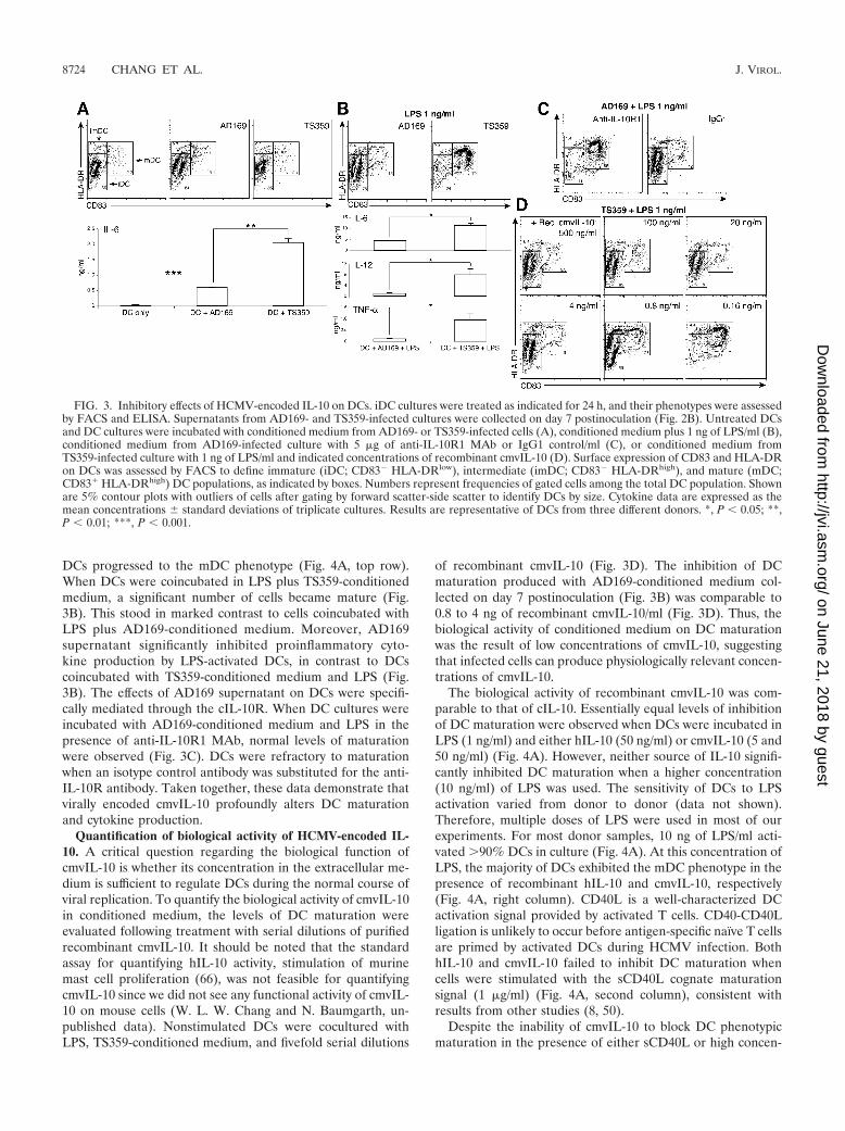
DCs progressed to the mDC phenotype (Fig. 4A, top row).When DCs were coincubated in LPS plus TS359-conditionedmedium, a significant number of cells became mature (Fig.3B). This stood in marked contrast to cells coincubated withLPS plus AD169-conditioned medium. Moreover, AD169supernatant significantly inhibited proinflammatory cyto-kine production by LPS-activated DCs, in contrast to DCscoincubated with TS359-conditioned medium and LPS (Fig.3B). The effects of AD169 supernatant on DCs were specifi-cally mediated through the cIL-10R. When DC cultures wereincubated with AD169-conditioned medium and LPS in thepresence of anti-IL-10R1 MAb, normal levels of maturationwere observed (Fig. 3C). DCs were refractory to maturationwhen an isotype control antibody was substituted for the anti-IL-10R antibody. Taken together, these data demonstrate thatvirally encoded cmvIL-10 profoundly alters DC maturationand cytokine production.
Quantification of biological activity of HCMV-encoded IL-10. A critical question regarding the biological function ofcmvIL-10 is whether its concentration in the extracellular me-dium is sufficient to regulate DCs during the normal course ofviral replication. To quantify the biological activity of cmvIL-10in conditioned medium, the levels of DC maturation wereevaluated following treatment with serial dilutions of purifiedrecombinant cmvIL-10. It should be noted that the standardassay for quantifying hIL-10 activity, stimulation of murinemast cell proliferation (66), was not feasible for quantifyingcmvIL-10 since we did not see any functional activity of cmvIL-10 on mouse cells (W. L. W. Chang and N. Baumgarth, un-published data). Nonstimulated DCs were cocultured withLPS, TS359-conditioned medium, and fivefold serial dilutions
of recombinant cmvIL-10 (Fig. 3D). The inhibition of DCmaturation produced with AD169-conditioned medium col-lected on day 7 postinoculation (Fig. 3B) was comparable to0.8 to 4 ng of recombinant cmvIL-10/ml (Fig. 3D). Thus, thebiological activity of conditioned medium on DC maturationwas the result of low concentrations of cmvIL-10, suggestingthat infected cells can produce physiologically relevant concen-trations of cmvIL-10.
The biological activity of recombinant cmvIL-10 was com-parable to that of cIL-10. Essentially equal levels of inhibitionof DC maturation were observed when DCs were incubated inLPS (1 ng/ml) and either hIL-10 (50 ng/ml) or cmvIL-10 (5 and50 ng/ml) (Fig. 4A). However, neither source of IL-10 signifi-cantly inhibited DC maturation when a higher concentration(10 ng/ml) of LPS was used. The sensitivity of DCs to LPSactivation varied from donor to donor (data not shown).Therefore, multiple doses of LPS were used in most of ourexperiments. For most donor samples, 10 ng of LPS/ml acti-vated �90% DCs in culture (Fig. 4A). At this concentration ofLPS, the majority of DCs exhibited the mDC phenotype in thepresence of recombinant hIL-10 and cmvIL-10, respectively(Fig. 4A, right column). CD40L is a well-characterized DCactivation signal provided by activated T cells. CD40-CD40Lligation is unlikely to occur before antigen-specific naïve T cellsare primed by activated DCs during HCMV infection. BothhIL-10 and cmvIL-10 failed to inhibit DC maturation whencells were stimulated with the sCD40L cognate maturationsignal (1 �g/ml) (Fig. 4A, second column), consistent withresults from other studies (8, 50).
Despite the inability of cmvIL-10 to block DC phenotypicmaturation in the presence of either sCD40L or high concen-
FIG. 3. Inhibitory effects of HCMV-encoded IL-10 on DCs. iDC cultures were treated as indicated for 24 h, and their phenotypes were assessedby FACS and ELISA. Supernatants from AD169- and TS359-infected cultures were collected on day 7 postinoculation (Fig. 2B). Untreated DCsand DC cultures were incubated with conditioned medium from AD169- or TS359-infected cells (A), conditioned medium plus 1 ng of LPS/ml (B),conditioned medium from AD169-infected culture with 5 �g of anti-IL-10R1 MAb or IgG1 control/ml (C), or conditioned medium fromTS359-infected culture with 1 ng of LPS/ml and indicated concentrations of recombinant cmvIL-10 (D). Surface expression of CD83 and HLA-DRon DCs was assessed by FACS to define immature (iDC; CD83� HLA-DRlow), intermediate (imDC; CD83� HLA-DRhigh), and mature (mDC;CD83� HLA-DRhigh) DC populations, as indicated by boxes. Numbers represent frequencies of gated cells among the total DC population. Shownare 5% contour plots with outliers of cells after gating by forward scatter-side scatter to identify DCs by size. Cytokine data are expressed as themean concentrations � standard deviations of triplicate cultures. Results are representative of DCs from three different donors. *, P � 0.05; **,P � 0.01; ***, P � 0.001.
8724 CHANG ET AL. J. VIROL.
on June 21, 2018 by guesthttp://jvi.asm
.org/D
ownloaded from

trations of LPS, recombinant cmvIL-10 inhibited proinflam-matory cytokine production for all stimulation conditions.Treatment of activated cmvIL-10 resulted in a dose-dependentinhibition of IL-12, IL-6, and TNF- production by DCs fol-lowing stimulation (Fig. 4B; only the IL-12 data are shown).This occurred even when most DCs had fully matured follow-ing sCD40L and LPS (10 and 100 ng/ml) stimulation. Inhibi-tion of cytokine production saturated at a concentration of 5ng of cmvIL-10/ml.
cmvIL-10–IL-10R engagement enhances cIL-10 production.Results of previous studies have led to the conclusion thatcIL-10 modulates its own expression, since neutralization ofendogenous cIL-10 diminishes IL-10 mRNA accumulation inactivated DCs (14). Based on this precedent for cIL-10, cmvIL-10-treated DCs were analyzed for changes in the levels ofcIL-10 expression. In contrast to the effects of cmvIL-10 onproinflammatory cytokines in DCs, cIL-10 production was in-creased in a dose-dependent manner by cmvIL-10 treatment
(Fig. 5A). Increased cIL-10 production was also observed insCD40L-stimulated DC cultures, despite the fact that sCD40Lis not a potent IL-10 stimulus, compared with LPS. The abso-lute level of cIL-10 produced by DCs varied from donor todonor, but the trend of cIL-10 induction by cmvIL-10 treat-ment was consistently observed in all tested donors (Fig. 5B).These data suggested that cIL-10 production by DCs could beup-regulated through a positive feedback mechanism in vivo,independent of whether IL-10 is encoded by the cells or byHCMV. Since activated DCs have a lower surface expressionof IL-10R1 than do iDCs (14, 34), iDCs accumulating at thesite of primary HCMV infection probably represent a promi-nent in vivo target of cmvIL-10.
Functional analogy of cmvIL-10 and cIL-10. Although thecmvIL-10 homodimer can bind to IL-10R1, it has evolved adifferent interdomain angle that may alter the orientation ofIL-10R1 on the cell surface (33). This structural change mayinterrupt IL-10R2 recruitment, and consequently, its biologicaleffects might be distinct from those of cIL-10. A central ques-tion is whether cmvIL-10 precisely mimics the function ofcIL-10 or whether there are functional differences commensu-rate with the sequence difference. Therefore, cIL-10 and cmvIL-10 were functionally compared to evaluate the effects of bothIL-10 molecules on multiple stages of the DC life cycle. iDCsare very efficient in antigen capture through several pathways,including macropinocytosis, receptor-mediated endocytosis,and phagocytosis (2). Although both forms of IL-10 stronglyinhibited maturation of iDCs (Fig. 4A), both recombinant hIL-10- and cmvIL-10-treated iDCs efficiently captured antigensvia endocytosis or phagocytosis. Endocytic activity followingIL-10 treatment was essentially equal to that observed in un-treated iDCs (Fig. 6A, top panel). Both IL-10 molecules slight-ly enhanced the phagocytic capacity of iDCs compared to thatof untreated cells (Fig. 6A, bottom panel).
Following LPS-mediated maturation of iDCs to mDCs, inaddition to CD83 and MHC class II, there was a concomitantincrease in surface expression of MHC class I (data not shown)and various costimulatory molecules involved in T-cell activa-
FIG. 4. Recombinant cmvIL-10 inhibits maturation of DCs and/oralters their cytokine expression profiles. (A) IL-10 inhibits LPS-stim-ulated but not CD40L-stimulated phenotypic maturation of DCs. Cellswere unstimulated or stimulated with sCD40L (1 �g/ml) or LPS (at theindicated concentration) for 24 h in the absence (top panel) or pres-ence of recombinant hIL-10 (second panel) or cmvIL-10 (bottom twopanels). Results are representative of 10 separate experiments. (B) cm-vIL-10 alters the IL-12 expression profiles of activated DCs. Cells werestimulated with 1 �g of sCD40L/ml or the indicated concentration ofLPS and concomitantly treated with recombinant cmvIL-10 (0, 0.5, 5,and 50 ng/ml). Data are expressed as the mean concentrations �standard deviations of triplicate cultures and are representative of fiveseparate experiments. *, P � 0.05; **, P � 0.01; ***, P � 0.001 relativeto result with no cmvIL-10 treatment.
FIG. 5. cmvIL-10 induces endogenous cIL-10 production. (A)cmvIL-10 enhances IL-10 secretion by activated DCs in a dose-depen-dent fashion. DCs treated with indicated concentrations of cmvIL-10were stimulated for 24 h with sCD40L or indicated doses of LPS.Shown are mean concentrations from duplicate cultures representativeof three separate experiments. (B) Shown are levels of cIL-10 pro-duced by DCs from various donors (n 9). Cells were stimulated with10 ng of LPS/ml in the presence or absence of recombinant cmvIL-10(50 ng/ml). Each symbol represents one individual donor. In all cases,cmvIL-10 stimulated the expression of cIL-10. The P value betweenuntreated and cmvIL-10-treated groups is shown.
VOL. 78, 2004 HCMV-ENCODED IL-10 HOMOLOG ALTERS DC FUNCTIONALITY 8725
on June 21, 2018 by guesthttp://jvi.asm
.org/D
ownloaded from

tion, including CD40, CD80, and CD86 (Fig. 6B, compare iDC[green open histogram] versus mDC [gray shaded histogram]).Since IL-10 potently inhibited maturation of iDCs, only theCD83� HLA-DRhi mDC subpopulation was gated to evaluatethe effects of both IL-10 molecules on the surface expression ofthese costimulatory molecules. Treatment of mDCs with eithercIL-10 or cmvIL-10 resulted in slightly reduced levels of CD40,CD80, and CD86, compared to those of mDCs that did notreceive IL-10-mediated signals (Fig. 6B, red and blue open his-tograms). In contrast, IL-10 treatment did not alter the surfaceexpression of CD1a (Fig. 6B), CD83, and HLA-DR (Fig. 4A)on the activated DC population. The effects of cellular andviral forms of IL-10 on alteration of the mDC phenotype wereindistinguishable from each other.
A direct comparison of both IL-10 molecules on proinflam-matory cytokine production by activated DCs was also per-formed (Fig. 6C). Quantification of secreted cytokines byELISA demonstrated that cIL-10 and cmvIL-10 had equivalentinhibitory activity for IL-6, IL-12, and TNF- expression. Thus,despite the considerable sequence evolution of cmvIL-10 andcIL-10, no functional distinctions were observed between thetwo in these assays.
Dual effects of IL-10 on DC migration. The migratory ca-pacity of DCs is tightly correlated with their maturation statusand is one of the most important features of DC physiology(21). DC trafficking is associated with the differential expres-sion of inflammatory chemokine receptors or the lymph nodehoming chemokine receptor CCR7 at distinct stages of the DClife span. The maturation process of DCs coordinates thedown-regulation of CCR1, CCR2, and CCR5 and up-regula-tion of CCR7 and therefore leads mDCs to migrate towardslymphoid tissues (21, 59, 63). To assess the effects of cmvIL-10on the migration capacity of the mDC population, chemotaxisof cmvIL-10-treated and untreated LPS-stimulated DCs toMIP-3� was assayed in a transwell system. FACS analysis ofinput and migrated cell populations revealed that only mDCs(CD83� HLA-DRhi) were able to migrate towards MIP-3�(Fig. 7A). Thus, because cmvIL-10 inhibits DC maturation, itreduced the extent of overall DC migration. Importantly, theDC subpopulation that overcame the restraint of cmvIL-10and differentiated into the mature phenotype of CD83� HLA-DRhi (Fig. 7A) and CCR1� CCR2� CCR5� CCR7� (data notshown) remained capable of migration towards MIP-3�.
Somewhat surprisingly, when migration capacity was com-pared between LPS-matured mDCs treated with cmvIL-10 andthose not treated with cmvIL-10 (Fig. 7B), cmvIL-10 consis-tently enhanced the frequency of mDCs that migrated (Fig.7C). Similar data were obtained when cells were treated withrecombinant hIL-10 (data not shown). cmvIL-10 treatment didnot appreciably affect the surface levels of CCR7 on mDCs(Fig. 7D). The observed increase in chemotaxis of IL-10-treated mDCs was, therefore, not due to an IL-10-inducedup-regulation of the MIP-3� receptor CCR7. In addition toMIP-3�, the inflammatory chemokine MIP-1 was used fortranswell migration assays. LPS-stimulated mDCs, either treatedor untreated with recombinant IL-10, did not respond to MIP-1, consistent with the surface chemokine receptor expressionpattern on those DCs (data not shown).
cmvIL-10 has long-term effects on activated DCs. It hasbeen suggested that terminal maturation of DCs occurs during
FIG. 6. cmvIL-10 and hIL-10 share comparable potency in modu-lating the functionality of DCs. (A) IL-10 enhances phagocytic capacityof iDCs. DC cultures left untreated or treated with recombinanthIL-10 or vIL-10 (50 ng/ml) were analyzed by FACS for their ability totake up FITC-conjugated dextran (top panels) or FITC-conjugatedbeads (bottom panels) for assessment of endocytic and phagocyticcapacity, respectively. Control panels on the left show untreated DCswith no FITC-dextran or FITC-conjugated beads added to the cul-tures. MFI, mean fluorescence intensity (FITC). Results are represen-tative of two separate experiments. (B) IL-10 suppresses the inductionof surface expression of costimulatory molecules on activated DCs.Cells were stimulated with 10 ng of LPS/ml in the presence (hIL-10,red open histogram; cmvIL-10, blue open histogram) or absence (grayshaded histogram) of recombinant IL-10 (5 ng/ml) for 24 h. Surfaceexpression of indicated molecules on DCs was analyzed by FACS andpresented as histograms after gating by forward scatter–side scatter–CD83–HLA-DR to identify mDC subsets. Expression levels of indi-cated molecules on the surface of iDCs are also shown (green openhistogram). Isotype control staining is shown by dashed lines. (C) IL-10alters cytokine secretion profiles of activated DCs. Levels of indicatedproinflammatory cytokines in supernatants of DC cultures 24 h afterLPS stimulation (10 ng/ml) and IL-10 treatment were determined byELISA. Data are expressed as the mean cytokine concentrations �standard deviations of triplicate cultures. Results are representative ofeight separate experiments. **, P � 0.01.
8726 CHANG ET AL. J. VIROL.
on June 21, 2018 by guesthttp://jvi.asm
.org/D
ownloaded from

their migration towards secondary lymphoid tissues and uponreceiving signals provided by activated T cells (2). This is sup-ported by our FACS analyses of those DCs that had migratedtowards MIP-3�. The cells that migrated in our transwell stud-ies showed higher expression levels of CD40, CD86, HLA-ABC, and HLA-DR than those of the input population (Fig.7E, gray-shaded histograms). Our data demonstrated that thisstep of DC maturation was also disrupted by the regulatoryevents triggered by early cmvIL-10–IL-10R engagement duringDC activation. The up-regulation of these surface moleculesfollowing cell migration was less pronounced in the cmvIL-10-treated DCs (Fig. 7E, open histograms). Whether the differ-ence in surface expression levels of these molecules betweenthe DCs that had migrated and those that had not was due topreferential migration of highly expressive cells or differences
in induction of surface markers could not be determined. How-ever, in some donors, CD86 and HLA-DR expression by cellsthat had migrated surpassed the levels of the input cell popu-lation (data not shown), suggesting that further up-regulationof these surface markers indeed occurred during DC migra-tion.
The long-term effects of cmvIL-10 on the functionality ofDCs were further shown by its continuing suppression of cy-tokine production by repeat-stimulated mDCs. A recent in vivostudy has demonstrated that CD40 ligation (of activatedDCs) and CD40L ligation (of activated T cells) facilitateproduction of IL-12 through preferential induction of p35transcription in DCs primed with microbial signals (61). Toevaluate the impact of cmvIL-10 on the CD40-dependentregulation of DC maturation, LPS-stimulated cells were
FIG. 7. Dual effects of cmvIL-10 on DC migration. (A) cmvIL-10 inhibits maturation of DCs and, thereby, inhibits their ability to migratetowards MIP-3�. Migration in response to MIP-3� (100 ng/ml) was assessed for DCs stimulated for 24 h with 1 ng of LPS/ml in the absence orpresence of recombinant cmvIL-10 (50 ng/ml). Shown are 5% contour plots of input cells (top panels) and cells present in the bottom chamber2 h after assay setup (bottom panels). Note that the only cells capable of migration were CD83� HLA-DRhigh mDCs. Cells were gated by forwardscatter-side scatter (FSC-SSC) to identify DCs by size. Results are representative of two separate experiments. (B to E) cmvIL-10 modulates thephenotype of migrating DCs. Cells stimulated with 10 ng of LPS/ml in the presence or absence of recombinant cmvIL-10 (50 ng/ml) for 24 h wereused for chemotaxis assays. One representative experiment of three separate experiments is shown. (B) Input DCs were analyzed for surfaceexpression of CD83–HLA-DR prior to chemotaxis. (C) Absolute numbers of CD83� HLA-DRhigh cells in each well that migrated. Data areexpressed as the mean numbers � standard deviations of cells that migrated from triplicate assays. ***, P � 0.001. (D) Shown are the overlaidhistograms for CCR7 expression on cmvIL-10-treated (open histogram) and untreated (gray shaded histogram) mDCs and the isotype controlstaining (dashed line). (E) The phenotypes of mDCs before (top panels) and after (bottom panels) cell migration towards MIP-3� were assessedby FACS. Surface expression of indicated molecules was analyzed by flow cytometry after gating by FSC–SSC–CD83–HLA-DR to identify mDCsubsets. Results are presented as histograms, and numbers indicate mean fluorescence intensities (MFI) for the respective fluorochromes (graynumber and shaded histogram, untreated; black number and open histogram, cmvIL-10 treated).
VOL. 78, 2004 HCMV-ENCODED IL-10 HOMOLOG ALTERS DC FUNCTIONALITY 8727
on June 21, 2018 by guesthttp://jvi.asm
.org/D
ownloaded from

harvested and restimulated with sCD40L to mimic physio-logical interactions between DCs and T cells in vivo. Afterculture for 24 h, supernatants and cells were collected forIL-12 ELISA and FACS analysis, respectively. Secondarystimulation of LPS-primed mDCs with sCD40L significantlyincreased their IL-12 production in a stimulation-dose-de-pendent manner (Fig. 8) but did not further up-regulate theexpression of MHC and costimulatory molecules (data notshown). In contrast, DCs exposed to cmvIL-10 during pri-mary LPS stimulation showed a strong reduction in IL-12secretion both prior to and following stimulation withsCD40L (Fig. 8). These data indicate that cmvIL-10-trig-gered inhibitory events continuously suppress their matura-tion and alter their cytokine expression profiles after acti-vated DCs migrate from the peripheral tissues toward thedraining lymphoid tissues. The affected DC population is,therefore, likely to be less capable of stimulating strongantiviral adaptive immune responses, even in the lymphnode, a site where cmvIL-10 is unlikely to be present in vivo.
DISCUSSION
HCMV establishes a persistent infection in the face of animmune system fully capable of initiating and maintaining an-tiviral responses. How this virus can evade immune surveil-lance remains to be fully elucidated. It is known that HCMVcan interrupt interactions between virus-infected cells and ef-fector cells, such as NK cells and T cells, by encoding multiple
viral gene products (13, 54, 67). Our study shows that theHCMV-encoded IL-10 homolog is expressed with late kineticsat a level that can inhibit DC functions. The temporal secretionof cmvIL-10 is contemporaneous with the release of progenyvirions from infected cells. Exposure of DCs to cmvIL-10 dem-onstrated that this virokine strongly inhibits the maturation ofDCs and irreversibly alters their functions. Since DCs are keyregulators of cell-mediated immunity, alterations in the extentof their activation and the quality of their responses likelyblunt the specific antiviral immunity induced against HCMV invivo.
An important implication of our study is that it does notrequire direct infection of DCs by HCMV to alter the host’simmune responses to virus infection. Rather, the results sug-gest that there is a spatial separation between the sites of virusinfection and perturbation of adaptive immune responses. Ourdata indicated that iDCs exposed to cmvIL-10 in vitro remaincapable of taking up antigens through endocytosis or phago-cytosis. Although cmvIL-10 reduces the levels of multiple co-stimulatory molecules on mDCs, their levels are still higherthan those on iDCs. Accordingly, activated DCs exposed tocmvIL-10 in vivo should be capable of priming naïve T cellsand stimulating de novo antiviral immune responses. This no-tion is further supported by the in vitro chemotaxis assay show-ing that DCs that overcome the IL-10 restraint can migratetowards MIP-3�. It should be noted that this result is in con-trast to the finding of D’Amico et al., who showed that con-comitant exposure of DCs to LPS and IL-10 blocks DC migra-tion due to the lack of chemokine receptor switch associatedwith DC maturation (16). Our results demonstrate that cmvIL-10 engenders a DC-directed cytokine milieu that would, seem-ingly, favor the establishment of a persistent infection. Earlyexposure of DCs to cmvIL-10 during their activation induced adistinct IL-10high IL-12low cytokine production profile. Theproduction of IL-12 by mDCs during T-cell priming is criticalfor polarizing CD4� T cells toward gamma interferon produc-tion (20, 40, 41). IL-12 is a key cytokine that also inducesproliferation and enhances cytotoxic activity of NK cells, oneof the crucial effector cells for the elimination of intracellularpathogens (70) and for controlling the spread of CMV (4).Thus, cmvIL-10-induced reduction of IL-12 production byDCs, at the site of viral infection or in secondary lymphoidtissues, might directly and indirectly reduce the activation ofcrucial antiviral defenses by CD4� T cells and CD8� CTLs aswell as NK cells.
A salient finding of our study is that cmvIL-10 coopts normalcellular processes to exert its effects. In addition to binding tothe IL-10Rs, cmvIL-10 significantly up-regulates the produc-tion of endogenous cIL-10 by LPS-activated DCs. Thus,HCMV may subvert the degree and quality of DCs activated bylocal virus replication though expression of the vIL-10 ho-molog and the induction of endogenous cIL-10 productionfrom cells that are not directly infected by the virus. In contrastto other cytokines expressed by DCs, the promoter activity ofIL-10 is independent of NF-�B activation. Instead, it is stronglydependent on two constitutively expressed transcription fac-tors, Sp1 and Sp3 (5, 68). IL-10 transcription would, therefore,be unaffected by an IL-10-induced NF-�B inhibition that af-fects many other DC cytokines (60). Sp1 and Sp3 are presentin many cell types and can be induced by LPS stimulation (38).
FIG. 8. Activated mDCs treated with cmvIL-10 do not respond tofurther stimulation via CD40L with increased IL-12 production. DCswere stimulated with LPS (10 ng/ml) in the absence or presence ofrecombinant cmvIL-10 (50 ng/ml) for 12 h. Cells were collected,washed with PBS, and then stimulated with indicated concentrations ofsCD40L for a further 24 h. Supernatants were harvested for measure-ment of IL-12 levels by ELISA. Results are expressed as the mean IL-12 concentrations � standard deviations of triplicate cultures. *, P �0.05; **, P � 0.01.
8728 CHANG ET AL. J. VIROL.
on June 21, 2018 by guesthttp://jvi.asm
.org/D
ownloaded from

Our data support and extend these findings by showing thatcIL-10 production by DCs is LPS dose dependent (Fig. 5A).Another checkpoint of cIL-10 expression is the control of itsown mRNA stability (51). Ligation of IL-10R triggers variousmechanisms that regulate cytokine or chemokine expressionposttranscriptionally (7, 35, 64). The mechanism that underliesthe positive feedback of IL-10–IL-10R ligation on IL-10 pro-duction in DCs is unknown but is likely to involve a number ofposttranscriptional mechanisms.
Somewhat surprisingly, given the high degree of divergenceat the sequence level, our results indicate that cmvIL-10 andcIL-10 share comparable potency in modulating DC activities.The local concentration of cmvIL-10 at the HCMV-infectedcell–DC interface would not need to be especially high toachieve its modulatory function, primarily because cmvIL-10binds to cIL-10R1 with an affinity essentially equal to that ofcIL-10 (33). As a consequence, secretion of cmvIL-10 frominfected cells in concentrations comparable to those of cIL-10should produce biologically relevant downstream effects. Al-though physiologically relevant levels of recombinant IL-10(0.5 to 5 ng/ml) were sufficient to inhibit DC maturation and toskew the cytokine expression schemes of activated DCs invitro, recombinant IL-10 was administered at a higher level (50ng/ml) for some experiments to ensure that IL-10-mediatedregulatory events were triggered in all cells in culture. Oneimportant question is whether cmvIL-10 secreted by virallyinfected cells can, indeed, inhibit DC maturation and alter itsfunctions in vivo. Although in vivo studies will be required toanswer this question fully, the biological activity of cmvIL-10expressed by HCMV-infected cells (Fig. 3) was higher thanthat of cIL-10 secreted by an equivalent number of DCs acti-vated by high doses of LPS (Fig. 5). LPS treatment is known topotently induce endogenous cIL-10 production that can inhibitDC activation in an autocrine fashion (14, 18). These data leadto the hypothesis that cmvIL-10 plays an important immuno-modulatory role during HCMV infection.
Support for a crucial role of cmvIL-10 in viral natural historycomes from a recent report on the remarkable degree to whichthe coding region and sequence for cmvIL-10 (ORF UL111A)are well conserved among all HCMV isolates examined thusfar (Hector and Davison, Abstr. 9th Int. CytomegalovirusWorkshop). The stability of the coding region in different clin-ical isolates implies that cmvIL-10 confers a selective advan-tage in vivo. Counterparts of HCMV UL111A have also beenfound in the genomes of other primate CMVs, including thoseisolated from rhesus macaques, African green monkeys, andbaboons (37). Intriguingly, the HCMV UL111A counterpart isnot present within the genome of chimpanzee CMV (CCMV),the closest known relative of HCMV, although the remainderof the genome is highly conserved (17). The reason for this isunknown, but it is possible that CCMV utilizes other pathwaysto achieve the same ends as those of the UL111A encoded byHCMV. Precedence for this idea can be found with MCMV,which also does not encode a vIL-10 homolog. Nevertheless,MCMV induces cIL-10 production within infected macro-phages (55), consistent with the idea that modulation of cIL-10is instrumental in CMV natural history. It is also interestingthat MCMV efficiently infects DCs and paralyzes their func-tions, thereby temporarily impairing the generation of antiviralimmunity (1). Since MCMV does not encode a vIL-10 ho-
molog, MCMV infection of DCs may be a necessary requisitefor efficient dissemination of the virus. The UL111A locus ofprimate CMV appears to be in a state of genetic flux (3). Oneinterpretation of the presence and absence of cmvIL-10 in theprimate and nonprimate CMV genomes is that both branchesof the CMV lineage evolved convergent mechanisms targetingthe same cell type. It is likely that CCMV, for reasons thatremain to be determined, has lost (or deleted) the cmvIL-10coding region after branching from its common ancestor withHCMV to replicate better in chimpanzees.
HCMV may have evolved distinct pathways for disruptingDC function that involve both indirect mechanisms (via cmvIL-10) and direct infection. HCMV establishes latency within asmall percentage of myeloid progenitors (28) and productivelyinfects differentiated macrophages (31, 42). Although disrup-tion of DC functions might be a key pathogenic mechanismamong many, if not all, CMV strains, the mechanism underly-ing the inhibition of DCs might vary among different CMVstrains. Previous studies have characterized infection of DCsby HCMV in vitro, although requiring very high MOIs ofendothelial cell-adapted strains (e.g., TB40/E) (46, 53, 58). Incontrast, DCs appear to be highly resistant to infection byfibroblast-adapted strains of HCMV (58, 62). The reason forthe differences in infectivity between viruses derived after pas-sage in different cell types is undoubtedly genetically based, butthe precise mechanisms remain to be determined. The 15-kbUL131-128 region, absent in the genome of a fibroblast-adapted HCMV strain, AD169, has been identified as essentialto viral tropism for both endothelial cells and leukocytes (G.Hahn, F. Baldanti, A. Gallina, G. Campanini, M. Wagner, M.Patrone, E. Percivalle, A. Sarasini, M. G. Revelo, and G.Gerna, Abstr. 28th Int. Herpesvirus Workshop, abstr. 1.02,2003). However, Gerna et al. recently reported that bothTowne and AD169 strains, lacking both endothelial cell tro-pism and leukotropism, reacquire both properties after a highnumber of sequential passages in endothelial cells (25, 26). Incontrast to the in vitro studies, in vivo data indicate that DCsdo not constitute a favored target of infection. A recent reportindicated that HCMV mainly infects cell types other than DCsand macrophages in vivo (48). Further studies are required toaddress the importance of DC infection during HCMV naturalhistory, particularly during the earliest stages of primary infec-tion.
Taken together, our in vitro data suggest that DCs areclearly an important, if indirect, target of HCMV. Since DCsrepresent the link between innate and adaptive immunity, ourresults also suggest that HCMV modulates host immune re-sponses at the earliest stage of primary infection. Inhibition orneutralization of cmvIL-10 through therapeutic or vaccinationapproaches might result in the generation of more vigorousand effective DC responses that can facilitate the induction offully protective cellular immunity to this virus.
ACKNOWLEDGMENTS
We thank T. Shenk, W. Britt, and J. Wiedeman for generouslyproviding the HCMV strains and the anti-gB hybridoma clone. Wealso acknowledge the invaluable assistance of A. Spinner with flowcytometry and M. Eberhardt with immunoblotting.
This work was supported by NIH grant AI49342 and CaliforniaNational Primate Research Center grant RR00169 to P.A.B.
VOL. 78, 2004 HCMV-ENCODED IL-10 HOMOLOG ALTERS DC FUNCTIONALITY 8729
on June 21, 2018 by guesthttp://jvi.asm
.org/D
ownloaded from

REFERENCES
1. Andrews, D. M., C. E. Andoniou, F. Granucci, P. Ricciardi-Castagnoli, andM. A. Degli-Esposti. 2001. Infection of dendritic cells by murine cytomega-lovirus induces functional paralysis. Nat. Immunol. 2:1077–1084.
2. Banchereau, J., F. Briere, C. Caux, J. Davoust, S. Lebecque, Y. J. Liu, B.Pulendran, and K. Palucka. 2000. Immunobiology of dendritic cells. Annu.Rev. Immunol. 18:767–811.
3. Barry, P. A., and W. L. Chang. Primate betaherpesviruses. In A. M. Arvin, G.Campadelli-Fumi, E. S. Mocarski, P. S. Moore, B. Roizman, R. Whitley, andK. Yamanishi (ed.), Human herpesviruses: biology, therapy and immuno-prophylaxis, in press. Cambridge University Press, Cambridge, United King-dom.
4. Biron, C. A., and L. Brossay. 2001. NK cells and NKT cells in innate defenseagainst viral infections. Curr. Opin. Immunol. 13:458–464.
5. Bondeson, J., K. A. Browne, F. M. Brennan, B. M. Foxwell, and M. Feld-mann. 1999. Selective regulation of cytokine induction by adenoviral genetransfer of I�B into human macrophages: lipopolysaccharide-induced, butnot zymosan-induced, proinflammatory cytokines are inhibited, but IL-10 isnuclear factor-�B independent. J. Immunol. 162:2939–2945.
6. Britt, W. J., and C. A. Alford. 1996. Cytomegalovirus, p. 2493–2522. In B. N.Fields, D. M. Knipe, and P. M. Howley (ed.), Fields virology, 3rd ed., vol. 2.Lippincott-Raven Publishers, Philadelphia, Pa.
7. Brown, C. Y., C. A. Lagnado, M. A. Vadas, and G. J. Goodall. 1996. Differ-ential regulation of the stability of cytokine mRNAs in lipopolysaccharide-activated blood monocytes in response to interleukin-10. J. Biol. Chem.271:20108–20112.
8. Buelens, C., V. Verhasselt, D. De Groote, K. Thielemans, M. Goldman, andF. Willems. 1997. Human dendritic cell responses to lipopolysaccharide andCD40 ligation are differentially regulated by interleukin-10. Eur. J. Immunol.27:1848–1852.
9. Carlquist, J. F., L. Edelman, D. W. Bennion, and J. L. Anderson. 1999.Cytomegalovirus induction of interleukin-6 in lung fibroblasts occurs inde-pendently of active infection and involves a G protein and the transcriptionfactor, NF-�B. J. Infect. Dis. 179:1094–1100.
10. Caux, C., C. Massacrier, B. Vanbervliet, C. Barthelemy, Y. J. Liu, and J.Banchereau. 1994. Interleukin 10 inhibits T cell alloreaction induced byhuman dendritic cells. Int. Immunol. 6:1177–1185.
11. Chang, W. L., V. Kirchoff, G. S. Pari, and P. A. Barry. 2002. Replication ofrhesus cytomegalovirus in life-expanded rhesus fibroblasts expressing humantelomerase. J. Virol. Methods 104:135–146.
12. Chang, W. L., A. F. Tarantal, S. S. Zhou, A. D. Borowsky, and P. A. Barry.2002. A recombinant rhesus cytomegalovirus expressing enhanced greenfluorescent protein retains the wild-type phenotype and pathogenicity infetal macaques. J. Virol. 76:9493–9504.
13. Chapman, T. L., A. P. Heikeman, and P. J. Bjorkman. 1999. The inhibitoryreceptor LIR-1 uses a common binding interaction to recognize class I MHCmolecules and the viral homolog UL18. Immunity 11:603–613.
14. Corinti, S., C. Albanesi, A. la Sala, S. Pastore, and G. Girolomoni. 2001.Regulatory activity of autocrine IL-10 on dendritic cell functions. J. Immu-nol. 166:4312–4318.
15. Cosman, D., N. Fanger, and L. Borges. 1999. Human cytomegalovirus, MHCclass I and inhibitory signalling receptors: more questions than answers.Immunol. Rev. 168:177–185.
16. D’Amico, G., G. Frascaroli, G. Bianchi, P. Transidico, A. Doni, A. Vecchi, S.Sozzani, P. Allavena, and A. Mantovani. 2000. Uncoupling of inflammatorychemokine receptors by IL-10: generation of functional decoys. Nat. Immu-nol. 1:387–391.
17. Davison, A. J., A. Dolan, P. Akter, C. Addison, D. J. Dargan, D. J. Alcendor,D. J. McGeoch, and G. S. Hayward. 2003. The human cytomegalovirusgenome revisited: comparison with the chimpanzee cytomegalovirus ge-nome. J. Gen. Virol. 84:17–28.
18. Demangel, C., P. Bertolino, and W. J. Britton. 2002. Autocrine IL-10 impairsdendritic cell (DC)-derived immune responses to mycobacterial infection bysuppressing DC trafficking to draining lymph nodes and local IL-12 produc-tion. Eur. J. Immunol. 32:994–1002.
19. De Smedt, T., B. Pajak, E. Muraille, L. Lespagnard, E. Heinen, P. DeBaetselier, J. Urbain, O. Leo, and M. Moser. 1996. Regulation of dendriticcell numbers and maturation by lipopolysaccharide in vivo. J. Exp. Med.184:1413–1424.
20. De Smedt, T., M. Van Mechelen, G. De Becker, J. Urbain, O. Leo, and M.Moser. 1997. Effect of interleukin-10 on dendritic cell maturation and func-tion. Eur. J. Immunol. 27:1229–1235.
21. Dieu, M. C., B. Vanbervliet, A. Vicari, J. M. Bridon, E. Oldham, S. Ait-Yahia,F. Briere, A. Zlotnik, S. Lebecque, and C. Caux. 1998. Selective recruitmentof immature and mature dendritic cells by distinct chemokines expressed indifferent anatomic sites. J. Exp. Med. 188:373–386.
22. Engelmayer, J., M. Larsson, M. Subklewe, A. Chahroudi, W. I. Cox, R. M.Steinman, and N. Bhardwaj. 1999. Vaccinia virus inhibits the maturation ofhuman dendritic cells: a novel mechanism of immune evasion. J. Immunol.163:6762–6768.
23. Fleming, S. B., C. A. McCaughan, A. E. Andrews, A. D. Nash, and A. A.
Mercer. 1997. A homolog of interleukin-10 is encoded by the poxvirus orfvirus. J. Virol. 71:4857–4861.
24. Fugier-Vivier, I., C. Servet-Delprat, P. Rivailler, M. C. Rissoan, Y. J. Liu,and C. Rabourdin-Combe. 1997. Measles virus suppresses cell-mediatedimmunity by interfering with the survival and functions of dendritic and Tcells. J. Exp. Med. 186:813–823.
25. Gerna, G., E. Percivalle, A. Sarasini, F. Baldanti, G. Campanini, and M. G.Revello. 2003. Rescue of human cytomegalovirus strain AD169 tropism forboth leukocytes and human endothelial cells. J. Gen. Virol. 84:1431–1436.
26. Gerna, G., E. Percivalle, A. Sarasini, F. Baldanti, and M. G. Revello. 2002.The attenuated Towne strain of human cytomegalovirus may revert to bothendothelial cell tropism and leuko- (neutrophil- and monocyte-) tropism invitro. J. Gen. Virol. 83:1993–2000.
27. Grigoleit, U., S. Riegler, H. Einsele, K. Laib Sampaio, G. Jahn, H. Hebart,P. Brossart, F. Frank, and C. Sinzger. 2002. Human cytomegalovirus inducesa direct inhibitory effect on antigen presentation by monocyte-derived im-mature dendritic cells. Br. J. Haematol. 119:189–198.
28. Hahn, G., R. Jores, and E. S. Mocarski. 1998. Cytomegalovirus remainslatent in a common precursor of dendritic and myeloid cells. Proc. Natl.Acad. Sci. USA 95:3937–3942.
29. Hsu, D. H., R. de Waal Malefyt, D. F. Fiorentino, M. N. Dang, P. Vieira, J.de Vries, H. Spits, T. R. Mosmann, and K. W. Moore. 1990. Expression ofinterleukin-10 activity by Epstein-Barr virus protein BCRF1. Science 250:830–832.
30. Huang, Q., D. Liu, P. Majewski, L. C. Schulte, J. M. Korn, R. A. Young, E. S.Lander, and N. Hacohen. 2001. The plasticity of dendritic cell responses topathogens and their components. Science 294:870–875.
31. Ibanez, C. E., R. Schrier, P. Ghazal, C. Wiley, and J. A. Nelson. 1991. Humancytomegalovirus productively infects primary differentiated macrophages.J. Virol. 65:6581–6588.
32. Jenkins, C., A. Abendroth, and B. Slobedman. 2004. A novel viral transcriptwith homology to human interleukin-10 is expressed during latent humancytomegalovirus infection. J. Virol. 78:1440–1447.
33. Jones, B. C., N. J. Logsdon, K. Josephson, J. Cook, P. A. Barry, and M. R.Walter. 2002. Crystal structure of human cytomegalovirus IL-10 bound tosoluble human IL-10R1. Proc. Natl. Acad. Sci. USA 99:9404–9409.
34. Kalinski, P., J. H. Schuitemaker, C. M. Hilkens, and M. L. Kapsenberg.1998. Prostaglandin E2 induces the final maturation of IL-12-deficientCD1a�CD83� dendritic cells: the levels of IL-12 are determined during thefinal dendritic cell maturation and are resistant to further modulation. J. Im-munol. 161:2804–2809.
35. Kontoyiannis, D., A. Kotlyarov, E. Carballo, L. Alexopoulou, P. J. Black-shear, M. Gaestel, R. Davis, R. Flavell, and G. Kollias. 2001. Interleukin-10targets p38 MAPK to modulate ARE-dependent TNF mRNA translationand limit intestinal pathology. EMBO J. 20:3760–3770.
36. Kotenko, S. V., S. Saccani, L. S. Izotova, O. V. Mirochnitchenko, and S.Pestka. 2000. Human cytomegalovirus harbors its own unique IL-10 ho-molog (cmvIL-10). Proc. Natl. Acad. Sci. USA 97:1695–1700.
37. Lockridge, K. M., S. S. Zhou, R. H. Kravitz, J. L. Johnson, E. T. Sawai, E. L.Blewett, and P. A. Barry. 2000. Primate cytomegaloviruses encode and ex-press an IL-10-like protein. Virology 268:272–280.
38. Ma, W., W. Lim, K. Gee, S. Aucoin, D. Nandan, M. Kozlowski, F. Diaz-Mitoma, and A. Kumar. 2001. The p38 mitogen-activated kinase pathwayregulates the human interleukin-10 promoter via the activation of Sp1 tran-scription factor in lipopolysaccharide-stimulated human macrophages.J. Biol. Chem. 276:13664–13674.
39. Macatonia, S. E., T. M. Doherty, S. C. Knight, and A. O’Garra. 1993.Differential effect of IL-10 on dendritic cell-induced T cell proliferation andIFN-gamma production. J. Immunol. 150:3755–3765.
40. Macatonia, S. E., N. A. Hosken, M. Litton, P. Vieira, C. S. Hsieh, J. A.Culpepper, M. Wysocka, G. Trinchieri, K. M. Murphy, and A. O’Garra.1995. Dendritic cells produce IL-12 and direct the development of Th1 cellsfrom naive CD4� T cells. J. Immunol. 154:5071–5079.
41. Maldonado-Lopez, R., T. De Smedt, P. Michel, J. Godfroid, B. Pajak, C.Heirman, K. Thielemans, O. Leo, J. Urbain, and M. Moser. 1999. CD8�
and CD8� subclasses of dendritic cells direct the development of distinct Thelper cells in vivo. J. Exp. Med. 189:587–592.
42. Minton, E. J., C. Tysoe, J. H. Sinclair, and J. G. Sissons. 1994. Humancytomegalovirus infection of the monocyte/macrophage lineage in bone mar-row. J. Virol. 68:4017–4021.
43. Mitra, R. S., T. A. Judge, F. O. Nestle, L. A. Turka, and B. J. Nickoloff. 1995.Psoriatic skin-derived dendritic cell function is inhibited by exogenous IL-10.Differential modulation of B7–1 (CD80) and B7–2 (CD86) expression. J. Im-munol. 154:2668–2677.
44. Mocarski, E. S. 1996. Cytomegaloviruses and their replication, p. 2447–2492.In B. N. Fields, D. M. Knipe, and P. M. Howley (ed.), Fields virology, 3rd ed.,vol. 2. Lippincott-Raven Publishers, Philadelphia, Pa.
45. Moore, K. W., R. de Waal Malefyt, R. L. Coffman, and A. O’Garra. 2001.Interleukin-10 and the interleukin-10 receptor. Annu. Rev. Immunol. 19:683–765.
46. Moutaftsi, M., A. M. Mehl, L. K. Borysiewicz, and Z. Tabi. 2002. Human
8730 CHANG ET AL. J. VIROL.
on June 21, 2018 by guesthttp://jvi.asm
.org/D
ownloaded from

cytomegalovirus inhibits maturation and impairs function of monocyte-de-rived dendritic cells. Blood 99:2913–2921.
47. Peguet-Navarro, J., C. Moulon, C. Caux, C. Dalbiez-Gauthier, J. Banche-reau, and D. Schmitt. 1994. Interleukin-10 inhibits the primary allogeneic Tcell response to human epidermal Langerhans cells. Eur. J. Immunol. 24:884–891.
48. Pereira, L., E. Maidji, S. McDonagh, O. Genbacev, and S. Fisher. 2003.Human cytomegalovirus transmission from the uterus to the placenta cor-relates with the presence of pathogenic bacteria and maternal immunity.J. Virol. 77:13301–13314.
49. Pierre, P., S. J. Turley, E. Gatti, M. Hull, J. Meltzer, A. Mirza, K. Inaba,R. M. Steinman, and I. Mellman. 1997. Developmental regulation of MHCclass II transport in mouse dendritic cells. Nature 388:787–792.
50. Poe, J. C., D. H. Wagner, Jr., R. W. Miller, R. D. Stout, and J. Suttles. 1997.IL-4 and IL-10 modulation of CD40-mediated signaling of monocyte IL-1�synthesis and rescue from apoptosis. J. Immunol. 159:846–852.
51. Powell, M. J., S. A. Thompson, Y. Tone, H. Waldmann, and M. Tone. 2000.Posttranscriptional regulation of IL-10 gene expression through sequences inthe 3�-untranslated region. J. Immunol. 165:292–296.
52. Pulendran, B., K. Palucka, and J. Banchereau. 2001. Sensing pathogens andtuning immune responses. Science 293:253–256.
53. Raftery, M. J., M. Schwab, S. M. Eibert, Y. Samstag, H. Walczak, and G.Schonrich. 2001. Targeting the function of mature dendritic cells by humancytomegalovirus: a multilayered viral defense strategy. Immunity 15:997–1009.
54. Reddehase, M. J. 2002. Antigens and immunoevasins: opponents in cyto-megalovirus immune surveillance. Nat. Rev. Immunol. 2:831–844.
55. Redpath, S., A. Angulo, N. R. Gascoigne, and P. Ghazal. 1999. Murinecytomegalovirus infection down-regulates MHC class II expression on mac-rophages by induction of IL-10. J. Immunol. 162:6701–6707.
56. Ricciardi-Castagnoli, P., and F. Granucci. 2002. Opinion: interpretation ofthe complexity of innate immune responses by functional genomics. Nat.Rev. Immunol. 2:881–889.
57. Riddell, S. R., K. S. Watanabe, J. M. Goodrich, C. R. Li, M. E. Agha, andP. D. Greenberg. 1992. Restoration of viral immunity in immunodeficienthumans by the adoptive transfer of T cell clones. Science 257:238–241.
58. Riegler, S., H. Hebart, H. Einsele, P. Brossart, G. Jahn, and C. Sinzger.2000. Monocyte-derived dendritic cells are permissive to the complete rep-licative cycle of human cytomegalovirus. J. Gen. Virol. 81:393–399.
59. Sallusto, F., P. Schaerli, P. Loetscher, C. Schaniel, D. Lenig, C. R. Mackay,S. Qin, and A. Lanzavecchia. 1998. Rapid and coordinated switch in che-mokine receptor expression during dendritic cell maturation. Eur. J. Immu-nol. 28:2760–2769.
60. Schottelius, A. J., M. W. Mayo, R. B. Sartor, and A. S. Baldwin, Jr. 1999.
Interleukin-10 signaling blocks inhibitor of �B kinase activity and nuclearfactor �B DNA binding. J. Biol. Chem. 274:31868–31874.
61. Schulz, O., A. D. Edwards, M. Schito, J. Aliberti, S. Manickasingham, A.Sher, and C. Reis e Sousa. 2000. CD40 triggering of heterodimeric IL-12 p70production by dendritic cells in vivo requires a microbial priming signal.Immunity 13:453–462.
62. Soderberg-Naucler, C., K. N. Fish, and J. A. Nelson. 1998. Growth of humancytomegalovirus in primary macrophages. Methods 16:126–138.
63. Sozzani, S., P. Allavena, G. D’Amico, W. Luini, G. Bianchi, M. Kataura, T.Imai, O. Yoshie, R. Bonecchi, and A. Mantovani. 1998. Differential regula-tion of chemokine receptors during dendritic cell maturation: a model fortheir trafficking properties. J. Immunol. 161:1083–1086.
64. Sozzani, S., S. Ghezzi, G. Iannolo, W. Luini, A. Borsatti, N. Polentarutti, A.Sica, M. Locati, C. Mackay, T. N. Wells, P. Biswas, E. Vicenzi, G. Poli, andA. Mantovani. 1998. Interleukin 10 increases CCR5 expression and HIVinfection in human monocytes. J. Exp. Med. 187:439–444.
65. Spencer, J. V., K. M. Lockridge, P. A. Barry, G. Lin, M. Tsang, M. E.Penfold, and T. J. Schall. 2002. Potent immunosuppressive activities ofcytomegalovirus-encoded interleukin-10. J. Virol. 76:1285–1292.
66. Thompson-Snipes, L., V. Dhar, M. W. Bond, T. R. Mosmann, K. W. Moore,and D. M. Rennick. 1991. Interleukin 10: a novel stimulatory factor for mastcells and their progenitors. J. Exp. Med. 173:507–510.
67. Tomasec, P., V. M. Braud, C. Rickards, M. B. Powell, B. P. McSharry, S.Gadola, V. Cerundolo, L. K. Borysiewicz, A. J. McMichael, and G. W.Wilkinson. 2000. Surface expression of HLA-E, an inhibitor of natural killercells, enhanced by human cytomegalovirus gpUL40. Science 287:1031.
68. Tone, M., M. J. Powell, Y. Tone, S. A. Thompson, and H. Waldmann. 2000.IL-10 gene expression is controlled by the transcription factors Sp1 and Sp3.J. Immunol. 165:286–291.
69. Tortorella, D., B. E. Gewurz, M. H. Furman, D. J. Schust, and H. L. Ploegh.2000. Viral subversion of the immune system. Annu. Rev. Immunol. 18:861–926.
70. Trinchieri, G. 2003. Interleukin-12 and the regulation of innate resistanceand adaptive immunity. Nat. Rev. Immunol. 3:133–146.
71. Ulbrecht, M., S. Martinozzi, M. Grzeschik, H. Hengel, J. W. Ellwart, M. Pla,and E. H. Weiss. 2000. Cutting edge: the human cytomegalovirus UL40 geneproduct contains a ligand for HLA-E and prevents NK cell-mediated lysis.J. Immunol. 164:5019–5022.
72. Walter, E. A., P. D. Greenberg, M. J. Gilbert, R. J. Finch, K. S. Watanabe,E. D. Thomas, and S. R. Riddell. 1995. Reconstitution of cellular immunityagainst cytomegalovirus in recipients of allogeneic bone marrow by transferof T-cell clones from the donor. N. Engl. J. Med. 333:1038–1044.
73. Yu, D., M. C. Silva, and T. Shenk. 2003. Functional map of human cyto-megalovirus AD169 defined by global mutational analysis. Proc. Natl. Acad.Sci. USA 100:12396–12401.
VOL. 78, 2004 HCMV-ENCODED IL-10 HOMOLOG ALTERS DC FUNCTIONALITY 8731
on June 21, 2018 by guesthttp://jvi.asm
.org/D
ownloaded from