human eif4e promotes mrna restructuring by stimulating
TRANSCRIPT
Human eIF4E promotes mRNA restructuring bystimulating eIF4A helicase activityKateryna Feoktistova, Enkhee Tuvshintogs, Angelie Do, and Christopher S. Fraser1
Department of Molecular and Cell Biology, College of Biological Sciences, University of California, Davis, CA 95616
Edited* by Jennifer A. Doudna, University of California, Berkeley, CA, and approved July 5, 2013 (received for review February 27, 2013)
Elevated eukaryotic initiation factor 4E (eIF4E) levels frequentlyoccur in a variety of human cancers. Overexpression of eIF4E pro-motes cellular transformation by selectively increasing the trans-lation of proliferative and prosurvival mRNAs. These mRNAs possesshighly structured 5′-UTRs that impede ribosome recruitment andscanning, yet the mechanism for how eIF4E abundance elevatestheir translation is not easily explained by its cap-binding activity.Here, we show that eIF4E possesses an unexpected second functionin translation initiation by strongly stimulating eukaryotic initiationfactor 4A (eIF4A) helicase activity. Importantly, we demonstratethat this activity promotes mRNA restructuring in a manner thatis independent of its cap-binding function. To explain these find-ings, we show that the eIF4E-binding site in eukaryotic initiationfactor 4G (eIF4G) functions as an autoinhibitory domain to modu-late its ability to stimulate eIF4A helicase activity. Binding of eIF4Ecounteracts this autoinhibition, enabling eIF4G to stimulate eIF4Ahelicase activity. Finally, we have successfully separated the twofunctions of eIF4E to show that its helicase promoting activityincreases the rate of translation by a mechanism that is distinctfrom its cap-binding function. Based on our results, we proposethat maintaining a connection between eIF4E and eIF4G through-out scanning provides a plausible mechanism to explain how eIF4Eabundance selectively stimulates the translation of highly struc-tured proliferation and tumor-promoting mRNAs.
protein synthesis | DEAD-box | ATPase
Recruitment of mRNAs to the ribosome must be tightly con-trolled in human cells because the dysregulation of protein
synthesis has a direct impact on cancer development and pro-gression (1, 2). Eukaryotic initiation factor 4F (eIF4F) is theprotein complex that binds the 5′ 7-methyl guanosine cap foundon all cellular mRNAs and is comprised of the cap-bindingprotein, eIF4E, the DEAD-box helicase, Eukaryotic initiationfactor 4A (eIF4A), and the scaffold protein Eukaryotic initiationfactor 4F (eIF4G) (3). The eIF4E component of eIF4F is gen-erally considered to be the rate-limiting factor in translationinitiation (4). Consistent with this, eIF4E availability is tightlycontrolled through regulated interaction with eIF4E-binding pro-teins (4E-BPs). These proteins function as competitive inhibitorsof eIF4E binding to eIF4G and are regulated through phosphor-ylation events coordinated by the PI3K–AKT–mammalian targetof rapamycin (mTOR) signaling pathway (5, 6). Activation ofmTOR complex 1 (mTORC1) releases eIF4E from 4E-BPs,leading to an increase in the translation of a pool of mRNAs oftenreferred to as “eIF4E-sensitive” (2, 5). These mRNAs possessregulatory elements in their 5′-UTRs that somehow confer theirsensitivity to eIF4E levels. The overwhelming majority of thesemRNAs possess long structured 5′-UTRs that must be unwoundto allow ribosome recruitment and scanning (2, 5, 7, 8). Accord-ingly, overexpression of eIF4E selectively increases the translationof highly structured proliferative and prosurvival mRNAs that cantransform immortalized cells and form tumors in mice (9–12).However, the mechanism by which the availability of eIF4E se-lectively controls translation initiation of mRNAs containingstructured 5′-UTRs is unknown.
Unwinding of secondary structure in the mRNA 5′-UTRinvolves the activity of the eIF4A helicase component of eIF4F(13, 14). Human eIF4A functions as an RNA-dependent ATPasethat bidirectionally unwinds RNA duplexes (15–17). Consistentwith other DEAD-box proteins, strand separation is promotedthrough well-defined conformational rearrangements of its RecAhomology domains (18–20). Although eIF4A is a relatively poorhelicase on its own, its ATPase and duplex unwinding activitiesare stimulated by eIF4G and the helicase accessory protein,eIF4B (15, 17, 21–24). In addition, the interaction of the poly(A)binding protein (PABP) with eIF4G also stimulates the ATPaseand duplex unwinding activity of eIF4A (25). Despite the factthat an increase in eIF4E availability stimulates the rate oftranslation initiation on highly structured mRNAs, no evidenceexists to link the presence of eIF4E in the eIF4F complex withthe activity of the eIF4A (15, 17). Previous attempts to study thehelicase activity of eIF4A in the eIF4F complex did not controlfor eIF4E abundance, raising a fundamental question regardingthe possible role of eIF4E in controlling the helicase activity ofthe eIF4F complex.Here, we have used a real-time fluorescence assay to reveal
the kinetic parameters of duplex unwinding by the human eIF4Fcomplex. Our data demonstrate that, in addition to its role incap-binding, eIF4E stimulates eIF4A duplex unwinding activityin the eIF4F complex. We further show that this unexpectedactivity of eIF4E increases the rate of translation initiation bya mechanism that is distinct from its role in cap-binding. Thisregulatory function of eIF4E provides a plausible mechanism toexplain how eIF4E can selectively stimulate the translation ofmRNAs that possess structured 5′-UTRs.
ResultseIF4E Stimulates the Rate of Duplex Unwinding by eIF4A. To de-termine the kinetic framework of duplex unwinding by the hu-man eIF4F complex, we generated a modified version of ourpreviously described fluorescence unwinding assay that uses anuncapped RNA duplex substrate (Fig. 1A) (21). This assay de-sign provides greater flexibility compared with our previous assaybecause it does not require a fluorescently modified RNA loadingstrand. The assay can be used to accurately measure the kineticsof RNA strand separation by monitoring an increase in totalfluorescence in real time by using highly purified initiation factors(Fig. S1). Consistent with our previous results, efficient strandseparation by eIF4A is observed in the presence of fixed amountsof eIF4B and an eIF4G truncation that spans amino acids 682to 1105 (eIF4G682–1105; Fig. 1B and Table S1 ) (21). AlthougheIF4G682–1105 constitutes a conserved region of eIF4G that is ableto stimulate eIF4A duplex unwinding, we wanted to determine if
Author contributions: K.F. and C.S.F. designed research; K.F., E.T., and A.D. performedresearch; K.F. and C.S.F. analyzed data; and K.F. and C.S.F. wrote the paper.
The authors declare no conflict of interest.
*This Direct Submission article had a prearranged editor.1To whom correspondence should be addressed. E-mail: [email protected].
This article contains supporting information online at www.pnas.org/lookup/suppl/doi:10.1073/pnas.1303781110/-/DCSupplemental.
www.pnas.org/cgi/doi/10.1073/pnas.1303781110 PNAS | August 13, 2013 | vol. 110 | no. 33 | 13339–13344
BIOCH
EMISTR
Y
Dow
nloa
ded
by g
uest
on
Dec
embe
r 29
, 202
1
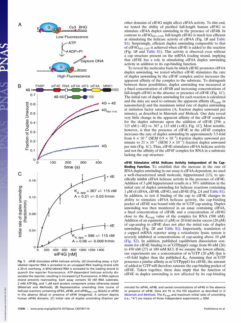
other domains of eIF4G might affect eIF4A activity. To this end,we tested the ability of purified full-length human eIF4G tostimulate eIF4A duplex unwinding in the presence of eIF4B. Incontrast to eIF4G682–1105, full-length eIF4G is much less efficientat stimulating the helicase activity of eIF4A (Fig. 1B and TableS1). Surprisingly, efficient duplex unwinding comparable to thatof eIF4G682–1105 is achieved when eIF4E is added to the reaction(Fig. 1B and Table S1). This activity is observed even withouta cap structure present on the mRNA loading strand, implyingthat eIF4E has a role in stimulating eIF4A duplex unwindingactivity in addition to its cap-binding function.To reveal the molecular basis by which eIF4E promotes eIF4A
duplex unwinding, we tested whether eIF4E stimulates the rateof duplex unwinding by the eIF4F complex and/or increases theapparent affinity of the complex to the substrate. To distinguishbetween these possibilities, duplex unwinding was measured ata fixed concentration of eIF4B and increasing concentrations offull-length eIF4G in the absence or presence of eIF4E (Fig. 1C).The initial rate of duplex unwinding for each reaction is calculatedand the data are used to estimate the apparent affinity (Kd,app, innanomolars]) and the maximum initial rate of duplex unwindingat initiation factor saturation (A; fraction duplex unwound perminute), as described in Materials and Methods. Our data revealvery little change in the apparent affinity of the eIF4F complexfor the duplex substrate upon the addition of eIF4E [596 ±115 nM (−4E) vs. 367 ± 115 nM (+4E); Fig. 1C]. Most notable,however, is that the presence of eIF4E in the eIF4F complexincreases the rate of duplex unwinding by approximately 3.5-foldfrom 6 × 10⁻² (SEM 0.9 × 10⁻²) fraction duplex unwound perminute to 21 × 10−2 (SEM 3 × 10⁻²) fraction duplex unwoundper min (Fig. 1C). Thus, eIF4E stimulates eIF4A helicase activityand not the affinity of the eIF4F complex for RNA in a substratelacking the cap structure.
eIF4E Stimulates eIF4A Helicase Activity Independent of Its Cap-Binding Function. To establish that the increase in the rate ofRNA duplex unwinding in our assay is eIF4A-dependent, we useda well-characterized small molecule, hippuristanol (13), to spe-cifically inhibit eIF4A helicase activity in the presence of eIF4E.Addition of 3 μM hippuristanol results in a 70% inhibition in theinitial rate of duplex unwinding for helicase reactions containing1 μM of eIF4A, eIF4B, eIF4G, and eIF4E (Fig. 2A and Table S1).In addition, to test if binding of the cap to eIF4E changes itsability to stimulate eIF4A helicase activity, the cap-bindingpocket of eIF4E was bound with the m7GTP cap-analog. Duplexunwinding was then monitored in an assay containing eIF4A,a fixed concentration of eIF4B, and a concentration of eIF4Gclose to the Kd,app value of the complex for RNA (500 nM).Addition of an equimolar (1 μM) or 20-fold molar excess (20 μM)of cap-analog to eIF4E does not alter the initial rate of duplexunwinding (Fig. 2B and Table S1). Importantly, translation ofa capped mRNA reporter using a reticulocyte lysate system isseverely inhibited at concentrations of cap-analog above 10 μM(Fig. S2). In addition, published equilibrium dissociation con-stants for eIF4E binding to m7GTPpppG range from 80 nM (26)to 450 nM (27) at 100 mM KCl. If we assume the lowest affinity,our experiments use a concentration of m7GTP (20 μM) that is∼45-fold higher than the published Kd. Assuming that m7GTPpossesses a similar affinity as m7GTPpppG for eIF4E, the amountof added m7GTP will therefore saturate the cap-binding pocket ofeIF4E. Taken together, these data imply that the function ofeIF4E in duplex unwinding is not affected by its cap-binding
AE G
Low Fluorescence
High Fluorescence
A B
E GA B
capture DNA
ATP
ADP+Pi
Cy3 BHQ
B
4G
4G682-1105
4G + 4E
682 11054G
1 16004GeIF4A eIF4AeIF4E eIF3 MNK1RNAPABP
time (s)
Frac
tion
of D
uple
x U
nwou
nd
Initi
al ra
te o
f dup
lex
unw
indi
ng(fr
actio
n/m
in)
eIF4G [nM]
C
– 4E
+ 4E
Kd, app = 367 +/- 115 nM
Kd, app = 596 +/- 115 nM
A = 0.21 +/- 0.03 fr/min
A = 0.06 +/- 0.009 fr/min
Fig. 1. eIF4E stimulates eIF4A helicase activity. (A) Unwinding assay: a Cy3-labeled reporter RNA is annealed to an uncapped RNA loading strand witha 20-nt overhang. A BHQ-labeled RNA is annealed to the loading strand toquench the reporter fluorescence. ATP-dependent helicase activity dis-sociates the reporter, resulting in increased Cy3 fluorescence. A DNA capturestrand prevents reannealing. Each assay contains 50 nM RNA substrate,2 mM ATP-Mg, and 1 μM each protein component unless otherwise stated(Materials and Methods). (B) Representative unwinding time course ofhelicase reactions containing eIF4A, eIF4B, and eIF4G682–1105 (black) or eIF4Gin the absence (blue) or presence of eIF4E (magenta). A cartoon depictshuman eIF4G domains. (C) Initial rate of duplex unwinding (fraction per
minute) for eIF4A, eIF4B, and varied concentrations of eIF4G in the absenceor presence of eIF4E. Data are fit to the Hill equation as described in SIMaterials and Methods. The Kd,app and maximum initial rates of unwinding(i.e., “A”) are means of three independent experiments ± SEM.
13340 | www.pnas.org/cgi/doi/10.1073/pnas.1303781110 Feoktistova et al.
Dow
nloa
ded
by g
uest
on
Dec
embe
r 29
, 202
1
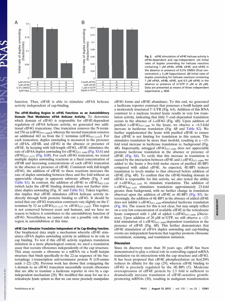
function. Thus, eIF4E is able to stimulate eIF4A helicaseactivity independent of cap-binding.
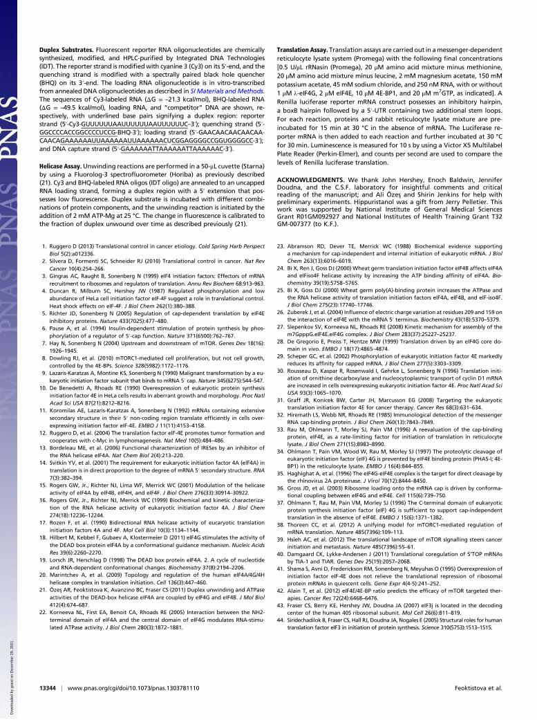
The eIF4E-Binding Region in eIF4G Functions as an AutoinhibitoryDomain That Modulates eIF4A Helicase Activity. To determinewhich domain of eIF4G is responsible for eIF4E-dependentregulation of eIF4A helicase activity, we generated two addi-tional eIF4G truncations. One truncation removes the N-termi-nal 556 aa (eIF4G557–1600), whereas the second truncation removesan additional 463 aa from the C terminus (eIF4G557–1137). Foreach truncation, duplex unwinding is measured in the presenceof eIF4A, eIF4B, and eIF4G in the absence or presence ofeIF4E. In keeping with full-length eIF4G, eIF4E stimulates therate of eIF4A duplex unwinding for eIF4G557–1600 (Fig. S3A) andeIF4G557–1137 (Fig. S3B). For each eIF4G truncation, we testedmultiple duplex unwinding reactions at a fixed concentration ofeIF4B and increasing concentrations of each eIF4G truncationin the absence or presence of eIF4E. Consistent with full-lengtheIF4G, the addition of eIF4E to these reactions increases therate of duplex unwinding between three and five fold without anappreciable change in apparent substrate affinity (Fig. 3 andTable S1). In contrast, the addition of eIF4E to eIF4G682–1105(which lacks the eIF4E binding domain) does not further stim-ulate duplex unwinding (Fig. 3C and Table S1). Taken together,this implies that eIF4E stimulates eIF4A helicase activity in-directly through both proteins binding to eIF4G. It should benoted that our eIF4G truncation constructs vary slightly on the Cterminus by 32 aa (eIF4G557–1137 vs. eIF4G557–1105). This regionis not conserved between yeast and humans, and we have noreason to believe it contributes to the autoinhibitory function ofeIF4G. Nevertheless, we cannot rule out a possible role of thisregion in autoinhibition at this time.
eIF4E Can Stimulate Translation Independent of Its Cap-Binding Function.Our biophysical data imply a mechanism whereby eIF4E stim-ulates eIF4A duplex unwinding activity in addition to its role incap binding. To test if this eIF4E activity regulates translationinitiation in a more physiological context, we used a translationassay that recruits ribosomes independently of the cap structure.This assay recruits a ribosome to a mRNA via a boxB hairpinstructure that binds specifically to the 22-aa sequence of the bac-teriophage λ-transcription anti-terminator protein N (λN-aminoacids 1–22) (28). Previous work has shown that conjugating theλ-domain to an eIF4G truncation successfully recruits ribosomesthat are able to translate a luciferase reporter in vivo by a cap-independent mechanism (28). We modified this assay for use in areticulocyte lysate system so that we can more precisely manipulate
eIF4G forms and eIF4E abundance. To this end, we generateda luciferase reporter construct that possesses a boxB hairpin anda moderately structured 5′-UTR (Fig. 4A). Addition of this RNAconstruct to a nuclease treated lysate results in very low trans-lation activity, indicating that little 5′-end–dependent translationoccurs in the absence of λ-eIF4G (Fig. 4B). Upon addition ofpurified λ-eIF4G557–1600 to the lysate, we observe a ∼6.5-foldincrease in luciferase translation (Fig. 4B and Table S2). Wefurther supplemented the lysate with purified eIF4E to ensurethat eIF4E is not limiting for translation in this system. Thisstimulates translation by more than twofold, resulting in a ∼15-fold total increase in luciferase translation vs. background (Fig.4B). Importantly, untagged eIF4G557–1600 does not appreciablypromote luciferase translation in the absence or presence ofeIF4E (Fig. S4). To verify that this increase in translation iscaused by the interaction between eIF4E and λ-eIF4G557–1600, weadded to the lysate a five-fold molar excess of purified 4E-BP1compared with added eIF4E. As expected, 4E-BP1 reducestranslation to levels similar to that observed before addition ofeIF4E (Fig. 4B). To confirm that the eIF4E-binding domain ineIF4G is responsible for this activity, we also tested the abilityof λ-eIF4G682–1105 to stimulate translation. The addition ofλ-eIF4G682–1105 stimulates translation approximately 23-foldgreater than background, with no further change in translationobserved upon the addition of eIF4E or 4E-BP1 (Fig. S5). In-terestingly, the addition of 4E-BP1 in the absence of added eIF4Edoes not inhibit λ-eIF4G557–1600-stimulated luciferase translation(Fig. S6). The reason for this is not clear, but may simply reflecton a very low concentration of available eIF4E in the reticulocytelysate compared with 1 μM of added λ-eIF4G557–1600 (Discus-sion). Upon addition of 20 μM m7GTP, we still observe a ∼12-fold stimulation of λ-eIF4G557–1600-dependent translation in thepresence of eIF4E (Fig. 4B). This further demonstrates thateIF4E stimulation of eIF4A duplex unwinding and cap-bindingevents are independent functions that together promote ribosomerecruitment, scanning, and translation initiation.
DiscussionSince its discovery more than 30 years ago, eIF4E has beendemonstrated to play a critical role in controlling capped mRNAtranslation via its interactions with the cap structure and eIF4G.It has been proposed that eIF4E phosphorylation on Ser(209)reduces its affinity for the cap (29), whereas eIF4E binding toeIF4G is precisely regulated by the 4E-BPs (5). Moderateoverexpression of eIF4E protein by 2.5 fold is sufficient todramatically increase translation of eIF4E-sensitive growth-promoting mRNAs (30), resulting in malignant transformation
Initi
al r
ate
of
du
ple
x u
nw
ind
ing
(fra
ctio
n/m
in)
+
–+
+ +++
++
– ––
+
–+
+ +++
++
– –
Initi
al r
ate
of
du
ple
x u
nw
ind
ing
(fra
ctio
n/m
in)
+
A B
Fig. 2. eIF4E stimulation of eIF4F helicase activity iseIF4A-dependent and cap-independent. (A) Initialrates of duplex unwinding for helicase reactionscontaining 1 μM eIF4A, eIF4B, eIF4E, and eIF4G inthe absence or presence of 0.3% DMSO (final con-centration) ± 3 μM hippuristanol. (B) Initial rates ofduplex unwinding for helicase reactions containing1 μM eIF4A, eIF4B, eIF4E, and 0.5 μM eIF4G in theabsence or presence of m7GTP (1 μM or 20 μM).Data are presented as means of three independentexperiments ± SEM.
Feoktistova et al. PNAS | August 13, 2013 | vol. 110 | no. 33 | 13341
BIOCH
EMISTR
Y
Dow
nloa
ded
by g
uest
on
Dec
embe
r 29
, 202
1

of immortalized cells and tumor formation in mice (9, 10, 12).Consistent with this, 30% of human cancers show similar ele-vated eIF4E levels, underscoring the importance of eIF4Eoverexpression in cancer progression (31). Despite this, a mech-anism to explain why a subset of cellular mRNAs requires higherlevels of available eIF4E for their translation had not beenidentified. One can speculate that some mRNAs may have theircap structure occluded by 5′-UTR secondary structure, causingthem to be poor cap-binding substrates for eIF4E. Thus, anincrease in the total amount of eIF4F complex would prefer-entially stimulate this type of mRNA pool. This potentialmechanism, however, has not been rigorously demonstrated foreIF4E-sensitive mRNAs, and awaits further investigation.Unexpectedly, we have uncovered a second function of eIF4E:
the stimulation of eIF4A helicase activity in the eIF4F complex.In addition to increasing the total amount of eIF4F, our datanow show that the resulting complex is in fact more active withregard to mRNA restructuring (Fig. 1). Importantly, we showthat the ability of eIF4E to stimulate eIF4A helicase activity isindependent of its cap-binding function (Fig. 2). Interestingly,the stimulation of eIF4A helicase activity occurs despite a rela-tively small effect of eIF4E on the affinity of the eIF4G/4Acomplex for RNA substrate. However, as our calculated affini-ties represent apparent equilibrium dissociation constants, it willbe important in the future to determine the direct equilibrium
dissociation constant for eIF4G/4A binding to RNA substratein the absence and presence of eIF4E. This information willbe necessary to determine if eIF4E, in addition to stimulatingeIF4A helicase activity, also increases the direct affinity ofeIF4G/4A for RNA independently of cap binding. Moreover, byusing a tethered-RNA assay to bypass the need for the capstructure in ribosome recruitment, we further show that cap-analog does not change the ability of eIF4E to stimulate the rateof protein synthesis (Fig. 4). It is worth noting that we observea small (∼20%) inhibition of λ-eIF4G557–1600-dependent trans-lation upon m7GTP addition in the presence of eIF4E (Fig. 4B).As we do not observe a change in eIF4E stimulated eIF4Ahelicase activity in the presence of m7GTP (Fig. 2), this mayindicate that cap binding may influence another translation ini-tiation step that is yet to be determined. Interestingly, we findthat the addition of 4E-BP1 does not inhibit the stimulation oftranslation by 1 μM λ-eIF4G557–1600 in the absence of exoge-nously added eIF4E (Fig. S6). The reported concentration ofeIF4E in reticulocyte lysate preparations varies between 8 nM(32) and 400 nM (33). Even at the highest eIF4E concentrationreported, 4E-BP1 is present at a ratio of 1:1 with eIF4E (33).Thus, the failure of added 4E-BP1 to inhibit λ-eIF4G557–1600-dependent translation might simply reflect the presence of verylittle free endogenous eIF4E to bind the exogenously addedλ-eIF4G557–1600. Alternatively, this finding may imply that the
Kd, app = 412 +/- 84 nM
Kd, app = 563 +/- 101 nM
– 4E
+ 4E
gnidniwnu xelpud fo etar laitinI
)nim/noitcarf(
eIF4G557-1600 [nM]
1600557eIF4A eIF4AeIF4E eIF3 MNK1RNA
Kd, app = 374 +/- 87 nM
Kd, app = 370 +/- 66 nM
– 4E
+ 4E
eIF4G557-1137 [nM]
– 4E + 4E – 4E + 4E – 4E + 4E – 4E + 4E
eIF4G682-1105 eIF4G557-1137 eIF4G557-1600 eIF4G1-1600
)nim/noitcarf( gnidni
wnu xelpud fo etar laitinI557
eIF4AeIF4E eIF3RNA1137
A = 0.33 +/- 0.03 fr/min
A = 0.065 +/- 0.008 fr/min
A = 0.32 +/- 0.03 fr/min
A = 0.09 +/- 0.009 fr/min
A
C
B
Fig. 3. The eIF4E-binding domain in eIF4G reg-ulates eIF4A activity. Initial rates of duplex un-winding (fraction per minute) for each reactioncontaining 1 μM eIF4A and eIF4B are plotted vs.increasing concentrations of eIF4G557–1600 (A) oreIF4G557–1137 (B) in the absence or presence of 1 μMeIF4E. Data are fit to the Hill equation (SI Materialsand Methods). The Kd,app and maximum initial ratesof unwinding (i.e., “A”) are shown as means ofthree independent experiments ± SEM. Cartoonsare shown to depict human eIF4G domains in theconstructs used. (C) Initial rates of duplex un-winding for different truncations of eIF4G ± eIF4E.Each reaction contains 1 μM eIF4G, eIF4A, eIF4B,and eIF4E.
13342 | www.pnas.org/cgi/doi/10.1073/pnas.1303781110 Feoktistova et al.
Dow
nloa
ded
by g
uest
on
Dec
embe
r 29
, 202
1

stimulation of λ-eIF4G557–1600-dependent translation by exoge-nous eIF4E is only relevant to situations in which eIF4E isoverexpressed (e.g., in certain tumors). Future work will hope-fully distinguish between these two possible models.We have used eIF4G truncations in our unwinding assay to
demonstrate that the eIF4E-binding site in eIF4G functions asa classic autoinhibitory domain (Fig. 3). In the absence of eIF4Ebinding, this domain reduces the ability of eIF4G to stimulateeIF4A helicase activity. The binding of eIF4E to this inhibito-ry domain counteracts the autoinhibition, enabling eIF4G tostimulate eIF4A helicase activity (Figs. 1 and 3). Consistent withthis, removal of this domain results in an eIF4G truncation(eIF4G682–1105) that is constitutively active with regard to stim-ulating eIF4A helicase activity (Fig. 1 and ref. 21). Interestingly,it has previously been shown that eIF4E binding induces a struc-tural change in eIF4G, as revealed by increased sensitivity to viralprotease cleavage (34–36). Therefore, it is likely that eIF4Einduces an eIF4G conformation that stimulates eIF4A helicaseactivity, as depicted in Fig. 5. In light of this model, maintainingan eIF4E/eIF4G interaction throughout scanning provides aplausible mechanism to explain how eIF4E abundance promotes
translation of highly structured mRNAs. Moreover, this activityof eIF4E further explains why uncapped mRNA translation issensitive to eIF4E availability (37).Recently, the translation of 5′-terminal oligopyrimidine tract
(5′TOP) containing messages has been shown to be sensitive toeIF4E levels, despite not possessing obvious predicted secondarystructure (38, 39). Our data imply that these mRNAs are stronglydependent on efficient eIF4A activity for their translation, per-haps through removal of 5′TOP-specific trans-acting factors (40).It should be noted, however, the degree to which 5′TOP mRNAtranslation requires elevated eIF4E levels is not clear, as pre-vious work found no change in their translation rate uponoverexpression of eIF4E (41). Chemotherapeutic drugs are cur-rently being developed to target eIF4E and mTOR as a meansto control tumor formation (1). It has recently been shown thatcancer cells can acquire resistance to mTOR inhibitors by down-regulating 4E-BPs so that eIF4E availability increases (42).By revealing an additional activity of eIF4E we describe here, weanticipate that our work may aid in the development of moreeffective cancer therapeutic agents that can target the inde-pendent functions of this central component of the translationmachinery.
Materials and MethodsPurified Components. Detailed sample purification protocols are described inSI Materials and Methods. Briefly, recombinant eIF4A isoform I (eIF4AI), 4E-BP1, eIF4G682–1105, and PABP proteins are expressed in bacteria as maltose-binding protein fusion constructs, cleaved by using recombinant tobaccoetch virus (TEV) protease, and purified by using established procedures togenerate untagged proteins (21, 43). Human eIF4E is expressed in bacteria asa protein G fusion construct, cleaved by using TEV protease, and purified byion-exchange chromatography to yield untagged protein. Human eIF4B isexpressed as a 6-histidine–tagged construct in insect cells (sf9) and purifiedas described previously (21). Human eIF4G557–1137 and eIF4G557–1600 areexpressed as 6-histidine–tagged constructs in sf9 cells and purified byimmobilized metal ion affinity chromatography (IMAC) and ion-exchangeand size-exclusion chromatography. Following elution from Ni-NTA Super-flow resin (Qiagen), purified human eIF4AI is added and incubated overnightat 4 °C to form a human eIF4G/eIF4A heterodimer. All λ-tagged eIF4G con-structs are generated as N-terminal 6-histidine–λ fusion constructs and puri-fied as described for the non–λ-tagged proteins. Endogenous eIF4F is purifiedfrom HeLa cell cytoplasmic extract by using established procedures (44), andstripped of its eIF4E component by using size-exclusion chromatography.
AUG L U CInhibitory Hairpin
boxB
B
A 43S preinitiationcomplex
Nor
mal
ized
Luc
ifera
se T
rans
latio
n
RNA557-1600eIF4E
m7GTP4E-BP1
+ + + +– + + +–––
–– –
++
– + + ++– +
– –
λ
λ-eIF4G
Fig. 4. eIF4E stimulates translation independent of cap-binding. (A) Sche-matic of the luciferase translation assay: the boxB RNA element recruitsλ-eIF4G557–1600 to the reporter RNA construct. The mRNA contains two ad-ditional stem loops between the boxB hairpin and the luciferase reportergene as described in Materials and Methods. An inhibitory hairpin is locatedupstream of the boxB element to prevent any 5′ end-dependent ribosomeloading. The binding of λ-eIF4G557–1600 enables eIF4F and eIF4B to unwindany secondary structure so that the 43S preinitiation complex (gray) can berecruited to the mRNA independent of cap-binding. (B) Bar graph of relativeluciferase translation rates measured for 30 min at 30 °C for reactions con-taining 250 nM RNA and 1 μM λ-eIF4G557–1600 with or without 2 μM eIF4E,10 μM 4E-BP1, and 20 μM m7GTP. Each reaction is normalized to basal non-specific levels of luciferase translation in the absence of λ-eIF4G557–1600.
4A
4A4E
4E
Low Helicase Activity
High Helicase Activity
Fig. 5. Proposed mechanism by which eIF4E enhances translation of struc-tured mRNAs. In the absence of eIF4E, the eIF4E-binding domain maintainsa conformation of eIF4G that possesses low eIF4A helicase stimulating ac-tivity. Upon eIF4E binding, a conformation of eIF4G is induced that possessesa high eIF4A helicase stimulating activity.
Feoktistova et al. PNAS | August 13, 2013 | vol. 110 | no. 33 | 13343
BIOCH
EMISTR
Y
Dow
nloa
ded
by g
uest
on
Dec
embe
r 29
, 202
1
Duplex Substrates. Fluorescent reporter RNA oligonucleotides are chemicallysynthesized, modified, and HPLC-purified by Integrated DNA Technologies(IDT). The reporter strand is modifiedwith cyanine 3 (Cy3) on its 5′-end, and thequenching strand is modified with a spectrally paired black hole quencher(BHQ) on its 3′-end. The loading RNA oligonucleotide is in vitro-transcribedfrom annealed DNA oligonucleotides as described in SI Materials and Methods.The sequences of Cy3-labeled RNA (ΔG = –21.3 kcal/mol), BHQ-labeled RNA(ΔG = –49.5 kcal/mol), loading RNA, and “competitor” DNA are shown, re-spectively, with underlined base pairs signifying a duplex region: reporterstrand (5′-Cy3-GUUUUUUAAUUUUUUAAUUUUUUC–3′); quenching strand (5′-GGCCCCACCGGCCCCUCCG-BHQ-3′); loading strand (5′-GAACAACAACAACAA-CAACAGAAAAAAUUAAAAAAUUAAAAAACUCGGAGGGGCCGGUGGGGCC-3′);and DNA capture strand (5′-GAAAAAATTAAAAAATTAAAAAAC-3′).
Helicase Assay.Unwinding reactions are performed in a 50-μL cuvette (Starna)by using a Fluorolog-3 spectrofluorometer (Horiba) as previously described(21). Cy3 and BHQ-labeled RNA oligos (IDT oligo) are annealed to an uncappedRNA loading strand, forming a duplex region with a 5′ extension that pos-sesses low fluorescence. Duplex substrate is incubated with different combi-nations of protein components, and the unwinding reaction is initiated by theaddition of 2 mM ATP-Mg at 25 °C. The change in fluorescence is calibrated tothe fraction of duplex unwound over time as described previously (21).
Translation Assay. Translation assays are carried out in amessenger-dependentreticulocyte lysate system (Promega) with the following final concentrations[0.5 U/μL rRNasin (Promega), 20 μM amino acid mixture minus methionine,20 μM amino acid mixture minus leucine, 2 mM magnesium acetate, 150 mMpotassium acetate, 45 mM sodium chloride, and 250 nM RNA, with or without1 μM λ-eIF4G, 2 μM eIF4E, 10 μM 4E-BP1, and 20 μM m7GTP, as indicated]. ARenilla luciferase reporter mRNA construct possesses an inhibitory hairpin,a boxB hairpin followed by a 5′-UTR containing two additional stem loops.For each reaction, proteins and rabbit reticulocyte lysate mixture are pre-incubated for 15 min at 30 °C in the absence of mRNA. The Luciferase re-porter mRNA is then added to each reaction and further incubated at 30 °Cfor 30 min. Luminescence is measured for 10 s by using a Victor X5 MultilabelPlate Reader (Perkin-Elmer), and counts per second are used to compare thelevels of Renilla luciferase translation.
ACKNOWLEDGMENTS. We thank John Hershey, Enoch Baldwin, JenniferDoudna, and the C.S.F. laboratory for insightful comments and criticalreading of the manuscript; and Ali Özes and Shirin Jenkins for help withpreliminary experiments. Hippuristanol was a gift from Jerry Pelletier. Thiswork was supported by National Institute of General Medical SciencesGrant R01GM092927 and National Institutes of Health Training Grant T32GM-007377 (to K.F.).
1. Ruggero D (2013) Translational control in cancer etiology. Cold Spring Harb PerspectBiol 5(2):a012336.
2. Silvera D, Formenti SC, Schneider RJ (2010) Translational control in cancer. Nat RevCancer 10(4):254–266.
3. Gingras AC, Raught B, Sonenberg N (1999) eIF4 initiation factors: Effectors of mRNArecruitment to ribosomes and regulators of translation. Annu Rev Biochem 68:913–963.
4. Duncan R, Milburn SC, Hershey JW (1987) Regulated phosphorylation and lowabundance of HeLa cell initiation factor eIF-4F suggest a role in translational control.Heat shock effects on eIF-4F. J Biol Chem 262(1):380–388.
5. Richter JD, Sonenberg N (2005) Regulation of cap-dependent translation by eIF4Einhibitory proteins. Nature 433(7025):477–480.
6. Pause A, et al. (1994) Insulin-dependent stimulation of protein synthesis by phos-phorylation of a regulator of 5′-cap function. Nature 371(6500):762–767.
7. Hay N, Sonenberg N (2004) Upstream and downstream of mTOR. Genes Dev 18(16):1926–1945.
8. Dowling RJ, et al. (2010) mTORC1-mediated cell proliferation, but not cell growth,controlled by the 4E-BPs. Science 328(5982):1172–1176.
9. Lazaris-Karatzas A, Montine KS, Sonenberg N (1990) Malignant transformation by a eu-karyotic initiation factor subunit that binds to mRNA 5′ cap. Nature 345(6275):544–547.
10. De Benedetti A, Rhoads RE (1990) Overexpression of eukaryotic protein synthesisinitiation factor 4E in HeLa cells results in aberrant growth and morphology. Proc NatlAcad Sci USA 87(21):8212–8216.
11. Koromilas AE, Lazaris-Karatzas A, Sonenberg N (1992) mRNAs containing extensivesecondary structure in their 5′ non-coding region translate efficiently in cells over-expressing initiation factor eIF-4E. EMBO J 11(11):4153–4158.
12. Ruggero D, et al. (2004) The translation factor eIF-4E promotes tumor formation andcooperates with c-Myc in lymphomagenesis. Nat Med 10(5):484–486.
13. Bordeleau ME, et al. (2006) Functional characterization of IRESes by an inhibitor ofthe RNA helicase eIF4A. Nat Chem Biol 2(4):213–220.
14. Svitkin YV, et al. (2001) The requirement for eukaryotic initiation factor 4A (elF4A) intranslation is in direct proportion to the degree of mRNA 5′ secondary structure. RNA7(3):382–394.
15. Rogers GW, Jr., Richter NJ, Lima WF, Merrick WC (2001) Modulation of the helicaseactivity of eIF4A by eIF4B, eIF4H, and eIF4F. J Biol Chem 276(33):30914–30922.
16. Rogers GW, Jr., Richter NJ, Merrick WC (1999) Biochemical and kinetic characteriza-tion of the RNA helicase activity of eukaryotic initiation factor 4A. J Biol Chem274(18):12236–12244.
17. Rozen F, et al. (1990) Bidirectional RNA helicase activity of eucaryotic translationinitiation factors 4A and 4F. Mol Cell Biol 10(3):1134–1144.
18. Hilbert M, Kebbel F, Gubaev A, Klostermeier D (2011) eIF4G stimulates the activity ofthe DEAD box protein eIF4A by a conformational guidance mechanism. Nucleic AcidsRes 39(6):2260–2270.
19. Lorsch JR, Herschlag D (1998) The DEAD box protein eIF4A. 2. A cycle of nucleotideand RNA-dependent conformational changes. Biochemistry 37(8):2194–2206.
20. Marintchev A, et al. (2009) Topology and regulation of the human eIF4A/4G/4Hhelicase complex in translation initiation. Cell 136(3):447–460.
21. Özes AR, Feoktistova K, Avanzino BC, Fraser CS (2011) Duplex unwinding and ATPaseactivities of the DEAD-box helicase eIF4A are coupled by eIF4G and eIF4B. J Mol Biol412(4):674–687.
22. Korneeva NL, First EA, Benoit CA, Rhoads RE (2005) Interaction between the NH2-terminal domain of eIF4A and the central domain of eIF4G modulates RNA-stimu-lated ATPase activity. J Biol Chem 280(3):1872–1881.
23. Abramson RD, Dever TE, Merrick WC (1988) Biochemical evidence supportinga mechanism for cap-independent and internal initiation of eukaryotic mRNA. J BiolChem 263(13):6016–6019.
24. Bi X, Ren J, Goss DJ (2000) Wheat germ translation initiation factor eIF4B affects eIF4Aand eIFiso4F helicase activity by increasing the ATP binding affinity of eIF4A. Bio-chemistry 39(19):5758–5765.
25. Bi X, Goss DJ (2000) Wheat germ poly(A)-binding protein increases the ATPase andthe RNA helicase activity of translation initiation factors eIF4A, eIF4B, and eIF-iso4F.J Biol Chem 275(23):17740–17746.
26. Zuberek J, et al. (2004) Influence of electric charge variation at residues 209 and 159 onthe interaction of eIF4E with the mRNA 5′ terminus. Biochemistry 43(18):5370–5379.
27. Slepenkov SV, Korneeva NL, Rhoads RE (2008) Kinetic mechanism for assembly of them7GpppG.eIF4E.eIF4G complex. J Biol Chem 283(37):25227–25237.
28. De Gregorio E, Preiss T, Hentze MW (1999) Translation driven by an eIF4G core do-main in vivo. EMBO J 18(17):4865–4874.
29. Scheper GC, et al. (2002) Phosphorylation of eukaryotic initiation factor 4E markedlyreduces its affinity for capped mRNA. J Biol Chem 277(5):3303–3309.
30. Rousseau D, Kaspar R, Rosenwald I, Gehrke L, Sonenberg N (1996) Translation initi-ation of ornithine decarboxylase and nucleocytoplasmic transport of cyclin D1 mRNAare increased in cells overexpressing eukaryotic initiation factor 4E. Proc Natl Acad SciUSA 93(3):1065–1070.
31. Graff JR, Konicek BW, Carter JH, Marcusson EG (2008) Targeting the eukaryotictranslation initiation factor 4E for cancer therapy. Cancer Res 68(3):631–634.
32. Hiremath LS, Webb NR, Rhoads RE (1985) Immunological detection of the messengerRNA cap-binding protein. J Biol Chem 260(13):7843–7849.
33. Rau M, Ohlmann T, Morley SJ, Pain VM (1996) A reevaluation of the cap-bindingprotein, eIF4E, as a rate-limiting factor for initiation of translation in reticulocytelysate. J Biol Chem 271(15):8983–8990.
34. Ohlmann T, Pain VM, Wood W, Rau M, Morley SJ (1997) The proteolytic cleavage ofeukaryotic initiation factor (eIF) 4G is prevented by eIF4E binding protein (PHAS-I; 4E-BP1) in the reticulocyte lysate. EMBO J 16(4):844–855.
35. Haghighat A, et al. (1996) The eIF4G-eIF4E complex is the target for direct cleavage bythe rhinovirus 2A proteinase. J Virol 70(12):8444–8450.
36. Gross JD, et al. (2003) Ribosome loading onto the mRNA cap is driven by conforma-tional coupling between eIF4G and eIF4E. Cell 115(6):739–750.
37. Ohlmann T, Rau M, Pain VM, Morley SJ (1996) The C-terminal domain of eukaryoticprotein synthesis initiation factor (eIF) 4G is sufficient to support cap-independenttranslation in the absence of eIF4E. EMBO J 15(6):1371–1382.
38. Thoreen CC, et al. (2012) A unifying model for mTORC1-mediated regulation ofmRNA translation. Nature 485(7396):109–113.
39. Hsieh AC, et al. (2012) The translational landscape of mTOR signalling steers cancerinitiation and metastasis. Nature 485(7396):55–61.
40. Damgaard CK, Lykke-Andersen J (2011) Translational coregulation of 5’TOP mRNAsby TIA-1 and TIAR. Genes Dev 25(19):2057–2068.
41. Shama S, Avni D, Frederickson RM, Sonenberg N, Meyuhas O (1995) Overexpression ofinitiation factor eIF-4E does not relieve the translational repression of ribosomalprotein mRNAs in quiescent cells. Gene Expr 4(4-5):241–252.
42. Alain T, et al. (2012) eIF4E/4E-BP ratio predicts the efficacy of mTOR targeted ther-apies. Cancer Res 72(24):6468–6476.
43. Fraser CS, Berry KE, Hershey JW, Doudna JA (2007) eIF3j is located in the decodingcenter of the human 40S ribosomal subunit. Mol Cell 26(6):811–819.
44. Siridechadilok B, Fraser CS, Hall RJ, Doudna JA, Nogales E (2005) Structural roles for humantranslation factor eIF3 in initiation of protein synthesis. Science 310(5753):1513–1515.
13344 | www.pnas.org/cgi/doi/10.1073/pnas.1303781110 Feoktistova et al.
Dow
nloa
ded
by g
uest
on
Dec
embe
r 29
, 202
1