human ischaemic cascade studies using sh-sy5y cells: a
TRANSCRIPT
ORIGINAL ARTICLE
Human Ischaemic Cascade Studies Using SH-SY5Y Cells:a Systematic Review and Meta-Analysis
Ye Liu1& Emma D. Eaton2
& Taryn E. Wills3 & Sarah K. McCann4& Ana Antonic5 & David W. Howells2,6
Received: 31 January 2018 /Revised: 3 March 2018 /Accepted: 6 March 2018 /Published online: 23 March 2018# The Author(s) 2018
AbstractLow translational yield for stroke may reflect the focus of discovery science on rodents rather than humans. Just how little isknown about human neuronal ischaemic responses is confirmed by systematic review and meta-analysis revealing that data forthe most commonly used SH-SY5Y human cells comprises only 84 papers. Oxygen-glucose deprivation, H2O2, hypoxia,glucose-deprivation and glutamate excitotoxicity yielded − 58, − 61, − 29, − 45 and − 49% injury, respectively, with a dose-response relationship found only for H2O2 injury (R2 = 29.29%, p < 0.002). Heterogeneity (I2 = 99.36%, df = 132, p < 0.0001)was largely attributable to the methods used to detect injury (R2 = 44.77%, p < 0.000) with cell death assays detecting greaterinjury than survival assays (− 71 vs − 47%, R2 = 28.64%, p < 0.000). Seventy-four percent of publications provided no descrip-tion of differentiation status, but in the 26% that did, undifferentiated cells were susceptible to greater injury (R2 = 4.13%,p < 0.047). One hundred and sixty-nine interventions improved average survival by 34.67% (p < 0.0001). Eighty-eight compar-isons using oxygen-glucose deprivation found both benefit and harm, but studies using glutamate and H2O2 injury reported onlyimprovement. In studies using glucose deprivation, intervention generally worsened outcome. There was insufficient data to rankindividual interventions, but of the studies reporting greatest improvement (> 90% effect size), 7/13 were of herbal medicineconstituents (24.85% of the intervention dataset).We conclude that surprisingly little is known of the human neuronal response toischaemic injury, and that the large impact of methodology on outcome indicates that further model validation is required. Lack ofevidence for randomisation, blinding or power analysis suggests that the intervention data is at substantial risk of bias.
Keywords Humanischaemiccascade .SH-SY5Ycells .Systematic reviewandmeta-analysis . Invitro ischaemia-relatedinjuries .
Study quality
Introduction
Translation in the field of stroke neuroprotection has proven tobe particularly challenging [1]. In part, this may be becauseour understanding of human stroke pathophysiology is incom-plete, and consequently, we may not have targeted the rightcells or the right molecular processes within these cells [2]. Atpresent, our understanding of the processes of the ischaemiccascade comes mainly from experiments in rodent grey matter[3] and remarkably little seems to be known of the humanneural ischaemic response.
To begin to systematise our knowledge of human neuronischaemic responses, we chose to identify the cell types stud-ied within the human dataset because their names are unam-biguous and then to select the most frequently used cell typesfor systematic review and meta-analysis. We used the SWIFT-Review software [4] developed by SCIOME to perform aword frequency analysis to identify the cell types (with thesearch term Bcells^) used within the titles and abstracts of the
Electronic supplementary material The online version of this article(https://doi.org/10.1007/s12975-018-0620-4) contains supplementarymaterial, which is available to authorized users.
* David W. [email protected]
1 The Florey Institute of Neuroscience and Mental Health, 30 RoyalParade, The University of Melbourne, Melbourne, VIC 3052,Australia
2 School of Medicine, Faculty of Health, University of Tasmania,Medical Sciences Precinct, 17 Liverpool Street, Hobart, TAS 7000,Australia
3 Melbourne Brain Centre, Florey Institute of Neuroscience andMental Health, 245 Burgundy St, Heidelberg, VIC 3084, Australia
4 Centre for Clinical Brain Sciences, University of Edinburgh,Edinburgh EH16 4SB, UK
5 Department of Neuroscience, Monash University,Melbourne, VIC 3004, Australia
6 School of Medicine, Faculty of Health, University of Tasmania,Medical Sciences Precinct, 17 Liverpool Street, Hobart, TAS 7000,Australia
Translational Stroke Research (2018) 9:564–574https://doi.org/10.1007/s12975-018-0620-4

PubMed component of our initial search (at this time SWIFTonly worked directly with PubMed). This search revealed thata much smaller number of papers explicitly identified the celltype they used, and amongst these, human neuroblastoma-derived SH-SY5Y cells are by far the most commonly usedhuman cell type (Table 1). Therefore, it was decided to con-centrate on this cell type and to systematically search the lit-erature for studies investigating ischaemia-related injuries inSH-SY5Y cell culture.
The ischaemic cascade starts with blood flow and energysupply reduction [5], which subsequently leads toexcitotoxicity, oxidative stress, and cell death [6, 7].Ischaemic injury to cells in vitro is induced by hypoxia [8],glucose deprivation [9] and the combination of oxygen andglucose deprivation (OGD) [10] to model energy deprivation.Application of glutamate/N-methyl-D-aspartate (NMDA) isused to model excitotoxicity [11], and H2O2 [12] and sodiumnitroprusside (SNP) [13] are used to model oxidative stress.Tumour necrosis factor-α (TNF-α) has been given to causenuclear factor-kappa (NF-κB)-mediated cell damage [14].Using these injury models, the responses of SH-SY5Y celllines have been studied to elucidate the roles of small mole-cules and pathways involving in the ischaemic cascade. Thesemodels have also been used as an in vitro platform to test theneuroprotective efficacy of several compounds [12, 15, 16].
Therefore, meta-analysis and meta-regression were conductedto investigate the responses of SH-SY5Y cells to differentinjury models and interventions.
Material and Methods
Identification of Relevant Studies
Searching
Three databases, Pubmed, Embase and Web of Science, weresearched for the terms BSHSY5Y OR SH-SY5Y OR SH-SY-5Y OR SHSY-5Y OR SH-SY^ AND Bbrain ischemia ORbrain ischaemia OR brain ischemic OR brain infarctions ORbrain infarction OR cerebral infarction OR cerebral infarctionsOR stroke OR ischemic stroke^ on 3 January 2017 (thedetailed search strategy is reported in Appendix 2).
The citations retrieved were pooled in Endnote 7, and du-plicates were identified and removed using the built-in dupli-cate removal tool. Screening of the titles and abstracts wasthen performed by two independent reviewers (E.E., T.W./S.M.) who were familiar with preclinical stroke research andable to apply the inclusion and exclusion criteria detailed be-low. Disagreements were adjudicated by a third person (A.A.)who also worked in the stroke research field. Full texts werethen assessed for their eligibility as described below beforedata extraction and meta-analysis.
Definition
A Bpublication^ was defined as a discrete piece of work (ex-cluding abstracts) containing data from one or more experi-ments, each of which may describe outcomes using multipleexperimental readouts. An Bindividual comparison^ was de-fined as the contrast between readouts in a single injury cohortcompared with that in an untreated cohort or a single interven-tion cohort compared with an appropriate injured controlcohort.
For many studies, the sample size and the number of tech-nical replicates were unclear. We believe that in most in-stances, the authors intended the Bn^ specified to indicate thenumber of times each experiment had been repeated indepen-dently on different days or in different experimental runs.Therefore, we took a pragmatic approach of using the origi-nating authors’ Bn^ to be the sample size applied to each ex-periment. Because this number was generally so low, we didnot adjust the numbers for weight and considered each to bean independent study. This was not the usual approach foundin systematic review and meta-analysis, but we felt that wehad little choice but to adopt this approach. The percentage ofpublications where the authors indicated a difference between
Table 1 Most frequently used human cell lines in aforementionedPubMed search identified by SWIFT
Human brain cell types used Abbreviateddesignators
Documentfrequency
Human neuroblastoma SH-SY5Y 139
SK-N-SH 27SK-N-MC
IMR-5 8IMR-32
IMR-90
Human terato-carcinoma-derivedneuron-like cells (Ntera2/D1)
NT2-N 11
Human glioblastoma cells U87 5
T98G 2
Human astrocytoma CCF-STTG1 2
U373 MG 1
1321N1 1
Human cortical neurons HCN-1A 2
Non-brain human cell lines used
Human umbilical vein endothelial cells HUVEC 28
Human umbilical cord blood cellsderived-mesenchymal stem cells
HUCB-MSC 3
Foetal human neural stem and progenitorcells
fNPCs 2
PubMed search performed in 12 August 2014; SWIFT search performedin May 2016
Transl. Stroke Res. (2018) 9:564–574 565
technical replicates and the number of such replicates weregiven in the BResults^ section.
Also, for the purposes of this study, we have considered theSH-SY5Y cell line to be a single biological entity. Bn^ is notthe same as the number of individuals recruited into a clinicaltrial or the number of animals used in an in vivo experiment,but the number of times a particular experimental contrast hasbeen assessed.
Inclusion and Exclusion Criteria
We included controlled studies that quantified cell death orsurvival as an outcome in SH-SY5Y cells after ischaemia-related injury. This was limited to models of glucose depriva-tion, hypoxia or combined oxygen glucose deprivation(OGD), oxidative stress imposed by the application of H2O2
or sodium nitroprusside (SNP), and injury caused by gluta-mate and TNF-α. Genetically modified SH-SY5Y studieswere only included under the aforementioned injury models.To be included, studies needed to report sample sizes, mean ofcontrol and injury, standard deviation or standard error, injuryduration and interventions. Study authors were contacted viae-mail if these data were absent in the full papers.
We excluded studies without control groups or notreporting any form of cell death/survival readout. We exclud-ed studies reporting non-ischaemic-related injury models suchas the Parkinson’s disease model created by the application ofMPP+ or 6-OHDA and the Alzheimer disease model createdby the application of A-β. Studies where relevance to ischae-mic injury was unclear were also excluded from analysis butrecorded for future examination. Toxicants targeting the finalcommon pathway of cell death were excluded from analysisbut also recorded for future analysis (Staurosporine/STS,Thapsigargin/TG or other cell death inducers). Conferenceabstracts were excluded.
Data Extraction
Outcomes of injury magnitude compared to uninjured cohortsand of interventions compared against injury controls wereextracted. Only the dose or time which achieved the bestoutcome/greatest injury was extracted from dose- or time-dependent curves to reflect the best outcome of oneintervention/greatest level of injury. When a publication re-ported more than one type of cell death or survival readout(such as reporting both MTT and LDH in one study), weconsidered these to be independent experiments and extracteddata for each of these. Where different outcome units or mea-suring methods were used for a single readout (such aspropidium iodide (PI) staining of individual cells counted un-der the microscope plus staining of PI measured by fluores-cence activated cell sorting), we nested them into one group,and the normalized mean differences were calculated.
Analysis
For each comparison of injury versus untreated controls, wedetermined the normalised mean difference by calculating thepercentage of damage in the injury group. For each compari-son of an intervention against injured controls, we calculated anormalised effect size (normalised mean difference) as thepercentage of improvement (B+^ sign) or worsening (B−^ sign)of outcomes [17]. Standard error was calculated as previouslydescribed [18].
DerSimonian and Laired random effects weighted meandifference meta-analysis was then used to calculate a summa-ry estimate of injury/improvement magnitude in each and allinjury models under various circumstances. Results were pre-sented as the percentage injury against untreated andimprovement/worsening of outcome after interventions in in-jured cohorts with the 95% confidence interval (CI). The var-iability of the outcomes assessed is presented as the heteroge-neity statistic (Q) with n − 1 degrees of freedom.
The effects of covariates explaining the heterogeneity be-tween studies were assessed using univariate meta-regressionwith themetareg function in STATA/SE10, with a significancelevel set at p < 0.05.
Study Quality
There were very few study quality checklists designed forin vitro studies. The CRIS Guidelines (Checklist forReporting In-vitro Studies) were a series of concept notesadapted from the CONSORT guidelines to improve studyquality in the dental research [19]. However, these were notsufficient for our purpose. Therefore, we adapted thereviewing criteria designed for the Nature PublicationQuality Improvement Project (NPQIP) study [20].
Eight categories were recorded: exclusion, randomisation,blinding, sample size, figures and statistical representation ofdata, definitions of statistical methods, implementation of sta-tistical methods and measures, as well as reagents and cellpreparations. Each category included one or more specificcriteria (Supplementary Table 1), and if the publication metthe criteria, then 1 point was scored for each item, with a totalpossible score of 20.
Results
Characteristics of Included Studies
Electronic searching identified 759 full publications with 429potentially relevant articles screened for inclusion after re-moval of duplicates. After screening, 150 met our prespecifiedcriteria, and 88 were eligible to be included in the analysis.Seven ischaemia-related injury models were described in the
566 Transl. Stroke Res. (2018) 9:564–574
included studies. Of these, five models (OGD, H2O2, hypoxiaalone, glucose deprivation alone, glutamate) from 84 publica-tions were included in the meta-analysis (Fig. 1).
OGD was the most commonly used injury model (48 pub-lications, 57.14%), followed by H2O2 (18 publications,21.43%), while the other three models made up the remaining21.43% of the publications (hypoxia only, 7 publications; glu-cose deprivation only, 6 publications; and glutamateexcitotoxicity, 5 publications). One publication reported uti-lizing both H2O2 and glutamate-induced injury [21]. Six ad-ditional publications were included in a qualitative analysis.Four of these publications used SNP to create oxidative injuryand another two used TNF-α in combination with hypoxicinjury in the same paper [22, 23]. In the publications providingdata for more than one injury model, the publication wascounted once and assigned to the injury model group whichsupplied the most data. However, the individual comparisonswere extracted and analysed independently.
Meta-Analysis of the Effects of Ischaemic InjuryInduction Compared with Uninjured Controls
84 publications exploring five ischaemia-related injuries re-ported cell damage/survival compared with that in uninjuredcontrols in 133 individual comparisons. Injury magnitude wasgrouped according to the models the authors reported andranked according to their effect size.
There was significant between-study heterogeneity (I2 =99.36%, degrees of freedom (df) = 132, p < 0.0001). The over-all injury caused by the different ischaemic-related modelswas very similar, with an average injury of − 55.04% (95%CI − 59.96, − 50.12) (Fig. 2).
OGD models contributed to the largest number of studiesto the dataset (48 publications, 70 comparisons) and exhibiteda full-range injury exploration from almost 0 to 100% injurywith a mean injury of − 58.51% (95% CI − 63.42, − 53.6).H2O2 injury (18 publications, 32 comparisons) was used overa narrower injury range with an apparent plateau at − 90%(three comparisons coming from one study) and a mean of− 60.77% (95% CI − 68.29, − 53.25).
Much less data was available for glucose deprivationalone (6 publications, 11 comparisons, − 45.2%, 95% CI− 64.16, − 26.27), glutamate excitotoxicity (5 publica-tions, 10 comparisons, − 49.43%, 95% CI − 61.4, −37.45) and hypoxia (7 publications, 10 comparisons, −28.98%, 95% CI − 71.04, 13.09). The glucose depriva-tion data showed an unusual clustering, suggesting au-thors to chose low, medium or high level of glucosedeprivation for their experiments. While all the afore-mentioned models reported only damaging effects, thehypoxia data was unusual in having three experimentalcomparisons (from one paper [24]) which suggested thepresence of a hypoxia preconditioning effect. Thesethree models reported less injury than seen after OGDand H2O2.
Fig. 1 Flow diagram of publication selection. N number of publications
Transl. Stroke Res. (2018) 9:564–574 567
Study Characteristics Accountingfor the Heterogeneity of Injury Magnitude
To investigate the factors that influenced injury efficacy, weused meta-regression to identify facets of the experimentaldesign that contributed to outcome heterogeneity. The threeoutliers from the hypoxia model data suggested a precondi-tioning effect were excluded from this meta-regression. Threestudy characteristics accounted for a significant proportion ofthe between-study heterogeneity in reporting cell damage.
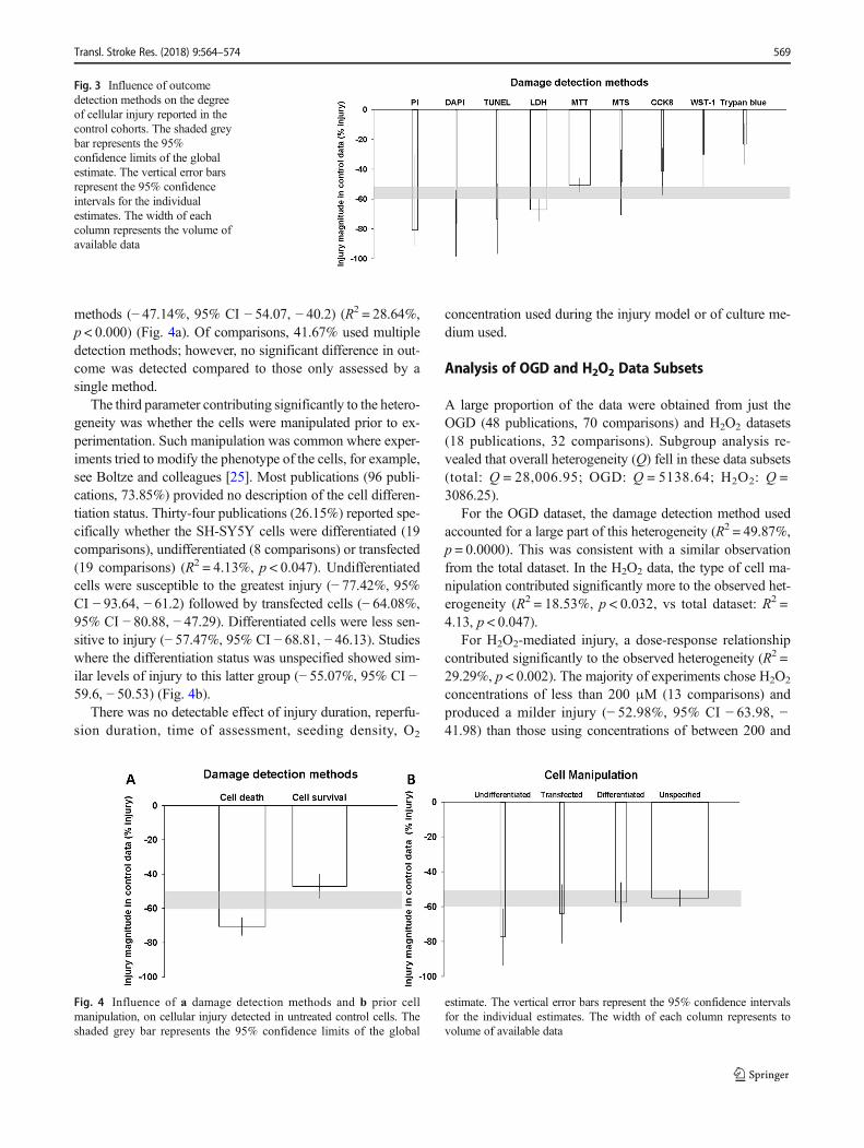
The damage detection methodology accounted for most ofthe observed heterogeneity (R2 = 44.77%, p < 0.000). Ninemethods were used to detect cellular injury in the 130 com-parisons. The MTT assay was the most frequently used meth-od (52 comparisons, 40% of the data) and detected an averageinjury of − 50.42% (95% CI − 55.16, − 45.68). By contrast,the second most commonly used method, the LDH assay (41comparisons, 31.54% of the data), reported − 67.19% injury(95% CI − 74.62, − 59.76). The MTT and LDH assay aloneaccounted for 71.54% (93 comparisons) of the assessment
methods. Overall, methodologies depending on cell countingtend to report more damage (PI − 80.68%, 95% CI − 91.24, −30.72; DAPI − 76.39%, 95% CI − 98.37, − 54.41). The onlyexception was the trypan blue exclusion assay (7 compari-sons), which appeared to underestimate the injury (−23.11%, 95% CI − 36.63, − 9.59). Of the other methods, theTUNEL assay reported the highest injury detection (3 com-parisons, − 73.52%, 95% CI − 96.63, − 49.94) while the MTSassay (3 comparisons, − 48.75%, 95% CI − 70.75, − 26.74),the CCK8 assay (5 comparisons, − 41.52%, 95% CI − 57.21,− 25.84) and the WST-1 assay (2 comparisons, − 30.02%,95% CI − 53.66, − 6.38) all reported smaller levels of injuryon average (Fig. 3).
Each of the nine detection methods could be classified aseither measures of cell death (60 comparisons, 46.15% of thedata: LDH, DAPI counting, PI (counting and FACS) andTUNEL assay) or survival (70 comparisons, 53.85% of thedata: MTT, CCK8, MTS, WST-1 and PI+FDA). Methods be-longing to the cell death category tended to report greaterinjury (− 70.57%, 95% CI − 75.8, − 65.33) than cell survival
Fig. 2 Summary of data includedin the meta-analysis withindividual comparisons groupedby five in vitro ischaemic models.Data are ranked according to theirinjury magnitude againstuntreated controls. The shadedgrey bar represents the 95% CI ofthe individual injuries. Thehorizontal error bars represent the95% CI for the individualestimates
568 Transl. Stroke Res. (2018) 9:564–574
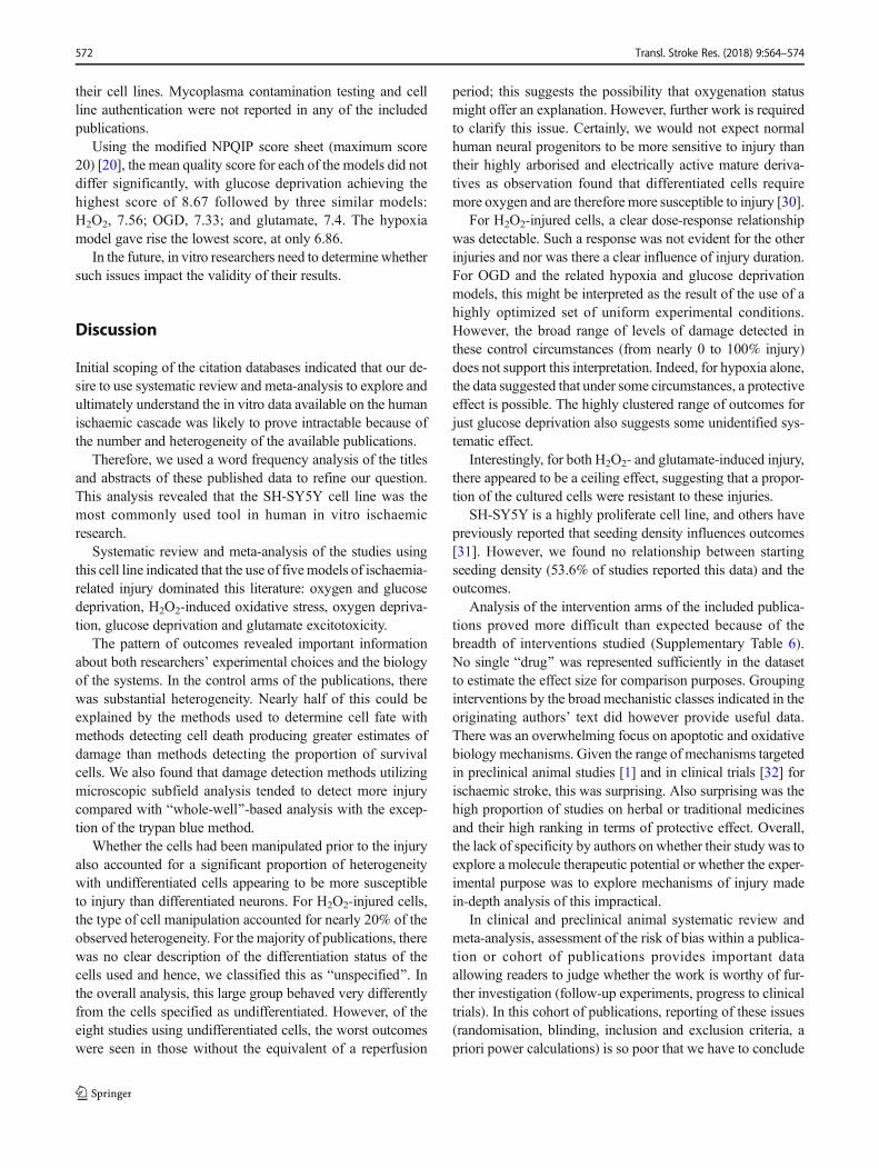
methods (− 47.14%, 95% CI − 54.07, − 40.2) (R2 = 28.64%,p < 0.000) (Fig. 4a). Of comparisons, 41.67% used multipledetection methods; however, no significant difference in out-come was detected compared to those only assessed by asingle method.
The third parameter contributing significantly to the hetero-geneity was whether the cells were manipulated prior to ex-perimentation. Such manipulation was common where exper-iments tried to modify the phenotype of the cells, for example,see Boltze and colleagues [25]. Most publications (96 publi-cations, 73.85%) provided no description of the cell differen-tiation status. Thirty-four publications (26.15%) reported spe-cifically whether the SH-SY5Y cells were differentiated (19comparisons), undifferentiated (8 comparisons) or transfected(19 comparisons) (R2 = 4.13%, p < 0.047). Undifferentiatedcells were susceptible to the greatest injury (− 77.42%, 95%CI − 93.64, − 61.2) followed by transfected cells (− 64.08%,95% CI − 80.88, − 47.29). Differentiated cells were less sen-sitive to injury (− 57.47%, 95% CI − 68.81, − 46.13). Studieswhere the differentiation status was unspecified showed sim-ilar levels of injury to this latter group (− 55.07%, 95% CI −59.6, − 50.53) (Fig. 4b).
There was no detectable effect of injury duration, reperfu-sion duration, time of assessment, seeding density, O2
concentration used during the injury model or of culture me-dium used.
Analysis of OGD and H2O2 Data Subsets
A large proportion of the data were obtained from just theOGD (48 publications, 70 comparisons) and H2O2 datasets(18 publications, 32 comparisons). Subgroup analysis re-vealed that overall heterogeneity (Q) fell in these data subsets(total: Q = 28,006.95; OGD: Q = 5138.64; H2O2: Q =3086.25).
For the OGD dataset, the damage detection method usedaccounted for a large part of this heterogeneity (R2 = 49.87%,p = 0.0000). This was consistent with a similar observationfrom the total dataset. In the H2O2 data, the type of cell ma-nipulation contributed significantly more to the observed het-erogeneity (R2 = 18.53%, p < 0.032, vs total dataset: R2 =4.13, p < 0.047).
For H2O2-mediated injury, a dose-response relationshipcontributed significantly to the observed heterogeneity (R2 =29.29%, p < 0.002). The majority of experiments chose H2O2
concentrations of less than 200 μM (13 comparisons) andproduced a milder injury (− 52.98%, 95% CI − 63.98, −41.98) than those using concentrations of between 200 and
Fig. 3 Influence of outcomedetection methods on the degreeof cellular injury reported in thecontrol cohorts. The shaded greybar represents the 95%confidence limits of the globalestimate. The vertical error barsrepresent the 95% confidenceintervals for the individualestimates. The width of eachcolumn represents the volume ofavailable data
Fig. 4 Influence of a damage detection methods and b prior cellmanipulation, on cellular injury detected in untreated control cells. Theshaded grey bar represents the 95% confidence limits of the global
estimate. The vertical error bars represent the 95% confidence intervalsfor the individual estimates. The width of each column represents tovolume of available data
Transl. Stroke Res. (2018) 9:564–574 569

400 μM (7 comparisons, − 55.07%, 95% CI − 73.57, − 36.58)or 600–800 μM (2 comparisons, − 75.33%, 95% CI − 104.65,− 46.01) and 800–1000 μM (8 comparisons, − 81.18%, 95%CI − 100.69, − 61.66) (Fig. 5). No dose response could bedetected for the other injury models.
Other experimental variables (OGD: cell manipulation, gascomposition, medium ingredients, injury duration or reperfu-sion status; H2O2: damage detection method, injury time orseeding density) did not account for significant proportions ofthe observed heterogeneity.
Meta-Analysis of Intervention After In Vitro IschaemicInjury
In addition to the analysis of experimental variables on theimpact on outcomes in untreated controls, we also sought toinvestigate the effects of the intervention arms of the includedstudies on outcome.
Eighty publications examined 169 intervention compari-sons from the top five in vitro ischaemia models. Four publi-cations (one using glucose deprivation [26], one using hypox-ia [25], and two using OGD [27, 28]) were excluded becauseno intervention was reported or it was reported in non-ischaemic models. The OGD model was used to study thelargest number of interventions (88 comparisons), followedby H2O2 (33 comparisons), hypoxia (15 comparisons), gluta-mate excitotoxicity (10 comparisons) and glucose deprivationalone (7 comparisons). Multiple interventions were frequentlyreported in one publication, and they were treated as individ-ual comparisons for the purposes of this analysis(Supplementary Table 6).
Heterogeneity was greater overall where an interventionwas applied (Q = 42,108, 169 comparisons) than for the datafor injury alone in untreated cohorts (Q = 28,006 over 133comparisons). Overall, the interventions improved cell
survival by 34.67% (95% CI 27.7, 41.62, I2 = 99.48%,p < 0.0001) across the five models. Interestingly, glutamateand H2O2 injury were the only two models where only im-proved outcomes were reported with 64% (95% CI 46.2,81.81) and 47.12% (95% CI 37.35, 56.88) improvement, re-spectively. In contrast, negative outcomes were common inthe models of OGD and its subcomponent models (hypoxiaand glucose deprivation only), and thus led to smaller effectsizes (OGD 35.84%, 95% CI 26.94, 44.73; hypoxia 6.8%,95% CI − 23.38, 36.98). Glucose deprivation was the onlymodel to give rise an overall worsening of outcome after theinterventions (− 20.8%, 95% CI − 54.32, 12.72) (Fig. 6).
Overall, we found that 21/169 comparisons reported nega-tive outcomes of which 10 involved genome manipulations.These studies appeared to be consistent with the authors whointent to study mechanisms.
Of all intervention comparisons, 24.85% utilized herbal ortraditional medicines or their derivates which improved out-come by 65.12% on average. Of the studies reporting thegreatest improvement (> 90% effect size), more than half(7/13 comparisons) were studies of the bioactive constituentsof herbal medicines (Supplementary Table 2).
Mechanism of Action of Interventions Studied
The most commonly studied molecules were only representedin two or three independent studies. Therefore, there was in-sufficient data for meta-analytical exploration of their compar-ative effects.
When we grouped intervention targets into single or mixedischaemia-related mechanisms according to the author’s orig-inal statements, we found that 91/169 (53.85%) experimentsstudied interventions targeting a single mechanism, of whichhalf (46/91 comparisons, 50.55%) targeted apoptosis. Thiswas followed by antioxidant-related mechanisms (32/91 com-parisons, 35.16%), which was also the only targeting mecha-nism studied in all the aforementioned models. The remainder(14%) studied excitotoxicity: 3 comparisons, mitochondrialprotection; 3 comparisons, growth factor secretion; 4 compar-isons; and axonal growth, 3 comparisons (SupplementaryTable 3).
Interventions targeting multiple mechanisms were investi-gated in 78 comparisons. Seventy-six comparisons of thesestudied two mechanisms in 11 different combinations. Onestudy referred to three potential mechanisms for their interven-tional drugs but no experiments to identify the mechanismwere performed [9]. Drugs modifying apoptosis and oxidativebiology were the most commonly studied pairs (32/76 com-parisons, 42.11%). This was particularly true for the H2O2
injury studies where 17/17 studies all used such a combination(Supplementary Table 4).
Researchers using the OGD injury model studied thegreatest range of intervention mechanisms in combination
Fig. 5 Influence of H2O2 concentration on injury magnitude in untreatedcontrol cohorts. Circle diameter is proportional to study size
570 Transl. Stroke Res. (2018) 9:564–574

(9/11 mechanisms). Apoptosis was the most commonly stud-ied mechanism both singly (40/52 comparisons, 77%) or incombination with antioxidant interventions (both have 13comparisons, 32.5%). This pattern was reversed for H2O2 in-jury (14/22 comparisons, 63.64%, studied antioxidant mech-anisms singly or 17/17 comparisons, 100%, studied antioxi-dant and anti-apoptotic mechanisms).
Study Quality
In systematic review and meta-analysis, which is most oftenused to determine a therapeutic effect in clinical studies, a keydeterminant of the value of the analysis is the quality of theincluded studies. This is most often ascertained by formal riskof bias analysis [29] or by application of a simple checklist inpre-clinical studies, where understanding of the need to reportsuch issues lags behind the clinical field. However, in in vitroresearch, these issues have not gained widespread recognitionas potential sources of experimental error. Therefore, in ourstudy, we had only a limited ability to assess study quality.
We reported and scored the items using our modified studyquality checklist for each included publication from the fivemost commonly used ischaemia- re la ted models(Supplementary Table 5). Study design features which helpreduce bias, such as randomisation, blinding, sample size cal-culation and description of inclusion and exclusion criteria,were extremely poorly reported. Only one publication inH2O2 injury and one publication on OGD injury out of a totalof 84 publications reported their exclusion criteria (2.38%),and the same study was also the only study to report a samplesize calculation (1.19%). None of the publications reportedeither randomisation or blinding in their full text. While BN^was reported in more than 95% of the publications, there wasgenerally a poor description of what BN^ stood for (wells/plates/microscope fields). Only 19/84 publications (22.62%)gave a clear statement of whether the samples representedtechnical or biologically relevant replicates, and 29/84(34.52%) described how many times the experiment was rep-licated. Cell preparation was also poorly reported, with onlyhalf of the publications (54.76%) describing the sources of
Fig. 6 Summary of interventionaldata included in the meta-analysisfrom five in vitro ischaemicmodels. Data are rankedaccording to their effect onchange after cell damage fromeach injury. The shaded grey barrepresents the 95% CI of theindividual injuries. The verticalerror bars represent the 95% CIfor the individual estimates
Transl. Stroke Res. (2018) 9:564–574 571
their cell lines. Mycoplasma contamination testing and cellline authentication were not reported in any of the includedpublications.
Using the modified NPQIP score sheet (maximum score20) [20], the mean quality score for each of the models did notdiffer significantly, with glucose deprivation achieving thehighest score of 8.67 followed by three similar models:H2O2, 7.56; OGD, 7.33; and glutamate, 7.4. The hypoxiamodel gave rise the lowest score, at only 6.86.
In the future, in vitro researchers need to determine whethersuch issues impact the validity of their results.
Discussion
Initial scoping of the citation databases indicated that our de-sire to use systematic review and meta-analysis to explore andultimately understand the in vitro data available on the humanischaemic cascade was likely to prove intractable because ofthe number and heterogeneity of the available publications.
Therefore, we used a word frequency analysis of the titlesand abstracts of these published data to refine our question.This analysis revealed that the SH-SY5Y cell line was themost commonly used tool in human in vitro ischaemicresearch.
Systematic review and meta-analysis of the studies usingthis cell line indicated that the use of fivemodels of ischaemia-related injury dominated this literature: oxygen and glucosedeprivation, H2O2-induced oxidative stress, oxygen depriva-tion, glucose deprivation and glutamate excitotoxicity.
The pattern of outcomes revealed important informationabout both researchers’ experimental choices and the biologyof the systems. In the control arms of the publications, therewas substantial heterogeneity. Nearly half of this could beexplained by the methods used to determine cell fate withmethods detecting cell death producing greater estimates ofdamage than methods detecting the proportion of survivalcells. We also found that damage detection methods utilizingmicroscopic subfield analysis tended to detect more injurycompared with Bwhole-well^-based analysis with the excep-tion of the trypan blue method.
Whether the cells had been manipulated prior to the injuryalso accounted for a significant proportion of heterogeneitywith undifferentiated cells appearing to be more susceptibleto injury than differentiated neurons. For H2O2-injured cells,the type of cell manipulation accounted for nearly 20% of theobserved heterogeneity. For the majority of publications, therewas no clear description of the differentiation status of thecells used and hence, we classified this as Bunspecified^. Inthe overall analysis, this large group behaved very differentlyfrom the cells specified as undifferentiated. However, of theeight studies using undifferentiated cells, the worst outcomeswere seen in those without the equivalent of a reperfusion
period; this suggests the possibility that oxygenation statusmight offer an explanation. However, further work is requiredto clarify this issue. Certainly, we would not expect normalhuman neural progenitors to be more sensitive to injury thantheir highly arborised and electrically active mature deriva-tives as observation found that differentiated cells requiremore oxygen and are therefore more susceptible to injury [30].
For H2O2-injured cells, a clear dose-response relationshipwas detectable. Such a response was not evident for the otherinjuries and nor was there a clear influence of injury duration.For OGD and the related hypoxia and glucose deprivationmodels, this might be interpreted as the result of the use of ahighly optimized set of uniform experimental conditions.However, the broad range of levels of damage detected inthese control circumstances (from nearly 0 to 100% injury)does not support this interpretation. Indeed, for hypoxia alone,the data suggested that under some circumstances, a protectiveeffect is possible. The highly clustered range of outcomes forjust glucose deprivation also suggests some unidentified sys-tematic effect.
Interestingly, for both H2O2- and glutamate-induced injury,there appeared to be a ceiling effect, suggesting that a propor-tion of the cultured cells were resistant to these injuries.
SH-SY5Y is a highly proliferate cell line, and others havepreviously reported that seeding density influences outcomes[31]. However, we found no relationship between startingseeding density (53.6% of studies reported this data) and theoutcomes.
Analysis of the intervention arms of the included publica-tions proved more difficult than expected because of thebreadth of interventions studied (Supplementary Table 6).No single Bdrug^ was represented sufficiently in the datasetto estimate the effect size for comparison purposes. Groupinginterventions by the broad mechanistic classes indicated in theoriginating authors’ text did however provide useful data.There was an overwhelming focus on apoptotic and oxidativebiology mechanisms. Given the range of mechanisms targetedin preclinical animal studies [1] and in clinical trials [32] forischaemic stroke, this was surprising. Also surprising was thehigh proportion of studies on herbal or traditional medicinesand their high ranking in terms of protective effect. Overall,the lack of specificity by authors on whether their study was toexplore a molecule therapeutic potential or whether the exper-imental purpose was to explore mechanisms of injury madein-depth analysis of this impractical.
In clinical and preclinical animal systematic review andmeta-analysis, assessment of the risk of bias within a publica-tion or cohort of publications provides important dataallowing readers to judge whether the work is worthy of fur-ther investigation (follow-up experiments, progress to clinicaltrials). In this cohort of publications, reporting of these issues(randomisation, blinding, inclusion and exclusion criteria, apriori power calculations) is so poor that we have to conclude
572 Transl. Stroke Res. (2018) 9:564–574
that the risk of bias is very high. A recent systematic review ofthe use of SH-SY5Y to model Parkinson’s disease did notperform meta-analysis and made no comments of the likelyquality of the studies included in the review [33].
Funding This study was supported by funding from the AustralianNational Health and Medical Research Council (NHMRC), grant num-bers 1013621 and 1037863.
Compliance with Ethical Standards
Conflict of Interest The authors declare that they have no conflict ofinterest.
Ethics Approval The article does not contain any studies with humanparticipants or animals performed by any of the authors.
Open Access This article is distributed under the terms of the CreativeCommons At t r ibut ion 4 .0 In te rna t ional License (h t tp : / /creativecommons.org/licenses/by/4.0/), which permits unrestricted use,distribution, and reproduction in any medium, provided you giveappropriate credit to the original author(s) and the source, provide a linkto the Creative Commons license, and indicate if changes were made.
References
1. O’CollinsVE,MacleodMR,DonnanGA, Horky LL, van derWorpBH, Howells DW. 1,026 experimental treatments in acute stroke.Ann Neurol. 2006;59(3):467–77. https://doi.org/10.1002/ana.20741.
2. Donnan GA, Howells DW. Neuroprotection: where to now? FutureNeurol. 2007;2(5):513–21. https://doi.org/10.2217/14796708.2.5.513.
3. Babusikova E, Beckett C, Dobrota D, Turner AJ, Nalivaeva NN.Ischaemia and oxidative stress modulate expression of amyloiddegrading enzymes. J Neurochem. 2013;125:211.
4. Howard BE, Phillips J, Miller K, Tandon A, Mav D, Shah MR,et al. SWIFT-Review: a text-mining workbench for systematic re-view. Syst Rev. 2016;5:87. https://doi.org/10.1186/s13643-016-0263-z.
5. Katsura K, Kristian T, Siesjo BK. Energy metabolism, ion homeo-stasis, and cell damage in the brain. Biochem Soc Trans.1994;22(4):991–6. https://doi.org/10.1042/bst0220991.
6. Dirnagl U, Iadecola C, Moskowitz MA. Pathobiology of ischaemicstroke: an integrated view. Trends Neurosci. 1999;22(9):391–7.https://doi.org/10.1016/S0166-2236(99)01401-0.
7. Brouns R, De Deyn PP. The complexity of neurobiological process-es in acute ischemic stroke. Clin Neurol Neurosurg. 2009;111(6):483–95. https://doi.org/10.1016/j.clineuro.2009.04.001.
8. Bando Y, Katayama T, Kasai K, Taniguchi M, Tamatani M,Tohyama M. GRP94 (94 kDa glucose-regulated protein) sup-presses ischemic neuronal cell death against ischemia/reperfusioninjury. Eur J Neurosci. 2003;18(4):829–40. https://doi.org/10.1046/j.1460-9568.2003.02818.x.
9. Vieira-Marques C, Arbo BD, Ruiz-Palmero I, Ortiz-Rodriguez A,Ghorbanpoor S, Kucharski LC, et al. Dehydroepiandrosterone pro-tects male and female hippocampal neurons and neuroblastomacells from glucose deprivation. Brain Res. 2016;1644:176–82.https://doi.org/10.1016/j.brainres.2016.05.014.
10. Jiang W, Fu F, Tian J, Zhu H, Hou J. Curculigoside A attenuatesexperimental cerebral ischemia injury in vitro and vivo.Neuroscience. 2011;192:572–9. https://doi.org/10.1016/j.neuroscience.2011.06.079.
11. Tian X, An L, Gao LY, Bai JP, Wang J, MengWH, et al. CompoundMQA, a caffeoylquinic acid derivative, protects against NMDA-induced neurotoxicity and potential mechanisms in vitro. CNSNeurosci Ther. 2015;21(7):575–84. https://doi.org/10.1111/cns.12408.
12. Seetapun S, Yaoling J, Wang Y, Zhu YZ. Neuroprotective effect ofDanshensu derivatives as anti-ischaemia agents on SH-SY5Y cellsand rat brain. Biosci Rep. 2013;33(4):677–88. https://doi.org/10.1042/bsr20130032.
13. Lim W, Kim JH, Gook E, Kim J, Ko Y, Kim I, et al. Inhibition ofmitochondria-dependent apoptosis by 635-nm irradiation in sodiumnitroprusside-treated SH-SY5Y cells. Free Radic Biol Med.2009;47(6):850–7. https://doi.org/10.1016/j.freeradbiomed.2009.06.023.
14. Luan HY, Kan ZC, Xu Y, Lv CJ, Jiang WL. Rosmarinic acid pro-tects against experimental diabetes with cerebral ischemia: relationto inflammation response. J Neuroinflammation 2013;10. https://doi.org/10.1186/1742-2094-10-157.
15. Zou YX, Liu YX, Ruan MH, Zhou Y, Wang JC, Chu ZY.Cordyceps sinensis oral liquid inhibits damage induced by oxygenand glucose deprivation in SH-SY5Y cells. Altern Ther HealthMed. 2016;22(2):37–42. https://doi.org/10.1007/s11064-016-1908-y.
16. Li Y, Li J, Li SS, Li Y, Wang XX, Liu BL, et al. Curcumin attenu-ates glutamate neurotoxicity in the hippocampus by suppression ofER stress-associated TXNIP/NLRP3 inflammasome activation in amanner dependent on AMPK. Toxicol Appl Pharmacol.2015;286(1):53–63. https://doi.org/10.1016/j.taap.2015.03.010.
17. Vesterinen HM, Sena ES, Egan KJ, Hirst TC, Churolov L, CurrieGL, et al. Meta-analysis of data from animal studies: a practicalguide. J Neurosci Methods. 2014;221:92–102. https://doi.org/10.1016/j.jneumeth.2013.09.010.
18. Antonic A, Sena ES, Lees JS, Wills TE, Skeers P, Batchelor PE,et al. Stem cell transplantation in traumatic spinal cord injury: asystematic review and meta-analysis of animal studies. PLoSBiol. 2013;11(12):e1001738. https://doi.org/10.1371/journal.pbio.1001738.
19. Krithikadatta J, Gopikrishna V, Datta M. CRIS Guidelines(Checklist for Reporting In-vitro Studies): a concept note on theneed for standardized guidelines for improving quality and trans-parency in reporting in-vitro studies in experimental dental re-search. J Conserv Dent. 2014;17(4):301–4. https://doi.org/10.4103/0972-0707.136338.
20. Cramond F, Irvine C, Liao J, Howells D, Sena E, Currie G, et al.Protocol for a retrospective, controlled cohort study of the impact ofa change in nature journals’ editorial policy for life sciences re-search on the completeness of reporting study design and execution.Scientometrics. 2016;108:315–28. https://doi.org/10.1007/s11192-016-1964-8.
21. Noh SJ, Lee SH, ShinKY, Lee CK, Cho IH, KimHS, et al. SP-8203reduces oxidative stress via SOD activity and behavioral deficit incerebral ischemia. Pharmacol Biochem Behav. 2011;98(1):150–4.https://doi.org/10.1016/j.pbb.2010.12.014.
22. TsurumaK, Nakagawa T,Morimoto N,MinamiM, Hara H, UeharaT, et al. Glucocorticoid modulatory element-binding protein 1 bindsto initiator procaspases and inhibits ischemia-induced apoptosis andneuronal injury. J Biol Chem. 2006;281(16):11397–404. https://doi.org/10.1074/jbc.M510597200.
23. Shen Y, Li R, Shiosaki K. Inhibition of p75 tumor necrosis factorreceptor by antisense oligonucleotides increases hypoxic injury andbeta-amyloid toxicity in human neuronal cell line. J Biol Chem.1997;272(6):3550–3. https://doi.org/10.1074/jbc.272.6.3550.
Transl. Stroke Res. (2018) 9:564–574 573
24. Romero JI, Hanschmann EM, Gellert M, Eitner S, Holubiec MI,Blanco-Calvo E, et al. Thioredoxin 1 and glutaredoxin 2 contributeto maintain the phenotype and integrity of neurons following peri-natal asphyxia. Biochim Biophys Acta. 2015;1850(6):1274–85.https://doi.org/10.1016/j.bbagen.2015.02.015.
25. Hau S, Reich DM, Scholz M, Naumann W, Emmrich F, KampradM, et al. Evidence for neuroprotective properties of human umbil-ical cord blood cells after neuronal hypoxia in vitro. BMCNeurosci. 2008;9:30. https://doi.org/10.1186/1471-2202-9-30.
26. Klacanova K, Pilchova I, Klikova K, Racay P. Short chemical is-chemia triggers phosphorylation of eIF2alpha and death of SH-SY5Y cells but not proteasome stress and heat shock protein re-sponse in both SH-SY5Y and T98G cells. J Mol Neurosci: MN.2016;58(4):497–506. https://doi.org/10.1007/s12031-015-0685-4.
27. Li G, Zou LY, Cao CM, Yang ES. Coenzyme Q10 protectsSHSY5Y neuronal cells from beta amyloid toxicity and oxygen-glucose deprivation by inhibiting the opening of the mitochondrialpermeability transition pore. Biofactors. 2005;25(1–4):97–107.https://doi.org/10.1002/biof.5520250111.
28. Liu SY, Zhang Y, Hu LS, Luo YN, Guan WM. Establishment andevaluation of OGD/R model in vitro inducing apoptosis of SH-
SY5Y cells. [Chinese]. J Jilin Univ Med Ed. 2012;38(4):658–64. http://lib.cqvip.com/qk/92319A/201204/42780844.html.
29. Sterne JA, Egger M, Smith GD. Systematic reviews in health care:investigating and dealing with publication and other biases in meta-analysis. BMJ. 2001;323(7304):101–5. https://www.ncbi.nlm.nih.gov/pmc/articles/PMC1120714/.
30. Candelario KM, Shuttleworth CW, Cunningham LA. Neural stem/progenitor cells display a low requirement for oxidativemetabolismindependent of hypoxia inducible factor-1alpha expression. JNeurochem. 2013;125(3):420–9. https://doi.org/10.1111/jnc.12204.
31. Mo WC, Zhang ZJ, Liu Y, Bartlett PF, He RQ. Magnetic shieldingaccelerates the proliferation of human neuroblastoma cell by pro-moting G1-phase progression. PLoS One. 2013;8(1):e54775.https://doi.org/10.1371/journal.pone.0054775.
32. Ginsberg MD. Neuroprotection for ischemic stroke: past, presentand future. Neuropharmacology. 2008;55(3):363–89. https://doi.org/10.1016/j.neuropharm.2007.12.007
33. Xicoy H, Wieringa B, Martens GJ. The SH-SY5Y cell line inParkinson’s disease research: a systematic review. MolNeurodegener. 2017;12(1):10. https://doi.org/10.1186/s13024-017-0149-0.
574 Transl. Stroke Res. (2018) 9:564–574