human molecular genetics - tauyossih/publications/ziv_s_2005.pdf · human molecular genetics...
TRANSCRIPT

Human Molecular Genetics
doi:10.1093/hmg/ddi324 14:2929-2943, 2005. First published 8 Sep 2005; Hum. Mol. Genet.
Rechavi, Yosef Shiloh and Ari Barzilai G. Sharma, Raj K. Pandita, Manjula Agarwal, Ran Elkon, Nirit Katzin, Irit Bar-Am, Tej K. Pandita, Raju Kucherlapati, Gideon Shelly Ziv, Ori Brenner, Ninette Amariglio, Nechama I. Smorodinsky, Ronit Galron, Danaise V. Carrion, Weijia Zhang, GirdharImpaired genomic stability and increased oxidative stress exacerbate different features of Ataxia-telangiectasia
Supplement/Special Issue http://hmg.oxfordjournals.org/cgi/content/full/ddi324/DC1
"Supplementary Material"This article is part of the following issue:
http://hmg.oxfordjournals.org/cgi/content/full/14/19/2929The full text of this article, along with updated information and services is available online at
References http://hmg.oxfordjournals.org/cgi/content/full/14/19/2929#BIBL
This article cites 43 references, 15 of which can be accessed free at
Cited by http://hmg.oxfordjournals.org/cgi/content/full/14/19/2929#otherarticles
This article has been cited by 3 articles at 23 June 2008 . View these citations at
Supplementary material http://hmg.oxfordjournals.org/cgi/content/full/ddi324/DC1
Data supplements for this article are available at
Reprints http://www.oxfordjournals.org/corporate_services/reprints.html
Reprints of this article can be ordered at
Email and RSS alerting Sign up for email alerts, and subscribe to this journal’s RSS feeds at http://hmg.oxfordjournals.org
image downloadsPowerPoint® Images from this journal can be downloaded with one click as a PowerPoint slide.
Journal informationat http://hmg.oxfordjournals.org Additional information about Human Molecular Genetics, including how to subscribe can be found
Published on behalf ofhttp://www.oxfordjournals.org Oxford University Press
at TEL AVIV UNIVERSITY on 23 June 2008 http://hmg.oxfordjournals.orgDownloaded from

Impaired genomic stability and increasedoxidative stress exacerbate different featuresof Ataxia-telangiectasia
Shelly Ziv1, Ori Brenner2, Ninette Amariglio3, Nechama I. Smorodinsky4, Ronit Galron1,
Danaise V. Carrion5, Weijia Zhang5, Girdhar G. Sharma6, Raj K. Pandita6, Manjula Agarwal6,
Ran Elkon7, Nirit Katzin8, Irit Bar-Am8, Tej K. Pandita6, Raju Kucherlapati5, Gideon Rechavi3,
Yosef Shiloh7,* and Ari Barzilai1,*
1Department of Neurobiochemistry, George S. Wise Faculty of Life Sciences, Tel Aviv University, Tel Aviv 69978,
Israel, 2Department of Veterinary Resources, the Weizmann Institute of Science, Rehovot 76100, Israel,3Sheba Cancer Research Center, Edmond and Lily Safra Children’s Hospital, Sheba Medical Center, Tel Hashomer
52126, and Sackler School of Medicine, Tel Aviv University, Israel, 4Alec and Myra Marmot Hybridoma Laboratory,
George S. Wise Faculty of Life Sciences, Tel Aviv University, Tel Aviv 69978, Israel, 5Departments of Medicine and
Genetics and Harvard Partners Center for Genetics and Genomics, Harvard Medical School, Boston, MA 02115, USA,6Department of Oncology and Radiation, Washington University, School of Medicine, St Louis, MO 63108, USA,7Department of Human Genetics and Molecular Medicine, Sackler School of Medicine, Tel Aviv University,
Tel Aviv 69978, Israel and 8Applied Spectral Imaging, Migdal Haemek 10551, Israel
Received June 22, 2005; Revised August 11, 2005; Accepted August 19, 2005
Ataxia–telangiectasia (A-T) is a multisystem, cancer-predisposing genetic disorder caused by deficiency ofthe ATM protein. To dissect the A-T phenotype, we augmented specific features of the human disease bygenerating mouse strains that combine Atm deficiency with dysfunction of other proteins. Increasingoxidative stress by combining deficiencies in Atm and superoxide dismutase 1 (Sod1) exacerbated growthretardation and markedly reduced the mean survival time following ionizing radiation. In contrast, increasinggenomic instability by combining deficiencies of Atm and the mismatch repair protein Mlh1 caused amoderate increase in radiation sensitivity and dramatic increase in aggressive lymphomas, compared withthes Atm2/2 single knockout. Remarkably, Atm, Mlh1 or Mlh1/Atm single or double heterozygosity didnot significantly affect the life span of the various genotypes. Mlh1/Atm double null tumors were polyclonal,whereas the tumors in other genotypes were mono- or oligoclonal, demonstrating the high predisposition ofthymocytes with this genotype to become malignant. Chromosomal aberrations in the tumors were localizedmainly in chromosomes 12 and 15. The genomic region on chromosome 15, which contains the gene for thec-Myc oncoprotein, was commonly amplified, and elevated levels of the c-Myc protein were subsequentlyobserved in the tumors. Our data suggest that impaired genomic instability is an important contributingfactor to cancer predisposition in A-T, whereas oxidative stress is more important in the radiation sensitivityand growth retardation facets of this disease.
INTRODUCTION
Ataxia–telangiectasia (A-T) is an autosomal recessive disordercharacterized by progressive cerebellar ataxia, oculo-facial
telangiectases (dilated blood vessels), immunodeficiency, pre-mature aging, gonadal dysgenesis, extreme sensitivity to ioniz-ing radiation (IR), genomic instability and high incidence oflymphoreticular malignancies (1). A-T is a prime example of
# The Author 2005. Published by Oxford University Press. All rights reserved.For Permissions, please email: [email protected]
*To whom correspondence should be addressed. Tel: þ972 36409782; Fax: þ972 36407643; Email: [email protected]
Human Molecular Genetics, 2005, Vol. 14, No. 19 2929–2943doi:10.1093/hmg/ddi324Advance Access published on September 8, 2005

a genomic instability syndrome caused by defective DNAdamage response. It is caused by loss or inactivation of theATM protein, a large nuclear serine/threonine kinase with aC-terminal domain containing PI3 kinase motifs. ATMbelongs to a conserved family of large proteins that share thePI3 kinase-like domain and are involved in various stressresponses, most notably the DNA damage response (2). ATMmediates the network of signaling pathways that are activatedby DNA double-strand breaks (DSBs) (3–5). The ATM-mediated response includes the cell cycle checkpoints andnumerous other pathways related to various aspects of cellularmetabolism.
Atm2/2 mice recapitulate many features of the human A-Tphenotype, including growth retardation, infertility, strikingpredisposition to thymic lymphomas and acute sensitivity toIR; mild neurological deficits are noted only in certainstrains of Atm2/2 animals (6–9). These mice thereforeprovide a model for exploring most of the features of theA-T phenotype.
We set out to augment oxidative stress and genomicinstability in Atm2/2 mice in order to link these two featuresof the cellular phenotype to specific features of the clinicalphenotype of A-T. We did this by generating mouse strainsthat combine Atm deficiency with dysfunction of other pro-teins: a strain deficient in both Atm and Sod1 and a doubleknockout strain lacking both Atm and the Mlh1 protein.Mice with targeted inactivation of the Sod1 gene, whichencodes copper/zinc superoxide dismutase (Sod1), one ofthe major intracellular enzymes that inactivate superoxideanions (10), live at least 18 months (11,12) and displaynormal motor behavior in the first 3 or 4 months and subtlemotor impairment by 6 months. Targeted inactivation of theMlh1 gene in mice leads to infertility and tumor susceptibility(13–15). Mlh12/2 mice exhibit reduced survival similar toMsh22/2 animals. Fifty percent of Mlh12/2 animals diebefore 6 months of age and all animals die by 13 monthsfrom T-cell lymphomas and gastrointestinal tumors.
We report here that increasing oxidative stress in Atm2/2mice moderately reduced their life span, markedly reducedtheir body weight and rendered them more sensitive to IR.Inactivation of the MMR system in Atm2/2 mice moderatelyincreased their sensitivity to IR but markedly reduced their lifespan due to a striking predisposition to develop aggressivethymic lymphomas at an early age. Collectively, our datasuggest that oxidative stress contributes to specific clinicalfeatures of A-T such as reduced body weight and radiosensi-tivity and may play a minor role in cancer predisposition,whereas enhanced genomic instability is a major contributingfactor to cancer predisposition in this disease.
RESULTS
Non-Mendelian ratios between various genotypes
The recoveries of the various genotypes from the two matingschemes significantly deviated from Mendelian distribution(P , 0.0002 for the Sod1/Atm, P , 0.007 for the Mlh1/Atmmice) (Table 1). There was a marked reduction in thenumber of the Mlh1/Atm double null mice, somewhat lower
than the expected number for Atm/Sod double null mice,and higher than expected for Atm-proficient mice (Table 1).
Markedly reduced life span in Mlh1/Atm double null micedue to increased tumorigenesis
Deletion of the Sod1 gene in Atm2/2 mice reduced the sur-vival rate compared with Atm2/2 genotype, especially at olderage (.1 year), although the reduction was not statistically sig-nificant (P ¼ 0.1): 50% of Sod12/2//Atm2/2 animals diedat 50 weeks, whereas 50% of the Atm2/2 mice died at55 weeks (Fig. 1Aa). The life span of Sod12/2//Atmþ/2 wassignificantly shorter than that of Sod1 null mice (P , 0.02)(Fig. 1Ab). Although Atmþ/2 animals have normal lifespan, Atm heterozygosity can have serious effects on theanimal under certain circumstances such as oxidative stress.Atm, Sod1 single or Sod/Atm double heterozygosity did notsignificantly affect mouse survival rate (Fig. 1Ac).
Mice lacking both Mlh1 and Atm displayed a significantlyreduced life span (P , 0.00001 between Mlh1/Atm doublenull versus Atm2/2 and Mlh12/2 genotypes): 50% died at11 weeks, 80% at 13 weeks and none survived beyond27 weeks, all succumbing to aggressive thymic lymphomas(Fig. 1Ad). The life span of Mlh1þ/2//Atm2/2 was signifi-cantly shorter than that of Atm null mice (P , 0.04), whereasAtm heterozygosity on Mlh12/2 background had no effecton mouse survival. Atm, Mlh1 single or Mlh1/Atm doubleheterozygosity did not significantly affect mouse survivalrate (Fig. 1Af). Tumor development was the leading causeof mortality in the Mlh1/Atm genotypes and in the Sod1/Atm double null mice. Thus, the cumulative survival curvesactually reflect the rate of tumor formation in the various geno-types. Post-mortem examination of the Atm2/2, Mlh12/2,Mlh12/2//Atm2/2 and Sod12/2//Atm2/2 mice revealed
Table 1. Genotype distribution of offspring in various matings
Expected Observed
Sod1/Atmþ/2//þ/þ 120 154 (128.0%)þ/2//þ/2 240 249 (103.8%)þ/2//2/2 120 111 (92.5%)2/2//þ/2 240 196 (81.7%)2/2//þ/þ 120 143 (119.2%)2/2//2/2 120 107 (89.2%)
Mlh1/Atmþ/þ//þ/þ 27.25 33 (121.1%)þ/þ//þ/2 54.50 61 (111.9%)þ/þ//2/2 27.25 23 (84.4%)þ/2//þ/þ 54.50 66 (121.1%)þ/2//þ/2 109.00 103 (94.5%)þ/2//2/2 54.50 45 (82.6%)2/2//þ/þ 27.25 42 (154.1%)2/2//þ/2 54.50 50 (91.7%)2/2//2/2 27.25 13 (47.7%)
Sod1/Atm mice were generated by crossing Sod12/2//Atmþ/2 maleswith Sod1þ/2//Atmþ/2 females. Mlh1/Atm mice were generated bycrossing Mlh1þ/2//Atmþ/2 males and females. Recovery is thenumber of mice at 21 days of age (at weaning) relative to the numberexpected from the Mendelian ratios. Most of the mice survived untilweaning and beyond.
2930 Human Molecular Genetics, 2005, Vol. 14, No. 19

aggressive thymic lymphoma with widespread visceral meta-stasis (in �90% of the animals examined) (Table 2). Histo-logically, neoplastic lymphocytes were large and of uniformappearance, consistent with large cell lymphoma. Immunohisto-chemical staining showed the bulk of the neoplastic cells to beCD3-positive and CD45R-negative, indicating a T-cell origin(Fig. 2).
Given that cancer was the major cause of death in all mutantgenotypes, the cumulative survival analysis indicates thatincreasing genomic instability on the background of Atmdeficiency had a much more marked effect than increasing oxi-dative stress on cancer predisposition. In contrast to humanpatients, Mlh1 heterozygosity in mice did not significantlyincrease cancer predisposition, nor did Atm heterozygosity.
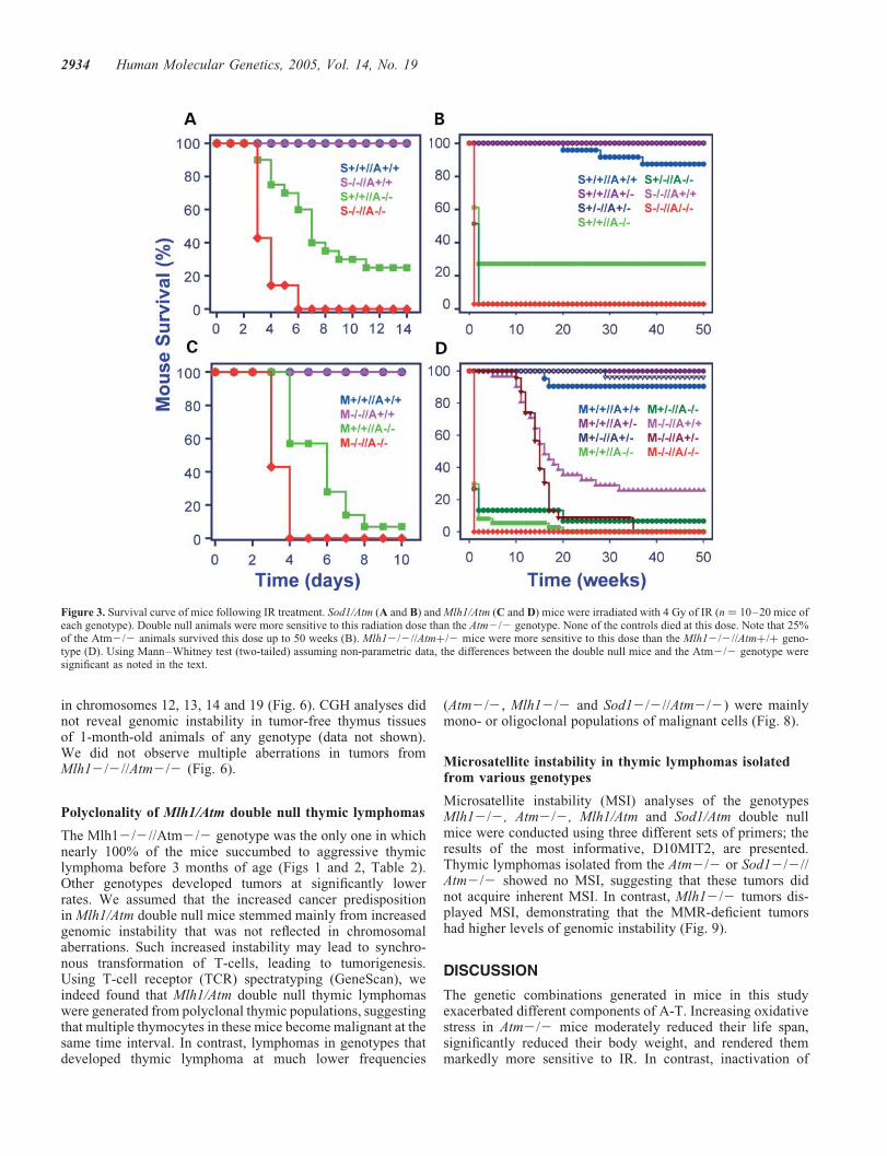
Figure 1. Life span and growth curve of mice with different genotypes. (A) The cumulative survival of the Sod1/Atm (a–c) and Mlh1/Atm (d–f) genotypesplotted according to Kaplan–Meier analysis, statistical significance calculated using log-rank. S stands for Sod1, M stands for Mlh1 and A stands for Atm.(b) Cumulative survival of Sod12/2//Atmþ/þ genotype (pink) and Sod12/2//Atmþ/2 (dark brown). The life span of the former was significantly shorterthan that of the latter (P , 0.02). (e) The life span of Mlh1þ/2//Atm2/2 was significantly shorter (P , 0.04) than that of Atm2/2 animals. (c and f) Thelife span of single and double heterozygote mice. (B) Body weight of mice measured from 4–52 weeks of age. Error bars represent +SD. Statistical analyseswere performed with two-tailed Student’s t-test (n ¼ 10–20 mice). Animals that lost over 4 g in two successive weeks were omitted. Genotypes colored asin (A).
Human Molecular Genetics, 2005, Vol. 14, No. 19 2931

Atm2/2 mice display a reduction in mature CD4þ/CD82or CD8þ/CD42 T-lymphocytes and an increase in immatureCD4þ/CD8þ cells relative to normal control mice (6).Atm2/2,Sod12/2//Atm2/2 and Mlh12/2//Atm2/2 mice displayeda significant decrease in mature CD4þ/CD82 (P , 0.02),whereas the level of mature CD8þ/CD42 in the thymus fluc-tuated in the various genotypes. (Supplementary Material,Table S1). A further and significant reduction in matureCD4þ/CD82 cells was detected in T-cell lymphomas isolatedfrom the various tumors (P , 0.0001). In parallel, increasedratios of double positive CD4þ/CD8þ in Atm2/2 and indouble null tumors were observed, reflecting a transition ofthe tumor cells into an earlier stage of differentiation (Sup-plementary Material, Table S1).
Growth retardation enhanced by increased oxidative stressbut not genomic instability in Atm2/2 mice
Atm2/2 mice are 10–20% smaller than their wild-type (WT)littermates (6). The inactivation of the Sod1 gene also affectedthe growth rate of the animals. The Sod12/2 mice were 9%smaller than their WT littermates at 4 weeks (P , 0.001), 12%smaller at 6 months (P , 0.001) and 22% smaller by 1 year(P , 0.001), suggesting that increased oxidative stressresulted in growth retardation. The combination of Sod1 andAtm deficiencies further reduced growth compared with allother genotypes. The gap in growth rate between Sod1/Atmdouble null mice and Atm2/2 genotype widened with age,from 5% at 4 weeks (P , 0.05) to 11% at 6 months(P , 0.001) to 18% at 1 year (P , 0.001) (Fig. 1Ba). Deletionof the Mlh1 gene did not affect the growth of the animals whencompared with their WT littermates. Deletion of the Atm generesulted in significant growth retardation: 10% (P , 0.05) at4 weeks and 12% (P , 0.001) at 6 months. Mlh1/Atmdouble null mice were 10% smaller than the Atm2/2 geno-type at 4 weeks, an insignificant difference (P . 0.05).Growth retardation in the various Atm2/2 genotypes wasnot caused by reduced caloric consumption (SupplementaryMaterial, Fig. S2).
Taken together, our data suggest that oxidative stress maybe an important contributing factor to the decrease in bodyweight observed in A-T patients.
Effect of oxidative stress and genomic instability on theradiosensitivity of Atm2/2 mice
As hypersensitivity to IR is one of the hallmarks of A-T (1),Atm2/2 mice and Atm2/2 cells (6), and as IR exerts someof its toxic effects through the generation of oxygen radicals,we examined the effect of Sod1 deficiency on the radiosensi-tivity of Atm2/2 mice. Their survival was monitored afterexposure to 4 Gy. Interestingly, the presumed increasedlevels of surperoxide anions (O2
2†) in Sod 1-deficient micedid not render them more vulnerable to IR when comparedwith control mice, suggesting that increased levels of thisradical were not sufficient to enhance the cytotoxic effectsof IR. Atm2/2 mice, in contrast, were severely affected.The mean survival time of Sod1-Atm double null mice post-irradiation was significantly shortened compared with that ofAtm2/2 mice (P , 0.007) (Fig. 3A and B).
Inactivation of the Mlh1 gene in Atm2/2 mice reduced themean survival time from 4.9 to 3.4 days (P , 0.02),suggesting that the impaired mismatch DNA repair pathwayhypersensitized Atm2/2 mice to IR as well (Fig. 3C).Mlh1þ/2//Atm2/2 mice displayed radiosensitivity similarto that of the Atm null mice. The radiosensitivity of theMlh12/2 genotype was significantly lower than that of theAtm2/2 animals; the I50 of these mice was 16 weeks(Fig. 3D). Interestingly, Atm heterozygosity seemed to hyper-sensitize the Mlh12/2 mice even though it did not reachstatistical significance (Fig. 3D).
Lack of increase in radiosensitivity of cultured fibroblastsof the Mlh12/2//Atm2/2 genotype
Eliminating the Mlh1 protein on an Atm-null backgroundmoderately increased the sensitivity of the animals to thelethal effect of IR. The degree of radiosensitivity conferredby a specific genotype can be assayed in vitro by measuringthe clonogenic survival of cultured cells following IR treatment.Although fibroblast lines could not be derived from either Sod1or Sod1/Atm double null mice, probably due to elevated oxi-dative stress, such cell lines could be derived from Mlh1/Atmdouble null animals. As expected, the Atm2/2 cells wereconsiderably more radiosensitive than WT and Mlh12/2cells (Fig. 4). Significantly, the Atm2/2//Mlh12/2 genotypedid not alter the radiosensitivity of skin fibroblasts comparedwith Atm-knockout cells (Fig. 4).
Chromosomal alterations and DNA amplification inthymic lymphoma isolated from the various genotypes
The karyotypic abnormalities of Atm2/2 and Mlh1/Atmdouble null tumorigenic cell lines were determined early inculture (passages 1–2) by spectral karyotyping (SKY), amethod that allows simultaneous, unambiguous identificationof each chromosome using individual chromosome-paintingprobes (16). The analysis of metaphase spreads from primarycultures of the thymic lymphomas isolated from two Atm2/2mice and three Mlh1/Atm double null mice revealed multiple,recurrent rearrangements: In tumor I (Atm2/2), all the meta-phases analyzed displayed t(13:14) and t(14:15) translocations(Fig. 5). Interestingly, these chromosomal translocations were
Table 2. Tumor incidence in mice from the various Mlh1/Atm genotypes andfrom the Sod1/Atm double null mice
Genotype Mouse age
,40 weeks .40 weeks
Mlh1þ/þ//Atm2/2 29/32 (91%) 8/8 (100%)Mlh12/2//Atmþ/þ 41/42 (98%) 11/12 (92%)Mlh12/2//Atm2/2 49/52 (94%) —Sod12/2//Atm2/2 28/32 (88%) 7/9 (78%)
Autopsies on random samples of animals to determine tumor incidencewere performed at different ages and are presented in two age groups:,40 weeks and .40 weeks. Numbers in brackets are the percentage ofcancer incidences. The table shows the leading cause of mousemortality to be oncogenesis.
2932 Human Molecular Genetics, 2005, Vol. 14, No. 19

previously reported in Atm2/2 mice (6). In tumor II, all themetaphases displayed the following chromosomal transloca-tions: t(4:11:3), t(11:10:11), t(14:12:14), t(14:1) andt(14:16:14) (Fig. 5). The number of chromosomes varied from39 to 79 in some metaphases and non-separated centromereswere seen in others. In all three Mlh1/Atm double nulltumors, all metaphases displayed aberrations in chromosomes12 and 15. Tumor I displayed two translocations t(9:15) andt(9:12) and loss of Y-chromosome; tumors II and III displayeda single translocation t(12:15). Certain metaphases from tumorsII and III also displayed non-separated centromeres. Becausethe analysis was performed on early passage cells, it mostlikely reflected karyotypic abnormalities intrinsic to the tumor.
Comparative genomic hybridization (aCGH) was used toassess chromosomal and sub-chromosomal copy numberalterations (CNA) in the thymic lymphomas in search of criti-cal changes that occur during tumor development. We usedoligonucleotide arrays that consisted of 18 644 oligomers(70mers, synthesized by Sigma) corresponding to 18 041genes deposited on glass, giving an average resolution of thegenome of 150 kb.
Two common and recurrent copy number changes wereobserved (Fig. 6): (1) all the lymphomas from
Mlh12/2//Atm2/2, Mlh12/2//Atmþ/þ and Mlh1þ/þ//Atm2/2 mice displayed a region of gain and loss on chromo-some 12 band 12F. Adjacent regions of gain and loss are thehallmark of chromosomal rearrangements resulting from DSBrepair. In the mouse, band 12F of chromosome 12 harborsthe immunoglobulin (Ig) genes and is involved in a region ofchromosomal aberrations in lymphoid tumors (17). (2) Theacquisition of one or more extra copies of chromosome 15was observed in all the Mlh1þ/þ//Atm2/2 lymphomas. Thegene encoding the c-Myc protein is located on mouse chromo-some 15 at region D2–D3, and elevated levels of c-Myc due toamplification are a feature of many tumors (18). Indeed,western blot analysis revealed variable elevation of c-Mycdue to over-expression of its gene levels in tumors with thevarious genotypes (Fig. 7). These tumors also showed amplifi-cation of a minimal common region of 14C1 (Fig. 6).
In addition to chromosome 15 amplification, Mlh1þ/þ//Atm2/2 tumors exhibited a high frequency of aberrationsin several other chromosomes (Fig. 6), including the ampli-fications on 4C5–C7, 5G2–G3, 12C3–D1, 12F1, 14Band 14B–C1, and frequent deletions on 12F1 and 12F2.Sod12/2//Atm2/2 thymic lymphomas exhibited amplifica-tions in chromosomes 12, 15, 16 and 18 and partial deletions
Figure 2. Histological analyses of lymphomas in various genotypes. Morphology and immunohistochemistry of metastatic thymic lymphoma in Atm2/2 (1–3),Mlh12/2 (4–6), Mlh12/2//Atm2/2 (7–9) and Sod12/2//Atm2/2 mice (10–12). (1–3) Atm2/2, lung metastasis: neoplastic lymphocytes form a cuffaround a pulmonary vein. L, vascular lumen. (4–6) Mlh2/2, kidney metastasis: neoplastic lymphocytes infiltrate among and displace cortical tubules,several of which are identified by an asterisk. Arrow marks a glomerulus. (7–9) Atm2/2//Mlh12/2, mediastinum: neoplastic lymphocytes surround alarge blood vessel (vascular lumen). (10–12) Sod12/2//Atm2/2, thymus: neoplastic cells have replaced normal lymphocytes. The corticomedullary junction(asterisk) is still discernable in this relatively early case. In all cases, the neoplastic lymphocytes are CD3þ and CD45R2, identifying this as a T-cell lymphoma.All micrographs were taken at �20 magnification. Bar ¼ 50 mm.
Human Molecular Genetics, 2005, Vol. 14, No. 19 2933
in chromosomes 12, 13, 14 and 19 (Fig. 6). CGH analyses didnot reveal genomic instability in tumor-free thymus tissuesof 1-month-old animals of any genotype (data not shown).We did not observe multiple aberrations in tumors fromMlh12/2//Atm2/2 (Fig. 6).
Polyclonality of Mlh1/Atm double null thymic lymphomas
The Mlh12/2//Atm2/2 genotype was the only one in whichnearly 100% of the mice succumbed to aggressive thymiclymphoma before 3 months of age (Figs 1 and 2, Table 2).Other genotypes developed tumors at significantly lowerrates. We assumed that the increased cancer predispositionin Mlh1/Atm double null mice stemmed mainly from increasedgenomic instability that was not reflected in chromosomalaberrations. Such increased instability may lead to synchro-nous transformation of T-cells, leading to tumorigenesis.Using T-cell receptor (TCR) spectratyping (GeneScan), weindeed found that Mlh1/Atm double null thymic lymphomaswere generated from polyclonal thymic populations, suggestingthat multiple thymocytes in these mice become malignant at thesame time interval. In contrast, lymphomas in genotypes thatdeveloped thymic lymphoma at much lower frequencies
(Atm2/2, Mlh12/2 and Sod12/2//Atm2/2) were mainlymono- or oligoclonal populations of malignant cells (Fig. 8).
Microsatellite instability in thymic lymphomas isolatedfrom various genotypes
Microsatellite instability (MSI) analyses of the genotypesMlh12/2, Atm2/2, Mlh1/Atm and Sod1/Atm double nullmice were conducted using three different sets of primers; theresults of the most informative, D10MIT2, are presented.Thymic lymphomas isolated from the Atm2/2 or Sod12/2//Atm2/2 showed no MSI, suggesting that these tumors didnot acquire inherent MSI. In contrast, Mlh12/2 tumors dis-played MSI, demonstrating that the MMR-deficient tumorshad higher levels of genomic instability (Fig. 9).
DISCUSSION
The genetic combinations generated in mice in this studyexacerbated different components of A-T. Increasing oxidativestress in Atm2/2 mice moderately reduced their life span,significantly reduced their body weight, and rendered themmarkedly more sensitive to IR. In contrast, inactivation of
Figure 3. Survival curve of mice following IR treatment. Sod1/Atm (A and B) and Mlh1/Atm (C and D) mice were irradiated with 4 Gy of IR (n ¼ 10–20 mice ofeach genotype). Double null animals were more sensitive to this radiation dose than the Atm2/2 genotype. None of the controls died at this dose. Note that 25%of the Atm2/2 animals survived this dose up to 50 weeks (B). Mlh12/2//Atmþ/2 mice were more sensitive to this dose than the Mlh12/2//Atmþ/þ geno-type (D). Using Mann–Whitney test (two-tailed) assuming non-parametric data, the differences between the double null mice and the Atm2/2 genotype weresignificant as noted in the text.
2934 Human Molecular Genetics, 2005, Vol. 14, No. 19

the MMR system reduced the life span of Atm2/2 mice andmoderately increased the sensitivity to IR compared withSod12/2//Atm2/2 mice. The leading cause of death in thisstudy was cancer, mainly thymic lymphoma. Indeed, themost pronounced effect of simultaneous inactivation of Mlh1and Atm in mice was the striking predisposition to developvery aggressive thymic lymphomas at an early age, due tothe high probability of the individual thymocyte with this geno-type to become malignant. Our results therefore suggest thatincreased genomic instability plays a more prominent rolethan oxidative stress in cancer predisposition in Atm2/2mice, and probably in human A-T patients.
Reduced body weight is one of the hallmarks of A-T and isrecapitulated in Atm2/2 mice. We found that increased oxi-dative stress, and to a lesser extent genomic instability, con-tributed to growth retardation of Atm2/2 mice, suggestingthat oxidative stress may be an important contributing factorto the decreased body weight of A-T patients. This notion issupported by findings that mice over-expressing Sod1 geneon the background of Atm deficiency are also significantlysmaller than Atm2/2 mice (19). Indeed, elevated levels ofSod1, same as Sod1 deficiency, increase oxidative stress andaugment growth retardation (20–22). Oxidative stress gener-ated by Atm deficiency may impose constant stress conditionson the animals, increasing their energy usage for maintainingbasic life functions.
It is widely accepted that radicals formed by IR are the maincause of IR-induced lethality. Thus, it is not surprising thatSod1/Atm mice show reduced mean survival time followingtreatment. In fact, the finding that normal radiosensitivity ofSod12/2 mice is similar to that of WT animals is surprisingand argues against a simplistic view of the role of free radicalsin IR-induced lethality (Fig. 3). Thus, the contribution ofoxidative stress to IR cytotoxicity in A-T is linked to theDNA damage response.
Mlh1 ablation contributed moderately to IR lethality inAtm-deficient mice compared with the more pronounced con-tribution of Sod1 deficiency. Surprisingly, this contributionwas not evident at all in fibroblast lines from the same geno-types. IR-induced animal death results primarily from acutedamage to the digestive tract epithelium and the bonemarrow. The differential sensitivity of tissues to genotoxicstress is well documented. In A-T patients, cerebellar andthymic tissues are the first to decay in the face of accumulatingDNA damage, and this differential sensitivity determines theclinical hallmarks of the disease. It appears that the incremen-tal abrogation of the DNA damage response conferred byMMR deficiency makes a difference primarily in the tissuesthat are most radiosensitive to high irradiation doses.
Cells with defective MMR have the so-called mutator phe-notype and accumulate genomic alterations at an increasedrate. In humans, Mlh1 deficiency leads to predisposition tocolon and endometrial carcinomas and lymphoma (23).Taken together, these observations argue for an importantrole for genomic instability in causing cancer predisposition.The MMR system, whose primary role is to eliminate basemismatches in the DNA, has been recently implicated in sig-naling pathways induced by other types of DNA damage,but the mechanistic aspects of this involvement are not clear(reviewed in 24). Cooperation between MMR proteins andATM or ATR in the activation of cell cycle checkpointsfollowing various genotoxic stresses has been suggested(25–31). In our study, the additive effect of Atm and Mlh1ablations on radiosensitivity of the animals indicates inde-pendent roles of Atm and Mlh1 in IR response rather thansubsequent action in the same pathway. A simple interpre-tation of this result is that the increase in genomic instabilityconferred by MMR deficiency contributes to the formationand persistence of radiation-induced genomic aberrationsleading to tumorigenesis.
These findings lend support to the notion that cancer predis-position in A-T is due to the inherent genomic instability inthis disease. It should be noted, however, that the lack ofcancer predisposition in certain mouse models of genomicinstability syndromes indicates that chromosomal instability(CIN) per se is insufficient to predispose to the initiation ofmalignancy. Examples are Mre11ATLD1/ATLD1 mice, whichhave a combination of checkpoint deficiency and CIN andare not prone to malignancy (32); the Nbs1DB/DB mouse, amurine model of Nijmegen breakage syndrome, which havea similar constellation of checkpoint defects but much lesspronounced CIN (33,34) and H2AX2/2 mice, which exhibitCIN, repair defects, and impaired recruitment of DNA repaircomplexes but have normal health and life span and noincreased cancer predisposition (35).
Despite the detection of higher levels of chromosomal aber-rations in tumors of Atm2/2 mice compared with Mlh1/Atmdouble null mice (Figs 5 and 6), cancer predisposition is sig-nificantly higher in the latter. Thus, our results are consistentwith the notion that increased CIN is not the most crucialfactor predisposing the animals to malignancy. As expected,Mlh1 deletion resulted in increased MSI (Fig. 9). CGH analy-sis revealed that all thymic lymphomas from various Atm andMlh1 genotypes displayed quantitative alterations in chromo-some 12, a region syntenic to human 14q23, suggesting
Figure 4. Cellular radiosensitivity of various Mlh1/Atm genotypes. Survivalcurves for fibroblast cells treated with IR while growing exponentially andasynchronously. WT cells (Mlh1þ/þ//Atmþ/þ) and Mlh12/2 cells did notsignificantly differ in radiosensitivity according to this assay. Atm2/2 cellsexhibited the expected radiosensitivity, whereas the response of Mlh1/Atmdouble null cells was indistinguishable from that of Atm-null cells. Thus, inac-tivation of Mlh1 on the background of Atm deficiency did not render the fibro-blasts more vulnerable to IR treatment when compared with Atm2/2 cells.
Human Molecular Genetics, 2005, Vol. 14, No. 19 2935

similar etiologies in human and mouse lymphomas. As thesealterations on chromosome 12 were detected in every thymiclymphoma tested, we believe this is a critical genomic alterationin the development of these tumors. Indeed, the 12F1 region,which was altered in Mlh12/2 or Mlh12/2//Atm2/2 lym-phomas (Fig. 6), contains several immune system genes and
is typically involved in chromosomal aberrations in lymphoidmalignancies. Another chromosomal alteration, one charac-terizing Mlh1þ/þ//Atm2/2 thymic lymphomas, was gain onchromosome 15, which harbors the murine gene encoding thec-Myc oncoprotein at region 15D2–D3. Indeed, we foundhigh levels of this protein, especially in Atm2/2 tumors and
Figure 5. SKY analysis of thymic lymphomas isolated from Atm2/2 and Mlh1/Atm double null animals. A representative metaphase from thymic lymphomasfrom Atm2/2 or Mlh1/Atm double null genotype. Left panel: the RGB display image with arrows indicating the chromosomal aberrations. Right panel: the fullkaryotype with each chromosome in its spectra-based classification color flanked by the DAPI and RGB images.
2936 Human Molecular Genetics, 2005, Vol. 14, No. 19
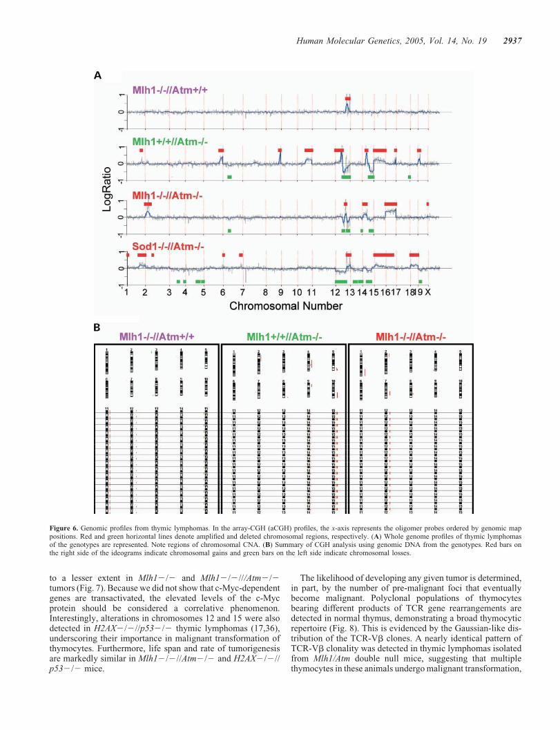
to a lesser extent in Mlh12/2 and Mlh12/2///Atm2/2tumors (Fig. 7). Because we did not show that c-Myc-dependentgenes are transactivated, the elevated levels of the c-Mycprotein should be considered a correlative phenomenon.Interestingly, alterations in chromosomes 12 and 15 were alsodetected in H2AX2/2//p532/2 thymic lymphomas (17,36),underscoring their importance in malignant transformation ofthymocytes. Furthermore, life span and rate of tumorigenesisare markedly similar in Mlh12/2//Atm2/2 and H2AX2/2//p532/2 mice.
The likelihood of developing any given tumor is determined,in part, by the number of pre-malignant foci that eventuallybecome malignant. Polyclonal populations of thymocytesbearing different products of TCR gene rearrangements aredetected in normal thymus, demonstrating a broad thymocyticrepertoire (Fig. 8). This is evidenced by the Gaussian-like dis-tribution of the TCR-Vb clones. A nearly identical pattern ofTCR-Vb clonality was detected in thymic lymphomas isolatedfrom Mlh1/Atm double null mice, suggesting that multiplethymocytes in these animals undergo malignant transformation,
Figure 6. Genomic profiles from thymic lymphomas. In the array-CGH (aCGH) profiles, the x-axis represents the oligomer probes ordered by genomic mappositions. Red and green horizontal lines denote amplified and deleted chromosomal regions, respectively. (A) Whole genome profiles of thymic lymphomasof the genotypes are represented. Note regions of chromosomal CNA. (B) Summary of CGH analysis using genomic DNA from the genotypes. Red bars onthe right side of the ideograms indicate chromosomal gains and green bars on the left side indicate chromosomal losses.
Human Molecular Genetics, 2005, Vol. 14, No. 19 2937

increasing the probability of tumorigenesis in the double nullmice. It also shows that in this genotype, the tumors do notdevelop from a single clone but from a variety of clones. Collec-tively, these results suggest that the type of TCR is not animportant factor in the tumorigenic process. A completelydifferent pattern was observed in the tumors of all other geno-types in which a monoclonal or oligoclonal pattern was noted.In this respect, the Atm2/2, Mlh12/2 and Sod12/2//Atm2/2 behave like human ATM2/2 lymphomas, whichare usually monoclonal in nature. It is thus conceivable thatthe accumulation of specific genomic alterations in Mlh1/Atmdouble null mice increases the probability of each thymocyticclone to become malignant.
By augmenting oxidative stress or genomic instability inAtm2/2 mice, we were able to exacerbate specific aspectsof this complex disease and link certain features of A-T to oxi-dative stress or genomic instability. Our data suggest thatoxidative stress has a role in radiation sensitivity and growthretardation of A-T patients, whereas genomic instability isan important factor in cancer predisposition and that specificalterations in chromosomes 12 and 15 are most probablyresponsible for the development of thymic lymphoma inAtm2/2 mice (Fig. 10). These experiments demonstrate thevalue of animal models in the physiological dissection ofhuman disease phenotypes.
EXPERIMENTAL PROCEDURES
Generation of various mouse genotypes
All mice in this study have SV129 background. Mice lackingSod1 were obtained from Jackson Laboratories (Bar Harbor,ME, USA). Sod12/2//Atm2/2 double mutant mice were gen-erated by crossing Sod12/2//Atmþ/2 males with Sod1þ/2//Atmþ/2 females. Three control genotypes were maintained:Sod1þ/þ//Atmþ/þ, Sod1þ/þ//Atm2/2 and Sod12/2//
Atmþ/þ. Additional genotypes were Sod12/2//Atmþ/2 andSod1þ/2//Atm2/2.
Mlh1 null mice are infertile. Mlh12/2//Atm2/2 micewere generated by crossing male and female Mlh1þ/2//Atmþ/2 genotypes. Control genotypes were Mlh12/2//Atmþ/þ, Mlh1þ/þ//Atm2/2 and Mlh1þ/þ//Atmþ/þ.Additional genotypes were Mlh1þ/þ//Atmþ/2, Mlh1þ/2//Atm2/2, Mlh1þ/2//Atmþ/2 and Mlh12/2//Atmþ/2.
Offspring were genotyped at age 3 weeks by polymerasechain reaction (PCR)-based assays using mouse-tail DNA,prepared with the GeneReleaserTM reagent (Bio-VenturesCo., Murfreesboro, TN, USA).
Growth measurements and pathology
Mice were weighed weekly from weaning (�3 weeks) toadulthood (100 weeks). Mice were perfused with 4% fixative(4% formaldehyde in PBS) or Bouin’s solution and post-fixedat 48C. Fixed tissues were removed, examined grossly, pro-cessed routinely, embedded in paraffin, sectioned at 6 mmand stained with hematoxylin and eosin.
Pathological analyses
Immunohistochemistry was performed on deparaffinized sec-tions of Bouin’s-fixed tissues using an immunoperoxidasecomplex procedure with diaminobenzidine as the chromogen.The primary antibodies against CD3 (rat anti-humanCD13-12, Serotec, Oxford, UK; diluted 1:50) and CD45R(biotin-conjugated rat anti-mouse CD45R/B220, PharMingen,San Diego, CA, USA; diluted 1:100) were used. HistofineUniversal Immunoperoxidase Polymer (Nichirei Corp.,Tokyo, Japan) was used as secondary antibody for CD3staining. Vector MOM Kit (Vector Laboratories, Burlingame,CA, USA) was used for the CD45R staining. Slides were micro-wave treated for antigen exposure prior to staining.
Figure 7. Elevated c-Myc levels in thymic lymphomas isolated from various genotypes. Total tumor proteins were extracted and immunoblotted with an anti-c-Myc antibody; the blots were reacted with an anti-total ERK antibody [anti-ERK2 (Santa Cruz) SC-154] to control for loading differences. WT, thymi isolatedfrom 1-month-old WT animals. A, Atm; M, Mlh1 and S, Sod1.
2938 Human Molecular Genetics, 2005, Vol. 14, No. 19

IR treatment
One-to-three-month-old mice were irradiated with 4 Gy ofX-ray using a linear accelerator (CLINAC1800; field size:30 � 30 cm2), housed together and examined daily for clinicalsymptoms.
Primary cell culture of mouse skin fibroblasts
Ear skin fibroblast cultures from 1–3-month-old mice wereprepared according to Stambrook et al. (37). Cell concen-tration was determined with a hemocytometer, and�5 � 106 cells were plated in 75 cm2 flasks containingDulbecco’s modified Eagle’s medium (DMEM), 15% fetalcalf serum (FCS), non-essential amino acids, gentamycin(50 mg/ml) and amphotericin B (2.5 mg/ml). Fibroblastsfrom Sod12/2 mice failed to proliferate in culture even inan atmosphere of 3% oxygen, suggesting that maintenanceof the redox state is critical for cell division and survival.
Primary cell culture of thymic lymphoma cells
Thymic lymphomas were dissected, chopped into small piecesand incubated with 0.5 mg/ml collagenase type I (Sigma,St Louis, MO, USA) for 30 min at 378C. Fragments weredissociated into a cell suspension by gentle pipetting througha 5 ml narrow-tipped pipette, and a single-cell suspension wasobtained by passing through 20 mm mesh filters. Cell con-centration was determined with a hemocytometer, and�5 � 106 cells were grown in suspension in 75 cm2 flasks con-taining DMEM, 15% FCS, non-essential amino acids, 2 mM
glutamine, penicillin/streptomycin (mg/ml) and amphotericin B(2.5 mg/ml).
Western blot analysis
Western blot analysis was performed as described by Harlowand Lane (38), using 12.5% polyacrylamide gel electropho-resis. Each lane was loaded with an equal amount of protein
Figure 8. Spectratyping analysis for TCR beta rearrangements. High resolution analysis and partial determination of Vb1 and Vb5.1 segment using PCR pro-ducts done by rendering them fluorescent prior to analysis on an automated DNA fragment analyzer. All the Vb segments exhibited a similar pattern (data notshown): the distribution of PCR products was Gaussian for the normal thymus and Mlh12/2//Atm2/2 thymic lymphoma, indicating polyclonality, and skewedfor the Mlh1þ/þ//Atm2/2, Mlh12/2//Atmþ/þ and Sod12/2//Atm2/2 thymic lymphomas, indicating mono- or oligoclonality. y-axis: fluorescent signalintensity. x-axis: PCR product size in base pairs, Vb1 fragment size150 bp, Vb5.1 fragment size 230 bp.
Human Molecular Genetics, 2005, Vol. 14, No. 19 2939

extract (35 mg) which, after electrophoresis, was transferred toa nitrocellulose membrane for 1.5 h. Blots were stained withPonceau to verify equal loading and transfer of proteins. Mem-branes were then probed with monoclonal anti-cMyc(C-33):sc-42 antibody (Santa Cruz) (1:500). For the detectionof c-Myc, horseradish peroxidase (goat anti mouse, 1:5000,Jackson) was used to enhance sensitivity. Intensity of thesignal was determined by ECL Plus detection system(Amersham Pharmacia Biotech, UK).
Metaphase preparations
Metaphases from exponentially growing cells were preparedby a standard procedure (39). Chromosome aberrations werescored as described in (40).
PCR amplification and GeneScan clonality analysis
For clonality identification, each sample was screened forTCRb gene rearrangements as described (41,42) using23 Vb primers. Because polyclonal lymphoid populationsgenerate products with a Gaussian distribution due to the vari-able length and nucleotide sequence of the heterogeneousV(D)J junctional sequences, a clonal population that has iden-tical V(D)J sequences will generate discrete bands dependingon the number of rearranged alleles (41,42). All tissues fromwhich RNA was isolated were indeed tumor tissues. Here,23 Vb primers were used to amplify TCRb recombinations.GeneScan analysis was performed using fluorescent primerslabeled at their 50 end with 6-carboxyfluorescein (FAM). Allreactions were carried out in a final volume of 50 ml with100 ng of cDNA sample, 200 nM of dNTPs, 12 pmol of eachprimer, and 2 U of Taq polymerase. Each PCR experiment
Figure 9. Analysis of MSI with a selected microsatellite marker D10MIT2 in tumors from the various genotypes. Samples of an MSI-positive tumor showedextra alleles when compared with matched normal samples. Fragment size (base pairs) is on the x-axis and fluorescent intensity on the y-axis.
2940 Human Molecular Genetics, 2005, Vol. 14, No. 19

included an appropriate positive clonal control, a polyclonalcontrol and a non-template negative control.
Aliquots of 1:25 diluted PCR products (1.5 ml) were mixedwith 13.5 ml formamide, and 1 ml of the internal size standard(Genescan-500 and Genescan-1000, Applied Biosystems,Foster City, CA, USA) was included for precise determinationof the length of the amplicons. After denaturation for 10 min at948C, the products were separated on POP-4 polymer (AppliedBiosystems) and analyzed by 310 GeneScan 3.1 software(Applied Biosystems). Scanning results of fluorescence-dye-labeled PCR products by GSA were interpreted as follows:one or two striking peaks indicated a dominant clone; anumber of peaks arranged in a normal (Gaussian) distributionindicated polyclonality.
SKY analysis
SKY was performed as previously described (43). Briefly,chromosome-specific libraries generated by PCR fromflow-sorted mouse chromosomes were directly labeled withnucleotides conjugated to five different dyes (FITC, Rhodamine,Texas Red, Cy5 and Cy5.5). All 21 chromosome libraries werehybridized simultaneously to the mouse metaphases. Afterwashing, the slides were stained with 406-diamidino-2 pheny-lindole (DAPI) in antifade medium. Discrimination betweenthe different spectra was done with the SD300 spectral bio-imaging system (Applied Spectral Imaging Ltd, MigdalHaemek, Israel) that uses a Sagnac interferometer to measurethe full visible light spectrum at each pixel of the image. Aclassification algorithm was used to differentiate between
different spectra in the image and to assign pseudocolors toall the pixels with similar spectral characteristics. The DAPIimage was captured separately and inverted to give aG-banding pattern. The chromosomes were then sortedautomatically into a karyotype table.
Analysis of MSI
Tumor tissues were manually microdissected from developedthymic lymphomas to ensure that most of the tissue containedneoplastic cells. Normal tissue controls were specimens fromcerebellum or kidneys isolated from the same animals orfrom the WT animals. Tumors and normal DNAs wereextracted and analyzed for MSI using a panel of threemicrosatellite markers, D1MIT36, D7MIT91 and D10MIT2,and performed as described (44). Oligonucleotide forwardmarkers were fluorescently 50 labeled. Multiple PCRs werecarried out in a 25 ml mixture containing �500–1000 ngDNA, 1 � PCR buffer, 0.4 mM of each PCR primer (AppliedBiosystems, Warrington, UK), 2 mM MgCl2, 3.2 mM of deoxy-nucleotide triphosphpate and 1 U of BIOTAK DNA polyme-rase (Bioline Ltd, London, UK). PCR amplification wasconducted in an Eppendorf master cycler gradient (Eppendorf,Hamburg, Germany). Samples were denatured for 10 min at948C and subjected to 35 cycles of 30 s each at 948C, 30 sat 588C, 30 s at 728C and a final extension step of 5 min at728C. Then 1 ml of fluorescently labeled PCR product ofpaired normal and tumor tissues was mixed with 12 ml ofdeionized formamide and 1 ml GeneScan TAMRA 500 SizeStandard (Applied Biosystems), respectively. The mixture
Figure 10. Differential contribution of various factors to specific features of A-T according to findings reported here.
Human Molecular Genetics, 2005, Vol. 14, No. 19 2941

was denatured for 5 min at 948C, cooled on ice and loaded onthe ABI PRISM 3100 Genetic Analyzer (Applied Biosystems).The data were collected automatically and analyzed using theGeneScan 3.1 and Genotyper analysis software (Applied Bio-systems), which automatically determined the actual size ofthe PCR product and the amount of fluorescent signal. MSIwas indicated by the presence of novel peaks in the tumortissue that were not seen in normal tissue from the sameanimal or by a difference in microsatellite length in the twosamples.
SUPPLEMENTARY MATERIAL
Supplementary Material is available at HMG Online.
ACKNOWLEDGEMENTS
This work was supported by research grants from the A-TChildren’s Project and the Israel Science Foundation to A.B.Work in the laboratory of Y.S. is supported by researchgrants from the A-T Medical Research Foundation, the A-TChildren’s Project, the A-T Medical Research Trust, theNational Institutes of Health (RO1 NS 31763) and the IsraelMinistry of Science. We wish to thank the Arison family fortheir donation to the center of the DNA microarrays.
Conflict of Interest statement. None declared.
REFERENCES
1. Chun, H.H. and Gatti, R.A. (2004) Ataxia-telangiectasia, an evolvingphenotype. DNA Repair (Amst), 3, 1187–1196.
2. Abraham, R.T. (2004) PI 3-kinase related kinases: ‘big’ players instress-induced signaling pathways. DNA Repair (Amst), 3, 883–887.
3. Shiloh, Y. (2003) ATM and related protein kinases: safeguarding genomeintegrity. Nat. Rev. Cancer, 3, 155–168.
4. Bakkenist, C.J. and Kastan, M.B. (2003) DNA damage activates ATMthrough intermolecular autophosphorylation and dimer dissociation.Nature, 421, 499–506.
5. Kurz, E.U. and Lees-Miller, S.P. (2004) DNA damage-induced activationof ATM and ATM-dependent signaling pathways. DNA Repair (Amst), 3,889–900.
6. Barlow, C., Hirotsune, S., Paylor, R., Liyanage, M., Eckhaus, M.,Collins, F., Shiloh, Y., Crawley, J.N., Ried, T., Tagle, D. et al. (1996)Atm-deficient mice: a paradigm of ataxia telangiectasia. Cell, 86,159–171.
7. Elson, A., Wang, Y., Daugherty, C.J., Morton, C.C., Zhou, F.,Campos-Torres, J. and Leder, P. (1996) Pleiotropic defects inataxia-telangiectasia protein-deficient mice. Proc. Natl Acad. Sci. USA,93, 13084–13089.
8. Xu, Y., Ashley, T., Brainerd, E.E., Bronson, R.T., Meyn, M.S. andBaltimore, D. (1996) Targeted disruption of ATM leads to growthretardation, chromosomal fragmentation during meiosis, immune defects,and thymic lymphoma. Genes Dev., 10, 2411–2422.
9. Borghesani, P.R., Alt, F.W., Bottaro, A., Davidson, L., Aksoy, S.,Rathbun, G.A., Roberts, T.M., Swat, W., Segal, R.A. and Gu, Y. (2000)Abnormal development of Purkinje cells and lymphocytes in Atm mutantmice. Proc. Natl Acad. Sci. USA, 97, 3336–3341.
10. Kohen, R. and Nyska, A. (2002) Oxidation of biological systems:oxidative stress phenomena, antioxidants, redox reactions, and methodsfor their quantification. Toxicol. Pathol., 30, 620–650.
11. Matzuk, M.M., Dionne, L., Guo, Q., Kumar, T.R. and Lebovitz, R.M.(1998) Ovarian function in superoxide dismutase 1 and 2 knockout mice.Endocrinology, 139, 4008–4011.
12. Lebovitz, R.M., Zhang, H., Vogel, H., Cartwright, J., Jr, Dionne, L.,
Lu, N., Huang, S. and Matzuk, M.M. (1996) Neurodegeneration,myocardial injury, and perinatal death in mitochondrial superoxidedismutase-deficient mice. Proc. Natl Acad. Sci. USA, 93, 9782–9787.
13. Baker, S.M., Plug, A.W., Prolla, T.A., Bronner, C.E., Harris, A.C.,
Yao, X., Christie, D.M., Monell, C., Arnheim, N., Bradley, A. et al.(1996) Involvement of mouse Mlh1 in DNA mismatch repair and meioticcrossing over. Nat. Genet., 13, 336–342.
14. Edelmann, W., Cohen, P.E., Kane, M., Lau, K., Morrow, B., Bennett, S.,
Umar, A., Kunkel, T., Cattoretti, G., Chaganti, R. et al. (1996) Meioticpachytene arrest in MLH1-deficient mice. Cell, 85, 1125–1134.
15. Kawate, H., Sakumi, K., Tsuzuki, T., Nakatsuru, Y., Ishikawa, T.,Takahashi, S., Takano, H., Noda, T. and Sekiguchi, M. (1998) Separation
of killing and tumorigenic effects of an alkylating agent in mice defectivein two of the DNA repair genes. Proc. Natl Acad. Sci. USA, 95,5116–5120.
16. Schrock, E., Badger, P., Larson, D., Erdos, M., Wynshaw-Boris, A.,
Ried, T. and Brody, L. (1996) The murine homolog of the human breastand ovarian cancer susceptibility gene Brca1 maps to mouse chromosome11D. Hum. Genet., 97, 256–259.
17. Bassing, C.H., Suh, H., Ferguson, D.O., Chua, K.F., Manis, J.,Eckersdorff, M., Gleason, M., Bronson, R., Lee, C. and Alt, F.W. (2003)Histone H2AX: a dosage-dependent suppressor of oncogenictranslocations and tumors. Cell, 114, 359–370.
18. Hurlin, P.J. and Dezfouli, S. (2004) Functions of myc:max in the controlof cell proliferation and tumorigenesis. Int. Rev. Cytol., 238, 183–226.
19. Peter, Y., Rotman, G., Lotem, J., Elson, A., Shiloh, Y. and Groner, Y.(2001) Elevated Cu/Zn-SOD exacerbates radiation sensitivity and
hematopoietic abnormalities of Atm-deficient mice. EMBO J., 20,1538–1546.
20. Bar-Peled, O., Korkotian, E., Segal, M. and Groner, Y. (1996)Constitutive overexpression of Cu/Zn superoxide dismutase exacerbates
kainic acid-induced apoptosis of transgenic-Cu/Zn superoxide dismutaseneurons. Proc. Natl Acad. Sci. USA, 93, 8530–8535.
21. Peled-Kamar, M., Lotem, J., Okon, E., Sachs, L. and Groner, Y. (1995)Thymic abnormalities and enhanced apoptosis of thymocytes and bone
marrow cells in transgenic mice overexpressing Cu/Zn-superoxidedismutase: implications for Down syndrome. EMBO J., 14, 4985–4993.
22. Peled-Kamar, M., Lotem, J., Wirguin, I., Weiner, L., Hermalin, A. andGroner, Y. (1997) Oxidative stress mediates impairment of muscle
function in transgenic mice with elevated level of wild-type Cu/Znsuperoxide dismutase. Proc. Natl Acad. Sci. USA, 94, 3883–3887.
23. Wei, K., Kucherlapati, R. and Edelmann, W. (2002) Mouse models forhuman DNA mismatch-repair gene defects. Trends Mol. Med., 8,
346–353.
24. Stojic, L., Brun, R. and Jiricny, J. (2004) Mismatch repair and DNAdamage signalling. DNA Repair (Amst), 3, 1091–1101.
25. Brown, K.D., Rathi, A., Kamath, R., Beardsley, D.I., Zhan, Q.,
Mannino, J.L. and Baskaran, R. (2003) The mismatch repair system isrequired for S-phase checkpoint activation. Nat. Genet., 33, 80–84.
26. Luo, Y., Lin, F.T. and Lin, W.C. (2004) ATM-mediated stabilization of
hMutL DNA mismatch repair proteins augments p53 activation duringDNA damage. Mol. Cell. Biol., 24, 6430–6444.
27. Yamane, K., Taylor, K. and Kinsella, T.J. (2004) Mismatchrepair-mediated G2/M arrest by 6-thioguanine involves the ATR-Chk1
pathway. Biochem. Biophys. Res. Commun., 318, 297–302.
28. Yan, T., Desai, A.B., Jacobberger, J.W., Sramkoski, R.M., Loh, T. andKinsella, T.J. (2004) CHK1 and CHK2 are differentially involved inmismatch repair-mediated 6-thioguanine-induced cell cycle checkpoint
responses. Mol. Cancer Ther., 3, 1147–1157.
29. Caporali, S., Falcinelli, S., Starace, G., Russo, M.T., Bonmassar, E.,Jiricny, J. and D’Atri, S. (2004) DNA damage induced by temozolomidesignals to both ATM and ATR: role of the mismatch repair system. Mol.
Pharmacol., 66, 478–491.
30. Cejka, P., Stojic, L., Marra, G. and Jiricny, J. (2004) Is mismatch repairreally required for ionizing radiation-induced DNA damage signaling?Nat. Genet., 36, 432–433; author reply 434.
31. Adamson, A.W., Beardsley, D.I., Kim, W.J., Gao, Y., Baskaran, R. andBrown, K.D. (2005) Methylator-induced, mismatch repair-dependent G2arrest is activated through Chk1 and Chk2. Mol. Biol. Cell, 16,1513–1526.
2942 Human Molecular Genetics, 2005, Vol. 14, No. 19

32. Petrini, J.H. and Theunissen, J.W. (2004) Double strand break metabolism
and cancer susceptibility: lessons from the mre11 complex. Cell Cycle, 3,
541–542.
33. Theunissen, J.W., Kaplan, M.I., Hunt, P.A., Williams, B.R.,
Ferguson, D.O., Alt, F.W. and Petrini, J.H. (2003) Checkpoint failure and
chromosomal instability without lymphomagenesis in Mre11(ATLD1/ATLD1) mice. Mol. Cell, 12, 1511–1523.
34. Williams, B.R., Mirzoeva, O.K., Morgan, W.F., Lin, J., Dunnick, W. and
Petrini, J.H. (2002) A murine model of Nijmegen breakage syndrome.Curr. Biol., 12, 648–653.
35. Celeste, A., Petersen, S., Romanienko, P.J., Fernandez-Capetillo, O.,
Chen, H.T., Sedelnikova, O.A., Reina-San-Martin, B., Coppola, V.,Meffre, E., Difilippantonio, M.J. et al. (2002) Genomic instability in mice
lacking histone H2AX. Science, 296, 922–927.
36. Celeste, A., Difilippantonio, S., Difilippantonio, M.J., Fernandez-Capetillo, O., Pilch, D.R., Sedelnikova, O.A., Eckhaus, M., Ried, T.,
Bonner, W.M. and Nussenzweig, A. (2003) H2AX haploinsufficiencymodifies genomic stability and tumor susceptibility. Cell, 114, 371–383.
37. Stambrook, P.J., Shao, C., Stockelman, M., Boivin, G., Engle, S.J. and
Tischfield, J.A. (1996) APRT: a versatile in vivo resident reporter of localmutation and loss of heterozygosity. Environ. Mol. Mutagen, 28,
471–482.
38. Harlow, E. and Lane, D. (1988) Antibodies: A Laboratory Manual. ColdSpring Harbor Laboratory.
39. Dhar, S., Squire, J.A., Hande, M.P., Wellinger, R.J. and Pandita, T.K.(2000) Inactivation of 14-3-3 sigma influences telomere behavior andionizing radiation-induced chromosomal instability. Mol. Cell. Biol., 20,7764–7772.
40. Hunt, C.R., Dix, D.J., Sharma, G.G., Pandita, R.K., Gupta, A., Funk, M.and Pandita, T.K. (2004) Genomic instability and enhancedradiosensitivity in Hsp70.1- and Hsp70.3-deficient mice. Mol Cell. Biol.,24, 899–911.
41. d’Auriol, L., Macintyre, E., Galibert, F. and Sigaux, F. (1989) In vitroamplification of T cell gamma gene rearrangements: a new tool for theassessment of minimal residual disease in acute lymphoblastic leukemias.Leukemia, 3, 155–158.
42. Delabesse, E., Burtin, M.L., Millien, C., Madonik, A., Arnulf, B.,Beldjord, K., Valensi, F. and Macintyre, E.A. (2000) Rapid,multifluorescent TCRG Vgamma and Jgamma typing: application toT cell acute lymphoblastic leukemia and to the detection of minor clonalpopulations. Leukemia, 14, 1143–1152.
43. Liyanage, M., Coleman, A., du Manoir, S., Veldman, T., McCormack, S.,Dickson, R.B., Barlow, C., Wynshaw-Boris, A., Janz, S., Wienberg, J.et al. (1996) Multicolour spectral karyotyping of mouse chromosomes.Nat. Genet., 14, 312–315.
44. Cai, K.Q., Albarracin, C., Rosen, D., Zhong, R., Zheng, W., Luthra, R.,Broaddus, R. and Liu, J. (2004) Microsatellite instability and alteration ofthe expression of hMLH1 and hMSH2 in ovarian clear cell carcinoma.Hum. Pathol., 35, 552–559.
Human Molecular Genetics, 2005, Vol. 14, No. 19 2943