human papillomavirus 16 oncoproteins downregulate the
TRANSCRIPT

Research ArticleHuman Papillomavirus 16 Oncoproteins Downregulate theExpression of miR-148a-3p, miR-190a-5p, and miR-199b-5p inCervical Cancer
Mi-Soon Han ,1,2 Jae Myun Lee,3 Soo-Nyung Kim,4
Jae-Hoon Kim ,5 and Hyon-Suk Kim 6
1Department of Medicine, Graduate School of Yonsei University, 50-1 Yonsei-ro, Seodaemun-gu, Seoul 03722, Republic of Korea2Department of Laboratory Medicine, U2 Clinical Laboratories, Jangwon Medical Foundation, 68 Geoma-ro, Songpa-gu,Seoul 05755, Republic of Korea
3Department of Microbiology and Immunology, Yonsei University College of Medicine, 50-1 Yonsei-ro, Seodaemun-gu,Seoul 03722, Republic of Korea
4Department of Obstetrics and Gynecology, Konkuk University Medical Center, 120-1 Neungdong-ro, Gwangjin-gu,Seoul 05030, Republic of Korea
5Department of Obstetrics and Gynecology, Gangnam Severance Hospital, Yonsei University College of Medicine,211 Eonju-ro, Gangnam-gu, Seoul 06273, Republic of Korea
6Department of Laboratory Medicine, Severance Hospital, Yonsei University College of Medicine, 50-1 Yonsei-ro, Seodaemun-gu,Seoul 03722, Republic of Korea
Correspondence should be addressed to Jae-Hoon Kim; [email protected] and Hyon-Suk Kim; [email protected]
Received 28 August 2018; Accepted 15 November 2018; Published 29 November 2018
Academic Editor: Sung-Hoon Kim
Copyright © 2018 Mi-Soon Han et al. This is an open access article distributed under the Creative Commons Attribution License,which permits unrestricted use, distribution, and reproduction in any medium, provided the original work is properly cited.
Almost all cervical cancers are associated with human papillomavirus (HPV); however, the majority of women infected with thisvirus do not develop cervical cancer. Therefore, new markers are needed for reliable screening of cervical cancer, especially inrelation to HPV infection. We aimed to identify potential microRNAs that may serve as diagnostic markers for cervical cancerdevelopment in high-risk HPV-positive patients. We evaluated the microRNA expression profiles in 12 cervical tissues using thehybridization method and verified them by quantitative polymerase chain reaction (qPCR). Finally, we evaluated the effects ofHPV16 oncoproteins on the expression of selectedmicroRNAs using cervical cancer cells (CaSki and SiHa) and RNA interference.With the hybridization method, eight microRNAs (miR-9-5p, miR-136-5p, miR-148a-3p, miR-190a-5p, miR-199b-5p, miR-382-5p,miR-597-5p, and miR-655-3p) were found to be expressed differently in the HPV16-positive cervical cancer group and HPV16-positive normal group (fold change ≥ 2). The results of qPCR showed that miR-148a-3p, miR-190a-5p, miR-199b-5p, and miR-655-3p levels significantly decreased in the cancer group compared with the normal group. Upon silencing of HPV16 E5 and E6/E7,miR-148a-3p levels increased in both cell lines. Silencing of E6/E7 in SiHa cells led to the increase inmiR-199b-5p andmiR-190a-5plevels.Three HPV16 oncoproteins (E5, E6, and E7) downregulate miR-148a-3p, while E6/E7 inhibit miR-199b-5p and miR-190a-5pexpression in cervical carcinoma. The three microRNAs, miR-148a-3p, miR-199b-5p, and miR-190a-5p, may be novel diagnosticbiomarkers for cervical cancer development in high-risk HPV-positive patients.
1. Introduction
The interaction between viral and host factors is importantin cervical carcinogenesis because it triggers tumor growth,invasion, and metastasis. Specifically, human papillomavirus(HPV) infection has been shown to be the most important
factor in cervical carcinogenesis: the transformation fromnormal cervical epithelium to cervical cancer tissue is mostlikely caused by HPVs, which are episomal, double-strandedDNA viruses that induce epithelial lesions. The oncogenicpotential of high-risk HPV is mostly attributed to the prod-ucts of three early genes: E5, E6, and E7. E6 and E7 exert their
HindawiBioMed Research InternationalVolume 2018, Article ID 1942867, 9 pageshttps://doi.org/10.1155/2018/1942867
2 BioMed Research International
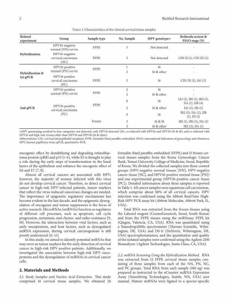
Table 1: Characteristics of the clinical cervical tissue samples.
Relatedexperiment Group Sample type No. Sample HPV genotype∗ Bethesda system&
FIGO stage (N)
Hybridization
HPV16-negativenormal (NN) cervix FFPE 3 Not detected -
HPV16-negativecervical carcinoma
(NC)FFPE 3 Not detected CIN II (1), CIN III (2)
Hybridization &1st qPCR
HPV16-positivenormal (PN) cervix FFPE 1 16 -
2 16 & other -HPV16-positive
cervical carcinoma(PC)
FFPE 3 16 CIN III (1), IA1 (2)
2nd qPCR
HPV16-positivenormal (PN) cervix FFPE
2 16 -2 16 & other -
HPV16-positivecervical carcinoma
(PC)
FFPE8 16 IA1 (1), 1B1 (1), IB2 (1),
IIA (1), IIB (4)2 16 & other IA1 (1), 1B1 (1)
Frozen
9 16 IB2 (5), IIA (2), IIB(1), III (1)
3 16 & 18 1B1 (1), IB2 (1), IIA (1)3 16 & other IB2 (2), IIA (1)
∗HPV genotyping resulted in four categories: not detected, only HPV16 detected (16), co-infected with HPV16 and HPV18 (16 & 18), and co-infected withHPV16 and high-risk viruses other than HPV16 and HPV18 (16 & other).Abbreviations: CIN, cervical intraepithelial neoplasia; FFPE, formalin-fixed paraffin-embedded; FIGO, international federation of gynecology and obstetrics;HPV, human papilloma virus; qPCR, quantitative PCR.
oncogenic effect by destabilizing and degrading retinoblas-toma protein (pRB) and p53 [1–6], while E5 is thought to playa role during the early steps of transformation in the basallayers of the epithelium and enhance the oncogenic effect ofE6 and E7 [7, 8].
Almost all cervical cancers are associated with HPV;however, the majority of women infected with this virusdo not develop cervical cancer. Therefore, to detect cervicalcancer in high-risk HPV-infected patients, tumor markersthat reflect the virus-induced cancerous changes are needed.The importance of epigenetic regulatory mechanisms hasbecome evident in the last decade, and the epigenetic dysreg-ulation of oncogenes and tumor suppressors is the focus ofactive research.MicroRNAs (miRNAs) function as regulatorsof different cell processes, such as apoptosis, cell cycleprogression, metastasis, and chemo- and radio-resistance [9,10]. However, the interaction between viral factors, such asearly oncoproteins, and host factors, such as dysregulatedmiRNA expression, during cervical carcinogenesis is stillpoorly understood [11–13].
In this study, we aimed to identify potential miRNAs thatmay serve as tumormarkers for the early detection of cervicalcancer in high-risk HPV-positive patients. Additionally, weinvestigated the association between high-risk HPV onco-proteins and the dysregulation of miRNAs in cervical cancercells.
2. Materials and Methods
2.1. Study Samples and Nucleic Acid Extraction. This studycomprised 41 cervical tissue samples. We obtained 26
formalin-fixed paraffin-embedded (FFPE) and 15 frozen cer-vical tissues samples from the Korea Gynecologic CancerBank, Yonsei University College of Medicine, Seoul, Republicof Korea. We divided the collected samples into three controlgroups (HPV-negative normal tissues [NN], HPV-negativecancer tissue [NC], and HPV16-positive normal tissue [PN])and one experimental group (HPV16-positive cancer tissue[PC]). Detailed information about these samples is reportedinTable 1. All cancer sampleswere squamous cell carcinomas,which comprise about 80% of all cervical cancers. HPVinfection was confirmed using the Abbott RealTime High-Risk HPV PCR assay kit (Abbott Molecular, Abbott Park, IL,USA).
Total RNA was extracted from the frozen tissues usingthe Labozol reagent (CosmoGenetech, Seoul, South Korea)and from the FFPE tissues using the miRNeasy FFPE kit(Qiagen, Valencia, CA, USA). RNA was quantitated usinga Nanodrop2000c spectrometer (Thermo Scientific, Wilm-ington, DE, USA) and DS-11 (DeNovix, Wilmington, DE,USA) spectrophotometers, and the quantitation and qualityof the isolated samples were confirmed using the Agilent 2100Bioanalyzer (Agilent Technologies, Santa Clara, CA, USA).
2.2. miRNA Screening Using the Hybridization Method. RNAwas extracted from 12 FFPE cervical tissue samples con-sisting of three samples from each of the NN, PN, NC,and PC groups. Total RNA from each sample (100 ng) wasprepared as instructed in the nCounter miRNA ExpressionAssay (NanoString Technologies, Seattle, WA, USA) usermanual. Mature miRNAs were ligated to a species-specific

BioMed Research International 3
tag sequence (miRtag). After enzymatic purification of non-ligated miRtags, the prepared samples were hybridized usingthe nCounter Human v3 miRNA Expression Assay CodeSetcontaining 800 human miRNA hybridization probes. Afterhybridization, the excess probes were removed by two-stepmagnetic bead-based purification on the nCounter Prepstation. Specific target molecules were quantified using thenCounter Digital Analyzer, by counting individual fluores-cent barcodes and assessing target molecule levels. For eachsample, a scan encompassing 280 fields of view was per-formed. The data were collected using the nCounter DigitalAnalyzer after taking images of the immobilized fluorescentreporters in the sample cartridge (NanoString Technologies).
2.3. Quantitative Polymerase Chain Reaction (qPCR) Analysesof Clinical Samples. To investigate the changes in miRNAexpression levels, we performed qPCR analysis of the samplesobtained with the hybridization method. Additionally, wevalidated eight miRNAs in the 29 clinical tissue samplesthat had not been used in the miRNA hybridization (4 PNand 25 PC). Reverse transcription was performed using themiScript II RT Kit (Qiagen, Hilden, Germany) according tothe manufacturer’s instructions. qPCR was performed on theStepOnePlus Real-Time PCR System (Applied Biosystems,Carlsbad, CA, USA) using the 2× QuantiTect SYBR GreenPCR Master Mix (Qiagen). Thermal cycling conditions wereas follows: 95∘C for 15 min, followed by 40 cycles of 94∘C for15 s and 55∘C for 30 s, and 70∘C for 30 s.The data was analyzedusing the StepOne software v2.2.2 (Applied Biosystems). AllqPCR reactions were run in triplicate, and gene expressionlevels of each miRNA were normalized to the levels of theendogenous control small RNAU6, using the 2-ΔΔCt method.
2.4. HPV16 E5/E6/E7 Silencing in Cancer Cells In Vitro. Weinvestigated the effect of HPV16 oncoproteins on humanmiRNA expression using two human cervical cancer celllines: HPV16-positive CaSki cells (ATCC CRL-1550; Amer-ican Type Culture Collection [ATCC], Manassas, VA, USA)and SiHa cells (ATCC HTB-35; ATCC). Cervical cancer cellswere cultured in RPMI-1640 medium (Gibco BRL, GrandIsland, New York, USA), supplemented with 10% fetal bovineserum (GibcoBRL) and 1%penicillin/streptomycin under 5%CO2at 37∘C. To investigate the role of HPV16 E5, E6, and E7
on the expression of miRNAs during cervical carcinogenesis,we silenced the E5 gene using small hairpin RNA (shRNA)overexpressed by lentiviral vectors and the bicistronic E6/E7genes using small interfering RNA (siRNA) in cervical cancercells, as previously reported [14, 15]. Scrambled shRNA orsiRNA sequences were used as a negative control.
Cells were transfected/infected in 12-well plates andcollected 0, 24, 48, and 72 h after. Total RNA was extractedwith the TRIzol reagent (Invitrogen, Carlsbad, CA, USA).The efficiency of knockdown was determined by measuringthe expression levels of HPV16 E5/E6/E7 mRNA three timesby qPCR at 72 h after transfection/infection. The levels ofmiRNA expression were also determined using qPCR atthe indicated time-points. Primer sets described previously[14] were used to amplify each miRNA. Glyceraldehyde
3-phosphate dehydrogenase (GAPDH) was used as an inter-nal control.
2.5. Data Analysis. For miRNA profiling, the reporter countswere collected using the nSolver software v3.0.22 (NanoStringTechnologies). miRNA profiling data were normalized bypositive reaction controls to a panel of five housekeepinggenes (actin B [ACTB], 𝛽2 microglobulin [B2M], GAPDH,and ribosomal proteins [RP] L19 and L10) and to miRNA-23a and miRNA-191 [16]. The R software v.3.1.1 was used foranalysis and graphics construction [17]. Differences betweenthe samples were considered significant at a fold change ≥ 2and P ≤ 0.01. For qPCR analysis, all data were expressed as themean± standard deviation. Statistical differences between thegroupswere assessed using Student’s two-tailed t-test.P< 0.05was considered to indicate a statistically significant difference.
3. Results
3.1. miRNA Profiling in Cervical Tissues. The expressionprofile of 800 human miRNAs in the PC and PN groups wasanalyzed using the hybridization method. We identified 99differentially expressed miRNAs (fold change ≥ 2 and P ≤0.01) between the two groups. Among these, eight miRNAshad significantly different expression in the PC group com-pared with pooled control group (combined NN, NC, andPN groups). Six miRNAs were upregulated and two weredownregulated in the PC group compared to the controlgroup. Figure 1 shows the heat map indicating these eightdifferentially expressed miRNAs.
3.2. Differentially Expressed miRNAs in Cervical Cancer. Toverify the results of the miRNA expression profiles, wereevaluated the expression of the eight identified miRNAs,by qPCR. We found that miR-148a-3p, miR-190a-5p, miR-199b-5p, and miR-655-3p levels were significantly decreasedin the PC group compared to the PN group (respective 0.22-fold, 0.11-fold, 0.11-fold, and 0.11-fold), while the levels ofother miRNAs did not significantly differ between the twogroups (Figure 2(a)). Notably, only the results regarding miR-655-3p expression were consistent with those obtained usingthe hybridization method. Analysis of additional clinicaltissue samples showed that miR-190a-5p, miR-199b-5p, andmiR-655-3p expression was significantly decreased in the PCgroup compared with the PN (control) group (respective0.32-fold, 0.12-fold, and 0.18-fold; Figure 2(b)).
Four miRNAs, whose expression was significantly differ-ent in the PC and PN groups, were analyzed according tothe International Federation of Gynecology and Obstetrics(FIGO) staging system classification of the tissue samples(Figure 3 & Table 2). We found that the expression of thesemiRNAs in the PC group significantly decreased in almost allFIGO stages compared to the PN group, with the exceptionof miR-148a-3p expression in the IB group, characterizedby clinically visible lesions confined to the cervix (stromalinvasion > 5.0mm in depth or >7.0mm in horizontal spread).The relative expression folds for each FIGO stage comparedto the PN group are shown in detail in Table 2. Furthermore,

4 BioMed Research International
3.00
2.00
1.00
0.00
-1.00-2.00-3.00
NN
1N
N2
NN
3N
C1N
C2N
C3PN
1PN
2PN
3PC
1PC
2PC
3
has-miR-382-5p
has-miR-9-5p
has-miR-199b-5p
has-miR-136-5p
has-miR-190a-5p
has-miR-148a-3p
has-miR-597-5p
has-miR-655-3p
Figure 1: Differential miRNA expression in cervical tissues. Heat map indicating the eight differentially expressed miRNAs in cervicalcancer tissues. NN, HPV16-negative normal; NC, HPV16-negative carcinoma; PN, HPV16-positive normal; PC, HPV16-positive carcinoma.
0
5
10
15
20
25
miR-9-5p miR-136-5p miR-190a-5p miR-382-5p0
50
100
150
200
250
300
350
miR-148a-3p miR-199b-5p
Rela
tive E
xpre
ssio
n of
miR
NA
s to
U6
0.00
0.50
1.00
1.50
2.00
2.50
miR-597-5p miR-655-3p
PN, ControlPC, IncreasedPC, Decreased
∗∗∗
∗∗∗
∗∗∗
∗∗
(a)
0
5
10
15
20
25
miR-9-5p miR-136-5p miR-190a-5p miR-382-5p0
50
100
150
200
250
300
miR-148a-3p miR-199b-5p
Rela
tive E
xpre
ssio
n of
miR
NA
s to
U6
0.00
0.50
1.00
1.50
2.00
miR-597-5p miR-655-3p
PN, ControlPC, IncreasedPC, Decreased
∗∗∗
∗∗∗
∗∗∗
(b)
Figure 2:Relative expression of selectedmiRNAs in cervical tissues. (a) Expression of the indicatedmiRNA assessedwith the hybridizationmethod (3 PN vs. 3 PC). (b) Expression of the indicated miRNA assessed with the hybridization method in additional samples (4 PN vs. 25PC). PN, HPV16-positive normal; PC, HPV16-positive carcinoma. ∗, P< 0.05; ∗∗, P< 0.001; ∗ ∗ ∗, P< 0.0001.

BioMed Research International 5
Table2:Th
erelativee
xpressionfold
ofselected
miRNAsw
ithdifferent
FIGOsta
ge.
FIGOsta
gemiR-148a-3p
miR-199b-5p
miR-190a-5p
miR-655-3p
(I)C
omparedwith
U6
2-ΔCg
Pvalue
2-ΔCg
Pvalue
2-ΔCg
Pvalue
2-ΔCg
Pvalue
PN(con
trol)
282.20
225.89
7.50
1.68
IA205.27
0.0385
62.03
0.0125
3.55
0.00
750.51
0.00
53IB
188.13
0.5729
23.88
<0.00
012.10
0.00
060.28
0.0157
IIA156.64
0.00
0327.69
<0.00
012.13
<0.00
010.28
<0.00
01IIB
andIII
102.17
<0.00
0116.59
<0.00
012.61
<0.00
010.21
<0.00
01(II)Com
paredwith
control
2-ΔΔCg
Pvalue
2-ΔΔCg
Pvalue
2-ΔΔCg
Pvalue
2-ΔΔCg
Pvalue
IA0.73
0.00
090.27
0.00
010.47
0.00
010.30
<0.00
01IB
0.67
0.4128
0.11
0.00
010.28
<0.00
010.17
0.00
56IIA
0.56
<0.00
010.12
0.00
010.28
<0.00
010.17
<0.00
01IIB
andIII
0.36
<0.00
010.07
<0.00
010.35
<0.00
010.12
<0.00
01ΔCg
=TargetCg
–RN
U6Cg
,2-Δ
Cg=Normalized
targetgene
amou
ntrelativ
etoRU
N6gene
amou
ntof
each
grou
p(m
ultip
liedby
1000
).ΔΔCg
=TargetsampleΔ
Cg-con
trolsam
pleΔ
Cg,2
-ΔΔCg
=Normalized
gene
amou
ntof
targetgrou
prelativ
etotargetgene
amou
ntof
controlgroup
.Ab
breviatio
ns:FIG
O,internatio
nalfederationof
gynecology
andob
stetrics;PN
,HPV
16-positive
norm
al.

6 BioMed Research International
0.050.0
100.0150.0200.0250.0300.0350.0400.0450.0
miR-148a-3p miR-199b-5p
Rela
tive E
xpre
ssio
n of
miR
NAs
to U
6
PNIAIB
IIAIIB+III
∗∗∗
∗∗∗ ∗∗∗∗∗∗
∗
∗
∗∗
(a)
0.0
2.0
4.0
6.0
8.0
10.0
12.0
miR-190a-5p miR-655-3p
Rela
tive E
xpre
ssio
n of
miR
NAs
to U
6
∗∗∗
∗∗∗
∗∗∗ ∗∗∗
∗
∗∗
∗∗
(b)
Figure 3:Relative expression of selectedmiRNA according to the International Federation of Gynecology andObstetrics (FIGO) stagesof the samples.TheHPV16-positive cancer (PC) group (IA [n = 4], 1B [n = 12], IIA [n = 5], IIB+III [n = 6]) were compared with the HPV16-positive normal group (PN; n = 5). ∗, P < 0.05; ∗∗, P < 0.001; ∗ ∗ ∗, P < 0.0001.
0.0
0.2
0.4
0.6
0.8
1.0
1.2
E5 E6 E7 E5 E6 E7Scramblesh/siRNA
Rela
tive m
RNA
expr
essio
n
CaSki cell SiHa cell
∗∗∗∗
∗∗
∗∗
Figure 4: E5,E6, and E7 expression levels afterRNA interference.Relative expression of E5, E6, and E7 mRNA 72 h after shRNA orsiRNA-mediated silencing in CaSki and SiHa cells. GAPDH wasused for normalization. ∗, P < 0.05; ∗∗, P < 0.01.
the expression levels of these miRNAwere shown to decreasegradually as the disease progressed.
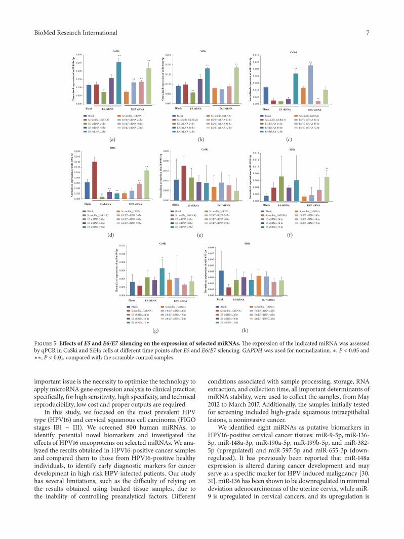
3.3. HPV16 E5/E6/E7 Effect on Host miRNA Expression.HPV16 E5, E6, and E7 silencing reduced the expression ofthese genes by 0.53-fold, 0.85-fold, and 0.49-fold in CaSkicells and by 0.74-fold, 0.62-fold, and 0.50-fold in SiHacells, compared to control samples, overexpressing scrambleshRNAor siRNA (Figure 4).We then determined the relativeexpression levels of the selected four miRNAs in Ca Skiand SiHa cervical cancer cells and compared them withthe miRNA levels in control samples, at the indicated timepoints after transfection/infection. miR-148a-3p expressionsignificantly increased in both cell lines 72 h after genesilencing, especially in the E6/E7 knockdown group, while
miR-199b-5p showed a variable expression pattern and itslevels were significantly increased only in SiHa cells 72 hafter E6/E7 silencing. miR-190a-5p expression significantlyincreased in CaSki cells 72 h after E5 silencing and inSiHa cells72 h after E6/E7 silencing. However, no significantchanges in miR-655-3p levels were observed (Figure 5).
4. Discussion
The correlation between changes in miRNA expression andcervical cancer development was originally described in 2009[18]. The authors of the study showed that the expression ofmiR-21 promotes HeLa cell proliferation, while its inhibitionsuppresses cell proliferation by inducing the overexpressionof the tumor-suppressor gene programmed cell death 4(PDCD4), a programmed cell death protein. miR-21 wassubsequently demonstrated to be an important oncomir,overexpressed in a wide variety of cancers, including cervicalcancer [19].
Several miRNAs, such as miR-34a, miR-886-5p, miR-143,miR-203, and miR-155, have been shown to have differentialexpression in cervical cancer and normal samples [20–24].Therefore, miRNAs have been studied as potential diagnosticbiomarkers in cancer development and progression and astherapeutic targets for cervical cancer treatment [25–27]. In2014, Sharma et al. [28] reviewed 246 differentially expressedmiRNAs involved in cervical cancer progression.
However, to date, no miRNAs are used practically asmarkers for the diagnosis of cervical cancer. One of thepossible reasons is the lack of consistency in the researchdata, which makes it difficult to determine the clinicalvalue of the identified miRNA. Inconsistency in miRNAexpression levels in cervical carcinogenesis may be attributedto patient-intrinsic variation, time and temperature changesduring sample collection, processing, contamination by cellsand blood components, RNA extraction method used, nor-malization, and storage time and conditions [29]. Another
BioMed Research International 7
0.000
0.050
0.100
0.150
0.200
0.250
0.300
Nor
mal
ized
expr
essio
n of
miR
-148
a-3p
CaSki
Blank E5 shRNA E6/7 siRNA
∗∗
∗∗ ∗∗
∗∗
∗
BlankScramble_(shRNA)E5 shRNA 24 hrE5 shRNA 48 hrE5 shRNA 72 hr
Scramble_(siRNA)E6/E7 siRNA 24 hrE6/E7 siRNA 48 hrE6/E7 siRNA 72 hr
(a)
0.000
0.050
0.100
0.150
0.200
0.250
Nor
mal
ized
expr
essio
n of
miR
-148
a-3p
SiHa
Blank E5 shRNA E6/7 siRNA
∗∗
∗∗∗∗
BlankScramble_(shRNA)E5 shRNA 24 hrE5 shRNA 48 hrE5 shRNA 72 hr
Scramble_(siRNA)E6/E7 siRNA 24 hrE6/E7 siRNA 48 hrE6/E7 siRNA 72 hr
(b)
E5 shRNABlank E6/7 siRNA
0.000
0.020
0.040
0.060
0.080
0.100
0.120
0.140CaSki
Nor
mal
ized
expr
essio
n of
miR
-190
a-5p
∗∗
∗∗
∗∗
BlankScramble_(shRNA)E5 shRNA 24 hrE5 shRNA 48 hrE5 shRNA 72 hr
Scramble_(siRNA)E6/E7 siRNA 24 hrE6/E7 siRNA 48 hrE6/E7 siRNA 72 hr
(c)
E5 shRNA
0.000
0.020
0.040
0.060
0.080
0.100
0.120
0.140
0.160
0.180SiHa
Blank E6/7 siRNA
Nor
mal
ized
expr
essio
n of
miR
-190
a-5p
∗∗
∗∗
∗∗∗∗
∗∗
∗
BlankScramble_(shRNA)E5 shRNA 24 hrE5 shRNA 48 hrE5 shRNA 72 hr
Scramble_(siRNA)E6/E7 siRNA 24 hrE6/E7 siRNA 48 hrE6/E7 siRNA 72 hr
(d)
E5 shRNABlank E6/7 siRNA
0.000
0.005
0.010
0.015
0.020
0.025 CaSki
Nor
mal
ized
expr
essio
n of
miR
-199
b-5p
BlankScramble_(shRNA)E5 shRNA 24 hrE5 shRNA 48 hrE5 shRNA 72 hr
Scramble_(siRNA)E6/E7 siRNA 24 hrE6/E7 siRNA 48 hrE6/E7 siRNA 72 hr
(e)
E5 shRNABlank E6/7 siRNA0.000
0.002
0.004
0.006
0.008
0.010
0.012
0.014SiHa
Nor
mal
ized
expr
essio
n of
miR
-199
b-5p
∗∗
BlankScramble_(shRNA)E5 shRNA 24 hrE5 shRNA 48 hrE5 shRNA 72 hr
Scramble_(siRNA)E6/E7 siRNA 24 hrE6/E7 siRNA 48 hrE6/E7 siRNA 72 hr
(f)
E5 shRNABlank E6/7 siRNA
0.000
0.002
0.004
0.006
0.008
0.010
0.012CaSki
Nor
mal
ized
expr
essio
n of
miR
-655
-3p
∗
BlankScramble_(shRNA)E5 shRNA 24 hrE5 shRNA 48 hrE5 shRNA 72 hr
Scramble_(siRNA)E6/E7 siRNA 24 hrE6/E7 siRNA 48 hrE6/E7 siRNA 72 hr
(g)
Blank E5 shRNA E6/7 siRNA0.000
0.001
0.002
0.003
0.004
0.005
0.006
0.007
0.008SiHa
Nor
mal
ized
expr
essio
n of
miR
-655
-3p
∗
BlankScramble_(shRNA)E5 shRNA 24 hrE5 shRNA 48 hrE5 shRNA 72 hr
Scramble_(siRNA)E6/E7 siRNA 24 hrE6/E7 siRNA 48 hrE6/E7 siRNA 72 hr
(h)
Figure 5: Effects of E5 and E6/E7 silencing on the expression of selected miRNAs.The expression of the indicated miRNA was assessedby qPCR in CaSki and SiHa cells at different time points after E5 and E6/E7 silencing. GAPDH was used for normalization. ∗, P < 0.05 and∗∗, P < 0.01, compared with the scramble control samples.
important issue is the necessity to optimize the technology toapplymicroRNA gene expression analysis to clinical practice;specifically, for high sensitivity, high specificity, and technicalreproducibility, low cost and proper outputs are required.
In this study, we focused on the most prevalent HPVtype (HPV16) and cervical squamous cell carcinoma (FIGOstages IB1 ∼ III). We screened 800 human miRNAs, toidentify potential novel biomarkers and investigated theeffects of HPV16 oncoproteins on selected miRNAs. We ana-lyzed the results obtained in HPV16-positive cancer samplesand compared them to those from HPV16-positive healthyindividuals, to identify early diagnostic markers for cancerdevelopment in high-risk HPV-infected patients. Our studyhas several limitations, such as the difficulty of relying onthe results obtained using banked tissue samples, due tothe inability of controlling preanalytical factors. Different
conditions associated with sample processing, storage, RNAextraction, and collection time, all important determinants ofmiRNA stability, were used to collect the samples, from May2012 to March 2017. Additionally, the samples initially testedfor screening included high-grade squamous intraepitheliallesions, a noninvasive cancer.
We identified eight miRNAs as putative biomarkers inHPV16-positive cervical cancer tissues: miR-9-5p, miR-136-5p, miR-148a-3p, miR-190a-5p, miR-199b-5p, and miR-382-5p (upregulated) and miR-597-5p and miR-655-3p (down-regulated). It has previously been reported that miR-148aexpression is altered during cancer development and mayserve as a specific marker for HPV-induced malignancy [30,31]. miR-136 has been shown to be downregulated inminimaldeviation adenocarcinomas of the uterine cervix, while miR-9 is upregulated in cervical cancers, and its upregulation is

8 BioMed Research International
associated with lymph nodemetastases and vascular invasion[32, 33]. miR-199b-5p has been reported to be downregulatedin squamous cell carcinoma and is associated with poorprognosis [34].
Among the eight identified miRNAs, miR-148a-3p, miR-190a-5p, miR-199b-5p, and miR-655 expression was shownto be significantly suppressed in the PC group by qPCR.The results for miR-148a, miR-190a, and miR-199b obtainedwith the hybridization and qPCR methods were discordant.One possible reason for the discrepancy is that the referencegenes used to normalize the data were different in the twomethods. All reference genes were selected according topreviously published studies [14, 16]. Notably, the differencein the expression levels might depend on the instability ofspecific miRNAs. Specifically, the stability of nucleic acidsextracted from FFPE samples may be reduced. While weverified the quality of extracted RNA, it should be notedthat we evaluated the quality of total isolated RNA, not thatof specific miRNAs. Finally, the small number of samplesused for screening increased the chances for nonsignificantresults.
On this note, Mestdag et al. [35] have compared 12available platforms formiRNA expression analysis and foundthat the concordance ofmiRNA expression was less than 70%between hybridization and qPCR. Particularly, the averagevalidation rate of miRNA levels when using any platformcombination was only 54.6% (95% confidential interval,52.5–56.7%). Notably, the silencing efficiency was about50–60% for E5 and E7 in CaSki cells and E6 and E7 inSiHa cells. This was due to the difference in HPV16 copynumber per cell and because of cell characteristics (such asrace and histologic type). The silencing of HPV16 E5 andE6/E7 was shown to inhibit miR-148a-3p expression in boththe cell lines, while the silencing of HPV16 E6/E7 in SiHacells increased miR-199b-5p and miR-190a-5p expressionlevels.
5. Conclusion
In this study, we found that three HPV16 oncoproteinswere associated with the downregulation of miR-148a-3pexpression, while HPV16 E6/E7 led to the downregulationof miR-199b-5p and miR-190a-5p in cervical carcinoma. Ourresults suggest that miR-148a, miR-199b, and miR-190a maybe novel biomarkers for cervical carcinogenesis after HPV16infection.
Data Availability
The [hybridization and qPCR] data used to support thefindings of this study are included within the supplementaryinformation files.
Ethical Approval
The research complied with the World Medical AssociationDeclaration of Helsinki regarding the ethical conduct ofresearch.
Consent
All study samples were obtained from patients who had giveninformed consent for using the samples in medical research.No confidential patient data will be used.
Conflicts of Interest
The authors declare that there is no conflict of interestsregarding the publication of this paper.
Authors’ Contributions
Jae-Hoon Kim and Hyon-Suk Kim contributed equally ascorrespondents in this work.
Acknowledgments
We would like to thank the Korea Gynecologic Cancer Bankof Medical Research Center, Gangnam Severance Hospital,Yonsei University College of Medicine, for providing tissuesamples.
Supplementary Materials
Table S1: Raw data Human microRNA lists (800) andexpression levels. Table S2: Raw data qRT PCR for 8selected human microRNAs. Table S3: Raw data HumanmicroRNA expression levels depend on stages. Table S4: Rawdata Relative expression of E5, E6, and E7 mRNA. Table S5:Raw data E5, E6, E7 silencing effects on human microRNAs.(Supplementary Materials)
References
[1] M. Scheffner, B. A. Werness, J. M. Huibregtse, A. J. Levine,and P. M. Howley, “The E6 oncoprotein encoded by humanpapillomavirus types 16 and 18 promotes the degradation ofp53,” Cell, vol. 63, no. 6, pp. 1129–1136, 1990.
[2] C. W. Menges, L. A. Baglia, R. Lapoint, and D. J. McCance,“Human papillomavirus type 16 E7 up-regulates AKT activitythrough the retinoblastoma protein,” Cancer Research, vol. 66,no. 11, pp. 5555–5559, 2006.
[3] J. M. Huibregtse, M. Scheffner, and P. M. Howley, “A cellularprotein mediates association of p53 with the E6 oncoprotein ofhuman papillomavirus types 16 or 18,” EMBO Journal, vol. 10,no. 13, pp. 4129–4135, 1991.
[4] M. S. Lechner, D. H. Mack, A. B. Finicle, T. Crook, K. H.Vousden, and L. A. Laimins, “Corrigendum: Human papil-lomavirus E6 proteins bind p53 in vivo and abrogate p53-mediated repression of transcription,” �e EMBO Journal, vol.11, no. 11, p. 4248, 1992.
[5] V. Band, S. Dalal, L. Delmolino, and E. J. Androphy, “Enhanceddegradation of p53 protein in HPV-6 and BPV-1 E6-immortalized human mammary epithelial cells,” EMBOJournal, vol. 12, no. 5, pp. 1847–1852, 1993.
[6] K. Munger and P. M. Howley, “Human papillomavirus immor-talization and transformation functions,”Virus Research, vol. 89,no. 2, pp. 213–228, 2002.
[7] D. Ranieri, F. Belleudi, A. Magenta, and M. R. Torrisi, “HPV16E5 expression induces switching from FGFR2b to FGFR2c

BioMed Research International 9
and epithelial-mesenchymal transition,” International Journal ofCancer, vol. 137, no. 1, pp. 61–72, 2015.
[8] N. Kivi, D. Greco, P. Auvinen, and E. Auvinen, “Genes involvedin cell adhesion, cellmotility andmitogenic signaling are altereddue to HPV 16 E5 protein expression,” Oncogene, vol. 27, no. 18,pp. 2532–2541, 2008.
[9] M. S. Kumar, J. Lu, K. L. Mercer, T. R. Golub, and T. Jacks,“ImpairedmicroRNA processing enhances cellular transforma-tion and tumorigenesis,”Nature Genetics, vol. 39, no. 5, pp. 673–677, 2007.
[10] V. Gonzalez-Quintana, L. Palma-Berre, A. D. Campos-Parra etal., “MicroRNAs are involved in cervical cancer development,progression, clinical outcome and improvement treatmentresponse (Review),” Oncology Reports, vol. 35, no. 1, pp. 3–12,2016.
[11] S. Liao, D. Deng, X. Hu et al., “HPV16/18 E5, a promising candi-date for cervical cancer vaccines, affects SCPs, cell proliferationand cell cycle, and forms a potential network with E6 and E7,”International Journal of Molecular Medicine, vol. 31, no. 1, pp.120–128, 2013.
[12] D. DiMaio and L. M. Petti, “The E5 proteins,”Virology, vol. 445,no. 1-2, pp. 99–114, 2013.
[13] J. P. Maufort, A. Shai, H. C. Pitot, and P. F. Lambert, “A role forHPV16 E5 in cervical carcinogenesis,” Cancer Research, vol. 70,no. 7, pp. 2924–2931, 2010.
[14] C. Liu, J. Lin, L. Li et al., “HPV16 early gene E5 specificallyreducesmiRNA-196a in cervical cancer cells,” Scientific Reports,vol. 5, no. 1, 2015.
[15] J. Zhou, C. Peng, B. Li et al., “Transcriptional gene silencing ofHPV16 E6/E7 induces growth inhibition via apoptosis in vitroand in vivo,” Gynecologic Oncology, vol. 124, no. 2, pp. 296–302,2012.
[16] M. d. Leitao, E. C. Coimbra, R. d. Lima et al., “QuantifyingmRNA and MicroRNA with qPCR in Cervical Carcinogenesis:AValidation ofReferenceGenes toEnsureAccurateData,”PLoSONE, vol. 9, no. 11, p. e111021, 2014.
[17] R Development Core Team, “R: A language and environmentfor statistical computing, R Foundation for Statistical Comput-ing,” Vienna, Austria, Download R-3.1.1 for Windows, The R-project for statistical computing (Updated on July 2014), 2014.
[18] R. C. Friedman, K. K. Farh, C. B. Burge, and D. P. Bartel, “Mostmammalian mRNAs are conserved targets of microRNAs,”Genome Research, vol. 19, no. 1, pp. 92–105, 2009.
[19] F. Wang, Y. Li, J. Zhou et al., “MiR-375 is down-regulatedin squamous cervical cancer and inhibits cell migration andinvasion via targeting transcription factor SP1,” �e AmericanJournal of Pathology, vol. 179, no. 5, pp. 2580–2588, 2011.
[20] R. T. K. Pang, C. O. N. Leung, T.-M. Ye et al., “MicroRNA-34a suppresses invasion through downregulation of Notch1and Jagged1 in cervical carcinoma and choriocarcinoma cells,”Carcinogenesis, vol. 31, no. 6, pp. 1037–1044, 2010.
[21] J.-H. Li, X. Xiao, Y.-N. Zhang et al., “MicroRNA miR-886-5pinhibits apoptosis by down-regulating Bax expression in humancervical carcinoma cells,” Gynecologic Oncology, vol. 120, no. 1,pp. 145–151, 2011.
[22] L. Liu, X. Yu, and X. Guo, “MiR-143 is downregulated in cervicalcancer and promotes apoptosis and inhibits tumor formationby targeting Bcl-2,”MolecularMedicine Reports, vol. 5, no. 3, pp.753–760, 2012.
[23] X. Zhu, K. Er, C. Mao et al., “miR-203 suppresses tumor growthand angiogenesis by targeting VEGFA in cervical cancer,”
Cellular Physiology and Biochemistry, vol. 32, no. 1, pp. 64–73,2013.
[24] G. Lao, P. Liu, Q. Wu et al., “Mir-155 promotes cervical cancercell proliferation through suppression of its target gene LKB1,”Tumor Biology, vol. 35, no. 12, pp. 11933–11938, 2014.
[25] S. Sharma, S. Hussain, K. Soni et al., “Novel MicroRNAsignatures in HPV-mediated cervical carcinogenesis in Indianwomen,” Tumor Biology, vol. 37, no. 4, pp. 4585–4595, 2016.
[26] Q. Kong, W. Wang, and P. Li, “Regulator role of HPV E7protein onmiR-21 expression in cervical carcinoma cells and itsfunctional implication,” in International Journal of Clinical andExperimental Pathology, vol. 8, pp. 15808–15813, 2015.
[27] Y.-X. Cheng, Q.-F. Zhang, L. Hong et al., “MicroRNA-200b sup-presses cell invasion and metastasis by inhibiting the epithelial-mesenchymal transition in cervical carcinoma,” MolecularMedicine Reports, vol. 13, no. 4, pp. 3155–3160, 2016.
[28] G. Sharma, P.Dua, and S.M. Agarwal, “A comprehensive reviewof dysregulated mirnas involved in cervical cancer,” CurrentGenomics, vol. 15, no. 4, pp. 310–323, 2014.
[29] J. Khan, J. A. Lieberman, and C. M. Lockwood, “Variability in,variability out: best practice recommendations to standardizepre-analytical variables in the detection of circulating andtissuemicroRNAs,”Clinical Chemistry and Laboratory Medicine(CCLM), vol. 55, no. 5, 2017.
[30] S. M. Wilting, P. J. F. Snijders, W. Verlaat et al., “AlteredmicroRNA expression associated with chromosomal changescontributes to cervical carcinogenesis,” Oncogene, vol. 32, no. 1,pp. 106–116, 2013.
[31] Z. Vojtechova, I. Sabol, M. Salakova et al., “Comparison of themiRNA profiles in HPV-positive and HPV-negative tonsillartumors and a model system of human keratinocyte clones,”BMC Cancer, vol. 16, no. 1, 2016.
[32] H. Lee, K. Kim, N. Cho et al., “MicroRNA expression profilingand Notch1 and Notch2 expression in minimal deviationadenocarcinoma of uterine cervix,” World Journal of SurgicalOncology, vol. 12, no. 1, p. 334, 2014.
[33] S. Azizmohammadi, A. Safari, S. Azizmohammadi et al.,“Molecular identification of miR-145 and miR-9 expressionlevel as prognostic biomarkers for early-stage cervical cancerdetection,” QJM: An International Journal of Medicine, vol. 110,no. 1, pp. 11–15, 2017.
[34] H. Park, M.-J. Lee, J.-Y. Jeong et al., “Dysregulated microRNAexpression in adenocarcinoma of the uterine cervix: Clinicalimpact ofmiR-363-3p,”Gynecologic Oncology, vol. 135, no. 3, pp.565–572, 2014.
[35] P. Mestdagh, N. Hartmann, and L. Baeriswyl, “Evaluation ofquantitative miRNA expression platforms in the microRNAquality control (miRQC) study,” Nature Methods, vol. 11, no. 8,pp. 809–815, 2014.

Stem Cells International
Hindawiwww.hindawi.com Volume 2018
Hindawiwww.hindawi.com Volume 2018
MEDIATORSINFLAMMATION
of
EndocrinologyInternational Journal of
Hindawiwww.hindawi.com Volume 2018
Hindawiwww.hindawi.com Volume 2018
Disease Markers
Hindawiwww.hindawi.com Volume 2018
BioMed Research International
OncologyJournal of
Hindawiwww.hindawi.com Volume 2013
Hindawiwww.hindawi.com Volume 2018
Oxidative Medicine and Cellular Longevity
Hindawiwww.hindawi.com Volume 2018
PPAR Research
Hindawi Publishing Corporation http://www.hindawi.com Volume 2013Hindawiwww.hindawi.com
The Scientific World Journal
Volume 2018
Immunology ResearchHindawiwww.hindawi.com Volume 2018
Journal of
ObesityJournal of
Hindawiwww.hindawi.com Volume 2018
Hindawiwww.hindawi.com Volume 2018
Computational and Mathematical Methods in Medicine
Hindawiwww.hindawi.com Volume 2018
Behavioural Neurology
OphthalmologyJournal of
Hindawiwww.hindawi.com Volume 2018
Diabetes ResearchJournal of
Hindawiwww.hindawi.com Volume 2018
Hindawiwww.hindawi.com Volume 2018
Research and TreatmentAIDS
Hindawiwww.hindawi.com Volume 2018
Gastroenterology Research and Practice
Hindawiwww.hindawi.com Volume 2018
Parkinson’s Disease
Evidence-Based Complementary andAlternative Medicine
Volume 2018Hindawiwww.hindawi.com
Submit your manuscripts atwww.hindawi.com