human pluripotent stem cells for the modelling and
TRANSCRIPT

Human pluripotent stem cells for the modelling and treatmentof respiratory diseases
Pien A. Goldsteen 1,2, Christina Yoseif1, Amalia M. Dolga1,2 and Reinoud Gosens1,2
1Dept of Molecular Pharmacology, University of Groningen, Groningen, The Netherlands. 2GRIAC Research Institute, University MedicalCentre Groningen, University of Groningen, Groningen, The Netherlands.
Corresponding author: Pien A. Goldsteen ([email protected])
Shareable abstract (@ERSpublications)Human pluripotent stem cells may help to develop animal-free, fully human in vitro models toadvance our understanding of disease mechanisms, for finding new potential targets by usinghigh-throughput screening platforms, and for personalised treatments. https://bit.ly/3cahaqz
Cite this article as: Goldsteen PA, Yoseif C, Dolga AM, et al. Human pluripotent stem cells for themodelling and treatment of respiratory diseases. Eur Respir Rev 2021; 30: 210042 [DOI: 10.1183/16000617.0042-2021].
AbstractRespiratory diseases are among the leading causes of morbidity and mortality worldwide, representing amajor unmet medical need. New chemical entities rarely make it into the clinic to treat respiratory diseases,which is partially due to a lack of adequate predictive disease models and the limited availability of humanlung tissues to model respiratory disease. Human pluripotent stem cells (hPSCs) may help fill this gap byserving as a scalable human in vitro model. In addition, human in vitro models of rare genetic mutationscan be generated using hPSCs. hPSC-derived epithelial cells and organoids have already shown greatpotential for the understanding of disease mechanisms, for finding new potential targets by using high-throughput screening platforms, and for personalised treatments. These potentials can also be applied toother hPSC-derived lung cell types in the future. In this review, we will discuss how hPSCs have brought,and may continue to bring, major changes to the field of respiratory diseases by understanding themolecular mechanisms of the pathology and by finding efficient therapeutics.
IntroductionMany drugs selected in early drug developmental phases to combat or prevent respiratory diseases fail tomeet the strict requirements of clinical studies. This is largely due to a lack of proper models to predictdrug efficacy and toxicity in pre-clinical studies [1]. Current pre-clinical models for respiratory medicineinclude: 1) in vitro models using cell lines or primary cells; 2) ex vivo models using precision cut lungslices; 3) in vivo models using mainly rodents; and 4) in vitro three-dimensional (3D) models such asorganoids and lung-on-a-chip models [2, 3]. However, current models do not fully recapitulate humanphysiology, and the translatability of animal models to the human situation is usually poor. Primary patientmaterial more adequately recapitulates the human situation, but accessibility to primary patient material isoften limited, or cells can be difficult to isolate or culture [1, 4]. A recent tool to complement ourunderstanding of respiratory diseases is via the use of human pluripotent stem cells (hPSCs). hPSCs are acombined term referring to human embryonic stem cells (ESCs) and induced pluripotent stem cells(iPSCs). An ESC is a type of pluripotent stem cell isolated from blastocysts, whereas an iPSC is a type ofpluripotent stem cell that can be generated directly from a somatic cell. For this, somatic cells aretransduced using a set of reprogramming factors, the so-called Yamanaka factors (figure 1) [5–7]. TheYamanaka factors are a set of transcription factors including Oct3/4, Sox2, Klf4 and c-Myc that arecommonly transduced using lentivirus into somatic cells like skin fibroblasts or urine-derived renalepithelial cells [8, 9]. In theory, the advantages of hPSCs include their essentially limitless supply, theirgeneral accessibility, the possibility to study the impact of rare mutations for personalised medicine, andthe possibility to differentiate them to any desired cell type, allowing co-cultures of different cell typesfrom the same patient-derived hPSC line. The past decade has shown that hPSCs are paving the waytoward animal-free modelling of diseases in vitro and will potentially further support the acceleration of
Copyright ©The authors 2021
This version is distributed underthe terms of the CreativeCommons Attribution Non-Commercial Licence 4.0. Forcommercial reproduction rightsand permissions [email protected]
Received: 19 Feb 2021Accepted: 26 May 2021
https://doi.org/10.1183/16000617.0042-2021 Eur Respir Rev 2021; 30: 210042
EUROPEAN RESPIRATORY REVIEWREVIEW
P.A. GOLDSTEEN ET AL.
drug development by enabling high-throughput drug screening, personalised medicine, and cell therapy [5,6, 10, 11]. This review will provide an in-depth review of the use of hPSCs for disease modelling, drugdiscovery, toxicology, and cell therapy for respiratory diseases, focusing on the current status of hPSCs inpersonalised medicine and highlighting their potential and limitations.
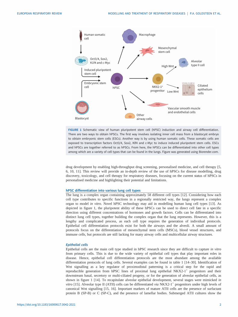
hPSC differentiation into various lung cell typesThe lung is a complex organ containing approximately 58 different cell types [12]. Considering how eachcell type contributes to specific functions in a regionally restricted way, the lungs represent a complexorgan to model in vitro. Novel hPSC technology may aid in modelling human lung cell types [13]. Asdepicted in figure 1, the pluripotent ability of these hPSCs can be used to direct cell fate in a specificdirection using different concentrations of hormones and growth factors. Cells can be differentiated intodistinct lung cell types, together building the complex organ that the lung represents. However, this is alengthy and complicated process, as each cell type requires the generation of individual protocols.Epithelial cell differentiation protocols exist for both the airways and the alveoli. A small amount ofprotocols focus on the differentiation of mesenchymal stem cells (MSCs), blood vessel structures, andimmune cells, but protocols are still lacking for many airway cells and multicellular airway structures.
Epithelial cellsEpithelial cells are the main cell type studied in hPSC research since they are difficult to capture in vitrofrom primary cells. This is due to the wide variety of epithelial cell types that play important roles indisease. Hence, epithelial cell differentiation protocols are the most abundant among the availabledifferentiation protocols of lung cells. Several examples can be found in table 1 [14–30]. Identification ofWnt signalling as a key regulator of proximodistal patterning is a critical step for the rapid andreproducible generation from hPSC lines of proximal lung epithelial NKX2-1+ progenitors and theirdownstream basal, secretory or multi-ciliated progeny, or for the generation of alveolar epithelial cells, asshown in figure 1 [14]. To recapitulate alveolar epithelial development, several stages were mimicked invitro [15]. Alveolar type II (ATII) cells can be differentiated via NKX2-1+ progenitors under high levels ofcanonical Wnt signalling [15, 16]. Important markers of mature ATII cells are the presence of surfactantprotein B (SP-B) or C (SP-C), and the presence of lamellar bodies. Submerged ATII cultures show the
Macrophage
Mesenchymal
stem cell
Alveolar
type II cellHigh Wnt
Low Wnt
Ciliated
epithelium
cells
Vascular smooth muscle
and endothelial cellsOther
airway cellsBlastocyst
Human somatic
cell
Oct3/4, Sox2,
K1f4 and c-Myc
Induced pluripotent
stem cell
Embryonic stem
cell hPSC NKX2-1+
progenitor
FIGURE 1 Schematic view of human pluripotent stem cell (hPSC) induction and airway cell differentiation.There are two ways to obtain hPSCs. The first way involves isolating inner cell mass from a blastocyst embryoto obtain embryonic stem cells (ESCs). Another way is by using human somatic cells. These somatic cells areexposed to transcription factors Oct3/4, Sox2, Klf4 and c-Myc to induce induced pluripotent stem cells. ESCsand hPSCs are together referred to as hPSCs. From here, the hPSCs can be differentiated into other cell typesamong which are a variety of cell types that can be found in the lungs. Figure was generated using Biorender.com.
https://doi.org/10.1183/16000617.0042-2021 2
EUROPEAN RESPIRATORY REVIEW MODELLING AND TREATMENT OF RESPIRATORY DISEASES | P.A. GOLDSTEEN ET AL.
presence of surfactant proteins [15]. It can be argued, however, that immature ATII cells can also expresssurfactant protein mRNA. The presence of lamellar bodies confirms a fully mature ATII phenotype, whichis only observed after the addition of an air–liquid interface (ALI) step; the air exposure step at the luminalside being an additional stimulus for differentiation [16].
Alveolar organoids were generated via NKX2-1+ progenitors as well [17]. Purified hPSC-derived SPC+
cells form monolayered alveolar organoids exhibit self-renewal capacity, and display additional ATIIfunctional capacities, including innate immune responsiveness and the production of lamellar bodies ableto package surfactant. It is important to note that the generated alveolar organoids mainly represent fetalATII cells, and not adult ATII cells, as shown by RNA-sequencing expression profiles. Moreover, thepresence of ATII cells was not observed in 3D culture models. ATII differentiation was only observedupon plating of the organoids in ALIs [17]. Next to alveolar organoids, proximal airway organoids weregenerated from hPSCs, showing ciliary beating in 3D ALI cultures [14, 18]. For the generation of proximalairway organoids, including multi-ciliated airway cells (MCACs) and pulmonary neuroendocrine cells,NKX2-1+ progenitors were treated with fibroblast growth factor-10 (FGF10) and a Wnt activator. ALIculture was crucial for the induced MCACs to demonstrate motile cilia generating flow for mucociliary
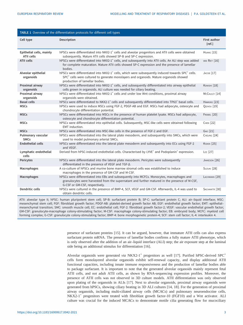
TABLE 1 Overview of the differentiation protocols for different cell types
Cell type Description First author[ref.]
Epithelial cells, mainlyATII cells
hPSCs were differentiated into NKX2-1+ cells and alveolar progenitors and ATII cells were obtainedsubsequently. Mature ATII cells showed SP-B and SP-C expression.
HUANG [15]
ATII cells hPSCs were differentiated into NKX2-1+ cells, and subsequently into ATII cells. An ALI step was addedfor complete maturation. Mature ATII cells showed SP-C expression and the presence of lamellarbodies.
VAN RIET [16]
Alveolar epithelialorganoids
hPSCs were differentiated into NKX2-1+ cells, which were subsequently induced towards SPC+ cells.SPC+ cells were cultured to generate monolayers and organoids. Mature organoids showedproduction of lamellar bodies.
JACOB [17]
Proximal airwayorganoids
hPSCs were differentiated into NKX2-1+ cells, and subsequently differentiated into airway epithelialcells grown in organoids. ALI culture was needed for ciliary beating.
KONISHI [18]
Proximal airwayorganoids
hPSCs were differentiated into NKX2-1+ cells and under low Wnt conditions, proximal airwayorganoids were obtained.
MCCAULEY [14]
Basal cells hPSCs were differentiated to NKX2-1+ cells and subsequently differentiated into TP63+ basal cells. HAWKINS [23]MSCs hPSCs were used to induce MSCs using FGF-2, PDGF-AB and EGF. MSCs had adipocyte, osteocyte and
chondrocyte differentiation potential.QIZHOU [19]
MSCs hPSCs were differentiated into MSCs in the presence of human platelet lysate. MSCs had adipocyte,osteocyte and chondrocyte differentiation potential.
FROBEL [20]
MSCs hPSCs were differentiated into epithelial cells. Subsequently, MSC-like cells were obtained followingEMT induction.
CHEN [22]
MSCs hPSCs were differentiated into MSC-like cells in the presence of FGF-2 and EGF. GAO [21]Pulmonary vascularSMCs
hPSCs were differentiated into the lateral plate mesoderm, and subsequently into SMCs, which wereused to model pulmonary arterial SMCs.
CHEUNG [24]
Endothelial cells hPSCs were differentiated into the lateral plate mesoderm and subsequently into ECs using FGF-2and VEGF.
KISKIN [25]
Lymphatic endothelialcells
Derived from hPSC-induced endothelial cells. Characterised by LYVE+ and Podoplanin+ expression. LEE [27]
Pericytes hPSCs were differentiated into the lateral plate mesoderm. Pericytes were subsequentlydifferentiated in the presence of VEGF and TGF-β.
JAMIESON [26]
Macrophages A co-culture of hPSCs and murine bone marrow stromal cells was established to inducemacrophages in the presence of GM-CSF and M-CSF.
SUZUKI [28]
Macrophages hPSCs were differentiated into EBs and subsequently into MCFCs. Monocytes, macrophages andgranulocytes were harvested from the supernatant and further matured in the presence of M-CSF,G-CSF or GM-CSF, respectively.
LACHMANN [29]
Dendritic cells hPSCs were cultured in the presence of BMP-4, SCF, VEGF and GM-CSF. Afterwards, IL-4 was used toobtain dendritic cells.
SACHAMITR [30]
ATII: alveolar type II; hPSC: human pluripotent stem cell; SP-B: surfactant protein B; SP-C: surfactant protein C; ALI: air–liquid interface; MSC:mesenchymal stem cell; FGF: fibroblast growth factor; PDGF-AB: platelet-derived growth factor AB; EGF: endothelial growth factor; EMT: epithelial–mesenchymal transition; SMC: smooth muscle cell; EC: endothelial cell; FGF-2: fibroblast growth factor-2; VEGF: vascular endothelial growth factor;GM-CSF: granulocyte-macrophage colony-stimulating factor; M-CSF: macrophage colony-stimulating factor; EB: embryoid body; MCFC: myeloid cellforming complex; G-CSF: granulocyte colony-stimulating factor; BMP-4: bone morphogenetic protein-4; SCF: stem cell factor; IL-4: interleukin 4.
https://doi.org/10.1183/16000617.0042-2021 3
EUROPEAN RESPIRATORY REVIEW MODELLING AND TREATMENT OF RESPIRATORY DISEASES | P.A. GOLDSTEEN ET AL.
transport. Alveolar and airway organoids both serve as great tools to study human epithelial cells in vitro,complementary in which air exposure seems crucial to obtain mature cells [14, 18]. The use of ALIcultures optimises in vitro cultures using hPSC-derived epithelial cells, as ALI cultures more closelyresemble the air-exchange physiology, mimicking the epithelial function seen in vivo.
MSCsMSCs are precursors of a variety of structural cells such as airway smooth muscle (ASM) cells andfibroblasts. Protocols for full differentiation of airway structural cells have been lacking so far. Still,protocols for hPSC-derived stromal MSCs have been established. However, the protocols are not abundantand the differentiation methods show high variation, reflecting the high degree of heterogeneity and tissuespecificity of MSCs. Different strategies include the addition of FGF-2, platelet-derived growth factor AB,epidermal growth factor or human platelet lysate [19–21], or generating stromal MSCs based onepithelial-to-mesenchymal transition [22]. The generated stromal MSCs are characterised by cellmorphology and potency for osteogenic, adipogenic and chondrogenic differentiation [19, 20], and areoften referred to as MSC-like cells as they are not tissue specific whereas endogenous MSCs are [21, 22].Several examples of protocols can be found in table 1. Interestingly, all the above-described protocols didnot yield ASM-specific differentiation. Stromal MSCs are precursors of ASM cells as well [31]. Theavailability of ASM cells would be useful for studies of bronchoconstriction mechanisms and airwayhyperresponsiveness.
Endothelial cells and blood vessel structuresBlood vessel structures such as smooth muscle cells (SMCs), endothelial cells (ECs) and pericytes can alsobe differentiated from hPSCs. Upon obtaining the lateral plate mesoderm, cells were cultured in smoothmuscle differentiation medium in the presence of platelet-derived growth factor BB and transforminggrowth factor-β (TGF-β) for an additional 12 days and mature SMCs were produced [24]. ECs wereobtained via the lateral plate mesoderm as well, followed by culture in FGF-2 and vascular endothelialgrowth factor (VEGF) [25]. In addition, pericytes can be generated to form the microvasculature. Pericytesare also obtained from the mesoderm and directed in the fate of early vascular cells in the presence ofVEGF and TGF-β continued with DMEM with 40% fetal bovine serum to induce pericyte maturation [26].The possibility of differentiating stem cell derived ECs to a lymphatic phenotype characterised by LYVE+Podoplanin+ expression and positivity for VEGF-A and -C has also been demonstrated, although such cellmodels have not yet been used extensively for respiratory research [27].
Macrophages and dendritic cellsMacrophages can be differentiated from hPSCs in the presence of colony stimulating factors (CSFs), suchas granulocyte-macrophage (GM) CSFs and macrophage (M) CSFs [28, 29]. After co-culturing hPSCs andmurine bone marrow stromal cells in the presence of GM-CSF and M-CSF, macrophage precursors wereisolated by sorting for CD235a−/CD41a− cells, followed by isolating CD45+ cells. Recovered cells werecultured in the presence of GM-CSF and M-CSF for another 7 days in order to generate maturedifferentiated macrophages. Mature hPSC-derived macrophages showed functions similar to in vivomacrophages, like tumour necrosis factor-α (TNF-α) release and surfactant clearance [28]. Another methodto derive macrophages is via embryoid body (EB) formation, in which EBs were cultivated in the presenceof interleukin (IL)-3 and CSFs (GM-CSF, M-CSF or granulocyte (G)-CSF) to generate myeloid cellforming complexes (MCFCs), producing type I macrophages, type II macrophages or granulocytes,respectively. Monocytes, macrophages or granulocytes generated by the MCFCs were harvested from thesupernatant. For further maturation, the monocytes, macrophages or granulocytes were cultured for another7–10 days in the presence of M-CSF, G-CSF or GM-CSF, respectively. Mature macrophages were capableof secretion of TNF-α and several cytokines like IL-8 [29]. The production of macrophages from MCFCshas already been shown to be available for up-scaled production in large stirred-tank bioreactors. MCFCswere induced from EBs in an orbital shaker, in which the MCFCs started to continuously produceimmature macrophages that could be harvested weekly for up to 3 months. After another week ofmaturation, the harvested macrophages represented a homogenous population of CD45+CD11b+ cells withclassical macrophage-like morphology and were able to phagocytose Escherichia coli bacteria [32].Differentiation of hPSCs to macrophages seems to be successful in the presence of M-CSF. However,dendritic cells (DCs; antigen-presenting cells) are still difficult to mimic in vitro. A protocol has beendeveloped to generate DCs but with a low efficiency. For hPSC-derived DCs, hPSCs were cultured in bonemorphogenetic protein 4 (BMP-4), stem cell factor, VEGF and GM-CSF. IL-4 was added sequentially inincreasing concentrations. By day 20, floating immature DCs were harvested and matured further toCD141+ cells in the presence of GM-CSF, IL-4, interferon-γ (IFN-γ), TNF-α, IL-1β, and prostaglandin E2.However, the levels of the specific lineage markers for CD141+ cells were quite low and the DCs do notsurpass the fetal phenotype [30]. An overview of available differentiation protocols is provided in table 1.
https://doi.org/10.1183/16000617.0042-2021 4
EUROPEAN RESPIRATORY REVIEW MODELLING AND TREATMENT OF RESPIRATORY DISEASES | P.A. GOLDSTEEN ET AL.

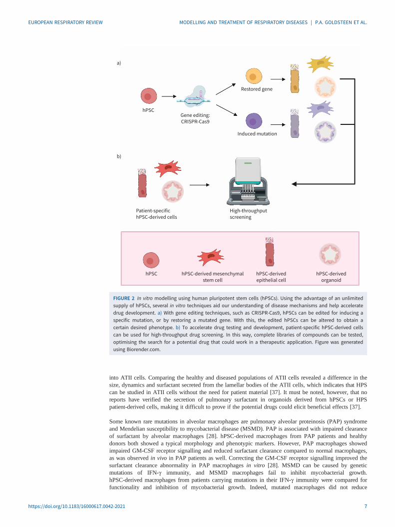
In vitro disease modelling and drug screeningUsing hPSC-derived cells, in vitro human disease modelling is now possible for genetic respiratorydiseases like cystic fibrosis (CF), and for modelling of pathologies like fibrosis and viral infections,including coronavirus disease 2019 (COVID-19) (see table 2 for examples). In addition, lung organoidshave proven to be highly suitable for modelling human lung development in vitro. Figure 2 shows some ofthe main advantages that hPSCs have over primary cells and cell lines. hPSCs can be genome-edited, bothto rescue the diseased phenotype or to incorporate mutations, which helps in the identification of keyregulatory genes. Modifying specific genes using, for example, CRISPR-Cas9 to study the mechanisms ofdisease-dependent genotype is possible in hPSCs without the ethical concerns one would face whenworking in vivo (figure 2a) [53]. Furthermore, hPSCs aid in drug discovery, for example by implementinghigh-throughput screening platforms (figure 2b). High-throughput screening platforms using automatedequipment can help to rapidly test a high number of samples, facilitating screening for new biomarkersand/or optimal treatment in in vitro disease models [34, 40].
CFCF is a respiratory disease that can be caused by mutations in the CF transmembrane conductanceregulator (CFTR) gene [54]. Due to its well-understood single-gene mutations, CF was one of the firstrespiratory diseases studied using hPSCs [55]. Patient-derived hPSCs enable: 1) the study of mechanismsunderlying CF in human in vitro models instead of mice; 2) the use of gene technology to repair specificCF mutations; and 3) the use of patient-derived CF-cells and organoids in screens for personalisedtreatment [55]. WONG et al. [33] were the first to show the potency of in vitro disease modelling usinghPSCs as a tool for personalised disease modelling, using hPSC-derived epithelial cells in ALI cultures.Epithelial cells harbouring the F508del mutation (which accounts for 70% of mutations in CF patients)appeared to have a dysfunctional CFTR. When treated in vitro with a small molecule, a so-called correctorcompound similar to VX-809, hPSC-derived CF-epithelial cells showed partial restoration of CFTRfunction. VX-809 is already known to correct CFTR function in patients who are homozygous for F508del[56]. Small patches of CFTR appeared on the cell surface following treatment with the VX-809-likecompound C18, while these CFTR channels were absent in untreated cultures [33]. Next, gene correctionof the F508del mutation resulted in full restoration of CFTR function in organoids. Compound screeningwith already known drugs partially rescued CFTR function by treatment with VX-770 and VX-809, as ithas been previously shown in CF patients, confirming the usefulness of hPSCs as an accurate human invitro model [34]. To take the automated compound screening to a next level, halide-sensitive yellowfluorescent protein was included into F508del CF organoids for automated quantitative measurement ofCFTR function. Fluorescent quantitative measurement enables high-throughput drug screening platformsthat may lead to the identification of novel drugs in a way that is superior compared to screening in celllines [34]. The study of the monogenic disease CF in hPSCs is more advanced compared to more complexdiseases. Current CF research using hPSCs has mainly focused on the predominant F508del mutation, butthere is no reason to expect that hPSC-derived CF epithelial cells could not be used for other mutations.Once hPSC models for in vitro disease modelling and drug research have been established for geneticdiseases like CF, researchers can look further into using these hPSC-derived models to expand theirunderstanding to more heterogeneous diseases like COPD and asthma.
Rare mutationsSeveral rare genetic mutations affect epithelial cells and are lethal because of altered surfactant production.SP-B deficiency is a fatal disease affecting neonates, as mutations in the SP-B gene result in a decrease insecretion and an abnormal composition of this surfactant. To demonstrate the effects of genome editing,hPSCs derived from SP-B-deficient patients were altered with a lentivirus carrying the healthy SP-B gene.Both SP-B-deficient and SP-B-corrected hPSCs were differentiated into lung organoids. Correctedorganoids showed SP-B protein, the presence of normal lamellar bodies, and secretion of surfactant in theculture medium, suggesting that the mutation can be targeted and modified to rescue the disease phenotypein vitro [35].
Hermansky–Pudlak syndrome (HPS) is a single-mutation disorder that can cause early onset pulmonaryfibrosis (PF). Using CRISPR-Cas9, several HPS mutations can be incorporated into hPSCs andsubsequently differentiated into lung organoids to induce fibrotic changes. Fibrotic changes werecharacterised by increased gene expression of collagens and markers for mesenchymal cells, and byextracellular matrix (ECM) deposition. Using these organoids, RNA expression profiles from genome-wideassociation studies were used to find common upregulated genes that cause fibrosis, like IL-11. IL-11induced ECM changes in control organoids as well, and its deletion prevented a fibrotic phenotype inHPS-mutant organoids. This way, hPSCs aid in understanding the molecular mechanism of HPS in vitro [36].Furthermore, hPSCs from HPS patients and their gene-corrected variant were generated and differentiated
https://doi.org/10.1183/16000617.0042-2021 5
EUROPEAN RESPIRATORY REVIEW MODELLING AND TREATMENT OF RESPIRATORY DISEASES | P.A. GOLDSTEEN ET AL.
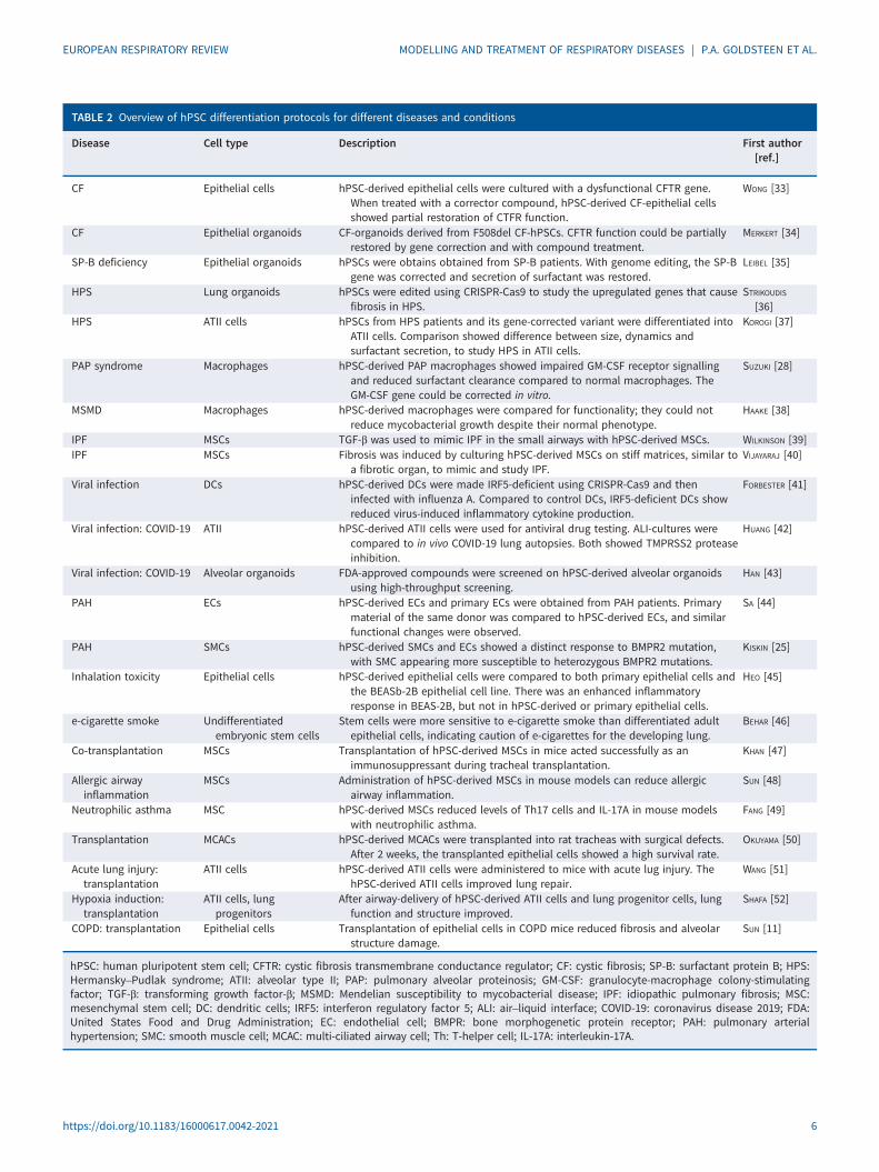
TABLE 2 Overview of hPSC differentiation protocols for different diseases and conditions
Disease Cell type Description First author[ref.]
CF Epithelial cells hPSC-derived epithelial cells were cultured with a dysfunctional CFTR gene.When treated with a corrector compound, hPSC-derived CF-epithelial cellsshowed partial restoration of CTFR function.
WONG [33]
CF Epithelial organoids CF-organoids derived from F508del CF-hPSCs. CFTR function could be partiallyrestored by gene correction and with compound treatment.
MERKERT [34]
SP-B deficiency Epithelial organoids hPSCs were obtains obtained from SP-B patients. With genome editing, the SP-Bgene was corrected and secretion of surfactant was restored.
LEIBEL [35]
HPS Lung organoids hPSCs were edited using CRISPR-Cas9 to study the upregulated genes that causefibrosis in HPS.
STRIKOUDIS[36]
HPS ATII cells hPSCs from HPS patients and its gene-corrected variant were differentiated intoATII cells. Comparison showed difference between size, dynamics andsurfactant secretion, to study HPS in ATII cells.
KOROGI [37]
PAP syndrome Macrophages hPSC-derived PAP macrophages showed impaired GM-CSF receptor signallingand reduced surfactant clearance compared to normal macrophages. TheGM-CSF gene could be corrected in vitro.
SUZUKI [28]
MSMD Macrophages hPSC-derived macrophages were compared for functionality; they could notreduce mycobacterial growth despite their normal phenotype.
HAAKE [38]
IPF MSCs TGF-β was used to mimic IPF in the small airways with hPSC-derived MSCs. WILKINSON [39]IPF MSCs Fibrosis was induced by culturing hPSC-derived MSCs on stiff matrices, similar to
a fibrotic organ, to mimic and study IPF.VIJAYARAJ [40]
Viral infection DCs hPSC-derived DCs were made IRF5-deficient using CRISPR-Cas9 and theninfected with influenza A. Compared to control DCs, IRF5-deficient DCs showreduced virus-induced inflammatory cytokine production.
FORBESTER [41]
Viral infection: COVID-19 ATII hPSC-derived ATII cells were used for antiviral drug testing. ALI-cultures werecompared to in vivo COVID-19 lung autopsies. Both showed TMPRSS2 proteaseinhibition.
HUANG [42]
Viral infection: COVID-19 Alveolar organoids FDA-approved compounds were screened on hPSC-derived alveolar organoidsusing high-throughput screening.
HAN [43]
PAH ECs hPSC-derived ECs and primary ECs were obtained from PAH patients. Primarymaterial of the same donor was compared to hPSC-derived ECs, and similarfunctional changes were observed.
SA [44]
PAH SMCs hPSC-derived SMCs and ECs showed a distinct response to BMPR2 mutation,with SMC appearing more susceptible to heterozygous BMPR2 mutations.
KISKIN [25]
Inhalation toxicity Epithelial cells hPSC-derived epithelial cells were compared to both primary epithelial cells andthe BEASb-2B epithelial cell line. There was an enhanced inflammatoryresponse in BEAS-2B, but not in hPSC-derived or primary epithelial cells.
HEO [45]
e-cigarette smoke Undifferentiatedembryonic stem cells
Stem cells were more sensitive to e-cigarette smoke than differentiated adultepithelial cells, indicating caution of e-cigarettes for the developing lung.
BEHAR [46]
Co-transplantation MSCs Transplantation of hPSC-derived MSCs in mice acted successfully as animmunosuppressant during tracheal transplantation.
KHAN [47]
Allergic airwayinflammation
MSCs Administration of hPSC-derived MSCs in mouse models can reduce allergicairway inflammation.
SUN [48]
Neutrophilic asthma MSC hPSC-derived MSCs reduced levels of Th17 cells and IL-17A in mouse modelswith neutrophilic asthma.
FANG [49]
Transplantation MCACs hPSC-derived MCACs were transplanted into rat tracheas with surgical defects.After 2 weeks, the transplanted epithelial cells showed a high survival rate.
OKUYAMA [50]
Acute lung injury:transplantation
ATII cells hPSC-derived ATII cells were administered to mice with acute lug injury. ThehPSC-derived ATII cells improved lung repair.
WANG [51]
Hypoxia induction:transplantation
ATII cells, lungprogenitors
After airway-delivery of hPSC-derived ATII cells and lung progenitor cells, lungfunction and structure improved.
SHAFA [52]
COPD: transplantation Epithelial cells Transplantation of epithelial cells in COPD mice reduced fibrosis and alveolarstructure damage.
SUN [11]
hPSC: human pluripotent stem cell; CFTR: cystic fibrosis transmembrane conductance regulator; CF: cystic fibrosis; SP-B: surfactant protein B; HPS:Hermansky–Pudlak syndrome; ATII: alveolar type II; PAP: pulmonary alveolar proteinosis; GM-CSF: granulocyte-macrophage colony-stimulatingfactor; TGF-β: transforming growth factor-β; MSMD: Mendelian susceptibility to mycobacterial disease; IPF: idiopathic pulmonary fibrosis; MSC:mesenchymal stem cell; DC: dendritic cells; IRF5: interferon regulatory factor 5; ALI: air–liquid interface; COVID-19: coronavirus disease 2019; FDA:United States Food and Drug Administration; EC: endothelial cell; BMPR: bone morphogenetic protein receptor; PAH: pulmonary arterialhypertension; SMC: smooth muscle cell; MCAC: multi-ciliated airway cell; Th: T-helper cell; IL-17A: interleukin-17A.
https://doi.org/10.1183/16000617.0042-2021 6
EUROPEAN RESPIRATORY REVIEW MODELLING AND TREATMENT OF RESPIRATORY DISEASES | P.A. GOLDSTEEN ET AL.
into ATII cells. Comparing the healthy and diseased populations of ATII cells revealed a difference in thesize, dynamics and surfactant secreted from the lamellar bodies of the ATII cells, which indicates that HPScan be studied in ATII cells without the need for patient material [37]. It must be noted, however, that noreports have verified the secretion of pulmonary surfactant in organoids derived from hPSCs or HPSpatient-derived cells, making it difficult to prove if the potential drugs could elicit beneficial effects [37].
Some known rare mutations in alveolar macrophages are pulmonary alveolar proteinosis (PAP) syndromeand Mendelian susceptibility to mycobacterial disease (MSMD). PAP is associated with impaired clearanceof surfactant by alveolar macrophages [28]. hPSC-derived macrophages from PAP patients and healthydonors both showed a typical morphology and phenotypic markers. However, PAP macrophages showedimpaired GM-CSF receptor signalling and reduced surfactant clearance compared to normal macrophages,as was observed in vivo in PAP patients as well. Correcting the GM-CSF receptor signalling improved thesurfactant clearance abnormality in PAP macrophages in vitro [28]. MSMD can be caused by geneticmutations of IFN-γ immunity, and MSMD macrophages fail to inhibit mycobacterial growth.hPSC-derived macrophages from patients carrying mutations in their IFN-γ immunity were compared forfunctionality and inhibition of mycobacterial growth. Indeed, mutated macrophages did not reduce
hPSC
hPSC hPSC-derived mesenchymal
stem cell
hPSC-derived
epithelial cell
hPSC-derived
organoid
Restored gene
Induced mutation
Gene editing:
CRISPR-Cas9
Patient-specific
hPSC-derived cells
High-throughput
screening
a)
b)
FIGURE 2 In vitro modelling using human pluripotent stem cells (hPSCs). Using the advantage of an unlimitedsupply of hPSCs, several in vitro techniques aid our understanding of disease mechanisms and help acceleratedrug development. a) With gene editing techniques, such as CRISPR-Cas9, hPSCs can be edited for inducing aspecific mutation, or by restoring a mutated gene. With this, the edited hPSCs can be altered to obtain acertain desired phenotype. b) To accelerate drug testing and development, patient-specific hPSC-derived cellscan be used for high-throughput drug screening. In this way, complete libraries of compounds can be tested,optimising the search for a potential drug that could work in a therapeutic application. Figure was generatedusing Biorender.com.
https://doi.org/10.1183/16000617.0042-2021 7
EUROPEAN RESPIRATORY REVIEW MODELLING AND TREATMENT OF RESPIRATORY DISEASES | P.A. GOLDSTEEN ET AL.
mycobacterial growth when challenged with bacteria, despite their normal phenotype [38]. Asdemonstrated in the examples above, hPSCs can be used to model the pathogenic molecular and cellularabnormalities in lung cells that drive genetic lung disease.
Lung development and neonatal diseasesMany hPSC-derived epithelial organoids do not fully mature and represent a fetal state [57]. The lungorganoids resemble the bronchi/bronchioles of the developing human airway in terms of cells and structureand are surrounded by lung mesenchyme and cells expressing alveolar cell markers. The bud tip progenitororganoids possess a population of highly proliferative multipotent cells with in vitro multilineagedifferentiation potential. After benchmarking the organoids models, their characterisation showed to berepresentative of human fetal-like tissue. This is a major problem, as many lung diseases are diseases thatare typical for a post-natal or adult lung. It is crucial that the lung airway organoids also represent thispost-natal/adult stage to accurately model the disease. There is a need to improve cell maturation. Strategiescan include time, medium supplementation, or interaction with other cells to promote cell maturation. Forexample, for alveolar cells, air exposure might be the way to go to induce maturation of ATI and ATIIepithelial cells [16]. Nevertheless, organoids serve as an excellent platform for the study of neonatal andinfant diseases, as primary material is mainly available from adult donors. This enables for the first timethe study of lung development in vitro in human models, looking at both healthy lung development anddefects upon genetic mutations that can affect lung development [57]. Also, the exact nature of thebranching process is not yet known. Currently, the process of branching seems spontaneous andspace-filling. These organoids can be used to study the mechanism of branching in the future [58].
Pulmonary fibrosisFibrosis can be mimicked in mesenchymal organoids using several techniques, without known geneticalterations [39, 40]. A first method to mimic PF in the small airways is by using TGF-β in concentrationsthat would induce cell alterations. hPSC-derived mesenchymal organoids were cultured in the presence offibroblast-coated alginate beads, which represented the lung alveolar structure. Organoids treated withTGF-β showed an increased expression of collagen 1 and α-smooth muscle actin (SMA), indicators offibrotic processes in organoids. Furthermore, the morphological and mechanical effects of PF weremimicked in vitro by an altered shape and increased contraction of the organoid [39]. Secondly, to inducefibrosis by altering the ECM scaffold to culture the cells, MSC-like cells were cultured on stiff (13 kPa)matrices that approximate the stiffness of a fibrotic organ. It was observed that MSC-like cells grown onthese substrates showed a highly proliferative phenotype and formed a dense, scar-like structure.Consistently, both the gene- and protein-expression levels of collagen I, α-SMA and TGF-β wereupregulated in hPSC-MSC-like cells compared to primary fibroblasts grown on stiff substrates. ThesehPSC-induced models of fibrosis using hPSC-derived MSCs can help to understand disease mechanismsand aid the identification of novel drugs using high-throughput screening platforms [34].
Viral infectionThe human respiratory system is often targeted by viral infections. hPSC-derived organoids and immunecells provide a great toolbox to understand the mechanisms of viral infection. In hPSC-derived DCs, thecrucial pathways in innate immune cells upon viral infections could be identified using targeted genomeediting. Interferon regulatory factor 5 (IRF5) is a gene suspected to play an important role in the antiviralresponse. CRISPR-Cas9 was used to generate IRF5-deficient hPSC-derived DCs, which were infected withinfluenza A. IRF5-deficient DCs showed reduced virus-induced inflammatory cytokine production outimpacting virus replication and a reduced type 1 IFN secretion, compared to DCs without the IRF5-mutation. Performing this study in parallel to studies of human lung cells and IRF5-deficient micedemonstrated that IRF5 indeed mediated viral pathogenesis in vivo [41]. Such studies aid the identificationof key regulatory genes upon a viral response. Moreover, the infant lung is prone to many viral infections,of which the exact mechanisms are not yet known. Current models to study viral infections consist ofprimary epithelial cells from the bronchus, obtained from adult donors, so little is known about paediatricresponse to viruses. Organoids that represent the infant or developing distal lung can be used torecapitulate these disease conditions. Infection of lung organoids with several viral strains led to differentresponses of the organoids, which were in accordance with the in vivo viral response. For example,infection with parainfluenza virus led to viral shedding without morphological changes, while respiratorysyncytial virus infection induced detachment and shedding of infected cells into the lung organoid lumens.Furthermore, infection with childhood viral strains like measles led to syncytium formation in theorganoids, showing lung organoids as a representative in vitro model to study infant respiratory diseases[58, 59]. It must be noted, however, that viral infections are simulated by adding viral titres to submergedorganoid cultures. Another limitation of working with organoids is that due to the 3D nature of manyorganoids, the viral particles are offered at the basolateral side of the epithelium. A model with lung
https://doi.org/10.1183/16000617.0042-2021 8
EUROPEAN RESPIRATORY REVIEW MODELLING AND TREATMENT OF RESPIRATORY DISEASES | P.A. GOLDSTEEN ET AL.
organoids derived from adult stem cells showed that the model could be improved when organoids areALI-cultured, in contrast to the 3D morphology of organoids. This ALI set-up ensures exclusive apicalexposure to the virus, similar to the human route of viral infection in the lungs [60]. Interestingly, theaforementioned detachment and shedding of infected cells seen in organoids following respiratory syncytialvirus infection was not seen in ALI-cultured epithelial cells, indicating additional differences betweenthese model systems [61].
The current COVID-19 pandemic is a perfect opportunity for the scientific community in hPSC research toshow how these hPSC-derived models can aid our understanding of disease mechanisms and drugdiscovery. For example, hPSC-derived ATII cells on ALI cultures were used for testing antiviral drugs likeremesdevir. Simultaneously, the hPSC-derived ALI cultures were compared to in vivo COVID-19 lungautopsies, showing TMPRSS2 protease inhibition in both models. This is a clear example of understandingthe importance of both using ALI cultures to mature the ATII cells and simultaneously validating hPSCmodels with patient material [42]. Next, a high-throughput system was tested in which US Food and DrugAdministration-approved compounds were screened on hPSC-derived alveolar organoids. This representsan efficient method to select potential drug candidates for further clinical trials [43]. The development ofhPSC models will further aid drug development and facilitate the deeper understanding of COVID-19pathogenesis.
Pulmonary hypertensionPulmonary arterial hypertension (PAH) can be modelled using hPSCs by focusing on EC function in thepulmonary arteries. hPSC-derived ECs were validated and compared with primary cells obtained from thesame donor. hPSCs showed similar responses in migration, adhesion, and tube formation compared toprimary ECs from the same donor. In addition, transcriptomic profiling revealed that high kisspeptin 1 canbe related to reduced migration and low carboxylesterase 1 to impaired survival. Given the fact that thecellular responses of primary ECs and hPSC-derived ECs overlap within the same donor, it is proposedthat hPSCs are a useful tool in uncovering disease mechanisms and in designing tailored therapies forpatients [44]. hPSC-derived SMCs have been studied in relation to PAH as well and compared to an ECresponse in both control and two isogenic BMP receptor type 2 ( BMPR2)-mutated hPSC lines.Interestingly, in co-cultures of SMCs and ECs, both cell types exhibit a distinct response to the BMPR2mutation, with the SMCs being more susceptible to heterozygous BMPR2 mutations than the ECs inresponse to disease-triggering agents like TNF-α [25].
Validation of modelsFor both hPSC-derived cells and organoids models, human in vitro models will have to be validated beforethe new models can be integrated into research and drug developmental studies. Many studies validatetheir human in vitro models in vivo in mice, ex vivo in human slices, and in vitro with primary cells [40,42, 62]. Another opportunity for validation is by running organoid studies next to clinical trials that havebeen scheduled already, to get a more accurate validation if human in vivo responses are similar to humanin vitro responses [63]. Several clinical trials are currently running for setting up hPSC-supported in vitromodels. The INVECCO study (https://clinicaltrials.gov/ NCT03181204) is currently exploring if COPDcan be modelled in vitro using iPSCs. Primary bronchial epithelial cells are compared with hPSC-derivedepithelial cells, in which COPD is induced using tobacco. Another study (NCT03750734) follows a similarapproach to investigate whether iPSCs can help to unravel molecular pathways and identify biomarkers inseveral lung diseases including idiopathic bronchiectasis, COPD and CF. iPSC-derived epithelial cells willbe compared to primary bronchial epithelial cells. One study (NCT02594917) is collecting iPSCs frompatients with PAH in parallel with another study involving patients and healthy controls. The PaCyFICstudy (NCT03754088) has collected CF-iPSCs and healthy iPSCs as well. In addition, validating iPSCmodels next to running clinical trials is accelerating and optimising the validation of hPSC models.
Pluripotent stem cells as cell therapyThe clinical potential of hPSCs in respiratory diseases extends even further as they may also be useddirectly as cell therapeutics. The first in vitro and in vivo animal studies show promise for hPSC-basedtherapy, contributing to personalised medicine [47–51, 64]. Despite the first successes in vivo in animalstudies, cell therapy using hPSCs is not yet available for the patient. This is mainly due to the fact thathPSCs can cause teratoma formation in mice when transplanted into the lungs [65].
hPSC-derived MSCs as a treatmentCo-transplantations of MSCs improve the success rates of lung transplantations. The vulnerability of thetransplantation procedure is the rejection of the donor lung caused by an immune response of the recipient.Primary MSCs are known to attenuate immune responses, making them promising tools for use during
https://doi.org/10.1183/16000617.0042-2021 9
EUROPEAN RESPIRATORY REVIEW MODELLING AND TREATMENT OF RESPIRATORY DISEASES | P.A. GOLDSTEEN ET AL.
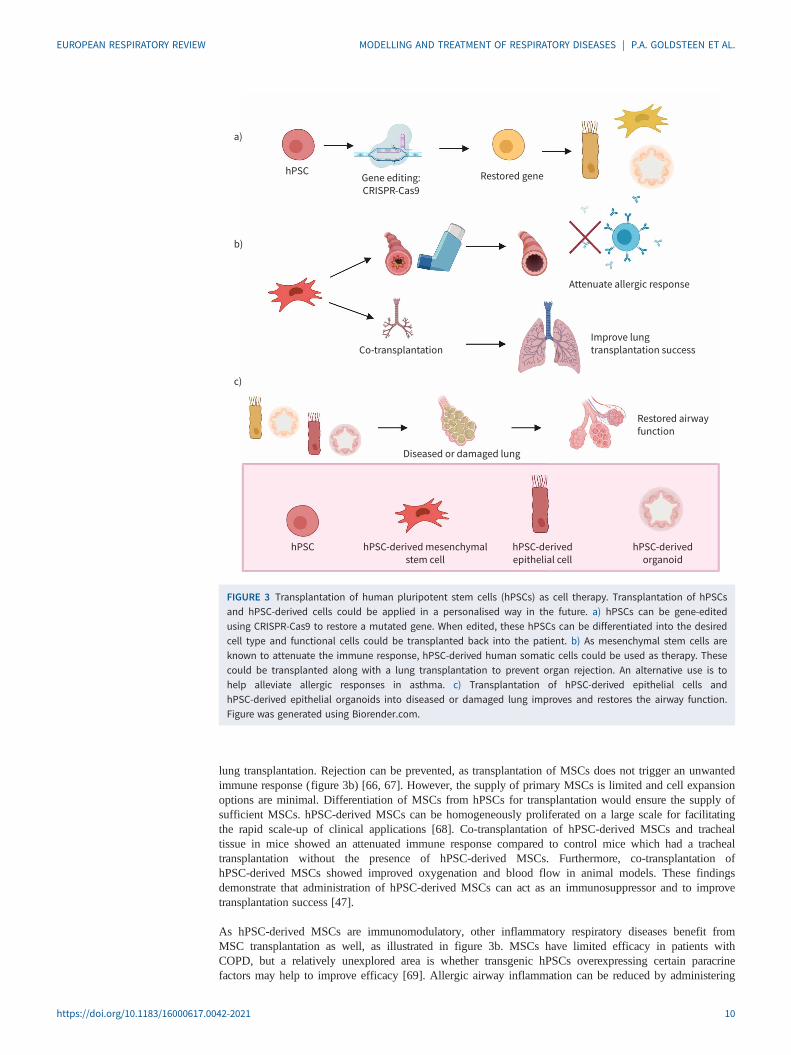
lung transplantation. Rejection can be prevented, as transplantation of MSCs does not trigger an unwantedimmune response (figure 3b) [66, 67]. However, the supply of primary MSCs is limited and cell expansionoptions are minimal. Differentiation of MSCs from hPSCs for transplantation would ensure the supply ofsufficient MSCs. hPSC-derived MSCs can be homogeneously proliferated on a large scale for facilitatingthe rapid scale-up of clinical applications [68]. Co-transplantation of hPSC-derived MSCs and trachealtissue in mice showed an attenuated immune response compared to control mice which had a trachealtransplantation without the presence of hPSC-derived MSCs. Furthermore, co-transplantation ofhPSC-derived MSCs showed improved oxygenation and blood flow in animal models. These findingsdemonstrate that administration of hPSC-derived MSCs can act as an immunosuppressor and to improvetransplantation success [47].
As hPSC-derived MSCs are immunomodulatory, other inflammatory respiratory diseases benefit fromMSC transplantation as well, as illustrated in figure 3b. MSCs have limited efficacy in patients withCOPD, but a relatively unexplored area is whether transgenic hPSCs overexpressing certain paracrinefactors may help to improve efficacy [69]. Allergic airway inflammation can be reduced by administering
hPSC hPSC-derived mesenchymal
stem cell
hPSC-derived
epithelial cell
hPSC-derived
organoid
Restored airway
function
Improve lung
transplantation success
Diseased or damaged lung
Co-transplantation
Attenuate allergic response
hPSCRestored geneGene editing:
CRISPR-Cas9
a)
b)
c)
FIGURE 3 Transplantation of human pluripotent stem cells (hPSCs) as cell therapy. Transplantation of hPSCsand hPSC-derived cells could be applied in a personalised way in the future. a) hPSCs can be gene-editedusing CRISPR-Cas9 to restore a mutated gene. When edited, these hPSCs can be differentiated into the desiredcell type and functional cells could be transplanted back into the patient. b) As mesenchymal stem cells areknown to attenuate the immune response, hPSC-derived human somatic cells could be used as therapy. Thesecould be transplanted along with a lung transplantation to prevent organ rejection. An alternative use is tohelp alleviate allergic responses in asthma. c) Transplantation of hPSC-derived epithelial cells andhPSC-derived epithelial organoids into diseased or damaged lung improves and restores the airway function.Figure was generated using Biorender.com.
https://doi.org/10.1183/16000617.0042-2021 10
EUROPEAN RESPIRATORY REVIEW MODELLING AND TREATMENT OF RESPIRATORY DISEASES | P.A. GOLDSTEEN ET AL.
hPSC-derived MSCs intravenously in mouse models. Short-term effects of an allergic mouse asthmamodel included an inhibition of inflammatory cell infiltration and mucus production in the lung and areduction in eosinophil infiltration in the nose. In addition, in the bronchoalveolar and nasal lavage fluids,there was a decrease in inflammatory cell infiltration plus reduced serum levels of T-helper cell (Th) type 2immunoglobulins and cytokines (including IL-4, IL-5 and IL-13) [48]. Long-term protective effects ofhPSC-MSCs in a chronic mouse asthma model included protection of the mice from the characteristics thatare typical for chronic allergic airway inflammation. hPSC-derived MSCs improved airway remodellingand prevented fibrosis. The TGF-β/Smad pathway is crucial in the immunomodulatory effects exerted byhPSC-derived MSCs in a chronic mice asthma model [64]. A PCR array performed in mice treated withhPSC-MSCs was used to identify the key regulatory genes involved in the immunomodulatorymechanisms of these MSCs. In mouse asthma models, chemokine (C-C motif ) ligand (Ccl) 11, Ccl24,IL-13, IL-33 and eosinophil-associated, ribonuclease A family member 11 (Ear11) were found to play aprominent role in MSC-immunomodulation [70]. Next to allergic asthma, in steroid-resistant neutrophilicasthma, MSC transplantation was presented as a promising therapy. Systemic administration ofhPSC-derived MSCs in a mouse model decreased the levels of Th17 cells and IL-17A. Furthermore,neutrophilic airway inflammation was significantly lower 48 h after ovalbumin and lipopolysaccharidechallenge following MSC administration, when compared to control and dexamethasone treatment [49].All the above studies have been performed in mice and do not show long-term side-effects at least for aperiod of 2 months. However, these studies did not try to further investigate any potential side-effects2 months after the systemic administration of the hPSC-derived MSCs. Also, the translation to the humansituation is lacking. Clinical trials have already demonstrated the safe use of bone marrow-derived MSCsin asthma patients and hPSC-derived MSCs might be a following step [71].
Epithelial transplantation for regenerationEpithelial cell transplantation can also serve as personalised transplantation upon acute lung injury for boththe upper and the lower airways. hPSC-derived MCACs (hPSC-MCACs), as found in the upper airways,were transplanted into the trachea of rats with surgical defects [50]. Most of the transplanted epithelial cellssurvived following 1–2 weeks of transplantation, as assessed by counting the cilia beat frequency. Thishigh survival rate of functional epithelial cells makes hPSC-MCACs a promising candidate for trachealreconstruction [50]. Long-term safety and efficacy studies need to be performed, however, as so far onlyshort-term effects (2 weeks) have been modelled [50]. For established ciliated cells, it has beendemonstrated that the half-life is ∼6 months in mouse trachea, after which basal cells are necessary togenerate new epithelial cells [72]. As an alternative strategy, hPSCs can be induced towards a basal cellphenotype for transplantation, possibly overcoming this limitation [13].
hPSC-derived ATII administration to mice models with bleomycin-induced lung injury showed that thehPSC-ATII cells differentiated and expressed markers for alveolar epithelial type I cells, which in part candifferentiate into ATII cells in situ. Moreover, transplantation of hPSC-derived ATII cells in acute lunginjury promoted an increased arterial blood flow into the airways in mice, when compared with controlmice without transplantation [51]. In addition, airway delivery of both hPSC-derived ATII cells and lungprogenitor cells showed improved lung structure and function after hypoxia-induced bronchopulmonarydysplasia in neonatal mice. The epithelial transplantation resulted in a long-term engraftment withoutevidence of tumour formation [52]. These studies indicate that hPSC therapy can be safe, as long as cellsare no longer in an undifferentiated state. In a mouse model of COPD, transplantation of epithelial cellswas more successful, as transplantation of epithelial cells reduced fibrosis and reduced alveolar structuraldamage [11]. However, transplantation of hPSCs and hPSC-derived epithelial cells has been investigatedwith little success in conditions of premature birth in mice. hPSCs give rise to teratoma formation, andhPSC-derived epithelial cells did not show any improvement in lung development [73].
For in vivo therapy, hPSCs are an exciting and fairly new way to treat respiratory diseases. Most studieshave used them in mice models, and only a few have gone into clinical trials. None of these studies haveyet been related to any lung diseases [74]. A promising approach for hPSCs in transplantation is by usingdifferentiated hPSCs, like hPSC-derived MSCs or epithelial cells. However, no human in vivo data existsso far, neither to replicate the transplantation successes in mice, nor to confirm the absence of teratomaformation [64, 75]. Moreover, there are still unknown factors for the transplantation of hPSC-derived cells.For example, the dose of cells for transplantation is not yet known, or whether the engrafted cells aresufficiently representative of the local cells [76, 77].
What is concerning, even for differentiated hPSCs, is that a small fraction of ESCs and hPSCs accumulatedominant-negative mutations in tumour-suppressor genes over time, presenting a cancer risk to patients if
https://doi.org/10.1183/16000617.0042-2021 11
EUROPEAN RESPIRATORY REVIEW MODELLING AND TREATMENT OF RESPIRATORY DISEASES | P.A. GOLDSTEEN ET AL.
hPSC-derived cells are used as a therapeutic. Thus, careful genetic characterisation of hPSCs and theirdifferentiated derivatives should be performed before clinical use [78].
Limitations of hPSCsIn terms of disease modelling, as protocols emerge, is it must be noted that the differentiation of cells andgrowth of organoids from hPSCs are still expensive, labour-intensive, and face difficulties in obtainingreproducible levels of mature cells with sometimes high degrees of variability between hPSC lines [57, 59,79]. Even for well-established differentiation of epithelial cells, mature cells were obtained with varyingefficiencies [5, 15, 33]. Moreover, the organ- and tissue-specific features of cell types are not alwaysrecapitulated using hPSC differentiation. By co-culturing ECs with human intestinal organoids, the ECsobtained a phenotype that appeared more related to native intestinal ECs compared to kidney and lung[80]. Similar advantages are to be expected when co-culturing different lung cells.
Some in vitro models require multiple cell types to be integrated for an improved resemblance to thehuman in vivo situation. Co-culturing multiple cell types might elucidate disease mechanisms that wouldnot be exposed by culturing either cell type alone. For example, the study of alveolar repair inhPSC-derived epithelial cells without the presence of endothelial or other supporting cells makes it lessrelevant [35]. Currently, the most accessible way of having multiple cell types in one model is by thegeneration of lung organoids. For co-cultures of hPSC-derived cells that have been obtained individually,differentiation protocols must be timed in order to be mature together, which can be precarious. For humanintestinal organoids, in which the intestinal organoids were cultured together with neural crest cells todevelop a nervous system in the organoids, co-culturing both improved maturation of the cell types andrevealed cellular interactions between different cell types [81].
As transplantable cells for regenerative medicine, their use could be limited by several factors, includingtumorigenic potential, immune rejection, genetic instability, heterogeneity of iPSC lineage phenotypiccells, and lack of full maturity in some iPSC-derived phenotypic cells [82]. Their tumorigenic potential isbased on shared iPSCs characteristics with cancer cells, such as unlimited proliferation capacity and theexpression of c-Myc, an oncogenic marker. Several strategies currently under investigation focus onreducing or elimination of iPSC tumorigenic potential and include the use of extracellular vesicles releasedby iPSCs [83], the use of differentiated cells instead of undifferentiated iPSCs, the use of reprogrammingtransgene-free methods for the generation of iPSCs or c-Myc-independent reprogramming methods byemploying small molecules (quercetin and YM155) or pluripotent cell-specific inhibitors (oleic acids) [84].
ConclusionhPSCs are emerging as a promising model to study respiratory diseases in vitro. They can be differentiatedinto various lung cell types, aiding in modelling different diseases in vitro. Differentiation protocols forepithelial cells have been established and showed robust ATII differentiation to represent alveoli orrecapitulate the beating of cilia in the larger airways. However, protocols for the many other cell typespresent in the lungs are still scarce. Developing protocols for these non-epithelial cells will be a valuabletool for further drug-developmental studies for the airways, as primary material can be limited in supply.Even though differentiation protocols can be lengthy, hPSCs are applicable in different fields of respiratorydisease research. hPSC-derived models can aid in the understanding of disease mechanisms and unravelthe pathological genetic signature. High-throughput drug screening platforms can accelerate drug testingand personalised treatments. For the first time, rare genetic mutations can be understood by combininghPSC and gene-editing technologies. The first steps are being taken towards cell therapy using hPSCs.Transplantation of hPSC-derived MSCs and epithelial cells show promising results in mice models.However, beneficial effects are still to be replicated in human in vivo experiments and the safety of hPSCtransplantation is still being investigated.
Provenance: Submitted article, peer reviewed.
Conflict of interest: P.A. Goldsteen has nothing to disclose. C. Yoseif has nothing to disclose. A.M. Dolga hasnothing to disclose. R. Gosens reports financial support from Aquilo, Boehringer Ingelheim and Longfonds duringthe conduct of this study and financial support from Sanofi Aventis and Chiesi outside the submitted works.
Support statement: The work was financially supported by the More Knowledge with Fewer Animals (Meer Kennismet Minder Dieren) programme of ZonMW (grant number 114021505) with co-financing from StichtingProefdiervrij, Aquilo BV, Boehringer Ingelheim and Longfonds. Funding information for this article has beendeposited with the Crossref Funder Registry.
https://doi.org/10.1183/16000617.0042-2021 12
EUROPEAN RESPIRATORY REVIEW MODELLING AND TREATMENT OF RESPIRATORY DISEASES | P.A. GOLDSTEEN ET AL.

References1 Barnes PJ, Bonini S, Seeger W, et al. Barriers to new drug development in respiratory disease. Eur Respir J
2015; 45: 1197–1207.2 Bhowmick R, Gappa-Fahlenkamp H. Cells and culture systems used to model the small airway epithelium.
Lung 2016; 194: 419–428.3 Liu G, Betts C, Cunoosamy DM, et al. Use of precision cut lung slices as a translational model for the study of
lung biology. Respir Res 2019; 20: 162.4 Holmes AM, Solari R, Holgate ST. Animal models of asthma: value, limitations and opportunities for
alternative approaches. Drug Discov Today 2011; 16: 659–670.5 Dye BR, Miller AJ, Spence JR. How to grow a lung: applying principles of developmental biology to generate
lung lineages from human pluripotent stem cells. Curr Pathobiol Rep 2016; 4: 47–57.6 Mianné J, Ahmed E, Bourguignon C, et al. Induced pluripotent stem cells for primary ciliary dyskinesia
modeling and personalized medicine. Am J Respir Cell Mol Biol 2018; 59: 672–683.7 Miller AJ, Spence JR. In vitro models to study human lung development, disease and homeostasis. Physiology
(Bethesda) 2017; 32: 246–260.8 Takahashi K, Tanabe K, Ohnuki M, et al. Induction of pluripotent stem cells from adult human fibroblasts by
defined factors. Cell 2007; 131: 861–872.9 Singh VK, Kumar N, Kalsan M, et al. Mechanism of induction: induced pluripotent stem cells (iPSCs). J Stem
Cells 2015; 10: 43–6210 Nadkarni RR, Abed S, Draper JS. Organoids as a model system for studying human lung development and
disease. Biochem Biophys Res Commun 2016; 473: 675–682.11 Sun Z, Li F, Zhou X, et al. Stem cell therapies for chronic obstructive pulmonary disease: current status of
pre-clinical studies and clinical trials. J Thorac Dis 2018; 10: 1084–1098.12 Travaglini KJ, Nabhan AN, Penland L, et al. A molecular cell atlas of the human lung from single-cell RNA
sequencing. Nature 2020; 587: 619–625.13 Hawkins F, Kotton DN. Embryonic and induced pluripotent stem cells for lung regeneration. Ann Am Thorac
Soc 2015; 12: Suppl. 1, S50–S53.14 McCauley KB, Hawkins F, Serra M, et al. Efficient derivation of functional human airway epithelium from
pluripotent stem cells via temporal regulation of Wnt signaling. Cell Stem Cell 2017; 20: 844–857.15 Huang SXL, Green MD, de Carvalho AT, et al. The in vitro generation of lung and airway progenitor cells from
human pluripotent stem cells. Nat Protoc 2015; 10: 413–425.16 van Riet S, Ninaber DK, Mikkers HMM, et al. In vitro modelling of alveolar repair at the air-liquid interface
using alveolar epithelial cells derived from human induced pluripotent stem cells. Sci Rep 2020; 10: 5499.17 Jacob A, Morley M, Hawkins F, et al. Differentiation of human pluripotent stem cells into functional lung
alveolar epithelial cells. Cell Stem Cell 2017; 21: 472–488.18 Konishi S, Gotoh S, Tateishi K, et al. Directed induction of functional multi-ciliated cells in proximal airway
epithelial spheroids from human pluripotent stem cells. Stem Cell Reports 2016; 6: 18–25.19 Qizhou L, Yuelin Z, Jinqiu Z, et al. Functional mesenchymal stem cells derived from human induced
pluripotent stem cells attenuate limb ischemia in mice. Circulation 2010; 121: 1113–1123.20 Frobel J, Hemeda H, Lenz M, et al. Epigenetic rejuvenation of mesenchymal stromal cells derived from
induced pluripotent stem cells. Stem Cell Reports 2014; 3: 414–422.21 Gao W-X, Sun Y-Q, Shi J, et al. Effects of mesenchymal stem cells from human induced pluripotent stem cells
on differentiation, maturation, and function of dendritic cells. Stem Cell Res Ther 2017; 8: 48.22 Chen YS, Pelekanos RA, Ellis RL, et al. Small molecule mesengenic induction of human induced pluripotent
stem cells to generate mesenchymal stem/stromal cells. Stem Cells Transl Med 2012; 1: 83–95.23 Hawkins FJ, Suzuki S, Beermann ML, et al. Derivation of airway basal stem cells from human pluripotent
stem cells. Cell Stem Cell 2021; 28: 79–95.24 Cheung C, Bernardo AS, Trotter MWB, et al. Generation of human vascular smooth muscle subtypes provides
insight into embryological origin-dependent disease susceptibility. Nat Biotechnol 2012; 30: 165–173.25 Kiskin FN, Chang C-H, Huang CJZ, et al. Contributions of BMPR2 mutations and extrinsic factors to cellular
phenotypes of pulmonary arterial hypertension revealed by induced pluripotent stem cell modeling. Am JRespir Crit Care Med 2018; 198: 271–275.
26 Jamieson J, Macklin B, Gerecht S. Pericytes derived from human pluripotent stem cells. Adv Exp Med Biol2018; 1109: 111–124.
27 Lee S-J, Park C, Lee JY, et al. Generation of pure lymphatic endothelial cells from human pluripotent stemcells and their therapeutic effects on wound repair. Sci Rep 2015; 5: 11019.
28 Suzuki T, Mayhew C, Sallese A, et al. Use of induced pluripotent stem cells to recapitulate pulmonary alveolarproteinosis pathogenesis. Am J Respir Crit Care Med 2013; 189: 183–193.
29 Lachmann N, Ackermann M, Frenzel E, et al. Large-scale hematopoietic differentiation of human inducedpluripotent stem cells provides granulocytes or macrophages for cell replacement therapies. Stem Cell Rep2015; 4: 282–296.
https://doi.org/10.1183/16000617.0042-2021 13
EUROPEAN RESPIRATORY REVIEW MODELLING AND TREATMENT OF RESPIRATORY DISEASES | P.A. GOLDSTEEN ET AL.
30 Sachamitr P, Leishman AJ, Davies TJ, et al. Directed differentiation of human induced pluripotent stem cellsinto dendritic cells displaying tolerogenic properties and resembling the CD141+ subset. Front Immunol 2018;8: 1935.
31 Whitsett JA, Kalin T V, Xu Y, et al. Building and regenerating the lung cell by cell. Physiol Rev 2019; 99:513–554.
32 Ackermann M, Kempf H, Hetzel M, et al. Bioreactor-based mass production of human iPSC-derivedmacrophages enables immunotherapies against bacterial airway infections. Nat Commun 2018; 9: 5088.
33 Wong AP, Bear CE, Chin S, et al. Directed differentiation of human pluripotent stem cells into mature airwayepithelia expressing functional CFTR protein. Nat Biotechnol 2012; 30: 876–882.
34 Merkert S, Schubert M, Olmer R, et al. High-throughput screening for modulators of CFTR activity based ongenetically engineered cystic fibrosis disease-specific iPSCs. Stem Cell Rep 2019; 12: 1389–1403.
35 Leibel SL, Winquist A, Tseu I, et al. Reversal of surfactant protein B deficiency in patient specific humaninduced pluripotent stem cell derived lung organoids by gene therapy. Sci Rep 2019; 9: 13450.
36 Strikoudis A, Cieslak A, Loffredo L, et al. Modeling of fibrotic lung disease using 3D organoids derived fromhuman pluripotent stem cells. Cell Rep 2019; 27: 3709–3723.
37 Korogi Y, Gotoh S, Ikeo S, et al. In vitro disease modeling of Hermansky-Pudlak syndrome type 2 usinghuman induced pluripotent stem cell-derived alveolar organoids. Stem Cell Rep 2019; 12: 431–440.
38 Haake K, Neehus A-L, Buchegger T, et al. Patient iPSC-derived macrophages to study inborn errors of theIFN-γ responsive pathway. Cells 2020; 9: 483.
39 Wilkinson DC, Alva-Ornelas JA, Sucre JMS, et al. Development of a three-dimensional bioengineeringtechnology to generate lung tissue for personalized disease modeling. Stem Cells Transl Med 2017; 6:622–633.
40 Vijayaraj P, Minasyan A, Durra A, et al. Modeling progressive fibrosis with pluripotent stem cells identifies ananti-fibrotic small molecule. Cell Rep 2019; 29: 3488–3505.
41 Forbester JL, Clement M, Wellington D, et al. IRF5 promotes influenza-induced inflammatory responses inhuman iPSC-derived myeloid cells and murine models. J Virol 2020; 94: e00121–20.
42 Huang J, Hume AJ, Abo KM, et al. SARS-CoV-2 infection of pluripotent stem cell-derived human lung alveolartype 2 cells elicits a rapid epithelial-intrinsic inflammatory response. Cell Stem Cell 2020; 27: 962–973.
43 Han Y, Yang L, Duan X, et al. Identification of candidate COVID-19 Therapeutics using hPSC-derived lungorganoids. bioRxiv 2020; preprint [https://doi.org/10.1101/2020.05.05.079095].
44 Sa S, Gu M, Chappell J, et al. Induced pluripotent stem cell model of pulmonary arterial hypertension revealsnovel gene expression and patient specificity. Am J Respir Crit Care Med 2017; 195: 930–941.
45 Heo H-R, Kim J, Kim WJ, et al. Human pluripotent stem cell-derived alveolar epithelial cells are alternativesfor in vitro pulmotoxicity assessment. Sci Rep 2019; 9: 505.
46 Behar RZ, Wang Y, Talbot P. Comparing the cytotoxicity of electronic cigarette fluids, aerosols and solvents.Tob Control 2018; 27: 325–333.
47 Khan MA, Alanazi F, Ahmed HA, et al. iPSC-derived MSC therapy induces immune tolerance and supportslong-term graft survival in mouse orthotopic tracheal transplants. Stem Cell Res Ther 2019; 10: 290.
48 Sun Y-Q, Deng M-X, He J, et al. Human pluripotent stem cell-derived mesenchymal stem cells prevent allergicairway inflammation in mice. Stem Cells 2012; 30: 2692–2699.
49 Fang S-B, Zhang H-Y, Jiang A-Y, et al. Human iPSC-MSCs prevent steroid-resistant neutrophilic airwayinflammation via modulating Th17 phenotypes. Stem Cell Res Ther 2018; 9: 147.
50 Okuyama H, Ohnishi H, Nakamura R, et al. Transplantation of multiciliated airway cells derived from humaniPS cells using an artificial tracheal patch into rat trachea. J Tissue Eng Regen Med 2019; 13: 1019–1030.
51 Wang D, Morales JE, Calame DG, et al. Transplantation of human embryonic stem cell-derived alveolarepithelial type II cells abrogates acute lung injury in mice. Mol Ther 2010; 18: 625–634.
52 Shafa M, Ionescu LI, Vadivel A, et al. Human induced pluripotent stem cell-derived lung progenitor andalveolar epithelial cells attenuate hyperoxia-induced lung injury. Cytotherapy 2018; 20: 108–125.
53 Barkauskas CE, Chung M-I, Fioret B, et al. Lung organoids: current uses and future promise. Development2017; 144: 986–997.
54 Boucher RC. Airway surface dehydration in cystic fibrosis: pathogenesis and therapy. Annu Rev Med 2007; 58:157–170.
55 Pollard BS, Pollard HB. Induced pluripotent stem cells for treating cystic fibrosis: state of the science. PediatrPulmonol 2018; 53: S12–S29.
56 Ren HY, Grove DE, De La Rosa O, et al. VX-809 corrects folding defects in cystic fibrosis transmembraneconductance regulator protein through action on membrane-spanning domain 1. Mol Biol Cell 2013; 24:3016–3024.
57 Miller AJ, Dye BR, Ferrer-Torres D, et al. Generation of lung organoids from human pluripotent stem cells invitro. Nat Protoc 2019; 14: 518–540.
58 Chen Y-W, Huang SX, de Carvalho ALRT, et al. A three-dimensional model of human lung development anddisease from pluripotent stem cells. Nat Cell Biol 2017; 19: 542–549.
https://doi.org/10.1183/16000617.0042-2021 14
EUROPEAN RESPIRATORY REVIEW MODELLING AND TREATMENT OF RESPIRATORY DISEASES | P.A. GOLDSTEEN ET AL.
59 Porotto M, Ferren M, Chen Y-W, et al. Authentic modeling of human respiratory virus infection in humanpluripotent stem cell-derived lung organoids. MBio 2019; 10: e00723–19.
60 Zhou J, Li C, Sachs N, et al. Differentiated human airway organoids to assess infectivity of emerging influenzavirus. Proc Natl Acad Sci USA 2018; 115: 6822–6827.
61 Persson BD, Jaffe AB, Fearns R, et al. Respiratory syncytial virus can infect basal cells and alter human airwayepithelial differentiation. PLoS One 2014; 9: e102368.
62 Chu p-H, Chen G, Kuo D, et al. Stem cell-derived endothelial cell model that responds to tobacco smoke likeprimary endothelial cells. Chem Res Toxicol 2020; 33: 751–763.
63 Sundarakrishnan A, Chen Y, Black LD, et al. Engineered cell and tissue models of pulmonary fibrosis. AdvDrug Deliv Rev 2018; 129: 78–94.
64 Zhong H, Fan X-LL, Fang S-B Bin Lin Y-DD, Wen W, Fu Q-LL. Human pluripotent stem cell-derivedmesenchymal stem cells prevent chronic allergic airway inflammation via TGF-β1-Smad2/Smad3 signalingpathway in mice. Mol Immunol 2019; 109: 51–57.
65 Deng J, Zhang Y, Xie Y, Zhang L, Tang P. Cell transplantation for spinal cord injury: tumorigenicity of inducedpluripotent stem cell-derived neural stem/progenitor cells. Stem Cells Int. 2018; 5653787.
66 Hu C, Li L. The immunoregulation of mesenchymal stem cells plays a critical role in improving the prognosisof liver transplantation. J Transl Med 2019; 17: 412.
67 Zhao L, Chen S, Yang P, et al. The role of mesenchymal stem cells in hematopoietic stem celltransplantation: prevention and treatment of graft-versus-host disease. Stem Cell Res Ther 2019; 10: 182.
68 Gao F, Chiu SM, Motan DAL, et al. Mesenchymal stem cells and immunomodulation: current status and futureprospects. Cell Death Dis 2016; 7: e2062.
69 Stolk J, Broekman W, Mauad T, et al. A phase I study for intravenous autologous mesenchymal stromal celladministration to patients with severe emphysema. QJM 2016; 109: 331–336.
70 Lin Y-D, Fan X-L, Zhang H, et al. The genes involved in asthma with the treatment of human embryonic stemcell-derived mesenchymal stem cells. Mol Immunol 2018; 95: 47–55.
71 Wang LT, Ting CH, Yen ML, et al. Human mesenchymal stem cells (MSCs) for treatment towards immune- andinflammation-mediated diseases: review of current clinical trials. J Biomed Sci 2016; 23: 1–13.
72 Rawlins EL, Hogan BLM. Ciliated epithelial cell lifespan in the mouse trachea and lung. Am J Physiol Cell MolPhysiol 2008; 295: L231–L234.
73 Mitchell A, Wanczyk H, Jensen T, et al. Human induced pluripotent stem cells ameliorate hyperoxia-inducedlung injury in a mouse model. Am J Transl Res 2020; 12: 292–307.
74 Martin U. Therapeutic application of pluripotent stem cells: challenges and risks. Front Med 2017; 4: 229.75 Zomer HD, Vidane AS, Gonçalves NN, et al. Mesenchymal and induced pluripotent stem cells: general insights
and clinical perspectives. Stem Cells Cloning 2015; 8: 125–134.76 Kang M, Thebaud B. Stem cell biology and regenerative medicine for neonatal lung diseases. Pediatr Res
2018; 83: 291–297.77 Beers MF, Moodley Y. When is an alveolar type 2 cell an alveolar type 2 cell? A conundrum for lung stem cell
biology and regenerative medicine. Am J Respir Cell Mol Biol 2017; 57: 18–27.78 Merkle FT, Ghosh S, Kamitaki N, et al. Human pluripotent stem cells recurrently acquire and expand
dominant negative P53 mutations. Nature 2017; 545: 229–233.79 Hiemstra PS, Grootaers G, van der Does AM, et al. Human lung epithelial cell cultures for analysis of inhaled
toxicants: lessons learned and future directions. Toxicol In Vitro 2018; 47: 137–146.80 Holloway EM, Wu JH, Czerwinski M, et al. Differentiation of human intestinal organoids with endogenous
vascular endothelial cells. Dev Cell 2020; 54: 516–528.81 Workman MJ, Mahe MM, Trisno S, et al. Engineered human pluripotent-stem-cell-derived intestinal tissues
with a functional enteric nervous system. Nat Med 2016; 23: 49–59.82 Doss MX, Sachinidis A. Current challenges of iPSC-based disease modeling and therapeutic implications. Cells
2019; 8: 403.83 Kobayashi H, Ebisawa K, Kambe M, et al. Effects of exosomes derived from the induced pluripotent stem
cells on skin wound healing. Nagoya J Med Sci 2018; 80: 141–153.84 Gorecka J, Kostiuk V, Fereydooni A, et al. The potential and limitations of induced pluripotent stem cells to
achieve wound healing. Stem Cell Res Ther 2019; 10: 87.
https://doi.org/10.1183/16000617.0042-2021 15
EUROPEAN RESPIRATORY REVIEW MODELLING AND TREATMENT OF RESPIRATORY DISEASES | P.A. GOLDSTEEN ET AL.