human tissue kallikrein 5 (hk5) is a member of a ... filehuman tissue kallikrein 5 (hk5) is a member...
TRANSCRIPT

HUMAN TISSUE KALLIKREIN 5 (HK5) IS A MEMBER OF A PROTEOLYTIC CASCADE PATHWAY INVOLVED IN SEMINAL CLOT LIQUEFACTION AND POTENTIALLY IN
PROSTATE CANCER PROGRESSION
Iacovos P Michaelsectpara Georgios PampalakisDagger Stephen D Mikolajczykdagger Johan Malmint Georgia SotiropoulouDagger Eleftherios P Diamandissectpara
sect Department of Pathology and Laboratory Medicine Mount Sinai Hospital 600 University
Avenue Toronto Ont Canada M5G 1X5 para Department of Laboratory Medicine and Pathobiology University of Toronto Toronto Ont Canada M5G 1L5 Dagger Department of Pharmacy School of Health Sciences University of Patras Rion 26500 Patras Greece dagger Beckman Coulter Inc 7330 Carrol Rd San Diego California 92121 USA int Section for Clinical Chemistry Department of Laboratory Medicine Lund University Malmouml University Hospital SE-205 02 Malmouml Sweden To whom correspondence should be addressed
Running title Kallikrein 5 and proteolytic cascade pathways Correspondence and Reprints Eleftherios P Diamandis Department of Pathology and
Laboratory Medicine Mount Sinai Hospital 600 University Avenue Toronto Ontario Canada M5G 1X5 Tel (416) 586-8443 Fax (416) 586-8628 email ediamandismtsinaionca
Human tissue kallikreins (hKs) are a family of fifteen serine proteases Several lines of evidence suggest that hKs participate in proteolytic cascade pathways Human kallikrein 5 (hK5) has trypsin-like activity is able to self-activate and is co-expressed in various tissues with other hKs In this study we examined the ability of hK5 to activate other hKs By using synthetic heptapeptides that encompass the activation site of each kallikrein and recombinant pro-hKs we demonstrated that hK5 is able to activate pro-hK2 and pro-hK3 We have then shown that following their activation hK5 can internally cleave and deactivate hK2 and hK3 Given the predominant expression of hK2 and hK3 in the prostate we examined the pathophysiological role of hK5 in this tissue We studied the regulation of hK5 activity by cations (Zn2+ Ca2+ Mg2+ Na+ K+) and citrate and showed that Zn2+ can efficiently inhibit hK5 activity at levels well below its normal concentration in the prostate We also show that hK5 can degrade semenogelins I and II the major components of the seminal clot Semenogelins can reverse the inhibition of hK5 by Zn2+ providing a novel regulatory mechanism of its serine protease activity hK5 is also able to internally cleave insulin-like growth factor binding proteins 1 2 3 4 and 5 but not 6 suggesting that it might be involved in prostate
cancer progression through growth factor regulation Our results uncover a kallikrein proteolytic cascade pathway in the prostate which participates in seminal clot liquefaction and probably in prostate cancer progression
Proteolytic cascade pathways are implicated in
many physiological functions such as blood coagulation fibrinolysis apoptosis digestion etc (1) Proteases are usually synthesized as inactive zymogens and require limited (auto)proteolysis of their propeptide in order to become active (2) Activation of a zymogen by the activated form of another protease can give rise to proteolytic cascades This allows rapid amplification of the initial signal followed by downstream control through inhibitors or (auto)digestion Serine proteases the second largest family of proteases are known to participate in proteolytic cascade pathways eg factors VII X and XI during the coagulation and fibrin formation cascade (1)
Human tissue kallikreins are 15 homologous serine protease genes that colocalize in tandem to chromosome 19q134 (3-5) The association of multiple members of this family with many cancer types such as prostate breast and ovarian as well their diagnosticprognostic value have been extensively studied (6-8) Human kallikrein 3 (hK3prostate-specific antigen) is a valuable biomarker for prostatic adenocarcinoma (9) Recently it has been realized that human
1
httpwwwjbcorgcgidoi101074jbcM600326200The latest version is at JBC Papers in Press Published on March 3 2006 as Manuscript M600326200
Copyright 2006 by The American Society for Biochemistry and Molecular Biology Inc
kallikreins may function through proteolytic cascades (10 11) KLK1 genes reside at a single locus many are regulated by steroids and are co-expressed in various tissues and fluids and concurrently up- or down- regulated during tumour progression (5 12) Similarly to hKs several serine proteases involved in sequential steps during the coagulation cascade are encoded by tandemly colocalized genes and some may share a common ancestor (1 13 14) Furthermore the fact that 14 of the 15 kallikreins (except hK4) require cleavage of their propeptide after lysine (hK6 hK7 hK8 hK12 hK13 hK14 and hK15) or arginine (hK1 hK2 hK3 hK5 hK9 hK10 and hK11) by a trypsin-like enzyme and 12 of them are predicted to have trypsin-like activity (except hK3 hK7 and hK9 which have chymotrypsin-like activity) strengthens the possibility that hKs are part of an as yet elusive proteolytic cascade
Currently it is known that hK2 hK4 and hK15 can activate pro-hK3 in vitro and that they may be involved in a proteolytic cascade in prostate tissue and seminal plasma(15-19) Other members of the family such as hK5 hK8 hK11 hK13 and hK14 are also expressed in the prostate and are secreted in seminal plasma so they might also participate in related cascades Brattsand et al have recently suggested that a proteolytic cascade of kallikreins operates in the stratum corneum and that hK5 in vitro can activate pro-hK7 (11) In addition some hKs ie hK2 (20 21) hK5 (11) hK6 (22 23) and hK13 (24) are capable of autoactivation and may therefore be involved in the initiation and maintenance of a cascade similar to factor XI of the intrinsic coagulation (25)
Human kallikrein 5 (hK5 encoded by the KLK5 gene) is a relatively new member of the
1 KLK kallikrein gene hK kallikrein protein AMC 7-amino-4-methyl-coumarin pNA p-nitroanilide ACT α1-antichymotrypsin HPLC high performance liquid chromatography TFA trifluoroacetic acid SDS-PAGE sodium dodecyl sulfate-polyacrylamide gel electrophoresis EDTA ethylenediaminetetraacetic acid IGFBP insulin-like growth factor binding protein IGF insulin-like growth factor IGF-IR insulin-like growth factor receptor I MMP metalloproteinase
human kallikrein family of serine proteases (26 27) Studies have shown that KLK5 is differentially regulated in a variety of hormone-dependent malignancies including ovarian (28) breast (29) prostate (30) and testicular (31) cancers By using an hK5-specific enzyme-linked immunosorbent assay (ELISA) we have recently shown that hK5 is a potential biomarker for ovarian and breast cancer (32 33) We have previously shown that hK5 has trypsin-like activity with strong preference for Arg over Lys for the P1 position and that its activity is inhibited by the serpins α2-antiplasmin and α1-antithrombin (34) Furthermore we proposed that hK5 is implicated in tumour progression by degrading components of the extracellular matrix such as collagen type I II III and IV fibronectin and laminin and by releasing angiostatin45 from plasminogen (34)
In this study we examined the interaction of hK5 with the remaining members of the kallikrein family and its ability to activate them We show that hK5 is able to activate pro-hK2 and pro-hK3 and subsequently deactivate them Our data also indicate that hK5 along with other members of the human kallikrein family may participate in a proteolytic cascade pathway that plays a role during seminal clot liquefaction and potentially in prostate cancer progression
Experimental procedures
Materials - The synthetic heptapeptides N-Ile-
Gln-Ser-Arg-Ile-Val-Gly-C N-Ile-Leu-Ser-Arg-Ile-Val-Gly-C N-Ser-Cys-Ser-Gln-Ile-Ile-Asn-C N-Ser-Ser-Ser-Arg-Ile-Ile-Asn-C N-Glu-Gln-Asn-Lys-Leu-Val-His-C N-Gln-Gly-Asp-Lys-Ile-Ile-Asp-C N-Gln-Glu-Asp-Lys-Val-Leu-Gly-C N-Asp-Thr-Arg-Ala-Ile-Gly-C N-Asn-Asp-Thr-Arg-Leu-Asp-Pro-C N-Glu-Thr-Arg-Ile-Ile-Lys-C N-Ala-Thr-Pro-Lys-Ile-Phe-Asn-C N-Glu-Ser-Ser-Lys-Val-Leu-Asn-C N-Asp-Glu-Asn-Lys-Ile-Ile-Gly-C N-Asp-Gly-Asp-Lys-Leu-Leu-Glu-C were purchased from Genemed Synthesis San Francisco CA The heptapeptides were diluted with water and stored at -20 degC The synthetic substrates Val- Pro-Arg-AMC (VPR-AMC) Suc-Ala-Ala-Pro-Phe-AMC (AAPF-AMC) and MeO-Suc-Arg-Pro-Tyr-pNAHCl (RPY-pNA) were purchased from BACHEM Bioscience (King of Prussia PA) and the latter one from Pharmacia-
2
Hepar-Chromogenix (Franklin OH USA) respectively The protease inhibitor ACT was purchased from Calbiochem (San Diego CA) its purity was ge 95 as verified by SDS-PAGE A 25 cm C18 column 50 Aring pore size was purchased from TOSOH Grove City OH Mutated pro[Val217]hK2 (pro-hK2mut) which abolishes its ability to auto activate and wild type active hK2 (hK2wt) were obtained from Hybritech Inc San Diego CA Pro-hK3 was obtained from Spectra Diagnostics Inc Toronto ON hK5 was produced in house as has been previously described (34) Semenogelins I and II were purified from seminal plasma as has been previously described (35) Insulin-like growth factor binding proteins 1-6 were purchased from Diagnostic System Laboratories Webster TX
Cleavage of heptapeptides by hK5 - Twenty-five micrograms of the heptapeptides were incubated with 1 microg of hK5 (15001 molar ratio) in assay buffer (hK5 optimal buffer 100 mM phosphate buffer 001 Tween 20 pH 80) at a final volume of 150 microl The reaction was incubated at 37 degC for 05 1 2 4 and 8 h and terminated by freezing the samples with liquid nitrogen 100 microl from each time point were diluted 15 fold with loading buffer (distilled H2O 01 TFA) and loaded to a C18 column connected to an HPLC system at a flow rate of 08 mlmin Elution was performed by using Buffer A (distilled H2O 01 TFA) and Buffer B (AcCN 01 TFA) with a linear gradient of 0-100 AcCN at a flow rate of 08 mlmin
Activation of pro-hK3 and pro-hK2mut by hK5 - The activation of pro-hK2mut and pro-hK3 was monitored by complex formation of hK2 and hK3 with the serpin ACT an inhibitor for the aforementioned kallikreins but not for hK5 After incubating pro-hK2mut and pro-hK3 with hK5 at different molar ratios and incubation times ACT and assay buffer for hK2 and hK3 (hK2 optimal buffer 01mM Tris-HCl 01mM NaCl 001Tween 20 pH 75 and hK3 optimal buffer 01mM Tris-HCl 3 mM NaCl 001 Tween 20 pH 75) were added in a final volume 150 microl The reaction was incubated for 4 h at 37 degC and terminated by freezing in liquid nitrogen Positive and negative controls were included as well 25 microl samples of each reaction were run on SDS-PAGE under reducing conditions and stained with
Coomassie blue in order to monitor the hK23-ACT complex formation
The activation of pro-hK3 by hK5 was also monitored by the release of AMC or pNA and the increase of fluorescence or absorbance respectively from hK3-specific substrates ie AAPF-AMC and RPY-pNA Pro-hK3 was incubated with hK5 at different molar ratios and incubation times in hK5 assay buffer at 37 degC The final volume was 50 microl and the reaction was terminated by the addition of aprotinin (1100 molar ratio) The activation of pro-hK3 was monitored by adding AAPF-AMC as above and assay buffer in a final volume of 200 microl Reactions were set up in microtiter wells and incubated at 37 degC Fluorescence or absorbance increase was measured for 20 min on a Wallac Victor fluorometer (Perkin-Elmer Wellesley MA) set at 355 nm for excitation and 460 nm for emission for the AMC substrate and absorbance at 405 nm for the pNA substrate Enzyme-free reactions for all substrate concentrations were used as negative controls and background fluorescence or absorbance was subtracted from each value A reaction with hK5 alone and pro-hK3 without incubation with hK5 were used as negative controls Duplicate reactions were run on SDS-PAGE under reducing conditions in two different gels The first was stained with Coomassie blue while the second was electroblotted onto nitrocellulose membranes (HybondTM-C Extra) Western blots were performed by using a polyclonal hK3 as primary antibody
Western blotting for detection of the hK3 fragments in seminal plasma and prostate - Seminal plasma from healthy individuals were leftovers of samples submitted for routine biochemical testing that had been stored at -80 degC The prostate extracts were the healthy samples from matched healthyndashcancerous prostatic tissue pairs were obtained from prostate cancer patients who had undergone radical retropubic prostatectomy for prostatic adenocarcinoma at the Charite University Hospital (Berlin Germany) The patients had not received hormonal therapy before surgery The tissue samples were dissected from cancerous and noncancerous (healthy) portions of the prostate immediately after surgical removal The samples were stored in liquid N2 until needed To determine the malignant or benign nature of the tissue samples a histologic
3
analysis was performed as described previously (36) These samples were approved for research purpose use by the Ethics Committee of the Charite Hospital The samples were resolved by SDS-PAGE (NuPAGE 4-12 Bis-Tris gels Invitrogen) and subsequently electroblotted onto nitrocellulose membranes (HybondTM-C Extra) Western blots were performed as above
Effect of cations (Zn2+ Ca2+ Mg2+ Na+ K+) and citrate on hK5 activity - In order to determine the effect of the cations (solutions made from salts of ZnCl2 NaCl KCl MgCl2 CaCl2) and citrate on hK5 activity reactions mixtures were set up as follows purified recombinant hK5 (final concentration of 12 nM) was incubated with each cation and citrate (final concentrations ranging from 0 12 60 120 1 200 and 12 000 nM) diluted in the assay buffer (01mM Tris-HCl 01mM NaCl 001 Tween 20 pH 80) at a final volume of 100 microl in microtubes for 10 min at 37 degC with gentle agitation After incubation the fluorogenic substrate VPR-AMC (final concentration of 1 mM) was applied to each hK5-cationcitrate mixture separately Reactions were set up in microtiter wells and incubated at 37 degC Fluorescence was measured for 15 min as described before Enzyme-free reactions for all cationscitrate and substrate were used as negative controls and background fluorescence was subtracted from each value All experiments were done in triplicate
Reversal of Zn2+ inhibition of hK5 activity by by semenogelin I and II - hK5 (final concentration 10 nM) was incubated with Zn2+ (final concentration 5 microM) to a final volume of 100 microl in microtubes for 10 min at 37 degC with gentle agitation After incubation the fluorogenic substrate VPR-AMC (final concentration of 08 mM) was added Reactions were set up in microtiter wells and incubated at 37 degC Fluorescence was measured for 20 min as described before At certain points during measurement semenogelin I or II (final concentration 005 microM) and EDTA (final concentration 10 mM) were added in each microtiter well Enzyme-free reactions were used as negative controls and background fluorescence was subtracted from each value All experiments were done in triplicate
Cleavage of semenogelins I and II by hK5 - Purified semenogelins I and II (5 microg) were
incubated separately with 165 ng of hK5 in 100 mM Na2HPO4 001 Tween 20 pH 80 02 M urea or with 165 ng of hK2 in 01 mM Tris-HCl 01 mM NaCl 001Tween 20 pH 75 02 M urea at 37 degC for 2 and 8 h The reactions were terminated by freezing in liquid nitrogen Subsequently the reactions were run on SDS-PAGE under reducing conditions and the gels were stained with silver staining
Cleavage of IGFBPs by hK5 - IGFBP1-6 (450 ng) were incubated separately with 50 ng of hK5 in 100 mM Na2HPO4 001 Tween 20 pH 80 at 37 degC for different time points The reactions were terminated by freezing in liquid nitrogen The reactions were run on SDS-PAGE under reducing conditions and the gels were stained with silver staining
N-terminal sequencing - Amino-terminal sequencing was performed with the Edman degradation method Proteins were transferred by electroblotting to a polyvinylidene difluoride membrane and visualized with Coomassie Blue stain
RESULTS
Cleavage of heptapeptides by hK5 - Since
fourteen out of the fifteen human kallikreins (except hK4) require cleavage after Arg (hK1 hK2 hK3 hK5 hK9 hK10 and hK11) or Lys (hK6 hK7 hK8 hK12 hK13 hK14 and hK15) for propeptide release and activation and hK5 has trypsin like activity (34) we designed heptapeptides encompassing the putative P1-P4 and Prsquo1-Prsquo3 positions of the activation site of each kallikrein We incubated these peptides with hK5 for various time intervals and the reactions were monitored with HPLC chromatography using a C18 column The time-dependent decrease of the height and the area under the main peak representing the intact heptapeptide and the generation of one or two new peaks representing the P1-P4 and Prsquo1-Prsquo3 fragments was indicative of the efficiency of hK5 to cleave each heptapeptide hK5 was able to cleave the heptapeptides encompassing the cleavage sites for hK1 hK2 and hK3 with high efficiency the heptapeptides for hK5 hK9 hK11 and hK12 with moderate efficiency and the heptapeptides for hK7 hK8 and hK15 with low efficiency No cleavage was observed for the heptapeptides for hK4 hK6
4
hK10 hK13 and hK14 The data are summarized in Table I
Activation and deactivation of pro-hK3 and pro-hK2mut by hK5 - Given the above findings (Table I) we examined the ability of hK5 to activate recombinant pro-hK2 and pro-hK3 After incubating both pro-hK2mut and pro-hK3 with hK5 we monitored their activation through binding to the serpin ACT Both pro-hK2mut and pro-hK3 after incubation with hK5 became active and formed a complex with ACT (Fig 1A amp 1B)
Since hK3 has chymotrypsin-like activity we monitored its activation by hK5 by adding a chymotrypsin-like fluorogenic or chromogenic substrate The activation was dependent on the pro-hK3hK5 molar ratio and the time of incubation (Fig 2A amp 2B) At a pro-hK3hK5 molar ratio of 101 the reaction rate decreased in a time dependent manner (Fig 2C) By SDS-PAGE and Coomassie Blue staining we observed generation of degradation products of hK3 (Fig 3A) The bands 1 2 and 5 had N-terminal sequence of I-V-G-G-W-E due to activation of pro-hK3 by hK5 (cleavage of the peptide bond at the activation site between R-1-I1) bands 3 and 4 had a sequence of F-L-R-P-G-D-D due to internal cleavage of hK3 between R85-F86 while band 6 had a sequence of S-T-C-S-G-D-S due to internal cleavage of hK3 between K182-S183 These results indicate that hK5 is able to activate and subsequently deactivate hK3 by internal cleavage
Clipped forms of the PSAhK3 in seminal plasma have been previously described Typically 20-30 of hK3 in this fluid is clipped between residues R85-F86 K145-K146 and K182-S183 (37-39) By using a polyclonal antibody against hK3 we performed western blots of seminal plasma prostate extracts and at different time points of the activation of pro-hK3 by hK5 We were able to detect four of the five predicted fragments generated by the incubation of pro-hK3 with hK5 (one fragment is probably not recognized by our antibodies) (Fig 3B) In seminal plasma we detected all four bands generated by the cleavage of pro-hK3 by hK5 at the peptide bonds R85-F86 and K182-S183 plus two bands generated by the cleavage at the peptide bond K145-K146 (Fig 3C) Interestingly we were able to detect the same fragments in prostate tissue extracts (Fig 3D) indicating that the deactivation of hK3 may initiate within the prostate after its secretion
During the activation of pro-hK2mut by hK5 we also observed the generation of two fragments (Fig 1A lane 6) The N-terminal sequence of band 1 was I-V-G-G-W-E due to activation of pro-hK2mut by hK5 (cleavage of the peptide bond at the activation site between R-1-I1) The N-terminal sequence of band 2 was S-L-Q-C-V-S-L due to internal cleavage of hK2mut between R145-S146 hK2 has been shown to internally cleave itself after autoactivation (40) However the overall catalytic efficiency of the mutated hK2 form ie hK2mut used in this study is less than 001 of wild-type hK2 (41) In the same study it has also been shown that hK2mut has a slightly altered P1 from Arg to Tyr These data led us to conclude that hK2mut fragmentation is due to hK5 activity and not to autolysis
Effect of cations and citrate on hK5 activity - The cations Zn2+ Ca2+ Mg2+ Na+ K+ and the anion citrate are present at high levels in seminal plasma and prostatic fluid and play an important role in the regulation of protease activity (42) After incubating each of the ions with hK5 we added the fluorogenic substrate VPR-AMC to monitor residual enzyme activity Citrate seems to enhance hK5 activity at relatively high amounts (Table II) From the cations tested Zn2+ showed strong inhibition of hK5 activity (Fig 4A amp Table II) At a 110 molar ratio (hK5 Zn2+) the inhibition was 975 indicating that the high levels of Zn2+ in seminal plasma and prostatic fluid could regulate hK5 activity The ability of Zn2+ to efficiently inhibit hK2 (20) and hK3 (43 44) has been previously investigated Sodium and other tested cations had no effect on hK5 activity (Table II) in contrast to hK3 whose activity increases with increasing Na+ (44)
Reversal of Zn2+ inhibition by semenogelins I and II - Semenogelins I and II are secreted by the seminal vesicles and are highly abundant proteins in human seminal plasma (35 45) Jonsson et al have been previously shown that semenogelins I and II have the ability to bind zinc and regulate the activity of hK3 (46) In this study after incubating hK5 with Zn2+ and accessing hK5 residual activity with the fluorogenic substrate VPR-AMC we demonstrated that addition of semenogelins I and II was able to rapidly reverse the inhibition likely by sequestration of Zn2+ by semenogelins (Fig 4B)
5
Cleavage of semenogelins I and II - Semenogelins have the tendency to aggregate therefore we used urea (02 M final concentration) in order to keep them in solution hK2 and hK3 are known to cleave semenogelins and play a crucial role in the processes that leads to the liquefaction of the seminal clot after the ejaculation (20 43 47) In this experiment the same amounts of hK5 and hK2wt (internal control) were incubated in separate reactions with semenogelins I and II After 8h incubation hK5 was able to fully digest semenogelin I and II (Fig 5A amp 5B) The generation of fragments was observed within 5 min after initiation of the reaction (data not shown)
Cleavage of IGFBP1-6 by hK5 - IGFBPs comprise a family of six soluble proteins with a primary physiological role to interact and regulate the bioavailability of insulin-like growth factors (IGF-I and IGF-II) by forming complexes and sequestering IGFs away from their receptors ie IGF-IR (48 49) Numerous studies have shown that their ability to bind IGFs is due to their amino and carboxy termini which are highly conserved among them (50) The middle (or linker) domain is the least conserved region of this protein family and is the target of many proteases eg human kallikrein 3 plasmin thrombin MMP1-3 and cathepsin L (48 51) Cleavage within this domain will result in the release of the two termini and abolishment of IGFBPsrsquo ability to bind IGFs We examined if hK5 is also able to cleave IGFBPs in vitro Indeed hK5 was able to cleave all IGFBPs (Fig 6) except IGFBP 6 (data not shown) All major cleavage sites were located within the linker domain (Table III) Two of these sites ie IG3c and IG5c were also cleavage sites for thrombin (52 53)
DISCUSSION
Human tissue kallikreins represent the largest
group of serine proteases in the human genome Members of this family are primarily known for their clinical applicability as cancer biomarkers (6-8) Their involvement in pathological (eg cancer progression neurodegeneration skin diseases) and physiological processes (eg seminal plasma liquefaction skin desquamation) is recently becoming apparent and represents a challenging area of investigation (34) Ample evidence
suggests the existence of cross-talk among members of the human kallikrein family (10) Here we examine the involvement of human kallikrein 5 in a kallikrein proteolytic cascade and the role of this pathway during physiological and pathological conditions in the prostate
We show for first time that hK5 is able to efficiently activate both prohK2 and prohK3 Previous reports have demonstrated that hK2 (15-17) hK4 (18) and hK15 (19) are also able to activate prohK3 However the activation by hK2 seems to be significantly lower than that of hK4 and hK15 (18 19) Human kallikrein 5 along with hK4 are the most efficient potential activators of prohK3 since both are able to activate it within 5 min at prohK3hK45 molar ratio of 101 with hK5 being able to completely convert prohK3 to its mature form Takayama et al (17) after fractionating seminal plasma with gel filtration were unable to activate prohK3 with the fraction predicted to contain hK2 Instead the activator was in the fraction of 60-120 kDa range indicating that the physiological activator has a higher molecular mass Human kallikrein 5 is a strong candidate since recombinant hK5 has molecular mass around 50 kDa when produced in a mammalian system Previous studies have shown that hK2 is able to autoactivate (21 40) In addition we here show that hK2 is also activated by hK5
Interestingly hK5 had the ability to also deactivate both hK3 and hK2 by internal cleavage Twenty to thirty per cent of human kallikrein 3 isolated from seminal plasma is known to be clipped between residues R85-F86 K145-K146 and K182-S183 (54-56) Until now the enzyme that was responsible for its cleavage and inactivation was unknown Here we showed that hK5 is able to internally cleave and inactivate hK3 between residues R85-F86 and K182-S183 (55) Furthermore we show that these clipped forms in addition to seminal plasma (54-56) they also exist in prostate tissue extracts Human kallikrein 2 once autoactivated is then autodeactivated by internal cleavage between residues R145-S146 and R226-K227 (40) Our results suggest that hK5 can also internally cleave between residues R145-S146 and deactivate hK2mut which abolishes its ability to autodeactivate (41) Deactivation by internal cleavage seems to be a common way of regulation of human kallikrein activity hK6 7 13 14 have
6
been shown to autodeactivate (23 24 57 58) hK11 to be internally cleaved probably by plasmin (our unpublished data) and hK5 to exist in clipped forms in vivo (27)
One of the main characteristics of the prostate epithelial cells is their ability to accumulate cellular levels of zinc that are three to ten-fold higher than other mammalian cells (59) Previous studies have shown that zinc has the ability to inhibit the activity of hK2 (20) and hK3 (43 44) Our results suggest that zinc efficiently inhibits hK5 activity as well This inhibition is reversible and for the first time we showed that semenogelins I and II are able to revert it Jonsson et al extensively studied the ability of semenogelins to bind zinc and regulate hK3 activity (46) The redistribution of zinc to semenogelins from human kallikreins may have an important physiological role during ejaculation and liquefaction of the seminal clot as discussed below Furthermore zinc seems to also have a major role in the pathogenesis of prostate cancer (59) Its levels decrease 10 and 20 times in the prostate tissue and prostatic fluid respectively (60 61) This is primarily due to the downregulation of the zinc-accumulating apparatus (ZIP1 and ZIP2) that are responsible for the uptake and accumulation of zinc in prostate cells in the malignant loci as compared to the adjacent normal tissue (62) The decrease of zinc levels during prostate cancer initiation will lead to higher human kallikrein activity which could contribute in prostate cancer progression
Semenogelins I and II are dominant proteins of human seminal plasma which together with fibronectin give rise to the gel-like coagulum of newly ejaculated semen (35 45 63) Various studies have shown that both hK2 (20) and hK3 (43 47) are able to degrade semenogelins I and II Here we show that hK5 is also able to efficiently degrade semenogelins Given that hK5 is able to also degrade fibronectin (34) it is possible that hK5 along with other human kallikreins
contributes in the liquefaction of the seminal plasma shortly after ejaculation
Our results also suggest that hK5 similar to other proteases (eg the serine proteases trypsin thrombin and plasmin the metalloproteinases MMP-1 2 and 3 cathepsin D and L) and other members of the human kallikrein family (ie hK1 2 3 and 4) (64-66) is also able to degrade IGFBP1 2 3 4 and 5 This will lead to an increase of bioavailable IGFs allowing them to bind and activate type 1 IGF receptor (48 51 67) and indirectly modulate cell survival mitogenesis and differentiation By this way hK5 might play a role in tumorigenesis (67 68) and particularly in prostate cancer progression (69)
Overall the results of this study point to the existence of a proteolytic cascade pathway in the prostate in which human kallikreins play a predominant role (Fig 7) Under physiological conditions hKs are activated in the prostate but are ldquosilencedrdquo by an allosteric reversible inhibition by Zn2+ After ejaculation hKs are reactivated due to Zn2+ redistribution to semenogelins and liquefy the seminal clot leading to the release of motile spermatozoa (70) On the other hand during prostate cancer initiation and progression the decrease of Zn2+ levels may result in an increase in hK activity and increased degradation of extracellular matrix components and insulin-like growth factor binding proteins These events could enhance prostate cancer progression and metastatic potential In addition to Zn2+ regulation this pathway seems to be regulated by two additional mechanisms (auto)degradation by internal cleavage and inhibition by serpins (4)
The predominant role of serine proteases in many enzymatic cascade pathways is well established Here we propose the involvement of human kallikreins in an enzymatic cascade pathway that leads to the liquefaction of seminal plasma The same cascade may play a major role in prostate cancer progression Further studies are necessary in order to further characterize this pathway and all of its participating components
REFERENCES
1 Davie E W Fujikawa K and Kisiel W (1991) Biochemistry 30 10363-10370 2 Khan A R and James M N (1998) Protein Sci 7 815-836 3 Borgono C A and Diamandis E P (2004) Nat Rev Cancer 4 876-890
7
4 Borgono C A Michael I P and Diamandis E P (2004) Mol Cancer Res 2 257-280 5 Yousef G M and Diamandis E P (2001) Endocr Rev 22 184-204 6 Obiezu C V and Diamandis E P (2005) Cancer Lett 224 1-22 7 Diamandis E P Yousef G M Luo L Y Magklara A and Obiezu C V (2000) Trends Endocrinol Metab 11 54-60 8 Diamandis E P and Yousef G M (2002) Clin Chem 48 1198-205 9 Diamandis E P (1998) Trends Endocrinol Metab 9 310-316 10 Yousef G M and Diamandis E P (2002) Biol Chem 383 1045-57 11 Brattsand M Stefansson K Lundh C Haasum Y and Egelrud T (2005) J Invest Dermatol 124 198-203 12 Yousef G M Polymeris M E Yacoub G M Scorilas A Soosaipillai A Popalis C Fracchioli S Katsaros D and Diamandis E P (2003) Cancer Res 63 2223-2227 13 Patthy L (1985) Cell 41 657-663 14 Davie E W Ichinose A and Leytus S P (1986) Cold Spring Harb Symp Quant Biol 51 Pt 1 509-514 15 Kumar A Mikolajczyk S D Goel A S Millar L S and Saedi M S (1997) Cancer Res 57 3111-4 16 Lovgren J Rajakoski K Karp M Lundwall A and Lilja H (1997) Biochem Biophys Res Commun 238 549-55 17 Takayama T K Fujikawa K and Davie E W (1997) J Biol Chem 272 21582-8 18 Takayama T K McMullen B A Nelson P S Matsumura M and Fujikawa K (2001) Biochemistry 40 15341-15348 19 Takayama T K Carter C A and Deng T (2001) Biochemistry 40 1679-87 20 Lovgren J Airas K and Lilja H (1999) Eur J Biochem 262 781-9 21 Denmeade S R Lovgren J Khan S R Lilja H and Isaacs J T (2001) Prostate 48 122-6 22 Little S P Dixon E P Norris F Buckley W Becker G W Johnson M Dobbins J R Wyrick T Miller J R MacKellar W Hepburn D Corvalan J McClure D Liu X Stephenson D Clemens J and Johnstone E M (1997) J Biol Chem 272 25135-42 23 Magklara A Mellati A A Wasney G A Little S P Sotiropoulou G Becker G W and Diamandis E P (2003) Biochem Biophys Res Commun 307 948-955 24 Sotiropoulou G Rogakos V Tsetsenis T Pampalakis G Zafiropoulos N Simillides G Yiotakis A and Diamandis E P (2003) Oncol Res 13 381-391 25 Naito K and Fujikawa K (1991) J Biol Chem 266 7353-7358 26 Yousef G M and Diamandis E P (1999) J Biol Chem 274 37511-6 27 Brattsand M and Egelrud T (1999) J Biol Chem 274 30033-40 28 Kim H Scorilas A Katsaros D Yousef G M Massobrio M Fracchioli S Piccinno R Gordini G and Diamandis E P (2001) Br J Cancer 84 643-50 29 Yousef G M Scorilas A Kyriakopoulou L G Rendl L Diamandis M Ponzone R Biglia N Giai M Roagna R Sismondi P and Diamandis E P (2002) Clin Chem 48 1241-50 30 Yousef G M Scorilas A Chang A Rendl L Diamandis M Jung K and Diamandis E P (2002) Prostate 51 126-32 31 Yousef G M Obiezu C V Jung K Stephan C Scorilas A and Diamandis E P (2002) Urology 60 714-8 32 Yousef G M Polymeris M E Grass L Soosaipillai A Chan P C Scorilas A Borgono C Harbeck N Schmalfeldt B Dorn J Schmitt M and Diamandis E P (2003) Cancer Res 63 3958-3965 33 Diamandis E P Borgono C A Scorilas A Yousef G M Harbeck N Dorn J Schmalfeldt B and Schmitt M (2003) Tumour Biol 24 299-309 34 Michael I P Sotiropoulou G Pampalakis G Magklara A Ghosh M Wasney G and Diamandis E P (2005) J Biol Chem 280 14628-14635
8
35 Malm J Hellman J Magnusson H Laurell C B and Lilja H (1996) Eur J Biochem 238 48-53 36 Meyer A Jung K Lein M Rudolph B Schnorr D and Loening S A (1997) Int J Cancer 74 630-6 37 Chen Z Chen H and Stamey T A (1997) J Urol 157 2166-2170 38 Bangma C H Kranse R Blijenberg B G and Schroder F H (1997) J Urol 157 544-547 39 Henricks W H England B G Giacherio D A Oesterling J E and Wojno K J (1998) Am J Clin Pathol 109 533-539 40 Lovgren J Tian S Lundwall A Karp M and Lilja H (1999) Eur J Biochem 266 1050-1055 41 Mikolajczyk S D Millar L S Marker K M Grauer L S Goel A Cass M M Kumar A and Saedi M S (1997) Eur J Biochem 246 440-6 42 Kavanagh J P (1985) J Reprod Fertil 75 35-41 43 Malm J Hellman J Hogg P and Lilja H (2000) Prostate 45 132-139 44 Hsieh M C and Cooperman B S (2000) Biochim Biophys Acta 1481 75-87 45 Lilja H and Laurell C B (1985) Scand J Clin Lab Invest 45 635-641 46 Jonsson M Linse S Frohm B Lundwall A and Malm J (2005) Biochem J 387 447-453 47 Robert M Gibbs B F Jacobson E and Gagnon C (1997) Biochemistry 36 3811-9 48 Firth S M and Baxter R C (2002) Endocr Rev 23 824-854 49 Hwa V Oh Y and Rosenfeld R G (1999) Endocr Rev 20 761-787 50 Rosenzweig S A (2004) Growth Horm IGF Res 14 329-336 51 Bunn R C and Fowlkes J L (2003) Trends Endocrinol Metab 14 176-181 52 Booth B A Boes M and Bar R S (1996) Am J Physiol 271 E465-E470 53 Zheng B Clarke J B Busby W H Duan C and Clemmons D R (1998) Endocrinology 139 1708-1714 54 Watt K W Lee P J MTimkulu T Chan W P and Loor R (1986) Proc Natl Acad Sci U S A 83 3166-70 55 Christensson A Laurell C B and Lilja H (1990) Eur J Biochem 194 755-63 56 Sensabaugh G F and Blake E T (1990) J Urol 144 1523-1526 57 Bernett M J Blaber S I Scarisbrick I A Dhanarajan P Thompson S M and Blaber M (2002) J Biol Chem 277 24562-24570 58 Hansson L Stromqvist M Backman A Wallbrandt P Carlstein A and Egelrud T (1994) J Biol Chem 269 19420-6 59 Costello L C Feng P Milon B Tan M and Franklin R B (2004) Prostate Cancer Prostatic Dis 7 111-117 60 Zaichick V Y Sviridova T V and Zaichick S V (1997) Int Urol Nephrol 29 565-574 61 Zaichick V Y Sviridova T V and Zaichick S V (1996) Int Urol Nephrol 28 687-694 62 Rishi I Baidouri H Abbasi J A Bullard-Dillard R Kajdacsy-Balla A Pestaner J P Skacel M Tubbs R and Bagasra O (2003) Appl Immunohistochem Mol Morphol 11 253-260 63 Lilja H Abrahamsson P A and Lundwall A (1989) J Biol Chem 264 1894-1900 64 Rehault S Monget P Mazerbourg S Tremblay R Gutman N Gauthier F and Moreau T (2001) Eur J Biochem 268 2960-2968 65 Cohen P Graves H C Peehl D M Kamarei M Giudice L C and Rosenfeld R G (1992) J Clin Endocrinol Metab 75 1046-53 66 Matsumura M Bhatt A S Andress D Clegg N Takayama T K Craik C S and Nelson P S (2005) Prostate 62 1-13 67 Yu H and Rohan T (2000) J Natl Cancer Inst 92 1472-1489 68 LeRoith D and Roberts C T Jr (2003) Cancer Lett 195 127-137 69 Pollak M Beamer W and Zhang J C (1998) Cancer Metastasis Rev 17 383-390 70 Robert M and Gagnon C (1999) Cell Mol Life Sci 55 944-960
9
FIGURE LEGENDS
Fig 1 Activation of pro-hK2mut (A) and pro-hK3 (B) by hK5 (A) hK5 was incubated with pro-hK2 (05 microM) at a 15 molar ratio (lane 6) Subsequently ACT at an hK2ACT molar ratio of 110 was added and the reaction was incubated for 2h at 37 degC The activation was monitored by formation of the ACT-hK2 complex (arrow) Active hK2 was incubated alone with ACT as a positive control (lane 5) while pro-hK2 was incubated with ACT (without hK5) as a negative control (lane 4) The kallikreins prohK2 and hK5 and the inhibitor ACT alone are shown in lanes 1 2 and 3 respectively Filled arrowheads show the two fragments generated from hK2 as a result of autodegradation (lane 5) or internal cleavage by hK5 (lane 6) For sequencing data see Table III The open arrowhead represents the low molecular weight fragment cleaved from ACT after the ACT-hK2 complex formation (B) hK5 was incubated with prohK3 (05 microM) at 15 molar ratio for 5 min at 37 degC (lanes 6) Subsequently ACT at an hK3ACT molar ratio of 110 was added and the reaction was incubated for 2h at 37 degC The activation was monitored by the ACT-hK3 complex formation (arrow) hK5 and pro-hK3 were incubated with ACT (without hK5) as negative control (lanes 4 and 5 respectively) The ACT inhibitor and the kallikreins hK5 and prohK3 alone are shown in lanes 1 2 and 3 respectively The open arrowhead represents the low molecular weight fragment generated from ACT after the ACT-hK3 complex formation M molecular mass standards with shown masses in kDa
Fig 2 Activation (A and B) and deactivation (C) of pro-hK3 by hK5 (A) Molar ratio-dependent activation hK5 (017 0085 and 00085 microM) was pre-incubated with pro-hK3 (085 microM) at 15 110 and 1100 molar ratio for 10 min at 37 degC (B) Time-dependent activation hK5 (085 nM) was pre-incubated with pro-hK3 (085 microM) at 11 000 molar ratio for 15 60 and 120 min at 37 degC (C) Time-dependent deactivation hK5 was pre-incubated with pro-hK3 (085 microM) at 110 molar ratio for 15 30 60 and 120 min at 37 degC All reactions were terminated by the addition of aprotinin (1100 molar ratio) and hK3 activity was monitored by the cleavage of the substrate RPY-pNA (panels A amp B) or AAPF-AMC (panel C)
Fig 3 Deactivation of hK3 by hK5 (A B) and detection of hK3 fragments in vivo (C D) Pro-hK3 was incubated with hK5 at a 110 molar ratio for 5 15 30 60 and 120 min at 37 degC (lanes 3 4 5 6 and 7 respectively) visualized by Coomassie Blue (A) and western blotting (B) The kallikreins hK5 and prohK3 alone are shown in lanes 1 and 2 respectively The fragments that have been subjected to NH2-terminal sequencing are indicated by arrows The hK3 fragments generated by hK5 can be detected by western blots in tissue extracts and seminal plasma (close arrowheads panels C and D respectively) The open arrowheads represent hK3 fragments generated by unknown protease(s) For more information see Table II and ldquoDiscussionrdquo
Fig 4 Inhibition of hK5 activity by Zn2+ (A) and reversion by semenogelins I and II (B) (A) Cleavage of VPR-AMC (02mM) by hK5 (12 nM) with only hK5 () with Zn2+ at 12 () 60 (nabla) 120 () and 1200 () nM (B) Cleavage of VPR-AMC (08mM) by hK5 (10nM) with only hK5 () hK5 and 001M EDTA () hK5 and 5uM Zn2+ () and with hK5 and 5uM Zn2+ before and after the addition of 005uM SgI (nabla) 005uM SgII () and 001M EDTA () The arrow indicates the time of addition of semenogelins and EDTA
Fig 5 Degradation of semenogelins (Sg) I (A) and II (B) hK5 (165 ng lanes 3 4 and 5) and hK2 (165 ng lanes 7 8 and 9) were incubated with 500 ng of semenogelins I and II for 0 1 and 4 h at 37 degC Semenogelins I and II incubated for 4h alone at 37 degC are shown in lane 1 in panels A and B respectively hK5 and 2 incubated for 4h alone at 37 degC are shown in lane 2 and 6 respectively in each panel Intact semenogelins are indicated by arrows
Fig 6 Degradation of insulin-like growth factor binding proteins (IGFBPs) 1 to 5 (panels A to E respectively) by hK5 hK5 (25ng) was incubated with IGFBPs (05ug) for 0 025 05 1 2 4 and 8h at 37 degC hK5 and IGFBPs incubated for 8h alone at 37 degC are shown in lanes 1 and 2 of each panel respectively The fragments that have been subjected to NH2-terminal sequencing are indicated by arrows For more information see Table II and ldquoDiscussionrdquo
10
Fig 7 Human kallikrein physiology and pathobiology in the prostate (A) Role of the proteolytic cascade pathway after ejaculation and during prostate cancer At ejaculation the sperm rich epididymal fluid mixes with prostatic fluid (including hK2 3 4 5 8 11 and 15) along with the secretions of the seminal vesicles (ie semenogelins I and II and fibronectin) constituting the seminal plasma The seminal vesicle secretion constitutes ~60 and the prostatic secretion ~30 of the ejaculated volume Semenogelins I and II along with fibronectin are the predominant structural proteins of seminal plasma aggregate together to form a gelatinous mass Semenogelins have the capacity to capture Zn2+ leading to hKs activation and subsequent degradation of the semenogelins and fibronectin resulting in seminal clot liquefaction During prostate cancer both the facts that zinc levels decrease primarily due to downregulation of zinc transporters and hK levels increase leading to increased serine protease activity and subsequent degradation of extracellular matrix components and IGFBPs (B) Regulation of the proteolytic cascade pathway by autodegradation of hKs and by inhibition of hKs by serpins This model was developed based on data presented here and previous literature reports For more details refer to the ldquoDiscussionrdquo section
11
Table I
Relative efficiency of cleavage of heptapeptides by hK5
Heptapeptide sequence (N rarr C) 1 Pro-hK Cleavage efficiency
I Q S R darr I V G 2
hK1 High
I Q S R darr I V G hK2 High
I L S R darr I V G hK3 High
S S S R darr I I N hK5 Moderate
D T R darr A I G hK9 Moderate
E T R darr I I K hK11 Moderate
A T P K darr I F N hK12 Moderate
Q G D K darr I I D hK7 Low
Q E D K darr V L G hK8 Low
D G D K darr L L E hK15 Low
S C S Q darr I I N hK4 No cleavage
E Q N K darr L V H hK6 No cleavage
N D T R darr L D P hK10 No cleavage
E S S K darr V L N hK13 No cleavage
D E N K darr I I G hK14 No cleavage
1 Single letter representation
2 Arrows indicate cleavage site Bold letters indicate P1 and P1rsquo sites
12
Table II
Regulation of hK5 activity by ions
Ion concentration (mM) (42) Ion Concentration tested (nM)
Molar ratio (hK5ion)
residual activity seminal plasma prostatic fluid
12 11 105
120 110 117
Citrate
1 200 11 000 132
19-48 7-208
12 11 25
60 15 65
120 110 25
Zn2+
1 200 1100 05
1-4 1-20
Ca2+ 1 200 1100 100 5-13 7-39
Mg2+ 1 200 1100 100 2-7 6-32
Na+ 1 200 1100 100 103-129 110-327
K+ 1 200 1100 100 18-39 28-157
13
Table III Amino-terminal sequences identified in the fragments of hK2 hK3 and IGFBP1-5 after
proteolytic digestion with hK5
Fragment Amino-terminal sequence1
hK2
K2a NH2-Ile-Val-Gly-Gly-Trp-Glu
K2b 137 (Lys)darrAla-Leu-His-Val-Thr-Asn
hK3
K3abe NH2-Ile-Val-Gly-Gly-Trp-Glu
K3cd 85 (Arg)darrPhe-Leu-Arg-Pro-Gly-Asp
K3f 182 (Lys)darrSer-Thr-Cys-Ser-Gly-Asp
IGFBP-1
IG1a NH2-Ala-Pro-Trp-Gln-Gly-Ala
IG1b 137 (Lys)darrAla-Leu-His-Val-Thr-Asn
IGFBP-2
IG2a NH2- Glu-Val-Leu-Phe-Arg
IG2b 164 (Arg)darrGln-Met-Gly-Lys-Gly-Leu
IGFBP-3
IG3a NH2- Gly-Ala-Ser-Asp-Gly
IG3b 158 (Arg)darrTyr-Lys-Val-Asp-Tyr-Glu
IG3c 206 (Arg)darrGly-Val-His-Ile-Pro-Asn
IGFBP-4
IG4a NH2- Asp-Glu-Ala-Ile-His
IG4b 170 (Arg)darrThr-His-Glu-Asp-Leu-Tyr
IGFBP-5
IG5a NH2- Glu-Pro-Cys-Asp-Glu-Lys
IG5b 123 (Arg) darrIle-Ser-Glu-Leu-Lys-Ala
IG5c 212 (Arg) darrAla-Val-Tyr-Leu-Pro-Asn
1 Arrow indicates internal cleavage site
14
Figure 1
15
Figure 2
16
17
Figure 3
Figure 4
18
Figure 5
19
Figure 6
20
Figure 7
21
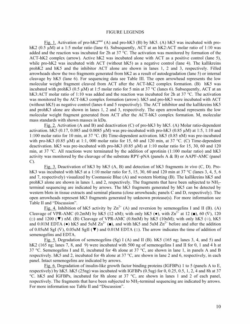

Figure 1
15
Figure 2
16
17
Figure 3
Figure 4
18
Figure 5
19
Figure 6
20
Figure 7
21

Figure 2
16
17
Figure 3
Figure 4
18
Figure 5
19
Figure 6
20
Figure 7
21

17
Figure 3
Figure 4
18
Figure 5
19
Figure 6
20
Figure 7
21

Figure 4
18
Figure 5
19
Figure 6
20
Figure 7
21

Figure 5
19
Figure 6
20
Figure 7
21

Figure 6
20
Figure 7
21

Figure 7
21