humanized mouse models to study cell -mediated immune
TRANSCRIPT

Humanized Mouse Models to Study Cell-Mediated ImmuneResponses to Liver-Stage Malaria Vaccines
Author
Good, Michael F, Hawkes, Michael T, Yanow, Stephanie K
Published
2015
Journal Title
Trends in Parasitology
Version
Accepted Manuscript (AM)
DOI
https://doi.org/10.1016/j.pt.2015.06.008
Copyright Statement
© 2015 Elsevier. Licensed under the Creative Commons Attribution-NonCommercial-NoDerivatives 4.0 International Licence (http://creativecommons.org/licenses/by-nc-nd/4.0/)which permits unrestricted, non-commercial use, distribution and reproduction in any medium,providing that the work is properly cited.
Downloaded from
http://hdl.handle.net/10072/125202
Griffith Research Online
https://research-repository.griffith.edu.au

1
Humanized mouse models to study cell-mediated immune responses to liver stage malaria vaccines
Michael F. Good1,2, Michael T. Hawkes2,3,4, Stephanie K. Yanow2,4,5
1Institute for Glycomics, Griffith University, Australia 2Department of Medical Microbiology and Immunology, University of Alberta, Edmonton, AB, Canada 3Department of Pediatrics, Division of Infectious Diseases, University of Alberta, Edmonton, AB, Canada 4School of Public Health, University of Alberta, Edmonton, AB, Canada 5Provincial Laboratory for Public Health, Edmonton, AB, Canada Corresponding author: Good, M. F. ([email protected]) Keywords: malaria, vaccines, humanized mice, liver-stage immunity.

2
Abstract
Malaria vaccine development is hampered by the lack of small animal models that
recapitulate human immune responses to Plasmodium falciparum. We review the
burgeoning literature on humanized mice for P. falciparum infection, including
challenges in engraftment of human immune cells, hepatocytes, and erythrocytes.
Recent advances in immune-compromised mouse models and stem cell technology
have already enabled proof-of-concept that the entire parasite life cycle can be
sustained in a murine model and that adaptive human immune responses to several
parasite stages can be measured. Nonetheless, optimization is needed to achieve a
reproducible and relevant murine model for malaria vaccine development. This
review is focused on the complexities of T cell development in a mouse humanized
with both a lymphoid system and hepatocytes. An understanding of this will facilitate
the utility of humanized mice in the development of liver stage vaccines.
Malaria vaccine approaches
Malaria is a major pathogen that is responsible for nearly one million deaths annually,
most of which occur in young children in sub-Saharan Africa [1]. The disease is caused
by an intracellular parasite (genus Plasmodium) that infects human hepatocytes and red
blood cells, and is transmitted by mosquitoes. In the human host, sporozoites exist
outside of a human cell; these are inoculated by the mosquito and enter a hepatocyte
where they develop over a period of 6-7 days into thousands of merozoites through a
process of schizogony (Figure 1). These rupture from the liver cell and invade a red
blood cell where, for P. falciparum, there is a 48 h developmental cycle. A single

3
merozoite gives rise to a schizont that contains 16-30 individual nuclei forming
merozoites. Rupture of the red blood cell releases merozoites that then invade new red
cells. Thus, there is exponential growth of the parasite within the red cell compartment.
Six different species of Plasmodium are known to infect humans; P. falciparum is the
most deadly.
More than 40 candidate malaria vaccines have reached the clinical trial stage [2].
Vaccine strategies may target the pre-erythrocytic stage, blood stage, or sexual stages
(transmission-blocking). An advantage of pre-erythrocytic vaccines is the potential to
completely block infection, preventing parasites from reaching the blood and causing
disease, as well as onward transmission. Current pre-erythrocytic vaccines exploit a
variety of approaches. (i) Genetically engineered surface antigens from the sporozoite
or infected hepatocyte. The RTS,S vaccine is the most advanced in the vaccine
development pipeline but has a protective efficacy of only 27-46% in infants and
children [3]. (ii) DNA and viral vectored delivery to induce a cellular immune
response [4, 5]. (iii) Live, attenuated whole sporozoites. The most advanced whole
parasite vaccine, known as PfSPZ, consists of the entire sporozoite, dissected from
mosquitoes, irradiated and vialed [6]. A recently published human study in a small
number of volunteers showed that it induced complete protection from challenge [7].
When tested in Rhesus monkeys, this vaccine induced an immune response in the
liver that involves T cells [8]. The findings from the viral-vectored and whole
sporozoite vaccine studies support an important role for CD8+ T-lymphocyte
responses in pre-erythrocytic vaccine efficacy. Pre-clinical development of the next
generation of anti-malaria vaccines will benefit from models that recapitulate cell-

4
mediated immunity to liver-stage parasites (see below) and the development of such
models will be the subject of this review.
Immunity to malaria in the liver
Our understanding of malaria immunity to liver stage parasites largely stems from
studies with irradiated sporozoites. Ruth Nussenzweig and colleagues first
demonstrated that irradiated sporozoites from a rodent malaria species could induce
immunity to subsequent sporozoite challenge [9]. In this and subsequent studies,
immunity was shown to be stage-specific (mice were not protected against challenge
with blood stage parasites), species-specific, and associated with the production of
antibodies to the circumsporozoite (coat) protein of the sporozoite. Sporozoites were
initially delivered by the bite of irradiated mosquitoes. Subsequent studies were
undertaken in human volunteers immunized by the bite of irradiated mosquitoes (e.g.
[10]) where immunity was also observed. More recently, human volunteers
immunized with large numbers of purified irradiated P. falciparum sporozoites were
protected from subsequent challenge infection with sporozoites [7].
T cell depletion studies in mice demonstrated that CD8+ T cells were critical for
immunity [11, 12] and studies in Rhesus monkeys immunized with P. falciparum
sporozoites showed that interferon-γ-producing parasite-specific CD8+ T cells were
found in the livers of immunized animals [8]. There has been debate as to where
CD8+ T cells are primed following immunization with irradiated sporozoites, with
evidence supporting priming in the draining lymph nodes of the skin and other data
suggesting that infected hepatocytes can directly prime T cells (reviewed in [13].

5
The mechanisms by which CD8+ T cells are then able to eliminate infected
hepatocytes are not well understood, but it appears likely that cytokines (IFN-γ, TNF-
α) are involved. Differences in critical immune responses may occur between
different individuals and between different species of Plasmodium [14]. Live imaging
of the liver during killing of infected hepatocytes by T cells shows that T cells recruit
other T cells to an infected hepatocyte and that a large cluster of T cells is involved in
killing the infected cell over a prolonged period of time (gang killing) [15].
These studies in human volunteers are extremely expensive and laborious. It is not
possible to study in detail the immune responses occurring in the livers of humans
immunized with irradiated sporozoites or with other malaria vaccines. Chimpanzees
and other primates are not easily available for both ethical and financial reasons.
Mice are widely used in research but human Plasmodium species do not infect mice,
leaving researchers with no choice but to use analogous rodent species. Extrapolations
from studies where mice are immunized with rodent parasites or vaccines to protect
against rodent parasites are not always reliable. The purpose of this review is to
describe the strategies behind the design of various humanized mouse models, along
with their limitations, as they may apply to the development of malaria vaccines that
target the human parasite during the liver stage of its life cycle.
The need for humanized leukocyte-liver (HLL) mice
‘Humanized’ mice are a revolutionary research tool for the pre-clinical study of
human infectious diseases. Through genetic manipulation, selective breeding, and
various approaches to avoid graft rejection, mice with high levels of human

6
chimerism can be routinely generated. Upon transplantation with specific human
tissues or cells (e.g. hepatocytes), these mice provide a localized human environment
that supports infection by human pathogens. Engraftment of human immune cells
could then permit the study of the host response to infection, thereby making these
animals ideally suited for pre-clinical evaluation of vaccines [16, 17].
For malaria, humanized leukocyte-liver (HLL) mice could be immunized with
experimental vaccines to stimulate an immune response and challenged with human
malaria infection to assess protection against liver stage parasites. Currently the only
ways to demonstrate liver stage immunity to a malaria vaccine are to either perform a
liver resection or use luminescent parasites and measure a reduction in parasite RNA
or protein expressed during the liver stage (e.g. [18]. While this cannot be done in
human studies, it is a straightforward procedure in mice.
Once an experimental vaccine is shown to induce protection against human malaria in
the HLL mouse, this model could further serve as a valuable tool to optimize the
dosage and delivery of the vaccine in preparation for human trials. Pre-clinical
optimization in the HLL mouse could become an integral component in selecting
vaccines to advance to clinical trials. Tremendous cost savings would be achieved and
the chances of success in human trials would be more favourable. Furthermore, the
possibility of testing combinations of vaccines with different targets is also feasible.
Another significant advantage is the ability to challenge the mice with both
homologous and heterologous parasite strains and species (eg. P. vivax). Mice could
be challenged by mosquito bite, injected sporozoites, or blood stage parasites to
evaluate the immune response to these different exposures to malaria. Feeding of

7
mosquitoes on the vaccinated mice could also assess transmission-blocking activity of
a vaccine. Collectively, the HLL mouse is a powerful tool to support the transition of
experimental vaccines from discovery research to clinical trials.
The HLL mouse is particularly relevant for studying vaccines that target the liver stage
of malaria. These include: irradiated, chemically attenuated, or genetically attenuated
whole sporozoite vaccines [7, 19, 20]; DNA prime boost and viral vectored subunit
vaccines [4]; the vaccine approach whereby liver stage immunity is induced following
exposure to low numbers of sporozoites with chloroquine chemoprophylaxis (CPS) [21];
and possibly other vaccines that induce CD8+ T cell responses (e.g. chemically
attenuated blood stage parasites [22]).
Developing HLL mice for vaccine research
While mice with a humanized immune system have been under development for nearly
30 years [23], and mice with a humanized liver were developed over the last 15 years
[24], attempts to construct mice with both a humanized immune system and liver have
been undertaken only recently. Many challenges remain but this is an exciting vaccine
research opportunity.
The first challenge is to identify or generate a suitable mouse strain that will permit the
engraftment of human immune cells and hepatocytes. It is critical to ensure that the
immune system of the mouse does not reject the human hepatocytes and the human
lymphoid tissue. The general principles are to engineer mice that are deficient in innate
and adaptive immune responses. The next challenge is to re-constitute the mouse with

8
appropriate human immune cells, or their precursors. This will lead to a humanized
immune system (HIS) mouse. Typically, stem cells (e.g. from the cord blood of a
newborn baby) are used. These give rise to antigen presenting cells (such as dendritic
cells) and to T cell progenitors. These progenitors must then mature in the mouse
thymus gland to generate mature T cells. This is a critical challenge in HLL mouse
construction. It is also necessary to ensure that the grafted human immune system does
not cause graft-versus-host disease in the mouse. The third challenge is to engraft
human hepatocytes. For this to occur, the mouse’s own hepatocytes must first be
destroyed. This is achieved by activating specific transgenes in the mice designed to
produce a liver toxin or by deleting genes required for hepatocyte survival. Donor
human hepatocytes are then injected into the recipient, usually via the intra-splenic route.
Generating the basic humanized immune system mouse
There are now many different types of immune-deficient mice that can be used to
accept human cells. Suitable mice have a phenotype of severe combined immune
deficiency (deficient in their own T and B cells). In one commonly used humanized
mouse model, recipient immune deficiency is conferred by a loss of function mutation
in the Prkdc gene, encoding a DNA-dependent protein kinase catalytic subunit which
functions in DNA non-homologous end-joining (commonly referred to as scid). Other
commonly used strains have genetic mutations in the recombination activating genes,
Rag1or Rag2, which encode enzymes that participate in immunoglobulin and T cell
receptor VDJ recombination. These genetic alterations are usually referred to as ‘S’
(SCID) or ‘R’ (Rag). However, SCID or Rag-/- mice are not fully suited to the
generation of HLL mice without further genetic manipulations. They have natural

9
killer (NK) cells and other innate immune responses that can limit human cell
engraftments.
A genetic knockout of the interleukin 2 receptor gamma (IL2Rγ) chain is commonly
used to control NK activity and reduce the effects of inflammatory cytokines and
chemokines. These mice are unable to signal via cytokines that require the IL2Rγ
chain (IL2, IL4, IL7, IL9, IL15 and IL21) thereby eliminating NK cell activity and
other inflammatory sequellae. This genetic alteration is usually referred to as ‘G’.
Mice with different genetic alterations in the IL2Rγ gene have been produced [16]).
Because they are so severely immune-compromised, they have other defects in lymph
node and B cell development even following immune reconstitution with human stem
cells.
Breeding scid mice onto the non-obese diabetic (NOD) background [25] further
generates a xenotransplantation-tolerant mouse with phagocytic dysfunction. This
occurs because the recipient mouse SIRPα gene closely resembles the human SIRPα
gene, thus reducing phagocytosis of xenogeneic cells (e.g. human stem cells) by
macrophages via the CD47-SIRPα interaction. SIRPα provides a signal to
macrophages not to remove self cells; thus if the SIRPα gene is similar to the human
gene, removal of foreign cells by macrophages will be lessened [26]. The NOD
background is usually referred to by the letter ‘N’.
These three basic genetic strategies come together to result in NSG and NRG mice.
Another mouse is the NOG mouse which is similar to the NSG mouse but with a
different IL2Rγ genetic alteration. While all these mice can readily accept human
stem cells, there are still obstacles in the differentiation from stem cell to mature

10
lymphocyte that must be overcome in order to use these mice for vaccine studies.
Other related immunodeficient strains have been used and recently reviewed [27, 28].
Human CD34+ stem cells are a source of progenitors for T cells and antigen
presenting cells that can be readily purified from newborn umbilical cord blood, adult
blood or human fetal tissue, and are commercially available. Sufficient stem cells to
generate 10-20 humanized mice can be readily obtained from one cord. The
progenitors must then develop into T cells. This happens in the thymus gland through
a process involving positive and negative selection to expand the repertoire of
antigen-specific T cells while removing self-reactive T cells [29]. T cells are selected
or ‘educated’ in the thymus to recognize foreign peptide epitopes presented by major
histocompatibility complex (MHC) molecules expressed in the thymus.
However, there are two significant issues in generating functional human T cells in a
mouse that can recognize malaria parasite-infected human liver cells.
(i) The T cells are generated in a thymus where the MHC type of the thymus gland
will play a major role in selecting the repertoire of antigen-specific T cells that
emerge. Cells that develop in a gland that expresses solely mouse MHC antigens will
have a repertoire that is skewed to recognize foreign antigenic peptides in association
with mouse MHC molecules. Some will recognize peptides in association with
human MHC molecules but these will be a minority. As discussed above, CD8+ T
cells are the principal effector cells in malaria liver-stage immunity and these
recognize antigenic peptides with MHC class I antigens. For efficient expansion and
recognition of infected human liver cells, the thymus in which the T cells are educated
should ideally express the same MHC class I antigens that are expressed in the
hepatocytes.

11
(ii) For expansion into effector and memory cells, CD8+ T cells will require antigen-
primed CD4+ T cells to provide cellular growth factors and ‘help’. CD4+ T cells
recognize antigenic peptides in association with MHC class II molecules. Cells that
express MHC class II peptides are typically professional antigen presenting cells such
as dendritic cells or tissue-resident macrophages such as the Kupffer cells in the liver.
Activation of malaria parasite-specific human CD4+ T cells requires presentation of
parasite antigenic epitopes in association with human MHC class II molecules. If the
CD4+ T cells were educated in a mouse thymus gland then there will be a skewing of
the CD4+ T cell repertoire to recognize antigen in association with the mouse MHC II
alleles present in that thymus. Education in human thymic tissue will be necessary if
the T cells are to recognize predominantly antigen in association with human MHC
molecules.
Engrafting human hepatocytes
Different models for hepatocyte engraftment have been described and are presented
briefly here and in Table 1.
The first mouse strain that would accept human hepatocytes was the SCID/bg mouse
with the urokinase type plasminogen activator gene (uPA) linked to an albumin promoter
[30]. The immunodeficient mice have sub-acute liver failure and are readily transplanted
with fresh or cryopreserved human hepatocytes at age 7-12 days via intrasplenic
injection. Six to 8 weeks after transplantation with human hepatocytes, large islands of
human liver tissue are produced within the mouse livers, creating a mouse with a
human/mouse chimeric liver. When the human hepatocytes are successfully engrafted,

12
the nodules of the human hepatocytes are visible and histologically integrated into the
mouse liver. The rate of successful engraftment is 60-70% as determined by human
serum albumin levels. This model has been used to study both viral hepatitis [24] and P.
falciparum [30-32] in the absence of a human immune system. Others have recently
attempted human lymphoid and myeloid cell engraftment in these mice[33].
In another model, mice were developed that are deficient in the gene for the tyrosine
catabolic enzyme, fumarylacetoacetate hydrolase (Fah). These Fah-/- mice maintain
their hepatocytes only in the presence of 2-(2-nitro-4-trifluoromethylbenzoyl)-1,3-
cyclohexanedione and lose them when the drug is withdrawn [34]. This gene deficiency
was bred into immuno-deficient mice to create the FRG™KO/NOD mouse. The mouse
was recently reported to permit the development of P. falciparum sporozoites into
exoerythrocytic forms in the liver [35]. Furthermore, when transplanted with human
erythrocytes, the life cycle could be continued into the red cells [36]. It was recently
shown that these mice can be given a human immune system [37] (Table 1).
An alternative model is the TK-NOG mouse in which mice express the herpes
simplex virus thymidine kinase (HSVtk) transgenic construct containing the mouse
albumin enhancer/promoter [38]. HSVtk mRNA is selectively expressed in the liver
of NOG mice and they develop severe parenchymal liver damage after gancyclovir
treatment [38]. Human liver cells are then injected intrasplenically (Table 1).
Caspase8-FK506 binding protein-albumin promoter transgenic mice offer a related
approach in which the inducible death of hepatocytes occurs when FK 506 is
dimerized. Balb/c RG mice were used to study hepatitis C infection in this study [39].

13
Mice have been engrafted with a human immune system (Table 1). Protection against
infection was not assessed.
A different approach, recently described, is to inject human fetal hepatoblasts into
immuoodeficient mice (NSG) which have been pre-treated with an anti-Fas antibody
which induces murine hepatocyte apoptosis via a Caspase-3-deependent pathway
(Bility MT et al., PLoS Pathogens e1004032, 2014). In this study, the mice also
carried the human HLA-A2 transgene and received human stem cells, giving rise to
functional human T cells (Table 1).
Engrafting human red cells or precursors
A powerful indicator of liver stage immunity from pre-erythrocytic malaria vaccines
is the absence of blood stage parasites emerging from the liver. To measure blood
stage P. falciparum infection in humanized mice, it is necessary to engraft mice with
human erythrocytes (RBCs). When developing this model, the first strategy involved
repeated injection with mature bovine erythrocytes into immune-compromised mice.
Engraftment of human erythrocytes in mice was less consistent and occurred at lower
levels of chimerism but such mice could sustain a P. falciparum infection [40-44].
The model was progressively improved with increasingly immune-deficient murine
hosts [45-47]. These mice can sustain higher parasitemia with P. falciparum [48] and
infection with multiple standard strains of P. falciparum without the need for species-
specific parasite adaptation [49]. A recent advance in murine models of blood stage P.
falciparum infection is the RBC-supplemented, immune cell-optimized humanized

14
(RICH) mouse [50]. The need for repeated intra-peritoneal or intra-venous injections
of uninfected human erythrocytes remains a limitation of this model.
The next anticipated major advance in humanized mouse models of blood stage
malaria will be the xenotransplantation of human stem cells capable of recapitulating
the entire cycle of erythrocyte development within the mouse. Recently, human
hematopoietic stem cells along with human erythrocytes and plasmids encoding
human erythropoietin and IL-3 were injected into recipient mice [51]. Significant
erythrocyte chimerism (~25%) was observed, as well as a large fraction (~40%) of
human PBMCs (peripheral blood mononuclear cells). In a further study with another
immune-compromised strain that received human stem cells, HLA-
DR4.RagKO.IL2RγcKO.NOD (DRAG) mice developed human erythrocytes and
reticulocytes that represented approximately 0.2% of the red cell pool and could
sustain an infection with P. falciparum following mosquito bite [52]. Low-level
human hematocrit was due to poor differentiation of stem cells toward the
erythrocytic lineage [52]. Further optimization of erythrocyte development in the
murine host is needed to add more value to this new mouse model of P. falciparum
blood stages.
Overcoming graft versus host disease and graft versus graft disease
Allogeneic haematopoietic stem cell transplantation in humans is sometimes
complicated by the development of graft versus host disease (GVHD) mediated by
donor immune cells responding to host antigens. In this disease the engrafted

15
immune cells recognize the host tissues as ‘foreign’ and the immune cells can cause
significant tissue injury and death. Tissues typically affected by GVHD include the
skin, the gut and the liver, but other tissues can be affected as well. Both innate and
adaptive immune mechanisms are implicated in human GVHD. A similar disease can
occur in mice transplanted with human immune cells; this is referred to as
‘xenogeneic GVHD’ in which human T cells that develop in the mouse respond to
murine tissue as foreign antigens. HLL mice can be screened for GVHD by
measuring a drop in serum human albumin, a rise in liver enzymes in the blood and
clinical manifestations (skin changes, hair loss). Human PBMCs can engraft at high
levels in NSG mice and produce a robust xeno-GVHD response [53-55]. Xeno-
GVHD in this model appears to be largely mediated by donor T cell recognition of
murine MHC molecules [53, 56]. Of interest, investigators did not report significant
levels of GVHD in a humanized mouse model of influenza using a mouse strain
deficient in murine MHC class 1 (beta-2 microglobulin (β-2m) knockout) [57]. β-2m
is a small peptide that stabilizes the expression of MHC class I molecules. Without
expression of MHC class I molecules, the human immune cells will not recognize nor
respond to the mouse cells.
Relative to PBMC and mature T-cells, cord blood CD34+ stem cells are associated
with reduced levels of lethal GVHD when injected into NSG mice [58]. However, in
the ‘BLT’ model where donor stem cells originate from fetal liver, late onset GVHD
was reported with similarities to chronic GVHD in humans. ‘BLT’ refers to immuno-
deficient mice that have been reconstituted with human hematopoietic stem cells, and
fragments of human fetal liver and thymus under the kidney capsule (Figure 2A)
(Wege, AK et al, Current Topics Microbiol Immunol 324:149, 2008). However,

16
further manipulations of BLT mice can reduce the levels of GVHD, such as the
deletion of CD47. Reduced levels of GVHD were observed in R,G,CD47-/--BLT
mice compared to NSG-BLT mice [59].
Secondary lymphoid tissue
Secondary lymphoid tissue and B cells do not develop normally in many HIS or HLL
mice. The reasons for this were recently reviewed [28] and relate in part to
deficiencies in human cytokines necessary for lymphoid development. There is
evidence that malaria sporozoites in the draining lymph nodes of skin can activate T
cells and eliminate infected hepatocytes [60]. Thus, secondary lymphoid tissue may
be critical to the development of optimal malaria-specific immune responses in HLL
mice. It was recently shown that C57 BL6 Rag-/-, IL2Rγ-/-, CD47-/- BLT mice
developed lymph nodes and spleens with follicles and normal B cells which
underwent class switching, although antibody levels were significantly lower than in
humans [59]. These mice, which require the implantation of human thymic tissue,
have not been used to study malaria. HLL mice with secondary lymphoid tissue
could also be ideal to study many malaria vaccines that rely on the production of
antibodies.
Perspective on the optimal HLL mouse for malaria vaccine research
With the above considerations, we suggest that appropriate mouse models can be
constructed for the study of liver stage immunity and vaccine development in HLL

17
mice. With the goal to reconstitute the mouse with a population of functional T cells
that can be stimulated by an experimental vaccine and which will then expand in
number and recognize a malaria parasite-infected human hepatocyte population,
consideration must be given to: (i) MHC matching between the CD34+ stem cells
which will ultimately develop into mature T cell populations, the thymus where the T
cells are educated, and the hepatocytes; and (ii) the frequency of human T cells that
are present in the mouse that can be activated by malaria antigen. These two issues
are related.
If the frequency of precursors is too low then it will be more difficult to activate
sufficient cells to eliminate malaria-infected liver cells. In non-humanized mice, a
large but defined number of T cells is required to effect immunity to the liver stages
of malaria [61]. If T cells are educated in a mouse thymus then the number of cells
that recognize foreign antigenic peptides in association with human MHC will be
reduced. Although it is not known how many specific T cells will develop in any
particular immunized HLL mouse, it seems reasonable to assume that education of T
cells in a human thymus or a mouse thymus expressing human HLA antigens would
be ideal. BLT mice offer this possibility but such mice would also need to express
uPA or have a deletion of Fah (or use a different hepatocyte suicide strategy) for
efficient engraftment with human hepatocytes. Figure 2A outlines a possible
structure. However, there are major technical challenges and ethical issues with using
human fetal tissue that limit the widespread suitability of this model.
Alternative models include transgenic mice that express human HLA antigens in their
thymus (e.g. [52]) or are infected with a virus expressing human HLA genes [62]
18
(Figure 2B). The T cells will develop from progenitors and be ‘educated’ in a HLA-
humanized mouse thymus resulting in the repertoire being skewed favourably to
recognize foreign antigens (e.g. malaria antigens) in association with human HLA. In
these models it is also necessary that the thymus gland and the hepatocytes share the
same human MHC class I antigen. This is not difficult to arrange if common HLA
alleles are chosen, such as A2. GVHD is not a limiting factor with these mice as the
T cells mature in a mouse where peripheral tissues (such as skin, kidney, pancreas,
etc) and thymic cells express the same (murine) MHC molecules.
A third approach is: (i) begin with immunodeficient mice (e.g. NRG mice) stably
engrafted with human CD34+ stem cells to generate antigen presenting cells; (ii)
transfuse these mice with HLA-matched mature T cells from the same donor (e.g.
from cord blood); (iii) use human hepatocytes that share at least one class I MHC
molecule with these cells which will enable them to present malaria antigen to
effector cytotoxic T-cells (Figure 2C). Breeding of the recipient mice to be deficient
in β-2m mice will likely prevent GVHD. As mentioned above, β-2m is a molecule
necessary to stabilize MHC class I molecules. Thus, the only cells that would possess
β-2m in β-2m-/- mice and be recognized by T cells would be the donor cells of human
origin. Mature human T cells have been transfused into β-2m -/- mice and GVHD
was not reported [57]. An advantage of this model is that the mature CD4+ T cells are
also educated in a thymus expressing human MHC antigens that are found on the
antigen presenting cells (APCs) providing help for CD8+ T cells to expand [63]. This
mouse may represent an ideal and relatively simple model for studying the immune
response to malaria where human T cell responses could be tested against liver stage
antigens and where vaccine efficacy could be monitored by measuring human red cell

19
infection. This model could also be used to test immune responses to and efficacy of
blood stage vaccines where T cell responses are critical [22]. Furthermore, the model
could be adapted to ask whether T cells from vaccinated humans could kill malaria
parasite-infected hepatocytes.
Concluding remarks
In this article, we summarized data relevant to the construction of mice humanized
with both a human lymphoid system and hepatocytes suitable for liver stage malaria
vaccine research. A current major impediment to progress in this field is the expense
of early phase human studies and the difficulties with analyzing relevant immune
responses that are occurring primarily in the liver. The HLL mouse offers a relatively
simple alternative. While there are still a number of obstacles to be addressed,
significant developments occurred over the last 5 years to enable the construction of
various models of HLL mice. It is our view that the last two models described in
Figure 2 represent the best current alternatives for further investigation.
While this review focused on the liver, a major tissue site for malaria parasites,
similar issues are relevant to engraftment of other human tissues along with a nascent
human immune system. Molecular, surgical and clinical tools are now available to
readily produce various humanized mice that can be used for pre-clinical evaluation
of novel vaccine approaches and to optimize delivery strategies.

20
Figure Legends
Figure 1. Life cycle of malaria parasites in the human host. (Reproduced from [64].

21
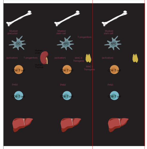
Figure 2. Models for generating a humanized leukocyte-liver (HLL) mouse. (A) The
‘BLT’ mouse (mice containing human stem cells, liver, and thymic grafts). This
immune-deficient mouse is reconstituted with human hematopoietic stem cells
(CD34+), and fragments of human fetal liver and thymus implanted under the kidney
capsule. Antigen presenting cells (APCs) would derive from the human stem cells.
Hepatocytes would need to express humanized major histocompatibility complex
(MHC) I molecules (e.g. HLA-A2) to be recognized by humanized CD8+ T cells.
Dendritic cells (DCs) and other APCs present antigen to CD4+ T cells in association
with MHC II (e.g. HLA-DR4). To engraft human hepatocytes it would be necessary
for these mice to express uPA, be deficient in Fah or use some other mouse
hepatocyte suicide strategy. (B) Progenitor T cells and myeloid stem cells (APC
progenitors) develop from human CD34+ (donor) stem cells. T cells develop in a
thymus that is engineered to express human MHC I (e.g. HLA-A2) and MHC II (e.g.
HLA-DR4). APCs (such as DCs or Kupffer cells) will express humanized MHC II
and present antigenic epitopes to CD4+ T cells that were educated to recognize
antigen in the context of human MHC II expressed in the thymus. (C) In this model,
the mouse is engrafted with mature human T cells (CD4+ and CD8+). APCs will
come from CD34+ stem cells that will be autologous to the donor T cells and allow
recognition by the CD4+ T cells. In this model, it is necessary to use β2m-/- recipient
mice to prevent GVHD (graft versus host disease). Mice without β2m will not express
mouse MHC I molecules and will not be susceptible to GVHD.
1A
Donor
Donor
Donor
Donor
CD34+
stem cells
uPA or Fah-/-
MHC II
DC APC APC
MHC IIrestricted
MHC Irestricted
Myeloidprecursor
HumanThymus
HumanLiver
CD4+ T cell
CD8+ T cell
CD4+ T cell
CD8+ T cell
CD4+ T cell
CD8+ T cell
Donor hepatocytes
(activation)
(help)
T progenitors
T progenitors
MHC IITransgene
MHC ITransgene
1B 1C
Donor
CD34+
stem cells
uPA or Fah-/-
MHC II Thymus
MHC IIrestricted
MHC Irestricted
Myeloidstem cell
Donor hepatocytes
(activation)
(help)
Donor
CD34+
stem cells
uPA or Fah-/-
MHC II Thymus
MHC IIrestricted
MHC Irestricted
Myeloidstem cell
Donor hepatocytes
(activation)
(help)

22
Acknowledgements
We thank Jahanara Rajwani for critically reviewing the manuscript. We thank Troy
Baldwin and D. Lorne Tyrrell for useful discussions. We acknowledge NHMRC
(Australia), Alberta Innovates – Health Solutions, and the Women & Children’s
Health Research Institute for grant support.
Glossary
Allogeneic – A mouse with a different genetic background.
APC – Antigen presenting cells (e.g. dendritic cells, Kupffer cells).
Autologous – Cells from the same animal.
β2m – beta-2 microglobulin. An MHC Class I associated polypeptide that stabilizes
the MHC molecule.
CD34 - Marker for pluripotent stem cells that can give rise to lymphoid and myeloid
progenitors. These cells are also CD45dim.
CD47 – A trans-membrane protein ubiquitously expressed on human cells and,
amongst other function, interacts with SIRPα on myeloid cells to inhibit phagocytosis.
Chimerism – Containing cells from two different individuals (here to mean mouse
and human).

23
Engraftment – Transplantation of foreign tissue.
Epitopes – Antigenic determinants from a protein or other molecule.
Fah-/- – These mice are deficient in fumarylacetoacetate hydrolase leading to
tyropsineamia and death of hepatocytes. Mie can be protected with the drug 2-(2-
nitro-4-trifluoromethylbenzoyl)-1,3-cyclohexanedione (NTBC) and hepatocyte death
will occur when the drug is withdrawn.
GVHD – Graft versus host disease.
Haematopoietic stem cells – Cells that give rise to all the other blood cells.
MHC I – Class I histocompatibility antigen expressed on all nucleated cells.
MHC II – Class II histocompatibility antigens expressed on professional antigen
present cells.
NOD – The Non Obese Diabetic mouse whose SIRPα gene more closely resembles
the human SIRPα gene, thus reducing phagocytosis of transplanted xenogeneic stem
cells by myeloid cells.
NOG, NSG – Forms of NOD/SCID IL2Rγ-/- mice.

24
Rag-/- – Recombination activating gene-deficient mice that cannot arrange
immunoglobulin and T cell receptor genes. These mice are deficient in T and B cell
function.
SCID – These mice are deficient in a DNA repair enzyme (Prkdc) preventing
recombination in humoral and cellular immune response genes. They lack T and B
cells and have similarities with the Rag-/- mice.
Secondary lymphoid tissue – Usually meant to indicate spleen and lymph nodes.
SIRPα - Signal regulatory protein alpha. It interacts with CD47 to negatively control
phagocytosis.
uPA – Mice bearing the urokinase type plasminogen activator gene linked to an
albumin promoter. These mice suffer from subacute liver disease and require
transplanted normal hepatocytes for survival.
VDJ recombination – Somatic recombination of Ig or T cell receptor genes during
lymphocyte development to generate diversity in the repertoire.
Xenotransplantation – Transplantation between two different species (here referring
to human into mouse).

25
References
1 Murray, C.J., et al. (2012) Global malaria mortality between 1980 and 2010: a
systematic analysis. Lancet 379, 413-431
2 Schwartz, L., et al. (2012) A review of malaria vaccine clinical projects based on
the WHO rainbow table. Malaria journal 11, 11
3 RTS,S Clinical Trials Partnership (2014) Efficacy and safety of the RTS,S/AS01
malaria vaccine during 18 months after vaccination: a phase 3 randomized, controlled
trial in children and young infants at 11 African sites. PLoS medicine 11, e1001685
4 Hill, A.V. (2011) Vaccines against malaria. Philosophical transactions of the Royal
Society of London. Series B, Biological sciences 366, 2806-2814
5 Ewer, K.J., et al. (2013) Protective CD8+ T-cell immunity to human malaria
induced by chimpanzee adenovirus-MVA immunisation. Nature communications 4,
2836
6 Hoffman, S.L., et al. (2002) Protection of humans against malaria by immunization
with radiation-attenuated Plasmodium falciparum sporozoites. The Journal of
infectious diseases 185, 1155-1164
7 Seder, R.A., et al. (2013) Protection against malaria by intravenous immunization
with a nonreplicating sporozoite vaccine. Science 341, 1359-1365
8 Epstein, J.E., et al. (2011) Live attenuated malaria vaccine designed to protect
through hepatic CD8(+) T cell immunity. Science 334, 475-480
9 Nussenzweig, R.S., et al. (1967) Protective immunity produced by the injection of
x-irradiated sporozoites of Plasmodium berghei. Nature 216, 160-162
10 Clyde, D.F., et al. (1973) Immunization of man against sporozite-induced
falciparum malaria. The American journal of the medical sciences 266, 169-177

26
11 Schofield, L., et al. (1987) Gamma interferon, CD8+ T cells and antibodies
required for immunity to malaria sporozoites. Nature 330, 664-666
12 Weiss, W.R., et al. (1988) CD8+ T cells (cytotoxic/suppressors) are required for
protection in mice immunized with malaria sporozoites. Proceedings of the National
Academy of Sciences of the United States of America 85, 573-576
13 Corradin, G. and Levitskaya, J. (2014) Priming of CD8(+) T cell responses to liver
stage malaria parasite antigens. Frontiers in immunology 5, 527
14 Tse, S.W., et al. (2011) Induction and maintenance of protective CD8+ T cells
against malaria liver stages: implications for vaccine development. Memorias do
Instituto Oswaldo Cruz 106 Suppl 1, 172-178
15 Cockburn, I.A., et al. (2013) In vivo imaging of CD8+ T cell-mediated elimination
of malaria liver stages. Proceedings of the National Academy of Sciences of the
United States of America 110, 9090-9095
16 Brehm, M.A., et al. (2010) Humanized mouse models to study human diseases.
Current opinion in endocrinology, diabetes, and obesity 17, 120-125
17 Legrand, N., et al. (2009) Humanized mice for modeling human infectious disease:
challenges, progress, and outlook. Cell host & microbe 6, 5-9
18 Ploemen, I.H., et al. (2009) Visualisation and quantitative analysis of the rodent
malaria liver stage by real time imaging. PloS one 4, e7881
19 Purcell, L.A., et al. (2008) Chemical attenuation of Plasmodium berghei
sporozoites induces sterile immunity in mice. Infection and immunity 76, 1193-1199
20 Mueller, A.K., et al. (2005) Genetically modified Plasmodium parasites as a
protective experimental malaria vaccine. Nature 433, 164-167
21 Bijker, E.M., et al. (2013) Protection against malaria after immunization by
chloroquine prophylaxis and sporozoites is mediated by preerythrocytic immunity.

27
Proceedings of the National Academy of Sciences of the United States of America
110, 7862-7867
22 Good, M.F., et al. (2013) Cross-species malaria immunity induced by chemically
attenuated parasites. The Journal of clinical investigation
23 Mosier, D.E., et al. (1988) Transfer of a functional human immune system to mice
with severe combined immunodeficiency. Nature 335, 256-259
24 Mercer, D.F., et al. (2001) Hepatitis C virus replication in mice with chimeric
human livers. Nature medicine 7, 927-933
25 Yamauchi, T., et al. (2013) Polymorphic Sirpa is the genetic determinant for
NOD-based mouse lines to achieve efficient human cell engraftment. Blood 121,
1316-1325
26 Takenaka, K., et al. (2007) Polymorphism in Sirpa modulates engraftment of
human hematopoietic stem cells. Nature immunology 8, 1313-1323
27 Kaushansky, A., et al. (2014) Of men in mice: the success and promise of
humanized mouse models for human malaria parasite infections. Cellular
microbiology 16, 602-611
28 Brehm, M.A., et al. (2014) Generation of improved humanized mouse models for
human infectious diseases. Journal of immunological methods 410, 3-17
29 Klein, L., et al. (2014) Positive and negative selection of the T cell repertoire: what
thymocytes see (and don't see). Nature reviews. Immunology 14, 377-391
30 Sacci, J.B., Jr., et al. (2006) Plasmodium falciparum infection and exoerythrocytic
development in mice with chimeric human livers. International journal for
parasitology 36, 353-360

28
31 VanBuskirk, K.M., et al. (2009) Preerythrocytic, live-attenuated Plasmodium
falciparum vaccine candidates by design. Proceedings of the National Academy of
Sciences of the United States of America 106, 13004-13009
32 Morosan, S., et al. (2006) Liver-stage development of Plasmodium falciparum, in
a humanized mouse model. The Journal of infectious diseases 193, 996-1004
33 Gutti, T.L., et al. (2014) Human hepatocytes and hematolymphoid dual
reconstitution in treosulfan-conditioned uPA-NOG mice. The American journal of
pathology 184, 101-109
34 Azuma, H., et al. (2007) Robust expansion of human hepatocytes in Fah-/-/Rag2-/-
/Il2rg-/- mice. Nature biotechnology 25, 903-910
35 Vaughan, A.M., et al. (2012) Complete Plasmodium falciparum liver-stage
development in liver-chimeric mice. The Journal of clinical investigation 122, 3618-
3628
36 Vaughan, A.M., et al. (2012) Development of humanized mouse models to study
human malaria parasite infection. Future microbiology 7, 657-665
37 Wilson, E.M., et al. (2014) Extensive double humanization of both liver and
hematopoiesis in FRGN mice. Stem cell research 13, 404-412
38 Hasegawa, M., et al. (2011) The reconstituted 'humanized liver' in TK-NOG mice
is mature and functional. Biochemical and biophysical research communications 405,
405-410
39 Washburn, M.L., et al. (2011) A humanized mouse model to study hepatitis C
virus infection, immune response, and liver disease. Gastroenterology 140, 1334-
1344

29
40 Tsuji, M., et al. (1995) Establishment of a SCID mouse model having circulating
human red blood cells and a possible growth of Plasmodium falciparum in the mouse.
Vaccine 13, 1389-1392
41 Moore, J.M., et al. (1995) Maintenance of the human malarial parasite,
Plasmodium falciparum, in scid mice and transmission of gametocytes to mosquitoes.
The Journal of experimental medicine 181, 2265-2270
42 Badell, E., et al. (1995) Human Plasmodium liver stages in SCID mice: a feasible
model? Parasitology today 11, 169-171
43 Badell, E., et al. (2000) Human malaria in immunocompromised mice: an in vivo
model to study defense mechanisms against Plasmodium falciparum. The Journal of
experimental medicine 192, 1653-1660
44 Moreno, A., et al. (2001) Human malaria in immunocompromised mice: new in
vivo model for chemotherapy studies. Antimicrobial agents and chemotherapy 45,
1847-1853
45 Moreno Sabater, A., et al. (2005) Experimental infection of immunomodulated
NOD/LtSz-SCID mice as a new model for Plasmodium falciparum erythrocytic
stages. Parasitology research 95, 97-105
46 Rochford, R., et al. (2013) Humanized mouse model of glucose 6-phosphate
dehydrogenase deficiency for in vivo assessment of hemolytic toxicity. Proceedings
of the National Academy of Sciences of the United States of America 110, 17486-
17491
47 Angulo-Barturen, I., et al. (2008) A murine model of falciparum-malaria by in vivo
selection of competent strains in non-myelodepleted mice engrafted with human
erythrocytes. PloS one 3, e2252

30
48 Jimenez-Diaz, M.B., et al. (2009) Improved murine model of malaria using
Plasmodium falciparum competent strains and non-myelodepleted NOD-scid
IL2Rgammanull mice engrafted with human erythrocytes. Antimicrobial agents and
chemotherapy 53, 4533-4536
49 Arnold, L., et al. (2011) Further improvements of the P. falciparum humanized
mouse model. PloS one 6, e18045
50 Chen, Q., et al. (2014) Human natural killer cells control Plasmodium falciparum
infection by eliminating infected red blood cells. Proceedings of the National
Academy of Sciences of the United States of America 111, 1479-1484
51 Chen, Q., et al. (2009) Expression of human cytokines dramatically improves
reconstitution of specific human-blood lineage cells in humanized mice. Proceedings
of the National Academy of Sciences of the United States of America 106, 21783-
21788
52 Wijayalath, W., et al. (2014) Humanized HLA-
DR4.RagKO.IL2RgammacKO.NOD (DRAG) mice sustain the complex vertebrate
life cycle of Plasmodium falciparum malaria. Malaria journal 13, 386
53 King, M.A., et al. (2009) Human peripheral blood leucocyte non-obese diabetic-
severe combined immunodeficiency interleukin-2 receptor gamma chain gene mouse
model of xenogeneic graft-versus-host-like disease and the role of host major
histocompatibility complex. Clinical and experimental immunology 157, 104-118
54 Abraham, S., et al. (2015) IL-10 exacerbates xenogeneic GVHD by inducing
massive human T cell expansion. Clinical immunology 156, 58-64
55 Ali, N., et al. (2012) Xenogeneic graft-versus-host-disease in NOD-scid IL-
2Rgammanull mice display a T-effector memory phenotype. PloS one 7, e44219

31
56 Covassin, L., et al. (2011) Human peripheral blood CD4 T cell-engrafted non-
obese diabetic-scid IL2rgamma(null) H2-Ab1 (tm1Gru) Tg (human leucocyte antigen
D-related 4) mice: a mouse model of human allogeneic graft-versus-host disease.
Clinical and experimental immunology 166, 269-280
57 Yu, C.I., et al. (2008) Broad influenza-specific CD8+ T-cell responses in
humanized mice vaccinated with influenza virus vaccines. Blood 112, 3671-3678
58 Gorin, N.C., et al. (2002) Increased risk of lethal graft-versus-host disease-like
syndrome after transplantation into NOD/SCID mice of human mobilized peripheral
blood stem cells, as compared to bone marrow or cord blood. Journal of
hematotherapy & stem cell research 11, 277-292
59 Lavender, K.J., et al. (2013) BLT-humanized C57BL/6 Rag2-/-gammac-/-CD47-/-
mice are resistant to GVHD and develop B- and T-cell immunity to HIV infection.
Blood 122, 4013-4020
60 Chakravarty, S., et al. (2007) CD8+ T lymphocytes protective against malaria liver
stages are primed in skin-draining lymph nodes. Nature medicine 13, 1035-1041
61 Schmidt, N.W., et al. (2008) Memory CD8 T cell responses exceeding a large but
definable threshold provide long-term immunity to malaria. Proceedings of the
National Academy of Sciences of the United States of America 105, 14017-14022
62 Huang, J., et al. (2014) An AAV vector-mediated gene delivery approach
facilitates reconstitution of functional human CD8+ T cells in mice. PloS one 9,
e88205
63 Janssen, E.M., et al. (2003) CD4+ T cells are required for secondary expansion
and memory in CD8+ T lymphocytes. Nature 421, 852-856
64 Good, M.F. (2013) Immunology. Pasteur approach to a malaria vaccine may take
the lead. Science 341, 1352-1353