huntingtin expression stimulates … · medicina, universidad auto ... huntingtin (ab1) (difiglia...
TRANSCRIPT
Huntingtin Expression Stimulates Endosomal–Lysosomal Activity,Endosome Tubulation, and Autophagy
Kimberly B. Kegel,1 Manho Kim,1 Ellen Sapp,1 Charmian McIntyre,1 Jose G. Castano,2 Neil Aronin,3 andMarian DiFiglia1
1Department of Neurology, Massachusetts General Hospital, Boston, Massachusetts 02114, 2Departamento deBioquımica e Instituto de Investigaciones Biomedicas del Consejo Superior de Investigaciones Cientıficas, Facultad deMedicina, Universidad Autonoma de Madrid, 28029 Madrid, Spain, and 3Departments of Medicine and Cell Biology,University of Massachusetts Medical Center, Worcester, Massachusetts 01655
An expansion of polyglutamines in the N terminus of huntingtincauses Huntington’s disease (HD) and results in the accrual ofmutant protein in the nucleus and cytoplasm of affected neurons.How mutant huntingtin causes neurons to die is unclear, butsome recent observations suggest that an autophagic processmay occur. We showed previously that huntingtin markedly ac-cumulates in endosomal–lysosomal organelles of affected HDneurons and, when exogenously expressed in clonal striatalneurons, huntingtin appears in cytoplasmic vacuoles causingcells to shrink. Here we show that the huntingtin-enriched cyto-plasmic vacuoles formed in vitro internalized the lysosomal en-zyme cathepsin D in proportion to the polyglutamine-length inhuntingtin. Huntingtin-labeled vacuoles displayed the ultrastruc-tural features of early and late autophagosomes (autolyso-somes), had little or no overlap with ubiquitin, proteasome, and
heat shock protein 70/heat shock cognate 70 immunoreactivities,and altered the arrangement of Golgi membranes, mitochondria,and nuclear membranes. Neurons with excess cytoplasmic hun-tingtin also exhibited increased tubulation of endosomal mem-branes. Exogenously expressed human full-length wild-type andmutant huntingtin codistributed with endogenous mouse huntingtinin soluble and membrane fractions, whereas human N-terminalhuntingtin products were found only in membrane fractions thatcontained lysosomal organelles. We speculate that mutant hunting-tin accumulation in HD activates the endosomal–lysosomal system,which contributes to huntingtin proteolysis and to an autophagicprocess of cell death.
Key words: Huntington’s disease; autophagy; lysosomes; en-dosome tubulation; cathepsin D; N-terminal huntingtin; hunting-tin proteolysis
Huntingtin is a protein of unknown function, is enriched in neu-rons, and resides mainly in the cytoplasm. An expanded polyglu-tamine tract in the N terminus of huntingtin causes Huntington’sdisease (HD), a neurological disorder associated with the selectiveloss of striatal and cortical neurons (Vonsattel and DiFiglia, 1998).Neurons affected in HD accumulate mutant huntingtin in thenucleus and cytoplasm (DiFiglia et al., 1997; Sapp et al., 1997;Gutekunst et al., 1998). Although the cause of protein accumula-tion in HD is unknown, experimental models developed in miceand in cell cultures have demonstrated that an excess of mutanthuntingtin, especially the N-terminal region, in the nucleus orcytoplasm can cause cellular dysfunction or cell death (Hackam etal., 1998; Saudou et al., 1998; Peters et al., 1999). Features ofapoptosis have been reported in some of these models, but theprocess of cell death in HD still remains unclear.
We showed that, in dying neurons in the HD brain, huntingtinaberrantly accumulated in perinuclear regions and in numerouspunctate cytoplasmic structures that resembled endosomal–lysoso-mal organelles (Sapp et al., 1997). These results may be importantin understanding how mutant huntingtin induces cell death be-cause the endosomal–lysosomal–vacuolar pathway has been tied tothe handling of other disease proteins, such as prions and Abpeptide 1–42 (Taraboulos et al., 1992; Cataldo et al., 1996), and tocell death by autophagy, a process whereby cells remove cytosolic
proteins and organelles and degrade themselves from within. Au-tophagy may precede and coexist with apoptosis, can be induced byapoptotic stimuli in the presence of caspase inhibitors (Xue et al.,1999), and may contribute to cell death in neurons through theregulation of lysosomal proteases cathepsin B and D (Ohsawa etal., 1998).
We described recently an in vitro model of HD using a clonalmouse striatal cell line transiently transfected with human hunting-tin (Kim et al., 1999a). The exogenous wild-type and mutanthuntingtins accumulated diffusely in the cytoplasm and formedcytoplasmic vacuoles or nuclear and cytoplasmic inclusions (mu-tant huntingtin only). Cells with cytoplasmic vacuoles becameshrunken, whereas cells with inclusions did not. Like inclusions, thevacuoles localized N-terminal fragments of huntingtin. Previously,we observed that endogenous wild-type and mutant huntingtinassociate with endosomes in primary fibroblasts (Velier et al., 1998)and that huntingtin accumulates in lysosomal-like organelles in theHD brain (Sapp et al., 1997). In view of these results, we undertookbiochemical, immunohistochemical, and electron microscopy stud-ies to further characterize vacuoles that accumulate exogenoushuntingtin and other effects of huntingtin expression in the cyto-plasm of clonal striatal cells. We found that the vacuoles associatedwith exogenous huntingtin incorporated the lysosomal enzymecathepsin D in proportion to polyglutamine length in huntingtinand had the ultrastructural features of autophagosomes. N-terminalfragments of huntingtin were mainly found in membrane fractionsthat contained vacuoles. Huntingtin expression also induced exten-sive tubulation of endosomal membranes. We speculate that theendosomal–lysosomal system is the main path for huntingtin deg-radation and proteolysis and that autophagy contributes to celldeath in HD.
MATERIALS AND METHODSAntibodies. Antisera used in this study were directed against the followingpeptides or proteins: huntingtin (Ab1) (DiFiglia et al., 1995); FLAG (clone
Received June 1, 2000; revised July 11, 2000; accepted July 13, 2000.This work was supported by National Institutes of Health Grants NS16367 and
NS35711 (to M.D.), NS38194 (to N.A.), T32-AG00222 (K.B.K.), and a grant from theHuntington’s Disease Society of America (to M.D.). We thank Mr. Lawrence Cherkasfor his assistance with the photography, Yun J. Kim for his assistance with confocalmicroscopy, Kristy Brown for advise with electron microscopy, and Yumei Wang andZheng-Hong Qin for help with the studies involving cathepsin D localization.
Correspondence should be addressed to Dr. Marian DiFiglia, Department ofNeurology, Massachusetts General Hospital East, 149 13th Street, Room 6604,Charlestown, MA 02129. E-mail: [email protected] © 2000 Society for Neuroscience 0270-6474/00/207268-11$15.00/0
The Journal of Neuroscience, October 1, 2000, 20(19):7268–7278
M5) and b-tubulin isotype III (monoclonals; Sigma, St. Louis, MO);calnexin C terminus (polyclonal; Stressgen, Victoria, British Columbia,Canada); GM130 and syntaxin 6 (monoclonals; Transduction Laborato-ries, Lexington, KY); transferrin receptor (monoclonal; Zymed Labora-tories, San Francisco, CA); histone (monoclonal; Boehringer Mannheim,Indianapolis, IN); ubiquitin (polyclonal; Dako, Carpinteria, CA); C2 sub-unit of proteasome and the whole complex (polyclonals) (Mengual et al.,1996); cathepsin D [d23, made in sheep, gift of Dr. Ann Cataldo (NathanS. Kline Institute for Psychiatric Research, Orangeburg, NY and NewYork University, New York, NY) (Cataldo et al., 1996); polyclonal Ab-2(Oncogene, Cambridge, MA)]; rab7 (goat polyclonal; Santa Cruz Biotech-nology, Santa Cruz, CA); and heat shock protein 70/heat shock cognate 70(HSC70) (HSP70/HSC70) and lysosome-associated membrane protein-2(LAMP2) (monoclonals; Stressgen). Secondary antibodies includedBodipy FL anti-mouse IgG (Molecular Probes, Eugene, OR), Cy3 anti-rabbit IgG (Jackson ImmunoResearch, West Grove, PA), and Texas Redanti-sheep IgG (Vector Laboratories, Burlingame, CA).
Expression constructs. The construction of expression plasmids used inthis study has been described previously (Kim et al., 1999a). Briefly, twoseries of constructs, FH3221 and FH9774, were made using pCDNA3 (In-vitrogen, Carlsbad, CA) in which the sequence for FLAG was followed inframe with the first 3221 base pairs or 9774 base pairs of wild-typehuntingtin cDNA. The FH constructs were modified to have variable CAGrepeat lengths (18, 46, or 100) and are designated FH3221-18, FH3221-46,and FH3221-100 and FH9774-18, FH9774-46, and FH9774-100. In addition, wesubcloned the first 3221 base pairs of the huntingtin cDNA with 18 or 100glutamines into pCDNA3 without the FLAG tag, and these constructs aredesignated H3221-18 and H3221-100. The plasmid containing the cDNA forthe c-Jun N-terminal protein kinase (JNK)-interacting protein 1 (JIP1)(Yasuda et al., 1999) preceded by a FLAG tag was a generous gift of Dr.R. J. Davis (Howard Hughes Medical Institute, University of Massachu-setts Medical School, Worcester, MA). All expression plasmids containedthe cytomegalovirus promoter.
Cell culture and transfections. Clonal striatal cells (X57) were producedby somatic cell fusion of embryonic day 18 mouse striatal neurons withneuroblastoma cells (N18TG2) (Wainwright et al., 1995). Cells werecultured in DMEM with high glucose (4.5 gm/l) supplemented with 10%fetal bovine serum and 50 U/ml penicillin–streptomycin at 37°C, with 5%CO2. All culture reagents were obtained from Life Technologies (GrandIsland, NY). Cells were transiently transfected using the activated den-drimer Superfect Transfection Reagent (Qiagen, Valencia, CA). Cellswere grown to 80% confluency in 100 mm tissue culture dishes, washedonce with serum-free medium (DMEM), and then incubated with amixture of 120 ml of Superfect reagent and 25 mg of DNA in 3 ml ofcomplete medium at 37°C for 3 hr. Transfection medium was removed andreplaced with normal growth medium. For immunocytochemistry, cellswere trypsinized and plated on to uncoated glass coverslips directly aftertransfection. Mock transfections included the expression vector with noinsert or the vector with FLAG sequence.
Subcellular f ractionation and Western blot analysis. Cells were washedthree times with ice-cold PBS and then scraped in 1 ml of homogenizationbuffer (in mM: 10 triethanolamine, 10 acetic acid, 250 sucrose, 1 EDTA,and 1 mM DTT, pH 7.4) with protease inhibitors (complete, mini-EDTA-free; Boehringer Mannheim). Cells were passed 15 times through a 26.5gauge needle and monitored by light microscopy until 95% disruption wasachieved. Crude homogenates (CH) were centrifuged at 2000 3 g at 4°Cfor 10 min to obtain a crude pellet (P1). The low-speed supernatant (S1)was centrifuged at 100,000 3 g for 1 hr at 4°C to obtain the high-speedpellet (P2) and the high speed supernatant (S2). All pellets were washedtwice in homogenization buffer and resuspended in a small volume of thesame buffer. Equal amounts of protein from each fraction were analyzed bySDS-PAGE on 10% acrylamide low-bis (0.05%) gels using a N,N9-diallyltartardiamide (DATD) stacking gel, and Western blot analysis wasperformed as described previously (DiFiglia et al., 1995). Antibody dilu-tions for Western blotting were as follows: Huntingtin, Ab1, 0.5 mg/ml;calnexin, 1:1000; GM130, 1:250; proteasome (C2 subunit), 1:1000; trans-ferrin receptor, 1:1000; histone, 5 mg/ml; cathepsin D (Ab-2), 2.5 mg/ml;HSP70/HSC70, 1 mg/ml; and b-tubulin, isotype III, 1:1000.
Isolation of nuclei. Cells were disrupted as described above, except thehomogenization buffer (in mM: 20 Tricine-NaOH, 250 sucrose, 25 KCl, and5 MgCl2, pH 7.8 plus protease inhibitors) was changed to favor theisolation of intact nuclei. Homogenization was monitored by light micros-copy and care was taken not to disrupt nuclei. Homogenates were centri-fuged at 2000 3 g at 4°C for 10 min. The supernatant (S1) was removed,the crude pellet was washed once, and nuclei were reisolated by centrifu-gation and resuspended in 1 ml of buffer (P1). This crude nuclear fractionwas brought to 25% iodixanol (Optiprep; Accurate Chemicals, Westbury,NY) and layered on a discontinuous iodixanol gradient (30%, 35%).Gradients were centrifuged in a SW41 swing bucket rotor at 10,000 3 g for20 min at 4°C. The first and second layers were collected separately. Thenuclei at the 30%/35% interface were collected, and the final layer wascollected. Equal volumes from each fraction were analyzed by SDS-PAGEand Western blot as described above.
Immunocytochemistry and confocal microscopy. Cells were grown onuncoated glass coverslips for the indicated times after transfection andwashed twice with PBS containing 1 mM CaCl2 and 1 mM MgCl2. Cellswere then fixed for 20 min with 4% paraformaldehyde in PBS containing
calcium and magnesium ions. Subsequent washes were done with PBSwithout Mg 21/Ca 21. Cells were washed twice, permeablized with 0.2%Triton X-100 in PBS for 30 min, and then incubated in blocking solution(4% normal goat serum in PBS) for 1 hr at room temperature. Fixed cellswere incubated in primary antibodies diluted in blocking solution over-night at 4°C. Antibodies were used at the following dilutions: Ab1, 0.5mg/ml; Ab585, 1:500; M5, 10 mg/ml; anti-ubiquitin, 1:100; anti-GM130,1:50; anti-cathepsin D (Oncogene), 1:500; anti-transferrin receptor, 4 mg/ml; HSP70/HSC70, 5 mg/ml; and anti-proteasome (whole complex), 1:500.Cells were then incubated with secondary antibodies (1:500) for 2 hr atroom temperature, washed five times with PBS, and then dehydratedstep-wise and mounted in Cytoseal 60 (Stephens Scientific, Riverdale, NJ).Individual images for each excitation wavelength (488 and 568 nm) wereobtained using a Bio-Rad (Hercules, CA) 1024 laser confocal microscopethrough a 1003 objective with oil immersion and merged in AdobeSystems (Salinas, CA) Photoshop to determine colocalization. Densitom-etry of cathepsin D labeling was performed using Sigma Scan Pro (JandelScientific, San Rafael, CA). For each labeled cell, the average signalintensity for cathepsin D in the region occupied by FLAG-positive vacu-oles was determined. The average signal intensity for cathepsin D in anequivalent area of a nearby FLAG-negative cell was obtained and sub-tracted from the average intensity obtained in the FLAG-positive cell.Student’s t test was used to compare the mean corrected average signalintensities.
Electron microscopy. Cells were plated and transfected in plastic tissueculture dishes, allowed to grow, fixed with 4% paraformaldehyde for 20min, permeabilized with 0.1% Triton X-100 for 5 min, and blocked 1 hr in4% normal goat serum. Fixed cells were incubated overnight at 4°C withthe monoclonal antibody M5 (10 mg/ml) in blocking solution. The primaryantibody was detected using an ABC kit (Vector Laboratories) with DABor with gold-labeled (5 nm) Protein A (Goldmark Biochemicals, Phillips-burg, NJ). Cells were post-fixed with 2.5% glutaraldehyde, treated with 1%osmium and 1% uranyl acetate, sequentially dehydrated through 50, 70, 90,and 100% ethanol, and embedded using an ethanol soluble Epon mix(Lx112; LADD). The plastic from the dish was broken away from theembedded cells, which were thin sectioned and viewed using a JEOL100CX electron microscope. The P1 pellet was prepared using the methoddescribed above for isolation of nuclei but without further purification overa discontinuous gradient. The pellet was washed three times with PBS withpartial resuspension and centrifugation at 1500 3 g and then fixed with 4%paraformaldehyde and 2.5% glutaraldehyde in PBS for 30 min. Afterseveral washes, the material was collected by centrifugation, treated enblock with 1% osmium and 1% uranyl acetate, dehydrated, embedded, thinsectioned, and examined as above.
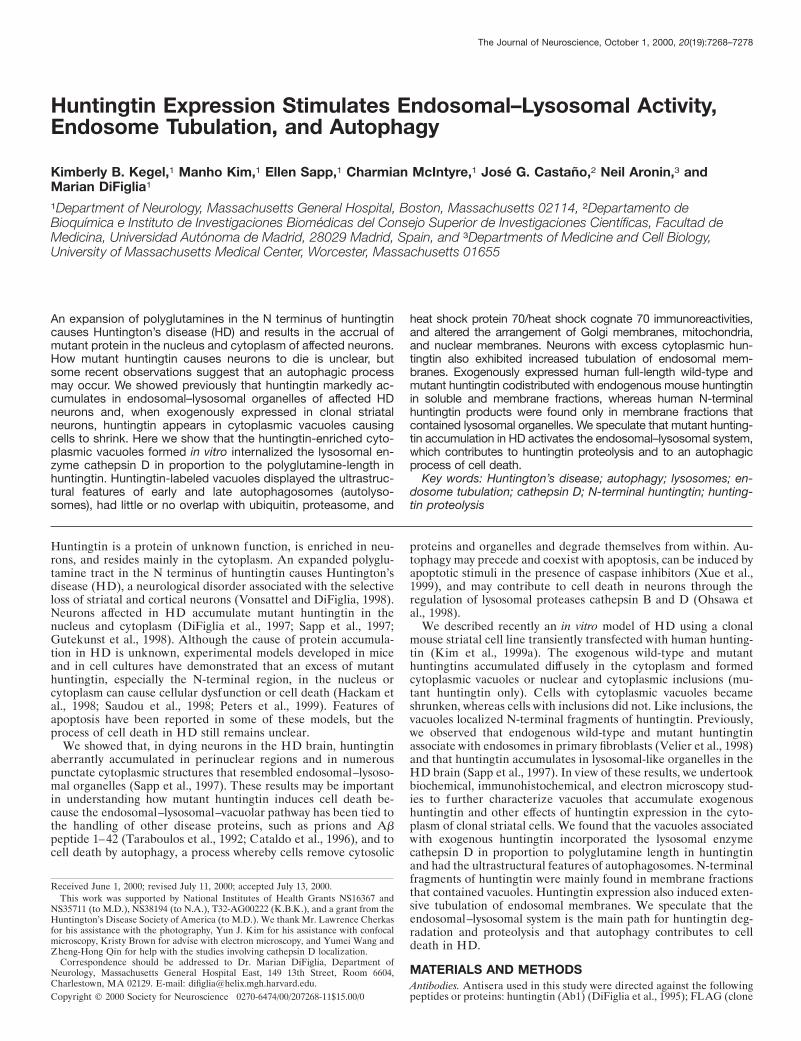
RESULTSMorphology, distribution, and time course ofappearance of dispersed and perinuclear vacuolesIn cells examined within 24 hr after transfection of truncated orfull-length FLAG-huntingtin cDNAs, the cytoplasmic staining de-tected with FLAG antibody in many cells was reticular and con-sisted of very small discrete tubular structures (Fig. 1a, shown fortruncated huntingtin). The immunoreactive vacuoles that appearedin a small proportion of cells had irregular shapes and sizes andoccurred in the cell bodies and proximal and distal portions ofneurites (Fig. 1b,c, shown for truncated huntingtin). Some vacuoleshad a ring-like appearance (Fig. 1d,e, f,g, shown for truncated andfull-length huntingtin). In some cells, coalescence of vacuoles inthe perinuclear region formed a single large complex (Fig. 1d,e,g,shown for truncated and full-length huntingtin). Vacuoles werealso detected using Ab585, an antibody to an internal site inhuntingtin (Fig. 1h). Expression of an untagged huntingtin cDNAproduced vacuoles detectable with Ab1, an antibody to the Nterminus of huntingtin (Fig. 1i, shown for truncated huntingtin).The vacuole formation was specific to huntingtin expression, be-cause overexpression of an unrelated protein, JIP1 (Yasuda et al.,1999), bearing a FLAG tag at the N terminus produced diffusecytoplasmic labeling but no vacuoles (Fig. 1h). In a previous study,we showed that N-terminal huntingtin fragments were localized tovacuoles 24–48 hr after transfection. We performed an experimentto see whether the appearance of dispersed and perinuclear vacu-oles coincided with the generation of N-terminal fragments, whichappeared at 9 hr and were maximal at 24 hr after transfection inWestern blots (Kim et al., 1999a). Cells were examined at 5, 7, 9,and 24 hr after transfection of the FLAG mutant huntingtin con-struct FH3221-100. At 5 and 7 hr, FLAG staining was cytoplasmicin 100% of labeled cells. At 9 hr, 7.6% of labeled cells haddispersed vacuoles. At 24 hr, 4.4% of labeled cells had dispersed
Kegel et al. • Huntingtin Stimulates Endosomes–Lysosomes J. Neurosci., October 1, 2000, 20(19):7268–7278 7269
vacuoles, and 11.8% displayed perinuclear vacuoles. (Additionalexperiments showed the same results and revealed that the perinu-clear vacuoles could be detected as early as 18 hr with FH3221-100.)Critically, as found in our previous study (Kim et al., 1999a), fewcells with nuclear or cytoplasmic inclusions were found in the first24 hr, accounting for only ;0.4% of the labeled cells at 24 hr. [Cellswith inclusions are maximal 4–6 d after transfection (Kim et al.,1999a).] Thus, the formation of vacuoles and not inclusions coin-cided with the production of N-terminal huntingtin fragments.
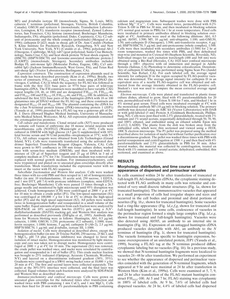
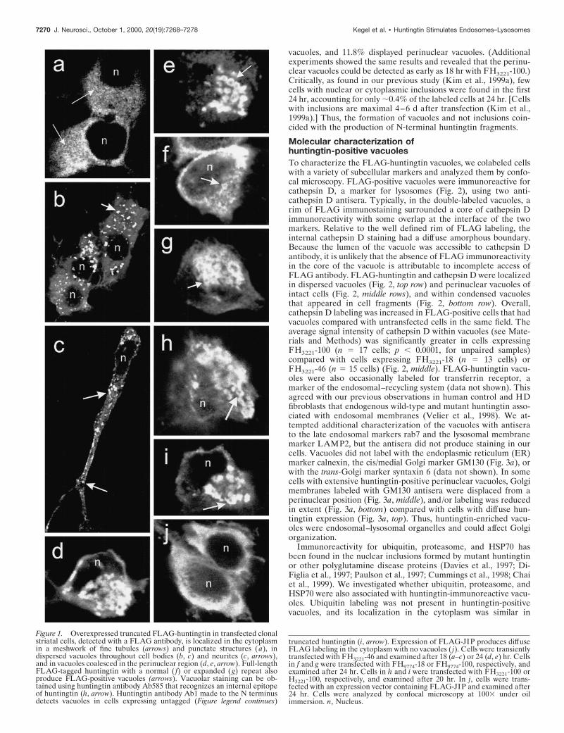
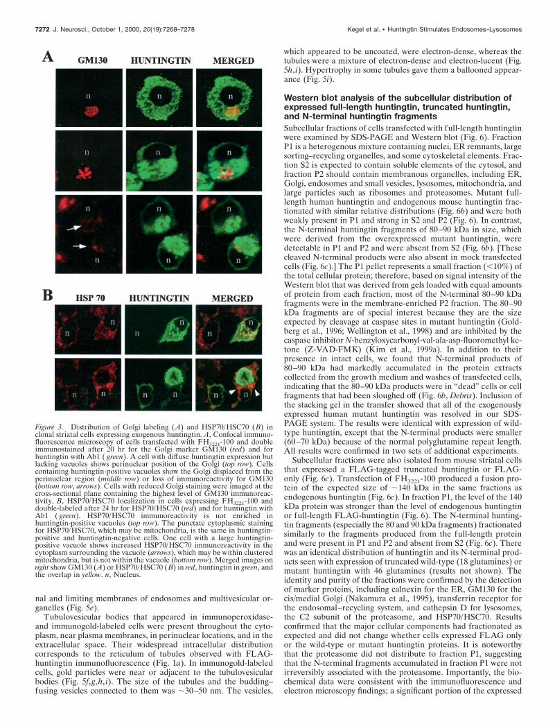
Molecular characterization ofhuntingtin-positive vacuolesTo characterize the FLAG-huntingtin vacuoles, we colabeled cellswith a variety of subcellular markers and analyzed them by confo-cal microscopy. FLAG-positive vacuoles were immunoreactive forcathepsin D, a marker for lysosomes (Fig. 2), using two anti-cathepsin D antisera. Typically, in the double-labeled vacuoles, arim of FLAG immunostaining surrounded a core of cathepsin Dimmunoreactivity with some overlap at the interface of the twomarkers. Relative to the well defined rim of FLAG labeling, theinternal cathepsin D staining had a diffuse amorphous boundary.Because the lumen of the vacuole was accessible to cathepsin Dantibody, it is unlikely that the absence of FLAG immunoreactivityin the core of the vacuole is attributable to incomplete access ofFLAG antibody. FLAG-huntingtin and cathepsin D were localizedin dispersed vacuoles (Fig. 2, top row) and perinuclear vacuoles ofintact cells (Fig. 2, middle rows), and within condensed vacuolesthat appeared in cell fragments (Fig. 2, bottom row). Overall,cathepsin D labeling was increased in FLAG-positive cells that hadvacuoles compared with untransfected cells in the same field. Theaverage signal intensity of cathepsin D within vacuoles (see Mate-rials and Methods) was significantly greater in cells expressingFH3221-100 (n 5 17 cells; p , 0.0001, for unpaired samples)compared with cells expressing FH3221-18 (n 5 13 cells) orFH3221-46 (n 5 15 cells) (Fig. 2, middle). FLAG-huntingtin vacu-oles were also occasionally labeled for transferrin receptor, amarker of the endosomal–recycling system (data not shown). Thisagreed with our previous observations in human control and HDfibroblasts that endogenous wild-type and mutant huntingtin asso-ciated with endosomal membranes (Velier et al., 1998). We at-tempted additional characterization of the vacuoles with antiserato the late endosomal markers rab7 and the lysosomal membranemarker LAMP2, but the antisera did not produce staining in ourcells. Vacuoles did not label with the endoplasmic reticulum (ER)marker calnexin, the cis/medial Golgi marker GM130 (Fig. 3a), orwith the trans-Golgi marker syntaxin 6 (data not shown). In somecells with extensive huntingtin-positive perinuclear vacuoles, Golgimembranes labeled with GM130 antisera were displaced from aperinuclear position (Fig. 3a, middle), and/or labeling was reducedin extent (Fig. 3a, bottom) compared with cells with diffuse hun-tingtin expression (Fig. 3a, top). Thus, huntingtin-enriched vacu-oles were endosomal–lysosomal organelles and could affect Golgiorganization.
Immunoreactivity for ubiquitin, proteasome, and HSP70 hasbeen found in the nuclear inclusions formed by mutant huntingtinor other polyglutamine disease proteins (Davies et al., 1997; Di-Figlia et al., 1997; Paulson et al., 1997; Cummings et al., 1998; Chaiet al., 1999). We investigated whether ubiquitin, proteasome, andHSP70 were also associated with huntingtin-immunoreactive vacu-oles. Ubiquitin labeling was not present in huntingtin-positivevacuoles, and its localization in the cytoplasm was similar in
truncated huntingtin (i, arrow). Expression of FLAG-JIP produces diffuseFLAG labeling in the cytoplasm with no vacuoles ( j). Cells were transientlytransfected with FH3221-46 and examined after 18 (a–c) or 24 (d, e) hr. Cellsin f and g were transfected with FH9774-18 or FH9774-100, respectively, andexamined after 24 hr. Cells in h and i were transfected with FH3221-100 orH3221-100, respectively, and examined after 20 hr. In j, cells were trans-fected with an expression vector containing FLAG-JIP and examined after24 hr. Cells were analyzed by confocal microscopy at 1003 under oilimmersion. n, Nucleus.
Figure 1. Overexpressed truncated FLAG-huntingtin in transfected clonalstriatal cells, detected with a FLAG antibody, is localized in the cytoplasmin a meshwork of fine tubules (arrows) and punctate structures (a), indispersed vacuoles throughout cell bodies (b, c) and neurites (c, arrows),and in vacuoles coalesced in the perinuclear region (d, e, arrow). Full-lengthFLAG-tagged huntingtin with a normal ( f) or expanded (g) repeat alsoproduce FLAG-positive vacuoles (arrows). Vacuolar staining can be ob-tained using huntingtin antibody Ab585 that recognizes an internal epitopeof huntingtin (h, arrow). Huntingtin antibody Ab1 made to the N terminusdetects vacuoles in cells expressing untagged (Figure legend continues)
7270 J. Neurosci., October 1, 2000, 20(19):7268–7278 Kegel et al. • Huntingtin Stimulates Endosomes–Lysosomes
FLAG-negative and FLAG-positive cells (results not shown).Overall, similar results were obtained using an antibody against thewhole proteasome complex (data not shown). In some cells, how-ever, proteasome labeling was increased at the borders of con-densed perinuclear vacuoles and rarely within the vacuoles (datanot shown). Immunoreactivity for HSP70/HSC70 in the cytoplasmwas not different in most cells expressing exogenous huntingtincompared with cells expressing only endogenous huntingtin (Fig.3b, top). However, in some cells with very large huntingtin-labeledvacuoles, HSP70/HSC70 staining was markedly increased in thecytoplasm surrounding the vacuoles (Fig. 3b, bottom). The increasemay be attributable to vacuole-induced displacement and accumu-lation of organelles, especially mitochondria that contain HSP70family members (Kang et al., 1990) (see below).
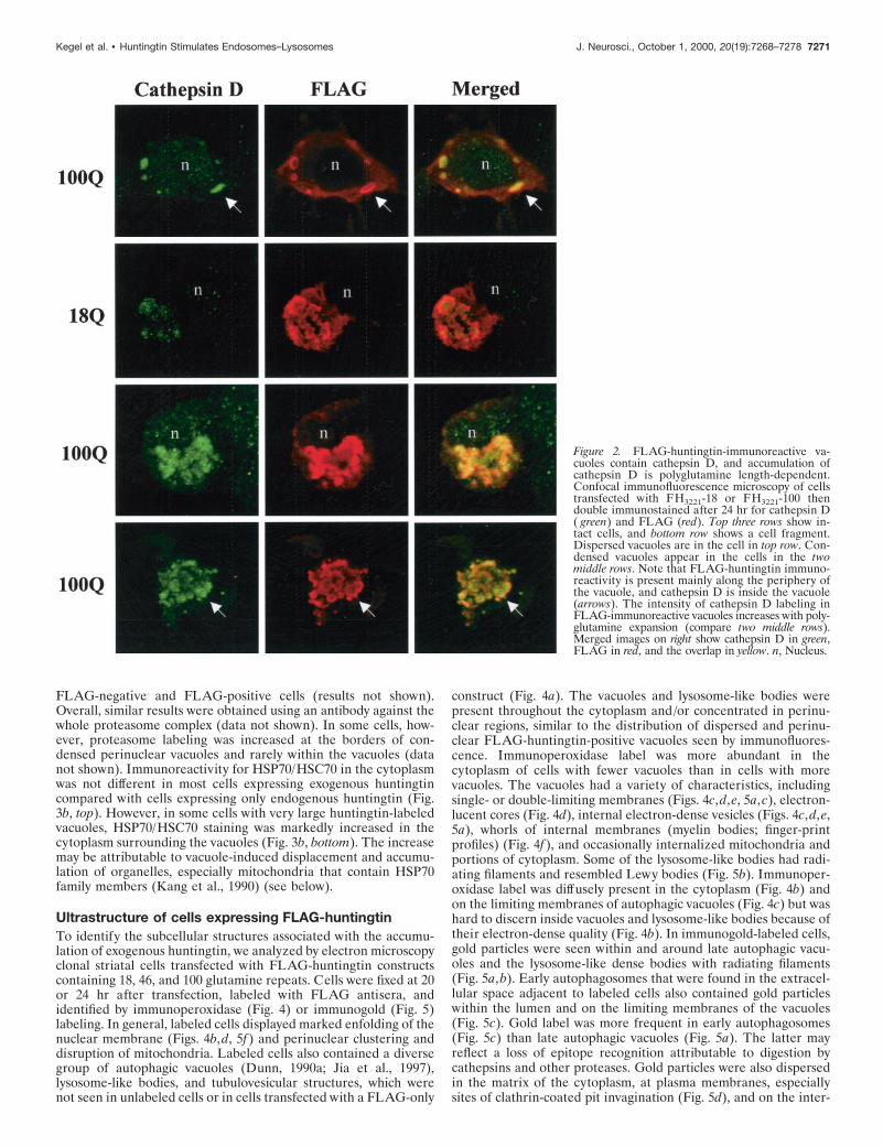
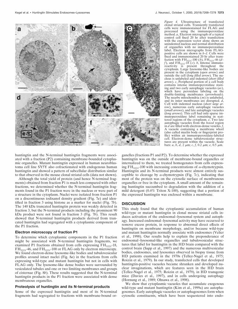
Ultrastructure of cells expressing FLAG-huntingtinTo identify the subcellular structures associated with the accumu-lation of exogenous huntingtin, we analyzed by electron microscopyclonal striatal cells transfected with FLAG-huntingtin constructscontaining 18, 46, and 100 glutamine repeats. Cells were fixed at 20or 24 hr after transfection, labeled with FLAG antisera, andidentified by immunoperoxidase (Fig. 4) or immunogold (Fig. 5)labeling. In general, labeled cells displayed marked enfolding of thenuclear membrane (Figs. 4b,d, 5f) and perinuclear clustering anddisruption of mitochondria. Labeled cells also contained a diversegroup of autophagic vacuoles (Dunn, 1990a; Jia et al., 1997),lysosome-like bodies, and tubulovesicular structures, which werenot seen in unlabeled cells or in cells transfected with a FLAG-only
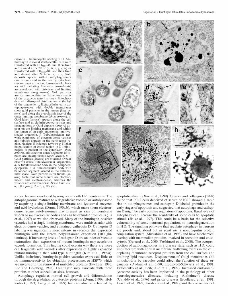
construct (Fig. 4a). The vacuoles and lysosome-like bodies werepresent throughout the cytoplasm and/or concentrated in perinu-clear regions, similar to the distribution of dispersed and perinu-clear FLAG-huntingtin-positive vacuoles seen by immunofluores-cence. Immunoperoxidase label was more abundant in thecytoplasm of cells with fewer vacuoles than in cells with morevacuoles. The vacuoles had a variety of characteristics, includingsingle- or double-limiting membranes (Figs. 4c,d,e, 5a,c), electron-lucent cores (Fig. 4d), internal electron-dense vesicles (Figs. 4c,d,e,5a), whorls of internal membranes (myelin bodies; finger-printprofiles) (Fig. 4f), and occasionally internalized mitochondria andportions of cytoplasm. Some of the lysosome-like bodies had radi-ating filaments and resembled Lewy bodies (Fig. 5b). Immunoper-oxidase label was diffusely present in the cytoplasm (Fig. 4b) andon the limiting membranes of autophagic vacuoles (Fig. 4c) but washard to discern inside vacuoles and lysosome-like bodies because oftheir electron-dense quality (Fig. 4b). In immunogold-labeled cells,gold particles were seen within and around late autophagic vacu-oles and the lysosome-like dense bodies with radiating filaments(Fig. 5a,b). Early autophagosomes that were found in the extracel-lular space adjacent to labeled cells also contained gold particleswithin the lumen and on the limiting membranes of the vacuoles(Fig. 5c). Gold label was more frequent in early autophagosomes(Fig. 5c) than late autophagic vacuoles (Fig. 5a). The latter mayreflect a loss of epitope recognition attributable to digestion bycathepsins and other proteases. Gold particles were also dispersedin the matrix of the cytoplasm, at plasma membranes, especiallysites of clathrin-coated pit invagination (Fig. 5d), and on the inter-
Figure 2. FLAG-huntingtin-immunoreactive va-cuoles contain cathepsin D, and accumulation ofcathepsin D is polyglutamine length-dependent.Confocal immunofluorescence microscopy of cellstransfected with FH3221-18 or FH3221-100 thendouble immunostained after 24 hr for cathepsin D( green) and FLAG (red). Top three rows show in-tact cells, and bottom row shows a cell fragment.Dispersed vacuoles are in the cell in top row. Con-densed vacuoles appear in the cells in the twomiddle rows. Note that FLAG-huntingtin immuno-reactivity is present mainly along the periphery ofthe vacuole, and cathepsin D is inside the vacuole(arrows). The intensity of cathepsin D labeling inFLAG-immunoreactive vacuoles increases with poly-glutamine expansion (compare two middle rows).Merged images on right show cathepsin D in green,FLAG in red, and the overlap in yellow. n, Nucleus.
Kegel et al. • Huntingtin Stimulates Endosomes–Lysosomes J. Neurosci., October 1, 2000, 20(19):7268–7278 7271
nal and limiting membranes of endosomes and multivesicular or-ganelles (Fig. 5e).
Tubulovesicular bodies that appeared in immunoperoxidase-and immunogold-labeled cells were present throughout the cyto-plasm, near plasma membranes, in perinuclear locations, and in theextracellular space. Their widespread intracellular distributioncorresponds to the reticulum of tubules observed with FLAG-huntingtin immunofluorescence (Fig. 1a). In immunogold-labeledcells, gold particles were near or adjacent to the tubulovesicularbodies (Fig. 5f,g,h, i). The size of the tubules and the budding–fusing vesicles connected to them was ;30–50 nm. The vesicles,
which appeared to be uncoated, were electron-dense, whereas thetubules were a mixture of electron-dense and electron-lucent (Fig.5h, i). Hypertrophy in some tubules gave them a ballooned appear-ance (Fig. 5i).
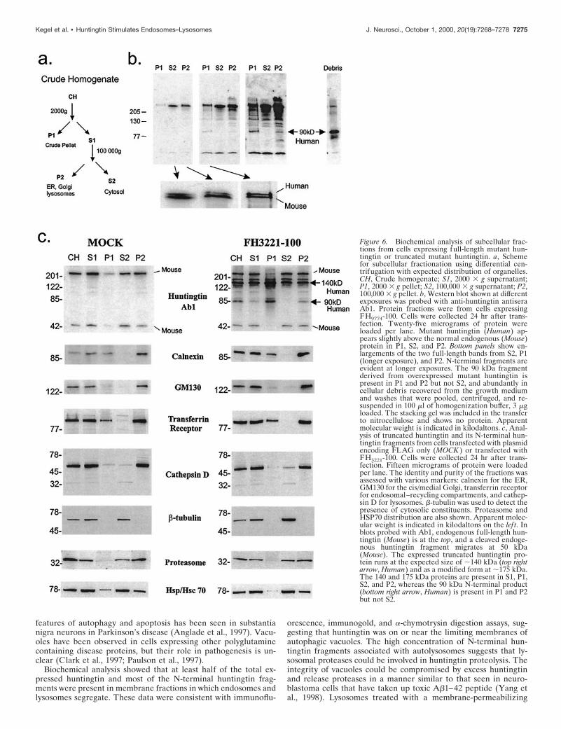
Western blot analysis of the subcellular distribution ofexpressed full-length huntingtin, truncated huntingtin,and N-terminal huntingtin fragmentsSubcellular fractions of cells transfected with full-length huntingtinwere examined by SDS-PAGE and Western blot (Fig. 6). FractionP1 is a heterogenous mixture containing nuclei, ER remnants, largesorting–recycling organelles, and some cytoskeletal elements. Frac-tion S2 is expected to contain soluble elements of the cytosol, andfraction P2 should contain membranous organelles, including ER,Golgi, endosomes and small vesicles, lysosomes, mitochondria, andlarge particles such as ribosomes and proteasomes. Mutant full-length human huntingtin and endogenous mouse huntingtin frac-tionated with similar relative distributions (Fig. 6b) and were bothweakly present in P1 and strong in S2 and P2 (Fig. 6). In contrast,the N-terminal huntingtin fragments of 80–90 kDa in size, whichwere derived from the overexpressed mutant huntingtin, weredetectable in P1 and P2 and were absent from S2 (Fig. 6b). [Thesecleaved N-terminal products were also absent in mock transfectedcells (Fig. 6c).] The P1 pellet represents a small fraction (,10%) ofthe total cellular protein; therefore, based on signal intensity of theWestern blot that was derived from gels loaded with equal amountsof protein from each fraction, most of the N-terminal 80–90 kDafragments were in the membrane-enriched P2 fraction. The 80–90kDa fragments are of special interest because they are the sizeexpected by cleavage at caspase sites in mutant huntingtin (Gold-berg et al., 1996; Wellington et al., 1998) and are inhibited by thecaspase inhibitor N-benzyloxycarbonyl-val-ala-asp-fluoromethyl ke-tone (Z-VAD-FMK) (Kim et al., 1999a). In addition to theirpresence in intact cells, we found that N-terminal products of80–90 kDa had markedly accumulated in the protein extractscollected from the growth medium and washes of transfected cells,indicating that the 80–90 kDa products were in “dead” cells or cellfragments that had been sloughed off (Fig. 6b, Debris). Inclusion ofthe stacking gel in the transfer showed that all of the exogenouslyexpressed human mutant huntingtin was resolved in our SDS-PAGE system. The results were identical with expression of wild-type huntingtin, except that the N-terminal products were smaller(60–70 kDa) because of the normal polyglutamine repeat length.All results were confirmed in two sets of additional experiments.
Subcellular fractions were also isolated from mouse striatal cellsthat expressed a FLAG-tagged truncated huntingtin or FLAG-only (Fig. 6c). Transfection of FH3221-100 produced a fusion pro-tein of the expected size of ;140 kDa in the same fractions asendogenous huntingtin (Fig. 6c). In fraction P1, the level of the 140kDa protein was stronger than the level of endogenous huntingtinor full-length FLAG-huntingtin (Fig. 6). The N-terminal hunting-tin fragments (especially the 80 and 90 kDa fragments) fractionatedsimilarly to the fragments produced from the full-length proteinand were present in P1 and P2 and absent from S2 (Fig. 6c). Therewas an identical distribution of huntingtin and its N-terminal prod-ucts seen with expression of truncated wild-type (18 glutamines) ormutant huntingtin with 46 glutamines (results not shown). Theidentity and purity of the fractions were confirmed by the detectionof marker proteins, including calnexin for the ER, GM130 for thecis/medial Golgi (Nakamura et al., 1995), transferrin receptor forthe endosomal–recycling system, and cathepsin D for lysosomes,the C2 subunit of the proteasome, and HSP70/HSC70. Resultsconfirmed that the major cellular components had fractionated asexpected and did not change whether cells expressed FLAG onlyor the wild-type or mutant huntingtin proteins. It is noteworthythat the proteasome did not distribute to fraction P1, suggestingthat the N-terminal fragments accumulated in fraction P1 were notirreversibly associated with the proteasome. Importantly, the bio-chemical data were consistent with the immunofluorescence andelectron microscopy findings; a significant portion of the expressed
Figure 3. Distribution of Golgi labeling (A) and HSP70/HSC70 ( B) inclonal striatal cells expressing exogenous huntingtin. A, Confocal immuno-fluorescence microscopy of cells transfected with FH3221-100 and doubleimmunostained after 20 hr for the Golgi marker GM130 (red) and forhuntingtin with Ab1 ( green). A cell with diffuse huntingtin expression butlacking vacuoles shows perinuclear position of the Golgi (top row). Cellscontaining huntingtin-positive vacuoles show the Golgi displaced from theperinuclear region (middle row) or loss of immunoreactivity for GM130(bottom row, arrows). Cells with reduced Golgi staining were imaged at thecross-sectional plane containing the highest level of GM130 immunoreac-tivity. B, HSP70/HSC70 localization in cells expressing FH3221-100 anddouble-labeled after 24 hr for HSP70/HSC70 (red) and for huntingtin withAb1 ( green). HSP70/HSC70 immunoreactivity is not enriched inhuntingtin-positive vacuoles (top row). The punctate cytoplasmic stainingfor HSP70/HSC70, which may be mitochondria, is the same in huntingtin-positive and huntingtin-negative cells. One cell with a large huntingtin-positive vacuole shows increased HSP70/HSC70 immunoreactivity in thecytoplasm surrounding the vacuole (arrows), which may be within clusteredmitochondria, but is not within the vacuole (bottom row). Merged images onright show GM130 (A) or HSP70/HSC70 (B) in red, huntingtin in green, andthe overlap in yellow. n, Nucleus.
7272 J. Neurosci., October 1, 2000, 20(19):7268–7278 Kegel et al. • Huntingtin Stimulates Endosomes–Lysosomes
huntingtin and the N-terminal huntingtin fragments were associ-ated with a fraction (P2) containing membrane-bounded cytoplas-mic organelles. Mutant huntingtin expressed in human neuroblas-toma cell line SY5Y also cofractionated with endogenous humanhuntingtin and showed a pattern of subcellular distribution similarto that observed in the mouse clonal striatal cells (data not shown).
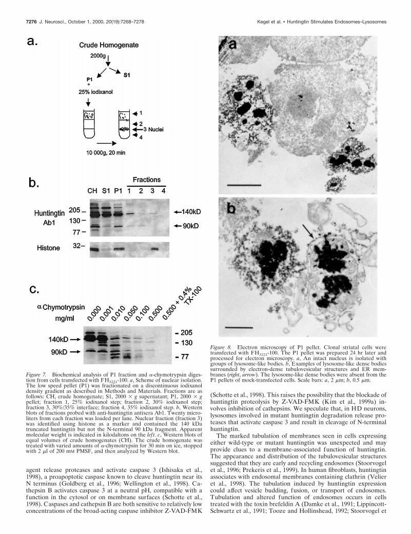
Although the total yield of protein (and hence N-terminal frag-ments) obtained from fraction P1 is much less compared with otherfractions, we determined whether the N-terminal huntingtin frag-ments found in the P1 fraction were in the nucleus or were part ofa structure in the cytoplasm. Nuclei were isolated from fraction P1on a discontinuous iodixanol density gradient (Fig. 7a) and iden-tified in fraction 3 using histone as a marker for nuclei (Fig. 7b).The 140 kDa truncated huntingtin protein was weakly detected infraction 3, but the N-terminal products including the prominent 90kDa product were not found in fraction 3 (Fig. 7b). This resultshowed that N-terminal huntingtin products derived from trun-cated huntingtin had segregated with a cytoplasmic component ofthe P1 fraction.
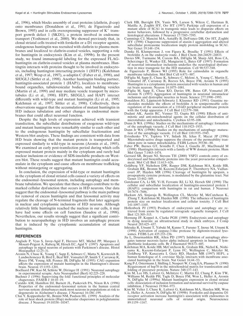
Electron microscopy of fraction P1To determine which cytoplasmic components in the P1 fractionmight be associated with N-terminal huntingtin fragments, weexamined P1 fractions obtained from cells expressing FH3221-18,FH3221-46, and FH3221-100 or FLAG only by electron microscopy.We found electron-dense lysosome-like bodies and tubulovesicularprofiles around intact nuclei (Fig. 8a) in the fractions from cellsexpressing wild-type and mutant huntingtin but not in cells withFLAG only. The lysosome-like dense bodies were surrounded byvesiculated tubules and one or two limiting membranes and groupsof cisternae (Fig. 8b). These results suggested that the N-terminalhuntingtin products in the P1 fraction were associated with densemembranous organelles.
Proteolysis of huntingtin and its N-terminal productsSome of the expressed huntingtin and most of its N-terminalfragments had segregated to fractions with membrane-bound or-
ganelles (fractions P1 and P2). To determine whether the expressedhuntingtin was on the outside of membrane-bound organelles orinternalized to them, we treated homogenates from cells express-ing FH3221-100 with increasing concentrations of a-chymotrypsin.Huntingtin and its N-terminal products were almost entirely sus-ceptible to cleavage by a-chymotrypsin (Fig. 7c), indicating thatmost of the protein was on the cytosolic surface of membranousorganelles or free in the cytoplasm. A small amount of the remain-ing huntingtin succumbed to degradation with the addition of amild detergent (0.4% Triton X-100), suggesting that a portion ofthe expressed huntingtin was enclosed within a membrane.
DISCUSSIONThis study found that the cytoplasmic accumulation of humanwild-type or mutant huntingtin in clonal mouse striatal cells in-duces activation of the endosomal–lysosomal system and autoph-agy. Increased endosomal–lysosomal activity may have occurred toremove excess protein, in response to abnormal direct effects ofhuntingtin on membrane morphology, and/or because wild-typeand mutant huntingtin normally associate with endosomes (Velieret al., 1998). Our results help to explain the preponderance ofendosomal–lysosomal-like organelles and tubulovesicular struc-tures that label for huntingtin in the HD brain compared with thecontrol brain (Sapp et al., 1997) and the numerous multivesicularbodies, endosomes, and lysosomes observed in biopsy tissue fromHD patients examined in the 1970s (Tellez-Nagel et al., 1975;Roizin et al., 1979). In our study, transfected cells that developedhuntingtin-positive vacuoles became shrunken and developed nu-clear invaginations, which are features seen in the HD brain(Tellez-Nagel et al., 1975; Roizin et al., 1979), in HD transgenicmice (Davies et al., 1997), and in cells undergoing autophagy(Hornung et al., 1989; Ohsawa et al., 1998).
We show that cytoplasmic vacuoles that accumulate exogenouswild-type and mutant huntingtin (Kim et al., 1999a) are autopha-gosomes. Early autophagic vacuoles or autophagosomes form whencytosolic constituents, which have been sequestered into endo-
Figure 4. Ultrastructure of transfectedclonal striatal cells. Transiently transfectedcells were immunostained for FLAG andprocessed using the immunoperoxidasemethod. a, Electron micrograph of a typicalcontrol cell fixed 20 hr after transfectionwith the expression vector alone shows anunindented nucleus and normal distributionof organelles with no immunoperoxidaselabel. Electron micrographs from FLAG-positive cells are shown in b–f. Cells werefixed and immunostained 20 hr after trans-fection with FH3221-100 (b), FH3221-46 (d–f ), and FH3221-18 (c). b, Intense immuno-reactivity is present throughout thecytoplasm. Large lysosome-like bodies arepresent in the cytoplasm (open arrow) andoutside the cell (long filled arrow). The nu-cleus is unlabeled and indented (short filledarrow). c, Peripheral portion of a cell bodycontains intense immunoperoxidase stain-ing and two early autophagic vacuoles (av),which have peroxidase labeling on thedouble-limiting membranes (arrowheads).The nearby mitochondria ( m) is unlabeled,and its inner membranes are disrupted. d,Cell with indented nucleus (short large ar-row), numerous early autophagic vacuoles(small arrows), and late autophagic vacuoles(long arrows). This cell had only sparse im-munoperoxidase label remaining in scat-tered regions of the cytoplasm. e, Two lateautophagic vacuoles from the boxed regionin d are filled with electron-dense vesicles. f,A vacuole containing a membrane whorl(also called myelin body or fingerprint pro-file) within an immunoperoxidase-labeledcell. Electron-dense tubulovesicular struc-tures are present within the vacuole. Scalebars: a, b, d, 2 mm; c, f, 0.2 mm; e, 0.5 mm.
Kegel et al. • Huntingtin Stimulates Endosomes–Lysosomes J. Neurosci., October 1, 2000, 20(19):7268–7278 7273
somes, become enveloped by rough or smooth ER membranes. Theautophagosome matures to a degradative vacuole or autolysosomeby acquiring a single-limiting membrane and lysosomal enzymesand acid hydrolases (Dunn, 1990a,b), which make them electron-dense. Some autolysosomes may present as sacs of membranewhorls or multivesicular bodies and can be extruded from cells (Jiaet al., 1997) as we also observed. Many of the huntingtin-positivevacuoles had a single-limiting membrane, were multivesicular withelectron-dense vesicles, and contained cathepsin D. Cathepsin Dlabeling was significantly more intense in vacuoles that expressedhuntingtin with the largest polyglutamine expansion (100 glu-tamines). If increasing levels of cathepsin D are an index of vacuolematuration, then expression of mutant huntingtin may acceleratevacuole formation. This finding could explain why there are morecell fragments with vacuoles after expression of highly expandedmutant huntingtin than wild-type huntingtin (Kim et al., 1999a).Unlike inclusions, huntingtin-positive vacuoles expressed little orno immunoreactivity for ubiquitin, proteasome, or HSP70, whichare involved in the removal of misfolded proteins (Buchner, 1996;Lee and Goldberg, 1998). Huntingtin may associate with theseproteins at other subcellular sites, however.
Autophagy regulates normal cell growth and differentiationthrough the degradation of cytosolic proteins (Dunn, 1990a; Hol-lenbeck, 1993; Liang et al., 1999) but can also be activated by
apoptotic stimuli (Xue et al., 1999). Ohsawa and colleagues (1998)found that PC12 cells deprived of serum or NGF showed a rapidrise in autophagosomes and cathepsin D-labeled granules in theearly stages of apoptosis and suggested that autophagy and cathep-sin D might be early positive regulators of apoptosis. Basal levels ofautophagy can increase the sensitivity of some cells to apoptoticstimuli (Jia et al., 1997). This could be a basis for the selectivevulnerability of some neuronal populations to neurodegenerationin HD. The signaling pathways that regulate autophagy in neuronsare poorly understood but in yeast use a nonubiquitin proteinconjugation system (Mizushima et al., 1998) and have biochemicaloverlap with mammalian proteins involved in secretion and endo-cytosis (Gerrard et al., 2000; Yoshimori et al., 2000). The overpro-duction of autophagosomes in a disease state, such as HD, couldalso interfere with normal membrane trafficking events in the cell,depleting membrane receptor proteins from the cell surface anddraining lipid resources. Displacement of Golgi membranes andmitochondria by vacuoles could affect the function of these or-ganelles (Dudani et al., 1990; Lippincott-Schwartz et al., 1991;Evtodienko et al., 1996; Xue et al., 1999). Increased endosome–lysosome activity has been implicated in the pathology of otherneurodegenerative diseases, including Alzheimer’s disease(Cataldo et al., 1996) and prion diseases (Boellaard et al., 1991;Laszlo et al., 1992; Taraboulos et al., 1992), and the coexistence of
Figure 5. Immunogold labeling of FLAG-huntingtin in clonal striatal cells. Cells weretransfected with FH3221-46 and then fixedand stained after 20 hr (a, b, d, f, g, h) ortransfected with FH3221-100 and then fixedand stained after 24 hr (c, e, i). a, Golddeposits appear within autophagosomes(top arrows) and in the nearby cytoplasm(bottom right arrow). b, Lysosome-like bod-ies with radiating filaments (arrowheads)are enveloped with cisternae and limitingmembranes (long arrows). Gold particlesare scattered within the filamentous matrixof the organelle (short arrows). Mitochon-dria with disrupted cisternae are to the lef tof the organelle. c, Extracellular early au-tophagosomes with double membranesshow gold particles in the lumen (long ar-rows) and along the cytoplasmic face of theouter limiting membrane (short arrows). d,Gold label (arrows) appears along the cellsurface and at clathrin-coated vesicles andinvaginations. e, Gold deposits (arrows) ap-pear on the limiting membrane and withinthe lumen of an early endosomal–multive-sicular organelle. f, Tubulovesicular net-work composed of electron-dense vesiclesand tubules appears in the perinuclear re-gion. Nucleus is indented (arrow). g, Highermagnification of boxed region in f. Immu-nogold is present in the cytoplasm (shortarrows), and electron-dense segments of tu-bules are clearly visible (open arrow). h, i,Gold particles (arrows) are attached or nearelectron-dense tubulovesicular organelles.h, A tubulovesicular body in the peripheralcytoplasm. i, A tubulovesicular body withballooned segment located in the extracel-lular space. Gold particle is on tubule (ar-row). Note that some tubules are electron-lucent and electron-dense, whereas thevesicles are electron-dense. Scale bars: a–e,h, i, 0.2 mm; f, 2 mm, g, 0.5 mm.
7274 J. Neurosci., October 1, 2000, 20(19):7268–7278 Kegel et al. • Huntingtin Stimulates Endosomes–Lysosomes
features of autophagy and apoptosis has been seen in substantianigra neurons in Parkinson’s disease (Anglade et al., 1997). Vacu-oles have been observed in cells expressing other polyglutaminecontaining disease proteins, but their role in pathogenesis is un-clear (Clark et al., 1997; Paulson et al., 1997).
Biochemical analysis showed that at least half of the total ex-pressed huntingtin and most of the N-terminal huntingtin frag-ments were present in membrane fractions in which endosomes andlysosomes segregate. These data were consistent with immunoflu-
orescence, immunogold, and a-chymotrysin digestion assays, sug-gesting that huntingtin was on or near the limiting membranes ofautophagic vacuoles. The high concentration of N-terminal hun-tingtin fragments associated with autolysosomes suggests that ly-sosomal proteases could be involved in huntingtin proteolysis. Theintegrity of vacuoles could be compromised by excess huntingtinand release proteases in a manner similar to that seen in neuro-blastoma cells that have taken up toxic Ab1–42 peptide (Yang etal., 1998). Lysosomes treated with a membrane-permeabilizing
Figure 6. Biochemical analysis of subcellular frac-tions from cells expressing full-length mutant hun-tingtin or truncated mutant huntingtin. a, Schemefor subcellular fractionation using differential cen-trifugation with expected distribution of organelles.CH, Crude homogenate; S1, 2000 3 g supernatant;P1, 2000 3 g pellet; S2, 100,000 3 g supernatant; P2,100,000 3 g pellet. b, Western blot shown at differentexposures was probed with anti-huntingtin antiseraAb1. Protein fractions were from cells expressingFH9774-100. Cells were collected 24 hr after trans-fection. Twenty-five micrograms of protein wereloaded per lane. Mutant huntingtin (Human) ap-pears slightly above the normal endogenous (Mouse)protein in P1, S2, and P2. Bottom panels show en-largements of the two full-length bands from S2, P1(longer exposure), and P2. N-terminal fragments areevident at longer exposures. The 90 kDa fragmentderived from overexpressed mutant huntingtin ispresent in P1 and P2 but not S2, and abundantly incellular debris recovered from the growth mediumand washes that were pooled, centrifuged, and re-suspended in 100 ml of homogenization buffer, 3 mgloaded. The stacking gel was included in the transferto nitrocellulose and shows no protein. Apparentmolecular weight is indicated in kilodaltons. c, Anal-ysis of truncated huntingtin and its N-terminal hun-tingtin fragments from cells transfected with plasmidencoding FLAG only (MOCK ) or transfected withFH3221-100. Cells were collected 24 hr after trans-fection. Fifteen micrograms of protein were loadedper lane. The identity and purity of the fractions wasassessed with various markers: calnexin for the ER,GM130 for the cis/medial Golgi, transferrin receptorfor endosomal–recycling compartments, and cathep-sin D for lysosomes. b-tubulin was used to detect thepresence of cytosolic constituents. Proteasome andHSP70 distribution are also shown. Apparent molec-ular weight is indicated in kilodaltons on the lef t. Inblots probed with Ab1, endogenous full-length hun-tingtin (Mouse) is at the top, and a cleaved endoge-nous huntingtin fragment migrates at 50 kDa(Mouse). The expressed truncated huntingtin pro-tein runs at the expected size of ;140 kDa (top rightarrow, Human) and as a modified form at ;175 kDa.The 140 and 175 kDa proteins are present in S1, P1,S2, and P2, whereas the 90 kDa N-terminal product(bottom right arrow, Human) is present in P1 and P2but not S2.
Kegel et al. • Huntingtin Stimulates Endosomes–Lysosomes J. Neurosci., October 1, 2000, 20(19):7268–7278 7275
agent release proteases and activate caspase 3 (Ishisaka et al.,1998), a proapoptotic caspase known to cleave huntingtin near itsN terminus (Goldberg et al., 1996; Wellington et al., 1998). Ca-thepsin B activates caspase 3 at a neutral pH, compatible with afunction in the cytosol or on membrane surfaces (Schotte et al.,1998). Caspases and cathepsin B are both sensitive to relatively lowconcentrations of the broad-acting caspase inhibitor Z-VAD-FMK
(Schotte et al., 1998). This raises the possibility that the blockade ofhuntingtin proteolysis by Z-VAD-FMK (Kim et al., 1999a) in-volves inhibition of cathepsins. We speculate that, in HD neurons,lysosomes involved in mutant huntingtin degradation release pro-teases that activate caspase 3 and result in cleavage of N-terminalhuntingtin.
The marked tubulation of membranes seen in cells expressingeither wild-type or mutant huntingtin was unexpected and mayprovide clues to a membrane-associated function of huntingtin.The appearance and distribution of the tubulovesicular structuressuggested that they are early and recycling endosomes (Stoorvogelet al., 1996; Prekeris et al., 1999). In human fibroblasts, huntingtinassociates with endosomal membranes containing clathrin (Velieret al., 1998). The tubulation induced by huntingtin expressioncould affect vesicle budding, fusion, or transport of endosomes.Tubulation and altered function of endosomes occurs in cellstreated with the toxin brefeldin A (Damke et al., 1991; Lippincott-Schwartz et al., 1991; Tooze and Hollinshead, 1992; Stoorvogel et
Figure 7. Biochemical analysis of P1 fraction and a-chymotrypsin diges-tion from cells transfected with FH3221-100. a, Scheme of nuclear isolation.The low speed pellet (P1) was fractionated on a discontinuous iodixanoldensity gradient as described in Methods and Materials. Fractions are asfollows: CH, crude homogenate; S1, 2000 3 g supernatant; P1, 2000 3 gpellet; fraction 1, 25% iodixanol step; fraction 2, 30% iodixanol step;fraction 3, 30%/35% interface; fraction 4, 35% iodixanol step. b, Westernblots of fractions probed with anti-huntingtin antisera Ab1. Twenty micro-liters from each fraction was loaded per lane. Nuclear fraction (fraction 3)was identified using histone as a marker and contained the 140 kDatruncated huntingtin but not the N-terminal 90 kDa fragment. Apparentmolecular weight is indicated in kilodaltons on the lef t. c, Western blots ofequal volumes of crude homogenates (CH). The crude homogenate wastreated with varied amounts of a-chymotrypsin for 30 min on ice, stoppedwith 2 ml of 200 mM PMSF, and then analyzed by Western blot.
Figure 8. Electron microscopy of P1 pellet. Clonal striatal cells weretransfected with FH3221-100. The P1 pellet was prepared 24 hr later andprocessed for electron microscopy. a, An intact nucleus is isolated withgroups of lysosome-like bodies. b, Examples of lysosome-like dense bodiessurrounded by electron-dense tubulovesicular structures and ER mem-branes (right, arrow). The lysosome-like dense bodies were absent from theP1 pellets of mock-transfected cells. Scale bars: a, 2 mm; b, 0.5 mm.
7276 J. Neurosci., October 1, 2000, 20(19):7268–7278 Kegel et al. • Huntingtin Stimulates Endosomes–Lysosomes
al., 1996), which blocks assembly of coat proteins (clathrin, b-cop)onto membranes (Donaldson et al., 1991; de Figueiredo andBrown, 1995) and in cells overexpressing suppressor of K1 trans-port growth defect 1 (SKD1), a protein involved in endosometransport (Yoshimori et al., 2000). We showed previously that, inclonal striatal cells treated with forskolin or a D1 receptor agonist,endogenous huntingtin was recruited with clathrin to plasma mem-branes and localized to clathrin-coated vesicles, supporting a rolefor huntingtin in endocytosis (Kim et al., 1999b). In the presentstudy, we found immunogold labeling for the expressed FLAG-huntingtin on clathrin-coated vesicles at plasma membranes. Hun-tingtin interacts with proteins thought to be involved in endocyto-sis, including huntington interacting protein 1 (HIP1) (Kalchmanet al., 1997; Wesp et al., 1997), a-adaptin C (Faber et al., 1998), andSH3GL3 (Sittler et al., 1998). Another huntingtin binding partner,huntingtin-associated protein 1 (HAP1), localizes to membrane-bound organelles, tubulovesicular bodies, and budding vesicles(Martin et al., 1999) and may mediate vesicle transport by micro-tubules (Li et al., 1998). HIP1, SH3GL3, and HAP1 interactdifferently with wild-type and mutant huntingtin (Li et al., 1995;Kalchman et al., 1997; Sittler et al., 1998). Collectively, theseobservations suggest that the accumulation of mutant huntingtin inHD induces tubulation and other changes in endosomal mem-branes that could affect neuronal function.
Despite the high levels of expression achieved with transienttransfection, the subcellular distributions of exogenous wild-typeand mutant huntingtins were remarkably similar to each other andto the endogenous huntingtin by subcellular fractionation andWestern blot analysis. These findings are consistent with data fromHD brain showing that the mutant protein is transported andexpressed similarly to wild-type in neurons (Aronin et al., 1995).We examined an early post-transfection period during which cellsexpressed mutant protein mainly in the cytoplasm and rarely hadnuclear inclusions or evidence of SDS-insoluble products on West-ern blot. These results suggest that mutant huntingtin could accu-mulate in the cytoplasm and cause affects on membrane traffickingwithout mistargeting or aggregating.
In conclusion, the expression of wild-type or mutant huntingtinin the cytoplasm of clonal striatal cells caused a variety of effects onthe endosomal–lysosomal system, including autophagy and mem-brane tubulation. We speculate that these changes contribute to themarked cellular dysfunction that occurs in HD neurons. Our datasuggest that the endosomal–lysosomal pathway is the main pathwayfor removal of excess huntingtin and that lysosomal activity mayregulate the cleavage of N-terminal fragments that later aggregatein nuclear and cytoplasmic inclusions of HD neurons. Althoughrelatively little huntingtin reached the nucleus in our cells, it mayhave had some effects on cell function (Saudou et al., 1998).Nevertheless, our results strongly suggest that a significant contri-bution to neuropathology in HD involves an autophagic processthat is induced by the cytoplasmic accumulation of mutanthuntingtin.
REFERENCESAnglade P, Vyas S, Javoy-Agid F, Herrero MT, Michel PP, Marquez J,
Mouatt-Prigent A, Ruberg M, Hirsch EC, Agid Y (1997) Apoptosis andautophagy in nigral neurons of patients with Parkinson’s disease. HistolHistopathol 12:25–31.
Aronin N, Chase K, Young C, Sapp E, Schwarz C, Matta N, Kornreich R,Landwehrmeyer B, Bird E, Beal MF, Vonsattel JP, Smith T, Carraway R,Boyce FM, Young AB, Penney JB, DiFiglia M (1995) CAG expansionaffects the expression of mutant huntingtin in the Huntington’s diseasebrain. Neuron 15:1193–1201.
Boellaard JW, Kao M, Schlote W, Diringer H (1991) Neuronal autophagyin experimental scrapie. Acta Neuropathol (Berl) 82:225–228.
Buchner J (1996) Supervising the fold: functional principles of molecularchaperones. FASEB J 10:10–19.
Cataldo AM, Hamilton DJ, Barnett JL, Paskevich PA, Nixon RA (1996)Properties of the endosomal–lysosomal system in the human centralnervous system: disturbances mark most neurons in populations at risk todegenerate in Alzheimer’s disease. J Neurosci 16:186–199.
Chai Y, Koppenhafer SL, Bonini NM, Paulson HL (1999) Analysis of therole of heat shock protein (Hsp) molecular chaperones in polyglutaminedisease. J Neurosci 19:10338–10347.
Clark HB, Burright EN, Yunis WS, Larson S, Wilcox C, Hartman B,Matilla A, Zoghbi HY, Orr HT (1997) Purkinje cell expression of amutant allele of SCA1 in transgenic mice leads to disparate effects onmotor behaviors, followed by a progressive cerebellar dysfunction andhistological alterations. J Neurosci 17:7385–7395.
Cummings CJ, Mancini MA, Antalffy B, DeFranco DB, Orr HT, ZoghbiHY (1998) Chaperone suppression of ataxin-1 aggregation and alteredsubcellular proteasome localization imply protein misfolding in SCA1.Nat Genet 19:148–154.
Damke H, Klumperman J, von Figura K, Braulke T (1991) Effects ofbrefeldin A on the endocytic route. J Biol Chem 266:24829–24833.
Davies SW, Turmaine M, Cozens BA, DiFiglia M, Sharp AH, Ross CA,Scherzinger E, Wanker EE, Mangiarini L, Bates GP (1997) Formationof neuronal intranuclear inclusions underlies the neurological dysfunc-tion in mice transgenic for the HD mutation. Cell 90:537–548.
de Figueiredo P, Brown WJ (1995) Role for calmodulin in organellemembrane tubulation. Mol Biol Cell 6:871–887.
DiFiglia M, Sapp E, Chase K, Schwarz C, Meloni A, Young C, Martin E,Vonsattel JP, Carraway R, Reeves SA, Boyce FM, Aronin N (1995)Huntingtin is a cytoplasmic protein associated with vesicles in human andrat brain neurons. Neuron 14:1075–1081.
DiFiglia M, Sapp E, Chase KO, Davies SW, Bates GP, Vonsattel JP,Aronin N (1997) Aggregation of huntingtin in neuronal intranuclearinclusions and dystrophic neurites in brain. Science 277:1990–1993.
Donaldson JG, Lippincott-Schwartz J, Klausner RD (1991) Guanine nu-cleotides modulate the effects of brefeldin A in semipermeable cells:regulation of the association of a 110-kD peripheral membrane proteinwith the Golgi apparatus. J Cell Biol 112:579–588.
Dudani AK, Austin RC, Venner TJ, Gupta RS (1990) Effects of anti-mitotic and anti-mitochondrial agents on the cellular distribution ofmicrotubules and mitochondria. Cytobios 63:95–108.
Dunn Jr WA (1990a) Studies on the mechanisms of autophagy: formationof the autophagic vacuole. J Cell Biol 110:1923–1933.
Dunn Jr WA (1990b) Studies on the mechanisms of autophagy: matura-tion of the autophagic vacuole. J Cell Biol 110:1935–1945.
Evtodienko YV, Teplova VV, Sidash SS, Ichas F, Mazat JP (1996)Microtubule-active drugs suppress the closure of the permeability tran-sition pore in tumor mitochondria. FEBS Letters 393:86–88.
Faber PW, Barnes GT, Srinidhi F, Chen J, Gusella JF, MacDonald M(1998) Huntingtin interacts with a family of WW domain proteins. HumMol Genet 7:1463–1474.
Gerrard SR, Bryant NJ, Stevens TH (2000) VSP21 controls entry of en-docytosed and biosynthetic proteins into the yeast prevacuolar compart-ment. Mol Cell Biol 11:613–626.
Goldberg YP, Nicholson DW, Rasper DM, Kalchman MA, Koide HB,Graham RK, Bromm M, Kazemi-Esfarjani P, Thornberry NA, Vaillan-court JP, Hayden MR (1996) Cleavage of huntingtin by apopain, aproapoptotic cysteine protease, is modulated by the glutamine tract. NatGenet 13:442–449.
Gutekunst CA, Li SH, Yi H, Ferrante RJ, Li XJ, Hersch SM (1998) Thecellular and subcellular localization of huntingtin-associated protein 1(HAP1): comparison with huntingtin in rat and human. J Neurosci18:7674–7686.
Hackam AS, Sinjaraja R, Wellington CL, Metzler M, McCutcheon K,Zhang T, Kalchman M, Hayden MR (1998) The influence of huntingtinprotein size on nuclear localization and cellular toxicity. J Cell Biol141:1097–1105.
Hollenbeck PJ (1993) Products of endocytosis and autophagy are re-trieved from axons by regulated retrograde organelle transport. J CellBiol 121:305–315.
Hornung JP, Koppel A, Clarke PGH (1989) Endocytosis and autophagyin dying neurons: an ultrastructural study in chick embryos. J CompNeurol 283:425–437.
Ishisaka R, Utsumi T, Yabuki M, Kanno T, Furuno T, Inoue M, Utsumi K(1998) Activation of caspase-3-like protease by digitonin-treated lyso-somes. FEBS Lett 435:233–236.
Jia L, Dourmashkin RR, Allen PD (1997) Inhibition of autophagy abro-gates tumour necrosis factor alpha induced apoptosis in human T lym-phoblastic leukaemic cells. Br J Haematol 98:673–685.
Kalchman MA, Koide HB, McCutcheon K, Graham RK, Nichol K, Nishi-yama K, Kazemi-Esfarjani P, Lynn FC, Wellington C, Metzler M,Goldberg YP, Kanazawa I, Gietz RD, Hayden MR (1997) HIP1, ahuman homologue of S. cerevisiae Sla2p, interacts with membrane asso-ciated huntingtin in the brain. Nat Genet 16:44–53.
Kang P-J, Ostermann J, Shilling J, Neupert W, Craig EA, Pfanner N (1990)Requirement of hsp70 in the mitochondrial matrix for translocation andfolding of precursor proteins. Nature 348:137–143.
Kim M, Lee HS, Laforet G, McIntyre C, Martin EJ, Chang P, Kim TW,Williams M, Reddy PH, Tagle D, Boyce FM, Won L, Heller A, AroninN, DiFiglia M (1999a) Mutant huntingtin expression in clonal striatalcells: dissociation of inclusion formation and neuronal survival by caspaseinhibition. J Neurosci 19:964–973.
Kim M, Velier J, Chase K, Laforet G, Kalchman MA, Hayden MR, WonL, Heller A, Aronin N, DiFiglia M (1999b) Forskolin and dopamine D1receptor activation increase huntingtin’s association with endosomes inimmortalized neuronal cells of striatal origin. Neuroscience89:1159–1167.
Kegel et al. • Huntingtin Stimulates Endosomes–Lysosomes J. Neurosci., October 1, 2000, 20(19):7268–7278 7277

Laszlo L, Lowe J, Self T, Kenward N, Landon M, McBride T, Farquhar C,McConnell I, Brown J, Hope J, Mayers RJ (1992) Lysosomes as keyorganelles in the pathogenesis of prion encephalopathies. J Pathol166:333–341.
Lee DHL, Goldberg AL (1998) Proteasome inhibitors: valuable new toolsfor cell biologists. Trends Cell Biol 8:397–403.
Li SH, Gutekunst CA, Hersch SM, Li XJ (1998) Interaction of huntingtin-associated protein with dynactin P150Glued. J Neurosci 18:1261–1269.
Li XJ, Li SH, Sharp AH, Nucifora Jr FC, Schilling G, Lanahan A, WorleyP, Snyder SH, Ross CA (1995) A huntingtin-associated protein enrichedin brain with implications for pathology. Nature 378:398–402.
Liang XH, Jackson S, Seaman M, Brown K, Kempkes B, Hibshoosh H,Levine B (1999) Induction of autophagy and inhibition of tumorigenesisby beclin 1. Nature 402:672–676.
Lippincott-Schwartz J, Yuan L, Tipper C, Amherdt M, Orci L, KlausnerRD (1991) Brefeldin A’s effects on endosomes, lysosomes, and the TGNsuggest a general mechanism for regulating organelle structure andmembrane traffic. Cell 67:601–616.
Martin EJ, Kim M, Velier J, Sapp E, Lee H, Laforet G, Won L, Chase K,Bhide P, Heller A, Aronin N, DiFiglia M (1999) Analysis of huntingtin-associated protein 1 in mouse brain and immortalized striatal neurons.J Comp Neurol 403:421–430.
Mengual E, Arizti P, Rodrigo J, Gimenez-Amaya JM, Castano JG (1996)Immunohistochemical distribution and electron microscopic subcellularlocalization of the proteasome in the rat CNS. J Neurosci 16:6331–6341.
Mizushima N, Noda T, Yoshimori T, Tanaka Y, Ishii T, George MD,Klionsky DJ, Ohsumi M, Ohsumi Y (1998) A protein conjugation sys-tem essential for autophagy. Nature 395:395–398.
Nakamura N, Rabouille C, Watson R, Nilsson T, Hui N, Slusarewicz P,Kreis TE, Warren G (1995) Characterization of a cis-Golgi matrixprotein, GM130. J Cell Biol 131:1715–1726.
Ohsawa Y, Isahara K, Kanamori S, Shibata M, Kametaka S, Gotow T,Watanabe T, Kominami E, Uchiyama Y (1998) An ultrastructural andimmunohistochemical study of PC12 cells during apoptosis induced byserum deprivation with special reference to autophagy and lysosomalcathepsins. Arch Histol Cytol 61:395–403.
Paulson HL, Perez MK, Trottier Y, Trojanowski JQ, Subramony SH, DasSS, Vig P, Mandel JL, Fischbeck KH, Pittman RN (1997) Intranuclearinclusions in expanded polyglutamine protein in spinocerebellar ataxiatype 3. Neuron 19:333–344.
Peters MF, Nucifora Jr FC, Kushi J, Seaman HC, Cooper JK, Herring WJ,Dawson VL, Dawson TM, Ross CA (1999) Nuclear targeting of mutanthuntingtin increases toxicity. Mol Cell Neurosci 14:121–128.
Prekeris R, Folleti DL, Scheller RH (1999) Dynamics of tubulovesicularrecycling endosomes in hippocampal neurons. J Neurosci 19:10324–10327.
Roizin L, Stellar S, Liu JC (1979) Neuronal nuclear-cytoplasmic changesin Huntington’s chorea: electron microscope investigations. In: Advancesin neurology, Vol 23 (Chase TN, Wexler NS, Barbeau A, eds), pp 95–122.New York: Raven.
Sapp E, Schwarz C, Chase K, Bhide PG, Young AB, Penney J, VonsattelJP, Aronin N, DiFiglia M (1997) Huntingtin localization in brains ofnormal and Huntington’s disease patients. Ann Neurol 42:604–612.
Saudou F, Finkbeiner S, Devys D, Greenberg ME (1998) Huntingtin actsin the nucleus to induce apoptosis but death does not correlate with theformation of intranuclear inclusions. Cell 95:55–66.
Schotte P, Van Criekinge W, Van deCraen M, Van Loo G, Desmedt M,Grooten J, Cornelissen M, De Ridder L, Vandekerckhove J, Friers W,Vandenabeele P, Beyaert R (1998) Cathepsin B-mediated activation ofthe proinflammatory caspase-11. Biochem Biophys Res Commun251:379–387.
Sittler A, Walter S, Wedemeyer N, Hasenbank R, Scherzinger E, EickhoffH, Bates GP, Lehrach H, Wanker EE (1998) SH3GL3 associates withthe huntingtin Exon 1 protein and promotes the formation of polygln-containing protein aggregates. Mol Cell 2:427–436.
Stoorvogel W, Oorschot V, Geuze HJ (1996) A novel class of clathrin-coated vesicles budding from endosomes. J Cell Biol 132:21–33.
Taraboulos A, Raeber AJ, Borchelt DR, Serban D, Prusiner SB (1992)Synthesis and trafficking of prion proteins in cultured cells. Mol Biol Cell3:851–863.
Tellez-Nagel I, Johnson AB, Terry RD (1975) Studies on brain biopsies ofpatients with Huntington’s chorea. J Neuropathol Exp Neurol33:308–332.
Tooze J, Hollinshead M (1992) In AtT20 and HeLa cells brefeldin Ainduces the fusion of tubular endosomes and changes their distributionand some of their endocytic properties. J Cell Biol 118:813–830.
Velier J, Kim M, Schwarz C, Kim TW, Sapp E, Chase K, Aronin N,DiFiglia M (1998) Wild-type and mutant huntingtins function in vesicletrafficking in the secretory and endocytic pathways. Exp Neurol152:34–40.
Vonsattel JPG, DiFiglia M (1998) Huntington disease. J Neuropathol ExpNeurol 57:369–384.
Wainwright MS, Perry BD, Won LA, O’Malley KL, Wang WY, EhrlichME, Heller A (1995) Immortalized murine striatal neuronal cell linesexpressing dopamine receptors and cholinergic properties. J Neurosci15:676–688.
Wellington CL, Ellerby LM, Hackam AS, Margolis RL, Trifiro MA,Singaraja R, McCutcheon K, Salvesen GS, Propp SS, Bromm M, Row-land KJ, Zhang T, Rasper D, Roy S, Thornberry N, Pinsky L, KakizukaA, Ross CA, Nicholson DW, Bredesen DE, Hayden MR (1998) Caspasecleavage of gene products associated with triplet expansion disordersgenerates truncated fragments containing the polyglutamine tract. J BiolChem 273:9158–9167.
Wesp A, Hicke L, Palecek J, Lombardi R, Aust T, Munn AL, Riezman H(1997) End4p/Sla2p interacts with actin-associated proteins for endocy-tosis in Saccharomyces cerevisiae. Mol Biol Cell 8:2291–2306.
Xue L, Fletcher GC, Tolkovsky AM (1999) Autophagy is activated byapoptotic signalling in sympathetic neurons: an alternative mechanism ofdeath execution. Mol Cell Neurosci 14:180–198.
Yang AJ, Chandswangbhuvana D, Margol L, Glabe CG (1998) Loss ofearly endosomal/ lysosomal impermeability is an early event in amyloid Abeta1–42 pathogenesis. J Neurosci Res 52:691–698.
Yasuda J, Whitmarsh AJ, Cavanagh J, Sharma M, Davis RJ (1999) TheJIP group of mitogen-activated protein kinase scaffold proteins. MolCell Biol 19:7245–7254.
Yoshimori T, Yamagata F, Yamamoto A, Mizushima N, Kabeya Y, NaraA, Miwako I, Ohashi M, Ohsumi M, Ohsumi Y (2000) The mouseSKD1, a homologue of yeast vps4p, is required for normal endosomaltrafficking and morphology in mammalian cells. Mol Biol Cell 11:747–763.
7278 J. Neurosci., October 1, 2000, 20(19):7268–7278 Kegel et al. • Huntingtin Stimulates Endosomes–Lysosomes