hydrophilic co@au yolk/shell nanospheres: synthesis, assembly, and application to gene delivery
TRANSCRIPT

CO
www.MaterialsViews.comwww.advmat.de
MM
U
Hydrophilic Co@Au Yolk/Shell Nanospheres:Synthesis, Assembly, and Application to Gene Delivery
NIC
ATI
By Yang Lu, Yang Zhao, Le Yu, Liang Dong, Ce Shi, Ming-Jun Hu,
Yun-Jun Xu, Long-Ping Wen, and Shu-Hong Yu*
ON
[*] Prof. S.-H. Yu, Y. Lu, Y. Zhao, L. Dong, C. Shi, M.-J. HuDivision of Nanomaterials and ChemistryHefei National Laboratory for Physical Sciences at the MicroscaleDepartment of ChemistryUniversity of Science and Technology of ChinaHefei, Anhui 230026 (P. R. China)E-mail: [email protected]
L. Yu, Prof. L.-P. WenHefei National Laboratory for Physical Sciences at the MicroscaleSchool of Life SciencesUniversity of Science and Technology of ChinaHefei 230027 (P. R. China)
Y.-J. XuImaging CenterAnhui Provincial HospitalHefei 230001 (P. R. China)
DOI: 10.1002/adma.200903298
Adv. Mater. 2010, 22, 1407–1411 � 2010 WILEY-VCH Verlag G
Nowadays, realizing new combinations ofmaterial properties andperforming several technological tasks simultaneously, especiallythe combination of superparamagnetism with noble-metalnanomaterials,[1] have attracted a great deal of attention. Mostof these magnetic/optical hybrid nanocomposites have beengenerally prepared in nonpolar solvents at high temperature fromorganometallic species,[2] and ligand exchange or a micellesystem is necessary to transfer these acquired nanoparticles to theaqueous phase. Fabrication of yolk/shell structures is a facile andimportant procedure for the preparation of hybrids.[3] Besides itsmultifunctionality, the yolk/shell structure with a cavity presentsan efficient way to deliver proteins and nucleic acids.[4] Previously,hollow gold nanoparticles and hollow PtCo alloy nanoparticleshave been prepared by in situ replacement by a noble metal saltusing Co nanoparticles.[5] To the best of our knowledge, there isno report of the synthesis of Co@Au yolk/shell nanospheres witha cavity and their application for gene delivery.
In this Communication, we report the synthesis of a novel kindof cobalt yolk/gold shell nanospheres with a superparamagneticcore left within hollow gold nanoshells. In addition, ‘‘peas in apod’’-like assemblies of them can be prepared under an externalmagnetic field. Owing to their cavitary structure and positivesurface potential, these hybrid yolk/shell nanostructures showexcellent performance in applications as a nonviral vector forgene delivery and transfection.
Cobalt nanoparticles coated with a gold shell are facilelyprepared at room temperature by a one-step galvanic replacementreaction involving magnetic cobalt nanoparticles as a sacrificialtemplate and AuCl4
� ions, which exploits the differences in thereduction potentials, using deionized water as the solvent in
the presence of polyvinylpyrrolidone (PVP) (see details in theSupporting Information). This is different from the procedurespreviously used to prepare similar composites.[6]
The X-ray diffraction (XRD) pattern of the obtained samplescan be indexed as a cobalt phase with face-centered structure(JCPDS 15-080), and (111) and (220) planes of face-centered-cubic(fcc) gold (see Supporting Information, Fig. S1). The scanningelectron microscopy (SEM) image in Figure 1a shows that thesample is composed of numerous nanospheres. The particles areuniform and have an average diameter of about 112 nm, which isconsistent with that measured by dynamic light scattering(see Supporting Information, Fig. S2).
The transmission electron microscopy (TEM) image in Figure1b shows that these nanoparticles aremainly yolk/shell structuresand each has an obvious cavity, identified by reduced contrast.The high-resolution transmission electron microscopy (HRTEM)image shows that the continuous shells of these nanoparticleshave mainly the same lattice spacings, 2.40 A, corresponding tothe interplanar distance of {111} planes of fcc Au (Fig. 1c). Energydispersive X-ray spectroscopy (EDS) analysis performed on sideand central areas of a representative yolk/shell nanospheresuggests that the structures consist of a cobalt core and gold shellwith ameanmolar ratio of nearly 1:1. EDSmapping of two typicalneighboring yolk/shell-structured nanospheres shows the dis-tribution of the elements Co and Au, gold encircling the cobalt(Fig. 1d). The line scanning analysis data obviously indicate thatthere is a trough of the Au EDS curve, marked by an arrow(Fig. 1e), which is consistent with a gap between the two goldshells. These results demonstrate the formation of an outsidegold shell with a Co nanocrystal in the cavity.
The magnetic properties of the yolk/shell nanospheres wereinvestigated using a superconducting quantum interferencedevice (SQUID) magnetometer. The hysteresis loops of thenanospheres shown in Figure 1f indicate that they exhibitsuperparamagnetic behavior at 300 Kwith mass magnetization of41.9 emu g�1; no coercivity (Hc) is observed even within aenlarged view. We found that the cobalt-based nanospheres arecomposed of small nanoparticles, which display surprisingsuperparamagnetism, with a size of around 100 nm, which is ingood agreement with the finding of magnetite clusters reportedpreviously[7] (see Supporting Information, Fig. S3). Hybrid yolk/shell nanospheres of this kind have potential applications inT2-weight magnetic resonance (MR) imaging, as demonstrated bythe MR images obtained using different concentration gradi-ents[8] and external magnetic field guided targeting in vivo(see Supporting Information, Fig. S4).
The gold component of these obtained yolk/shell nanocom-posites could be directly investigated after strong acid etching,
mbH & Co. KGaA, Weinheim 1407

COM
MUNIC
ATIO
N
www.advmat.dewww.MaterialsViews.com
Figure 1. a) SEM, b) TEM, c) HRTEM, d) EDS mapping, e) line analysis along the red line in(b), and f) M–H test of the Co@Au yolk/shell nanospheres. Insets in (f): Quick response ofhybrid nanospheres to a magnet (upper left inset) and the amplified M–H curve (lower rightinset) of Co@Au yolk/shell nanospheres. Inset in (a): low-magnification SEM image. Inset in(b): photograph of the produced sample dispersion.
1408
and abundant uniform spherical nanocages were obtained, asshown in Figure 2, with the only composite of gold characterizedby EDS analysis of a spherical nanocage (see SupportingInformation, Fig. S5). The TEM image clearly shows that thenanocage is a nanoskeleton[9] made up of one-dimensional (1D)gold rods, which are crosslinked to form a firm structure thatwithstands ultrasonic dispersion. The UV–vis–near-infrared(NIR) spectrum of the gold nanocages indicates that theas-prepared yolk/shell nanoparticles display an obvious absorp-tion in the NIR region related to their structures (see SupportingInformation, Fig. S6), which is similar to that of gold nanoshellsreported previously, implying a potential for biomedical applica-tions, especially for optical imaging.[10] Previously, Au nanorodsand Ag heterostructures have been reported to act as two-photonimaging agents.[11] In this study we have performed two-photonfluorescence imaging. Strong two-photon fluorescence intensityresulting from the well-defined gold shell (see SupportingInformation, Fig. S7a) and the use of a near-infrared laser(800 nm) for the excitation, which shows lower scattering andadsorption in living tissues,[12] make the obtained nanocompo-sites eligible for effective cellular imaging and deep tissuebioimaging in vivo.
Figure 2. a,b) SEM images and c) TEM image of gold nanocages obtained by HCl (1:1) etching.
� 2010 WILEY-VCH Verlag GmbH & Co. KGaA, Weinhe
When an external magnetic field is applied,with two magnets fixed symmetrically at twosides of the three-necked flask, the magnetmoments of small cobalt nanoparticles in theas-synthesized assembled nanospheres canfreely rotate to orient along the direction ofthe external magnetic field.[13] Then neigh-boring large magnetic spheres line up due tothe magnetic dipole–dipole interactionbetween them,[14] resulting in linear assem-blies of chain-like Co yolk/Au shell particles,with the addition of gold salt, as shown inFigure 3. The formation process is schemati-cally illustrated in Figure 3a. Owing to thelower magnetic induction and gentle mechan-ical stirring, thesemagnetically induced chainsare interestingly uniform in length (approxi-mately 3mm) and straight, in contrast to thelonger and distorted magnetically inducedchains reported previously.[15] Obviousenhancement in the magnetic susceptibility(xm) and saturation magnetization (Ms) of thehybrid nanochains,Ms increasing from 41.9 to57.7 emu g�1, are observed as shown in Figure1f because of the formation of uniform 1D
assemblies under a weak external magnetic field, which results inan increase in MR imaging effect, especially at the concentrationof 0.625mM (see Supporting Information, Fig. S4). In addition,magnetic nanochains could enhance multivalent interactionsbetweenmonoclonal antibodies bioconjugated to nanochains andreceptors on the surface of cells, resulting in more chances forattachment to and targeting of tumor cells in vitro compared withspherical MR contrast agents.[16] Interestingly, the two-photonfluorescence signal of a monodisperse hybrid nanochain wasobserved using laser confocal microscopy (see SupportingInformation, Fig. S7b)
The formation process of such yolk/shell structures has beenexamined and is proposed as illustrated in Scheme 1. PVP as abiocompatible surfactant introduces amine groups to the surfaceof cobalt nanoparticles, resulting in a positively charged surfacefor electrostatic absorption of a gold salt (AuCl4
�).Then, owing tothe presence of seed cobalt nanoparticles, gold nanoparticles arereduced and deposited outside the cobalt nanospheres, which issupported by the standard reduction potentials for the Co2þ/Co(E0¼�0.277 eV vs the standard hydrogen electrode (SHE)) andAuCl4
�/Au (E0¼þ1.00 eV vs SHE) redox pairs. From then on,these gold nanoparticles undergo oriented growth to form a
complete network under gentle stirring andwith the weakly reductive cobalt nanospheres(Fig. 2, Scheme 1). Meanwhile, the cobaltnanoparticles, as a sacrificial template, incom-pletely dissolve, resulting in the transitionform hypothesized previously by Wan andothers.[5] When we adopted a SiO2 nanoparticlewith a diameter of 100nm instead of thereductive template, no similar gold-shell-coatedyolk/shell nanocrystals were obtained in thepresence of PVP and NaBH4, and few goldnanoparticles were observed on the surface of
im Adv. Mater. 2010, 22, 1407–1411

COM
MUNIC
ATIO
N
www.MaterialsViews.comwww.advmat.de
Figure 3. a) Schematic illustration of the formation of ‘‘peas in a pod’’-like assemblies from nanospheres. b) TEM image and c–f) a series of SEM imagesof the large-scale synthesized uniform pea-like assemblies of Co@Au yolk/shell nanospheres.
each SiO2� nanoparticle. The majority of particles were mono-
disperse gold nanoparticles, which is the same as without SiO2
templates (see Supporting Information, Fig. S8). This confirmsthat the formation of gold nanocages is a consequence of thereduction function of the weakly reactive core templates insynergy with the positively charged PVP molecules adsorbed ontheir surface.
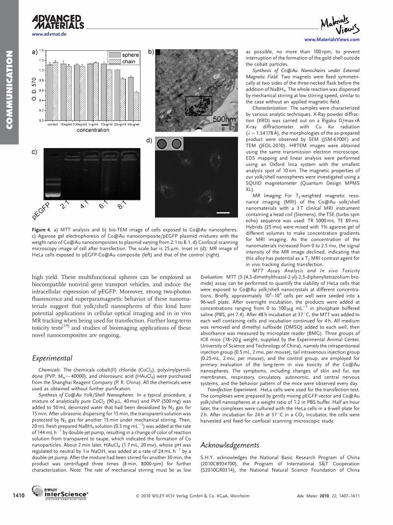
Good biocompatibility of the as-prepared Co@Au nanospheresand nanochains is confirmed by MTT cytotoxicity assay (Fig. 4a)even at the concentration of 20mg mL�1 (calculated by elementalCo) due to the gold shell and PVP coating, indicating that they arepromising for the following bioimaging and transfectionexperiments. The distribution of these spherical hybrids couldbe clearly imaged by bio-TEM analysis, and a large number ofthem can be found singly dispersed in endosomes and the cytosol
Scheme 1. Schematic illustration of the formation of the yolk/shell nanosassemblies (chains) under an external magnetic field.
Adv. Mater. 2010, 22, 1407–1411 � 2010 WILEY-VCH Verlag G
(intracellular fluid), as shown in Figure 4b. Cells were found tomaintain their structure and activity well when exposed to thenanocrystals at a concentration of 5mg mL�1. Three groups ofICR (imprinting control region) mice, i.e., those treated byintraperitoneal injection, tail intravenous injection, and thecontrol group, were employed for primary evaluation of thelong-term in vivo toxicity of our Co@Au nanospheres. No obviousabnormalities in the agent-treated groups were observed in fourweeks, and no deaths occurred.
The effective cellular uptake and relative safety of theas-prepared Co@Au nanospheres—in comparison with thepotential fatalness of viral vectors and the immune response tothem,[17] and the lack of multifunctionality of liposomecarriers[18]—make it possible to introduce them into genetransfection. The pEGFP plasmid vector, which is used as a report
pheres and linear
mbH & Co. KGaA, Wein
gene, can be efficiently combined with thenanospheres even at the weight ratio of 1:2 asa result of their positively charged surface,which is revealed by evident retardation of theDNA band in agarose gel electrophoresis incomparison with the three bands of purepEGFP plasmid (Fig. 4c). In the transfectionexperiment mediated by the present nano-spheres, obvious green fluorescence of greenfluorescent protein (GFP) was observed in theconfocal scanning microscopy images ofHeLa cells (Fig. 4d). However, no greenfluorescence was exhibited without the assis-tance of the nanocomposites, provingthe successful transportation and expressionof this report gene. The MR image of HeLacells exposed to the pEGFP-Co@Au compo-site shown in the inset of Figure 4d indicatesits potential ability to track the transfection.
In summary, we have reported here a facileroute to synthesize hydrophilic Co@Au yolk/shell nanospheres and their assemblies in
heim 1409
COM
MUNIC
ATIO
N
www.advmat.dewww.MaterialsViews.com
Figure 4. a) MTT analysis and b) bio-TEM image of cells exposed to Co@Au nanospheres.c) Agarose gel electrophoresis of Co@Au nanocomposite/pEGFP plasmid mixtures with theweight ratio of Co@Au nanocomposites to plasmid varying from 2:1 to 8:1. d) Confocal scanningmicroscopy image of cell after transfection. The scale bar is 25mm. Inset in (d): MR image ofHeLa cells exposed to pEGFP-Co@Au composite (left) and that of the control (right).
1410
high yield. These multifunctional spheres can be employed asbiocompatible nonviral gene transport vehicles, and induce theintracellular expression of pEGFP. Moreover, strong two-photonfluorescence and superparamagnetic behavior of these nanoma-terials suggest that yolk/shell nanospheres of this kind havepotential applications in cellular optical imaging and in in vivoMR tracking when being used for transfection. Further long-termtoxicity tests[19] and studies of bioimaging applications of thesenovel nanocomposites are ongoing.
Experimental
Chemicals: The chemicals cobalt(II) chloride (CoCl2), polyvinylpyrroli-done (PVP, Mw� 40000), and chloroauric acid (HAuCl4) were purchasedfrom the Shanghai Reagent Company (P. R. China). All the chemicals wereused as obtained without further purification.
Synthesis of Co@Au Yolk/Shell Nanospheres: In a typical procedure, amixture of analytically pure CoCl2 (90mL, 40mM) and PVP (500mg) wasadded to 50mL deionized water that had been deoxidized by N2 gas for15min. After ultrasonic dispersing for 15min, the transparent solution wasprotected by N2 gas for another 15min under mechanical stirring. Then,20mL fresh prepared NaBH4 solution (0.5mgmL�1) was added at the rateof 144mL h�1 by double-jet pump, resulting in a change of color of reactionsolution from transparent to taupe, which indicated the formation of Conanoparticles. About 2min later, HAuCl4 (1.7mL, 20mM), whose pH wasregulated to neutral by 1 M NaOH, was added at a rate of 24mL h�1 by adouble-jet pump. After the mixture had been stirred for another 30min, theproduct was centrifuged three times (8min, 8000 rpm) for furthercharacterization. Note: The rate of mechanical stirring must be as low
� 2010 WILEY-VCH Verlag GmbH & Co. KGaA, Weinhe
as possible, no more than 100 rpm, to preventinterruption of the formation of the gold shell outsidethe cobalt particles.
Synthesis of Co@Au Nanochains under ExternalMagnetic Field: Two magnets were fixed symmetri-cally at two sides of the three-necked flask before theaddition of NaBH4. The whole reaction was dispersedby mechanical stirring at low stirring speed, similar tothe case without an applied magnetic field.
Characterization: The samples were characterizedby various analytic techniques. X-Ray powder diffrac-tion (XRD) was carried out on a Rigaku D/max-rAX-ray diffractometer with Cu Ka radiation(l¼ 1.54178 A), the morphologies of the as-preparedproduct were observed by SEM (JSM-6700F) andTEM (JEOL-2010). HRTEM images were obtainedusing the same transmission electron microscope.EDS mapping and linear analysis were performedusing an Oxford Inca system with the smallestanalysis spot of 10 nm. The magnetic properties ofour yolk/shell nanospheres were investigated using aSQUID magnetometer (Quantum Design MPMSXL).
MR Imaging: For T2-weighted magnetic reso-nance imaging (MRI) of the Co@Au yolk/shellnanomaterials with a 3 T clinical MRI instrumentcontaining a head coil (Siemens), the TSE (turbo spinecho) sequence was used: TR 5000ms, TE 89ms.Hybrids (25mM) were mixed with 1% agarose gel ofdifferent volumes to make concentration gradientsfor MRI imaging. As the concentration of thenanomaterials increased from 0 to 2.5mM, the signalintensity of the MR image declined, indicating thatthis alloy has potential as a T2 MRI contrast agent forin vivo tracking during transfection.
MTT Assay Analysis and In vivo Toxicity
Evaluation: MTT (3-(4,5-dimethylthiazol-2-yl)-2,5-diphenyltetrazolium bro-mide) assay can be performed to quantify the viability of HeLa cells thatwere exposed to Co@Au yolk/shell nanocrystals at different concentra-tions. Briefly, approximately 103–104 cells per well were seeded into a96-well plate. After overnight incubation, the products were added atconcentrations ranging from 0 to 100mg mL�1 in phosphate bufferedsaline (PBS, pH 7.4). After 48 h incubation at 37 8C, the MTT was added toeach well containing cells and incubation continued for 4 h. All mediumwas removed and dimethyl sulfoxide (DMSO) added to each well, thenabsorbance was measured by microplate reader (BMG). Three groups ofICR mice (18–20 g weight, supplied by the Experimental Animal Center,University of Science and Technology of China), namely the intraperitonealinjection group (0.5mL, 2mM, per mouse), tail intravenous injection group(0.25mL, 2mM, per mouse), and the control group, are employed forprimary evaluation of the long-term in vivo toxicity of the Co@Aunanospheres. The symptoms, including changes of skin and fur, eyemembranes, respiratory, circulatory, autonomic, and central nervoussystems, and the behavior pattern of the mice were observed every day.Transfection Experiment: HeLa cells were used for the transfection test.The complexes were prepared by gently mixing pEGFP vector and Co@Auyolk/shell nanospheres at a weight ratio of 1:2 in PBS buffer. Half an hourlater, the complexes were cultured with the HeLa cells in a 6-well plate for2 h. After incubation for 24 h at 37 8C in a CO2 incubator, the cells wereharvested and fixed for confocal scanning microscopic study.
Acknowledgements
S.H.Y. acknowledges the National Basic Research Program of China(2010CB934700), the Program of International S&T Cooperation(S2010GR0314), the National Natural Science Foundation of China
im Adv. Mater. 2010, 22, 1407–1411

COM
MUNIC
ATIO
www.MaterialsViews.comwww.advmat.de
(nos. 50732006, 20621061, 20671085), and the Partner Group of theChinese Academy of Sciences and the Max Planck Society. SupportingInformation is available online fromWiley InterScience or from the authors.
Received: September 25, 2009
Revised: October 24, 2009
Published online: January 18, 2010
N
[1] a) F. Wetz, K. Soulantica, A. Falqui, M. Respaud, E. Snoeck, B. Chaudret,Angew. Chem. Int. Ed. 2007, 46, 7079. b) J. S. Choi, Y. W. Jun, S. I. Yeon,
H. C. Kim, J. S. Shin, J. W. Cheon, J. Am. Chem. Soc. 2006, 128, 15982.
c) Z. C. Xu, Y. L. Hou, S. H. Sun, J. Am. Chem. Soc. 2007, 129, 8698.
d) J. Bao, W. Chen, T. T. Liu, Y. L. Zhu, P. Y. Jin, L. Y. Wang, J. F. Liu,
Y. G. Wei, Y. D. Li, ACS Nano 2007, 1, 293.
[2] a) J. Jiang, H. Gu, H. Shao, E. Devlin, G. C. Papaefthymiou, J. Y. Ying, Adv.
Mater. 2008, 20, 4403. b) Z. C. Xu, Y. L. Hou, S. H. Sun, J. Am. Chem. Soc.
2007, 129, 8698.
[3] V. Salgueirino-Maceira, M. A. Correa-Duarte, Adv. Mater. 2007, 19, 4131.
[4] a) Y. D. Yin, R. M. Rioux, C. K. Erdonmez, S. Hughes, G. A. Somorjai,
A. P. Alivisatos, Science 2004, 304, 711. b) X. W. Lou, Y. Wang, C. Yuan,
J. Y. Lee, L. A. Archer, Adv. Mater. 2006, 18, 2325. c) Y. G. Sun, Y. N. Xia,
Science 2002, 298, 2176. d) J. Y. Chen, B. Wiley, J. McLellan, Y. J. Xiong,
Z. Y. Li, Y. N. Xia,Nano. Lett. 2005, 5, 2058. e) T. R. Zhang, J. P. Ge, Y. X. Hu,
Q. Zhang, S. Aloni, Y. D. Yin, Angew. Chem. Int. Ed. 2008, 47, 5806.
f) T. R. Zhang, Q. Zhang, J. P. Ge, J. Goebl, M. W. Sun, Y. S. Yan, Y. S. Liu,
C. L. Chang, J. H. Guo, Y. D. Yin, J. Phys. Chem. C 2009, 113, 3168.
g) J. H. Gao, G. L. Liang, J. S. Cheung, Y. Pan, Y. Kuang, F. Zhao, B. Zhang,
X. X. Zhang, E. X. Wu, B. Xu, J. Am. Chem. Soc. 2008, 130, 11828.
[5] a) H. P. Liang, H. M. Zhang, J. S. Hu, Y. G. Guo, L. J. Wan, C. L. Bai, Angew.
Chem. Int. Ed. 2004, 43, 1540. b) H. P. Liang, L. J. Wan, C. L. Bai, L. Jiang,
J. Phys. Chem. B 2005, 109, 7795. c) Y. Vasquez, A. K. Sra, R. E. Schaak,
J. Am. Chem. Soc. 2005, 127, 12504. d) J. Zeng, J. L. Huang, W. Lu,
X. P. Wang, B. Wang, S. Y. Zhang, J. G. Hou, Adv. Mater. 2007, 19, 2172.
Adv. Mater. 2010, 22, 1407–1411 � 2010 WILEY-VCH Verlag G
[6] a) Y. P. Bao, H. Calderon, K. M. Krishnan, J. Phys. Chem. C 2007, 111, 1941.
b) L. Bouchard, M. S. Anwar, G. L. Liu, B. Hann, Z. H. Xie, J. W. Gray,
X. D. Wang, A. Pines, F. F. Chen, Proc. Natl. Acad. Sci. USA 2009, 106, 4085.
[7] a) J. Liu, Z. Sun, Y. Deng, Y. Zou, C. Li, X. Guo, L. Xiong, Y. Gao, F. Li,
D. Y. Zhao, Angew. Chem. Int. Ed. 2009, 48, 5875. b) J. Ge, Y. Hu,M. Biasini,
W. P. Beyermann, Y. D. Yin, Angew. Chem. Int. Ed. 2007, 46, 43242.
[8] Y. W. Jun, J. H. Lee, J. W. Cheon, Angew. Chem. Int. Ed. 2008, 47, 5122.
[9] X. M. Lu, L. Au, J. McLellan, Z. Y. Li, M.Marquez, Y. N. Xia,Nano Lett. 2007,
7, 1764.
[10] a) C. J. Murphy, A. M. Gole, J. W. Stone, P. N. Sisco, A. M. Alkilany,
E. C. Goldsmith, S. C. Baxter, Acc. Chem. Res. 2008, 41, 1721. b) C. Loo,
L. Hirsch, M. H. Lee, E. Chang, J. West, N. Halas, R. Drezek, Opt. Lett.
2005, 30, 1012. c) R. D. Averitt, S. L. Westcott, N. Halas, J. Opt. Soc. Am. B
1999, 16, 1824. d) L. R. Hirsch, R. J. Stafford, J. A. Bankson, S. R. Sershen,
B. Rivera, R. E. Price, J. D. Hazle, N. J. Halas, Proc. Natl. Acad. Sci. USA
2003, 100, 13549.
[11] a) H. F. Wang, T. B. Huff, D. A. Zweifel, W. He, P. S. Low, A. Wei, J. X. Cheng,
Proc. Natl. Acad. Sci. USA 2005, 102, 15752. b) J. Jiang, H. W. Gu,
H. L. Shao, E. Devlin, G. C. Papaefthymiou, J. Y. Ying, Adv. Mater. 2008,
20, 4403.
[12] M. R. Hamblin, T. N. Demidova, Proc. SPIE 2006, 6140, 614001.
[13] a) M. J. Hu, Y. Lu, S. Zhang, S. R. Guo, B. Lin, M. Zhang, S. H. Yu, J. Am.
Chem. Soc. 2008, 130, 11606. b) M. J. Hu, B. Lin, S. H. Yu, Nano Res. 2008,
1, 303.
[14] V. Salgueirino-Maceira, M. A. Correa-Duarte, A. Hucht, M. Farle, J Magn.
Magn. Mater. 2006, 303, 163.
[15] a) A. M. Schwartzberg, T. Y. Olson, C. E. Talley, J. Z. Zhang, J. Phys. Chem. C
2007, 111, 16080. b) J. Zeng, J. L. Huang, W. Lu, X. P. Wang, B. Wang,
S. Y. Zhang, J. G. Hou, Adv. Mater. 2007, 19, 2172.
[16] J. H. Park, G. Maltzahn, L. L. Zhang, M. P. Schwartz, E. Ruoslahti,
S. N. Bhatia, M. J. Sailor, Adv. Mater. 2008, 20, 1630.
[17] C. E. Thomas, A. Ehrhardt, M. A. Kay, Nat. Rev. Genet. 2003, 4, 346.
[18] P. P. Karmali, A. Chaudburi, Med. Res. Rev. 2007, 27, 696.
[19] T. Hansen, G. Clermont, A. Alves, R. Eloy, C. Brochhausen, J. P. Boutrand,
A. M. Gatti, C. J. Kirkpatrick, J. R. Soc. Interface 2006, 3, 767.
mbH & Co. KGaA, Weinheim 1411