hydroxyl regioisomerization of anthracycline catalyzed by ... · hydroxyl regioisomerization of...
TRANSCRIPT

Hydroxyl regioisomerization of anthracycline catalyzedby a four-enzyme cascadeZhuan Zhanga,1, Yu-Kang Gonga,1, Qiang Zhoua, Yu Hua, Hong-Min Maa, Yong-Sheng Chena, Yasuhiro Igarashib,Lifeng Pana,2, and Gong-Li Tanga,2
aState Key Laboratory of Bio-organic and Natural Products Chemistry, Shanghai Institute of Organic Chemistry, Chinese Academy of Sciences, Shanghai200032, China; and bBiotechnology Research Center, Toyama Prefectural University, Toyama 939-0398, Japan
Edited by Jerrold Meinwald, Cornell University, Ithaca, NY, and approved January 4, 2017 (received for review June 21, 2016)
Ranking among the most effective anticancer drugs, anthracyclinesrepresent an important family of aromatic polyketides generated bytype II polyketide synthases (PKSs). After formation of polyketidecores, the post-PKS tailoring modifications endow the scaffold withvarious structural diversities and biological activities. Here we demon-strate an unprecedented four-enzyme-participated hydroxyl regioiso-merization process involved in the biosynthesis of kosinostatin. First,KstA15 and KstA16 function together to catalyze a cryptic hydroxyl-ation of the 4-hydroxyl-anthraquinone core, yielding a 1,4-dihydroxylproduct, which undergoes a chemically challenging asymmetricreduction-dearomatization subsequently acted by KstA11; then, KstA10catalyzes a region-specific reduction concomitant with dehydration toafford the 1-hydroxyl anthraquinone. Remarkably, the shunt productidentifications of both hydroxylation and reduction-dehydration reac-tions, the crystal structure of KstA11 with bound substrate andcofactor, and isotope incorporation experiments reveal mechanisticinsights into the redox dearomatization and rearomatization steps.These findings provide a distinguished tailoring paradigm for type IIPKS engineering.
biosynthesis | C-4 deoxyanthracycline | two-component hydroxylase |NmrA-like short-chain dehydrogenase | dehydroxylation
Anthracycline antibiotics, including doxorubicin, daunorubi-cin, idarubicin, epirubicin, and aclacinomycin (Fig. 1A),
rank among the most effective anticancer drugs. However, large-scale clinical application of them is often hampered by the risk ofinducing cardiomyopathy (1). Hence, there have been intensiveattempts to develop analogs with an improved therapeutic index.In the last 50 y, most modifications have been focused on the sugarmoiety, providing a successful drug, epirubicin, as well as severalpromising candidates under clinical trials (1, 2). In contrast, only afew analogs have been generated by altering the anthracyclinecore, most of which were obtained by chemical synthesis. There-fore, more efforts toward analogs, especially through enzymaticmodifications of the previously seldom-touched anthracycline core,are still urgently needed.Naturally occurring anthracycline antibiotics, usually produced
by Streptomyces, belong to a family of aromatic polyketides bio-synthesized by type II polyketide synthases (PKSs) (3). During thelast 3 decades, biosynthetic studies of this system in bacteria havegained a deeper insight into the anthracycline’s molecular logicand enzymatic mechanism (4–6). In general, the nascent poly-β-ketone generated by minimal PKS undergoes carbon-9 (C-9)reduction first and then cyclizations and dehydrations mediated bycyclases, followed by oxidation of the second ring to afford theanthraquinone portion presented in all anthracyclines. The finalhydrolytic release of the anthraquinone produces the nascentanthracycline core, which contains a C-4 hydroxyl group in D-ring(Fig. 1A) (3, 7). The anthracycline core is then decorated withvarious tailoring enzymes to generate types of anthracycline anti-biotics (8–10). Therefore, most of the anthracyclines possess a C-4hydroxyl group, and a very few C-4 deoxyanthracyclines have beenisolated in nature.
Kosinostatin (KST, 1), a rare C-4 deoxyanthracycline antibioticproduced by marine Micromonospora (M.) sp. TP-A0468, exhibitsstrong cytotoxicity against various cancer cell lines and an inhibitiontoward Gram-positive bacteria (11). During our previous studies,the anthracycline intermediate 4, bearing a C-1 hydroxyl group inD-ring, was identified from the kstB1-inactivation mutant strain(Fig. 1B) (12). The high sequence identity between KST genecluster and other anthracycline gene clusters indicates that KSTmaygenerate the C-4 hydroxyl anthracycline as primary anthracyclinecore; in addition, our labeled acetate feeding experiments furtherconfirmed the prediction that KST has undergone similar assemblyprocess to afford C-4 hydroxyl anthracycline (Fig. 1B) (12). If this isthe case, an apparent hydroxyl regioisomerization of anthracyclinemust be involved in the KST biosynthesis, which is a chemicallychallenging transformation process. Here, we report a four-enzyme-catalyzed regioselective hydroxylation-dehydroxylation process result-ing in the hydroxyl regioisomerization, which is totally distinct fromcurrently known tailoring steps in type II PKS platforms.
ResultsCryptic Hydroxylation by KstA15/KstA16. Bioinformatic analysis ofKST gene cluster revealed two closely linked genes, kstA15 andkstA16. The gene product of former is evolutionarily related topolyketide cyclase, and the latter encodes a short-chain alcoholdehydrogenase. Furthermore, the KstA15/KstA16 pair is homol-ogous to the AclR/AclQ and SnoaL2/SnoaW couples, which are
Significance
Enzymatic modifications of anthracycline antibiotics are urgentlyneeded in the fields of biosynthesis, biocatalysis, and even medi-cal chemistry. However, neither hydroxyl regioisomerization nordehydroxylation of anthracycline core was described previously.Here, we discover an unprecedented hydroxyl regioisomerizationprocess in the biosynthesis of a rare carbon-4 deoxyanthracycline,which includes three tailoring steps performed by a four-enzymecascade: two-component hydroxylases mediated a cryptic hydrox-ylation, and two NmrA-like short-chain dehydrogenase/reductasescatalyzed a reduction-dearomatization followed by a reduction-dehydration process. This study expands the enzymology andchemistry of type II polyketide synthase and provides tools togeneratemore analogs by engineering or enzymatic semisynthesis.
Author contributions: G.-L.T. designed research; Z.Z., Y.-K.G., Q.Z., Y.H., H.-M.M., and Y.-S.C.performed research; Y.I. contributed new reagents/analytic tools; Z.Z., Y.-K.G., L.P., and G.-L.T.analyzed data; and Z.Z., L.P., and G.-L.T. wrote the paper.
The authors declare no conflict of interest.
This article is a PNAS Direct Submission.
Data deposition: The atomic coordinates have been deposited in the Protein Data Bank,www.pdb.org (PDB ID code 5F5L and 5F5N).1Z.Z. and Y.-K.G. contributed equally to this work.2To whom correspondence may be addressed. Email: [email protected] or [email protected].
This article contains supporting information online at www.pnas.org/lookup/suppl/doi:10.1073/pnas.1610097114/-/DCSupplemental.
www.pnas.org/cgi/doi/10.1073/pnas.1610097114 PNAS Early Edition | 1 of 6
BIOCH
EMISTR
Y
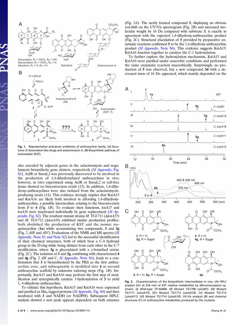
also encoded by adjacent genes in the aclacinomycin and noga-lamycin biosynthetic gene clusters, respectively (SI Appendix, Fig.S1). AclR or SnoaL2 was previously discovered to be involved inthe production of 1,4-dihydroxylated anthracyclines in vivo;however, in vitro experiment using AclR or SnoaL2 or cell-freelysate showed no bioconversion result (13). In addition, 1,4-dihy-droxy-anthracyclines were also isolated from the aclacinomycin-producing strain (14). This evidence strongly implies that KstA15and KstA16 are likely both involved in affording 1,4-dihydroxy-anthracycline, a possible intermediate relating to the bioconversionfrom 3 to 4 (Fig. 1B). To evaluate their functions, kstA15 andkstA16 were inactivated individually by gene replacement (SI Ap-pendix, Fig. S2). The resultant mutant strainsM. TG1711 (ΔkstA15)and M. TG1712 (ΔkstA16) exhibited similar production profiles:both abolished the production of KST and the isomer iso-quinocycline (1a) while accumulating two compounds, 3 and 3g(Fig. 2 AIII and AIV). Evaluations of the NMR and MS spectra (SIAppendix, Note S1 and Note S2) led to the successful identificationof their chemical structures, both of which bear a C-4 hydroxylgroup in the D-ring while being distinct from each other in the C-7modification, where 3g is glycosylated with a γ-branched octose(Fig. 2C). The isolation of 3 and 3g, combining with characterized 4and 4g (Fig. 2 AII and C, SI Appendix, Note S4), leads to a con-firmation that 3 is biosynthesized by the PKS as the first anthra-cycline core, and subsequently is modified into 4 as secondanthracycline scaffold by unknown tailoring steps (Fig. 1B). Im-portantly, KstA15 and KstA16 may perform the first step of mod-ification and synergistically catalyze 1-hydroxylation of 3 to yield1, 4-dihydroxy-anthracycline.To validate this hypothesis, KstA15 and KstA16 were expressed
and purified as His6-tagged proteins (SI Appendix, Fig. S4) and thenincubated with 3 and NADH (or NADPH). Subsequent HPLCanalysis showed a new peak appears dependent on both enzymes
(Fig. 3A). The newly formed compound 5, displaying an obviousred-shift on the UV/Vis spectrogram (Fig. 2B) and increased mo-lecular weight by 16 Da compared with substrate 3, is exactly inagreement with the expected 1,4-dihydroxy-anthracycline product(Fig. 2C). Structural elucidation of 5 provided by preparative en-zymatic reactions confirmed 5 to be the 1,4-dihydroxy-anthracyclineproduct (SI Appendix, Note S6). This evidence suggests KstA15/KstA16 function together to catalyze the C-1 hydroxylation.To further explore the hydroxylation mechanism, KstA15 and
KstA16 were purified under anaerobic conditions and performedthe same enzymatic reaction anaerobically. Surprisingly, no pro-duction of 5 was observed, but a new compound 3d with a de-creased mass of 16 Da appeared, which mainly depended on the
O S ACP
O
OOOOH
O O
2
O
OOH
OH
O
O
N
HN
O
OH
OHO
O OCH3
OHOHO
OH
3O
OHOHO
OH
4OOH
O OCH3
OH
1
4 7
10
4
101
10
Kosinostatin(KST, 1)
PKS-II
10 x MCoA
???
Isoquinocycline B(1a)
O
N
NH
A
1
4
77A
A
B
R1 OH
O
O O
OH
O
OHH2N
OH OR2
Doxorubicin, R1 = OCH3, R2 = OH;Daunorubicin, R1 = OCH3, R2 = H;Idarubicin, R1 = H, R2 = H.
1
47
10
AD9
9
OH
O
O OH
CO2CH3
O
OH
O
NO
O
HOO
O
O
Aclacinomycin AEpirubicin
9D
O OH
O
O O
OH
OHOH2N
OH O
D
6
11
5
12
BC
OH
D
Fig. 1. Representative anticancer antibiotics of anthracycline family. (A) Struc-tures of doxorubicin-like drugs and aclacinomycin A. (B) Biosynthetic pathway ofkosinostatin (KST).
A
B
C
Fig. 2. Characterization of the biosynthetic intermediates in vivo. (A) HPLCanalysis (UV at 254 nm) of KST relative metabolites by Micromonospora sp.strains. (I) Wild-type TP-A0468. (II) Mutant TG1708 (ΔkstB1). (III) MutantTG1711 (ΔkstA15). (IV) Mutant TG1712 (ΔkstA16). (V) Mutant TG1713(ΔkstA11). (VI) Mutant TG1714 (ΔkstA10). UV-Vis analysis (B) and chemicalstructures (C) of anthracycline metabolites produced by the mutants.
2 of 6 | www.pnas.org/cgi/doi/10.1073/pnas.1610097114 Zhang et al.
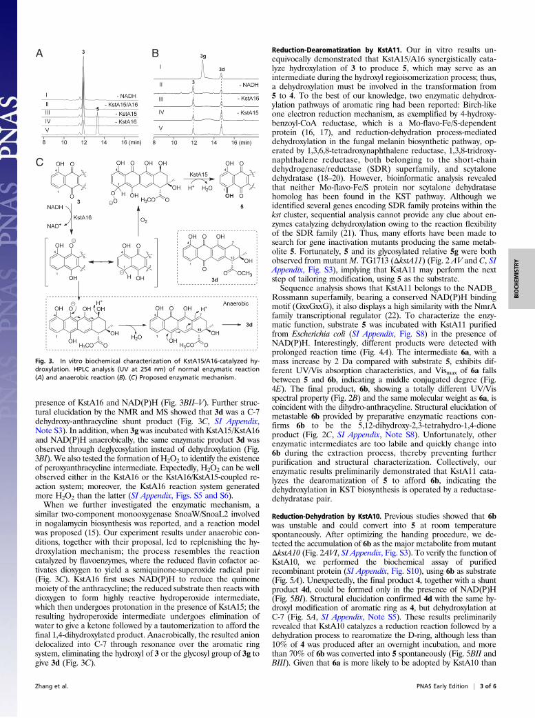
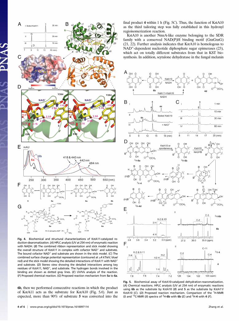
presence of KstA16 and NAD(P)H (Fig. 3BII–V). Further struc-tural elucidation by the NMR and MS showed that 3d was a C-7dehydroxy-anthracycline shunt product (Fig. 3C, SI Appendix,Note S3). In addition, when 3g was incubated with KstA15/KstA16and NAD(P)H anaerobically, the same enzymatic product 3d wasobserved through deglycosylation instead of dehydroxylation (Fig.3BI). We also tested the formation of H2O2 to identify the existenceof peroxyanthracycline intermediate. Expectedly, H2O2 can be wellobserved either in the KstA16 or the KstA16/KstA15-coupled re-action system; moreover, the KstA16 reaction system generatedmore H2O2 than the latter (SI Appendix, Figs. S5 and S6).When we further investigated the enzymatic mechanism, a
similar two-component monooxygenase SnoaW/SnoaL2 involvedin nogalamycin biosynthesis was reported, and a reaction modelwas proposed (15). Our experiment results under anaerobic con-ditions, together with their proposal, led to replenishing the hy-droxylation mechanism; the process resembles the reactioncatalyzed by flavoenzymes, where the reduced flavin cofactor ac-tivates dioxygen to yield a semiquinone-superoxide radical pair(Fig. 3C). KstA16 first uses NAD(P)H to reduce the quinonemoiety of the anthracycline; the reduced substrate then reacts withdioxygen to form highly reactive hydroperoxide intermediate,which then undergoes protonation in the presence of KstA15; theresulting hydroperoxide intermediate undergoes elimination ofwater to give a ketone followed by a tautomerization to afford thefinal 1,4-dihydroxylated product. Anaerobically, the resulted aniondelocalized into C-7 through resonance over the aromatic ringsystem, eliminating the hydroxyl of 3 or the glycosyl group of 3g togive 3d (Fig. 3C).
Reduction-Dearomatization by KstA11. Our in vitro results un-equivocally demonstrated that KstA15/A16 synergistically cata-lyze hydroxylation of 3 to produce 5, which may serve as anintermediate during the hydroxyl regioisomerization process; thus,a dehydroxylation must be involved in the transformation from5 to 4. To the best of our knowledge, two enzymatic dehydrox-ylation pathways of aromatic ring had been reported: Birch-likeone electron reduction mechanism, as exemplified by 4-hydroxy-benzoyl-CoA reductase, which is a Mo-flavo-Fe/S-dependentprotein (16, 17), and reduction-dehydration process-mediateddehydroxylation in the fungal melanin biosynthetic pathway, op-erated by 1,3,6,8-tetradroxynaphthalene reductase, 1,3,8-tridroxy-naphthalene reductase, both belonging to the short-chaindehydrogenase/reductase (SDR) superfamily, and scytalonedehydratase (18–20). However, bioinformatic analysis revealedthat neither Mo-flavo-Fe/S protein nor scytalone dehydratasehomolog has been found in the KST pathway. Although weidentified several genes encoding SDR family proteins within thekst cluster, sequential analysis cannot provide any clue about en-zymes catalyzing dehydroxylation owing to the reaction flexibilityof the SDR family (21). Thus, many efforts have been made tosearch for gene inactivation mutants producing the same metab-olite 5. Fortunately, 5 and its glycosylated relative 5g were bothobserved from mutant M. TG1713 (ΔkstA11) (Fig. 2 AV and C, SIAppendix, Fig. S3), implying that KstA11 may perform the nextstep of tailoring modification, using 5 as the substrate.Sequence analysis shows that KstA11 belongs to the NADB_
Rossmann superfamily, bearing a conserved NAD(P)H bindingmotif (GxxGxxG), it also displays a high similarity with the NmrAfamily transcriptional regulator (22). To characterize the enzy-matic function, substrate 5 was incubated with KstA11 purifiedfrom Escherichia coli (SI Appendix, Fig. S8) in the presence ofNAD(P)H. Interestingly, different products were detected withprolonged reaction time (Fig. 4A). The intermediate 6a, with amass increase by 2 Da compared with substrate 5, exhibits dif-ferent UV/Vis absorption characteristics, and Vismax of 6a fallsbetween 5 and 6b, indicating a middle conjugated degree (Fig.4E). The final product, 6b, showing a totally different UV/Visspectral property (Fig. 2B) and the same molecular weight as 6a, iscoincident with the dihydro-anthracycline. Structural elucidation ofmetastable 6b provided by preparative enzymatic reactions con-firms 6b to be the 5,12-dihydroxy-2,3-tetrahydro-1,4-dioneproduct (Fig. 2C, SI Appendix, Note S8). Unfortunately, otherenzymatic intermediates are too labile and quickly change into6b during the extraction process, thereby preventing furtherpurification and structural characterization. Collectively, ourenzymatic results preliminarily demonstrated that KstA11 cata-lyzes the dearomatization of 5 to afford 6b, indicating thedehydroxylation in KST biosynthesis is operated by a reductase-dehydratase pair.
Reduction-Dehydration by KstA10. Previous studies showed that 6bwas unstable and could convert into 5 at room temperaturespontaneously. After optimizing the handing procedure, we de-tected the accumulation of 6b as the major metabolite from mutantΔkstA10 (Fig. 2AVI, SI Appendix, Fig. S3). To verify the function ofKstA10, we performed the biochemical assay of purifiedrecombinant protein (SI Appendix, Fig. S10), using 6b as substrate(Fig. 5A). Unexpectedly, the final product 4, together with a shuntproduct 4d, could be formed only in the presence of NAD(P)H(Fig. 5BI). Structural elucidation confirmed 4d with the same hy-droxyl modification of aromatic ring as 4, but dehydroxylation atC-7 (Fig. 5A, SI Appendix, Note S5). These results preliminarilyrevealed that KstA10 catalyzes a reduction reaction followed by adehydration process to rearomatize the D-ring, although less than10% of 4 was produced after an overnight incubation, and morethan 70% of 6b was converted into 5 spontaneously (Fig. 5BII andBIII). Given that 6a is more likely to be adopted by KstA10 than
A
C
B
Fig. 3. In vitro biochemical characterization of KstA15/A16-catalyzed hy-droxylation. HPLC analysis (UV at 254 nm) of normal enzymatic reaction(A) and anaerobic reaction (B). (C) Proposed enzymatic mechanism.
Zhang et al. PNAS Early Edition | 3 of 6
BIOCH
EMISTR
Y
6b, then we performed consecutive reactions in which the productof KstA11 acts as the substrate for KstA10 (Fig. 5A). Just asexpected, more than 90% of substrate 5 was converted into the
final product 4 within 1 h (Fig. 5C). Thus, the function of KstA10as the third tailoring step was fully established in this hydroxylregioisomerization reaction.KstA10 is another NmrA-like enzyme belonging to the SDR
family with a conserved NAD(P)H binding motif (GxxGxxG)(21, 22). Further analysis indicates that KstA10 is homologous toNAD+-dependent nucleotide diphosphate sugar epimerases (23),which act on totally different substrates from that in KST bio-synthesis. In addition, scytalone dehydratase in the fungal melanin
Fig. 4. Biochemical and structural characterizations of KstA11-catalyzed re-duction-dearomatization. (A) HPLC analysis (UV at 254 nm) of enzymatic reactionwith NADH. (B) The combined ribbon representation and stick model showingthe overall structure of KstA11 in complex with cofactor NAD+ and substrate.The bound cofactor NAD+ and substrate are shown in the stick model. (C) Thecombined surface charge potential representation (contoured at ±4 kT/eV; blue/red) and the stick model showing the detailed interactions of KstA11 with NAD+
and substrate. (D) Stereo view showing the detailed interactions among keyresidues of KstA11, NAD+, and substrate. The hydrogen bonds involved in thebinding are shown as dotted gray lines. (E) UV/Vis analysis of the reaction.(F) Proposed chemical reaction. (G) Proposed reaction mechanism from 5a to 6a.
A
B
D
E
F
C
Fig. 5. Biochemical assay of KstA10-catalyzed dehydration-rearomatization.(A) Chemical reactions. HPLC analysis (UV at 254 nm) of enzymatic reactionsusing 6b as the substrate by KstA10 (B) and 5 as the substrate by KstA11/KstA10 (C). (D) Proposed reaction mechanism. Comparison of the 1H-NMR(I) and 13C-NMR (II) spectra of 2H-6b with 6b (E) and 2H-4 with 4 (F).
4 of 6 | www.pnas.org/cgi/doi/10.1073/pnas.1610097114 Zhang et al.
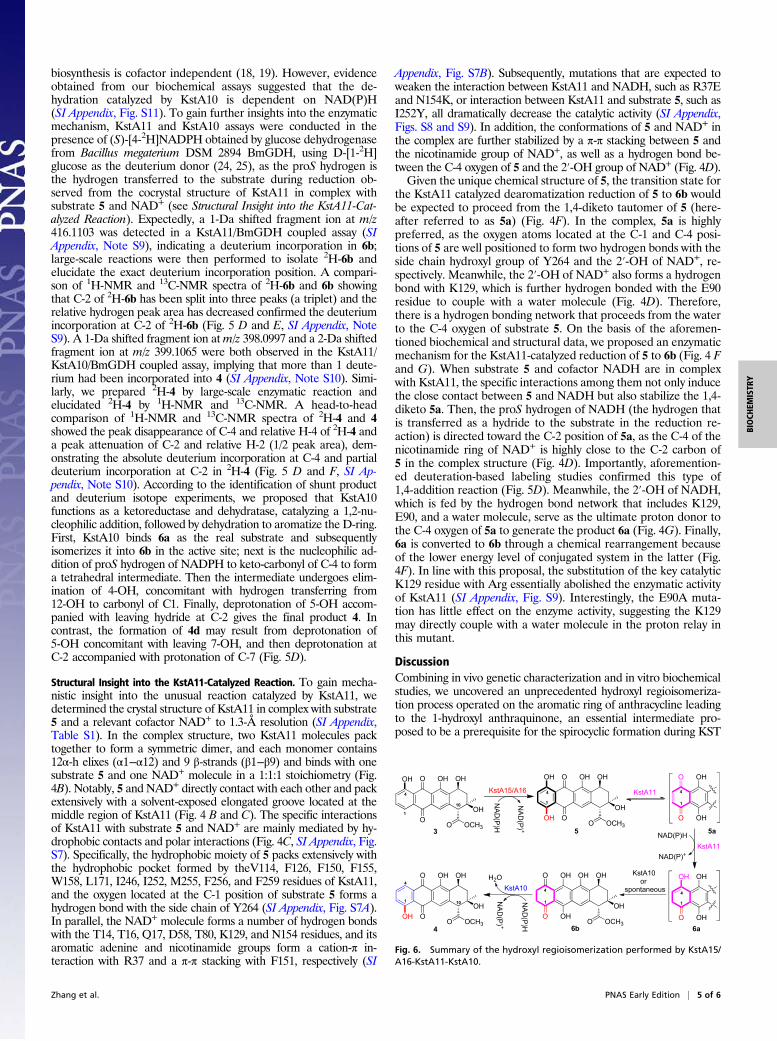
biosynthesis is cofactor independent (18, 19). However, evidenceobtained from our biochemical assays suggested that the de-hydration catalyzed by KstA10 is dependent on NAD(P)H(SI Appendix, Fig. S11). To gain further insights into the enzymaticmechanism, KstA11 and KstA10 assays were conducted in thepresence of (S)-[4-2H]NADPH obtained by glucose dehydrogenasefrom Bacillus megaterium DSM 2894 BmGDH, using D-[1-2H]glucose as the deuterium donor (24, 25), as the proS hydrogen isthe hydrogen transferred to the substrate during reduction ob-served from the cocrystal structure of KstA11 in complex withsubstrate 5 and NAD+ (see Structural Insight into the KstA11-Cat-alyzed Reaction). Expectedly, a 1-Da shifted fragment ion at m/z416.1103 was detected in a KstA11/BmGDH coupled assay (SIAppendix, Note S9), indicating a deuterium incorporation in 6b;large-scale reactions were then performed to isolate 2H-6b andelucidate the exact deuterium incorporation position. A compari-son of 1H-NMR and 13C-NMR spectra of 2H-6b and 6b showingthat C-2 of 2H-6b has been split into three peaks (a triplet) and therelative hydrogen peak area has decreased confirmed the deuteriumincorporation at C-2 of 2H-6b (Fig. 5 D and E, SI Appendix, NoteS9). A 1-Da shifted fragment ion atm/z 398.0997 and a 2-Da shiftedfragment ion at m/z 399.1065 were both observed in the KstA11/KstA10/BmGDH coupled assay, implying that more than 1 deute-rium had been incorporated into 4 (SI Appendix, Note S10). Simi-larly, we prepared 2H-4 by large-scale enzymatic reaction andelucidated 2H-4 by 1H-NMR and 13C-NMR. A head-to-headcomparison of 1H-NMR and 13C-NMR spectra of 2H-4 and 4showed the peak disappearance of C-4 and relative H-4 of 2H-4 anda peak attenuation of C-2 and relative H-2 (1/2 peak area), dem-onstrating the absolute deuterium incorporation at C-4 and partialdeuterium incorporation at C-2 in 2H-4 (Fig. 5 D and F, SI Ap-pendix, Note S10). According to the identification of shunt productand deuterium isotope experiments, we proposed that KstA10functions as a ketoreductase and dehydratase, catalyzing a 1,2-nu-cleophilic addition, followed by dehydration to aromatize the D-ring.First, KstA10 binds 6a as the real substrate and subsequentlyisomerizes it into 6b in the active site; next is the nucleophilic ad-dition of proS hydrogen of NADPH to keto-carbonyl of C-4 to forma tetrahedral intermediate. Then the intermediate undergoes elim-ination of 4-OH, concomitant with hydrogen transferring from12-OH to carbonyl of C1. Finally, deprotonation of 5-OH accom-panied with leaving hydride at C-2 gives the final product 4. Incontrast, the formation of 4d may result from deprotonation of5-OH concomitant with leaving 7-OH, and then deprotonation atC-2 accompanied with protonation of C-7 (Fig. 5D).
Structural Insight into the KstA11-Catalyzed Reaction. To gain mecha-nistic insight into the unusual reaction catalyzed by KstA11, wedetermined the crystal structure of KstA11 in complex with substrate5 and a relevant cofactor NAD+ to 1.3-Å resolution (SI Appendix,Table S1). In the complex structure, two KstA11 molecules packtogether to form a symmetric dimer, and each monomer contains12α-h elixes (α1−α12) and 9 β-strands (β1−β9) and binds with onesubstrate 5 and one NAD+ molecule in a 1:1:1 stoichiometry (Fig.4B). Notably, 5 and NAD+ directly contact with each other and packextensively with a solvent-exposed elongated groove located at themiddle region of KstA11 (Fig. 4 B and C). The specific interactionsof KstA11 with substrate 5 and NAD+ are mainly mediated by hy-drophobic contacts and polar interactions (Fig. 4C, SI Appendix, Fig.S7). Specifically, the hydrophobic moiety of 5 packs extensively withthe hydrophobic pocket formed by theV114, F126, F150, F155,W158, L171, I246, I252, M255, F256, and F259 residues of KstA11,and the oxygen located at the C-1 position of substrate 5 forms ahydrogen bond with the side chain of Y264 (SI Appendix, Fig. S7A).In parallel, the NAD+ molecule forms a number of hydrogen bondswith the T14, T16, Q17, D58, T80, K129, and N154 residues, and itsaromatic adenine and nicotinamide groups form a cation-π in-teraction with R37 and a π-π stacking with F151, respectively (SI
Appendix, Fig. S7B). Subsequently, mutations that are expected toweaken the interaction between KstA11 and NADH, such as R37Eand N154K, or interaction between KstA11 and substrate 5, such asI252Y, all dramatically decrease the catalytic activity (SI Appendix,Figs. S8 and S9). In addition, the conformations of 5 and NAD+ inthe complex are further stabilized by a π-π stacking between 5 andthe nicotinamide group of NAD+, as well as a hydrogen bond be-tween the C-4 oxygen of 5 and the 2′-OH group of NAD+ (Fig. 4D).Given the unique chemical structure of 5, the transition state for
the KstA11 catalyzed dearomatization reduction of 5 to 6b wouldbe expected to proceed from the 1,4-diketo tautomer of 5 (here-after referred to as 5a) (Fig. 4F). In the complex, 5a is highlypreferred, as the oxygen atoms located at the C-1 and C-4 posi-tions of 5 are well positioned to form two hydrogen bonds with theside chain hydroxyl group of Y264 and the 2′-OH of NAD+, re-spectively. Meanwhile, the 2′-OH of NAD+ also forms a hydrogenbond with K129, which is further hydrogen bonded with the E90residue to couple with a water molecule (Fig. 4D). Therefore,there is a hydrogen bonding network that proceeds from the waterto the C-4 oxygen of substrate 5. On the basis of the aforemen-tioned biochemical and structural data, we proposed an enzymaticmechanism for the KstA11-catalyzed reduction of 5 to 6b (Fig. 4 Fand G). When substrate 5 and cofactor NADH are in complexwith KstA11, the specific interactions among them not only inducethe close contact between 5 and NADH but also stabilize the 1,4-diketo 5a. Then, the proS hydrogen of NADH (the hydrogen thatis transferred as a hydride to the substrate in the reduction re-action) is directed toward the C-2 position of 5a, as the C-4 of thenicotinamide ring of NAD+ is highly close to the C-2 carbon of5 in the complex structure (Fig. 4D). Importantly, aforemention-ed deuteration-based labeling studies confirmed this type of1,4-addition reaction (Fig. 5D). Meanwhile, the 2′-OH of NADH,which is fed by the hydrogen bond network that includes K129,E90, and a water molecule, serve as the ultimate proton donor tothe C-4 oxygen of 5a to generate the product 6a (Fig. 4G). Finally,6a is converted to 6b through a chemical rearrangement becauseof the lower energy level of conjugated system in the latter (Fig.4F). In line with this proposal, the substitution of the key catalyticK129 residue with Arg essentially abolished the enzymatic activityof KstA11 (SI Appendix, Fig. S9). Interestingly, the E90A muta-tion has little effect on the enzyme activity, suggesting the K129may directly couple with a water molecule in the proton relay inthis mutant.
DiscussionCombining in vivo genetic characterization and in vitro biochemicalstudies, we uncovered an unprecedented hydroxyl regioisomeriza-tion process operated on the aromatic ring of anthracycline leadingto the 1-hydroxyl anthraquinone, an essential intermediate pro-posed to be a prerequisite for the spirocyclic formation during KST
O OCH3
OHOHO
OH
3O
OHOHO
OH
4
OOHO OCH3
OH
1
4
10
1
4
10
OHOHO
OH
5
OOHO OCH3
1
4
OH
KstA10OH
6aOHO
1
4
OH
KstA15/A16
NAD(P) +
NAD(P)H
NAD(P)+
NAD(P)H
H2O OHOHOH
OH
6bOHO
O OCH3
1
4
O
OH
5a
OHO
O
KstA11
NAD(P) +
NAD(P)H
KstA11
KstA10or
spontaneous
1
4
Fig. 6. Summary of the hydroxyl regioisomerization performed by KstA15/A16-KstA11-KstA10.
Zhang et al. PNAS Early Edition | 5 of 6
BIOCH
EMISTR
Y
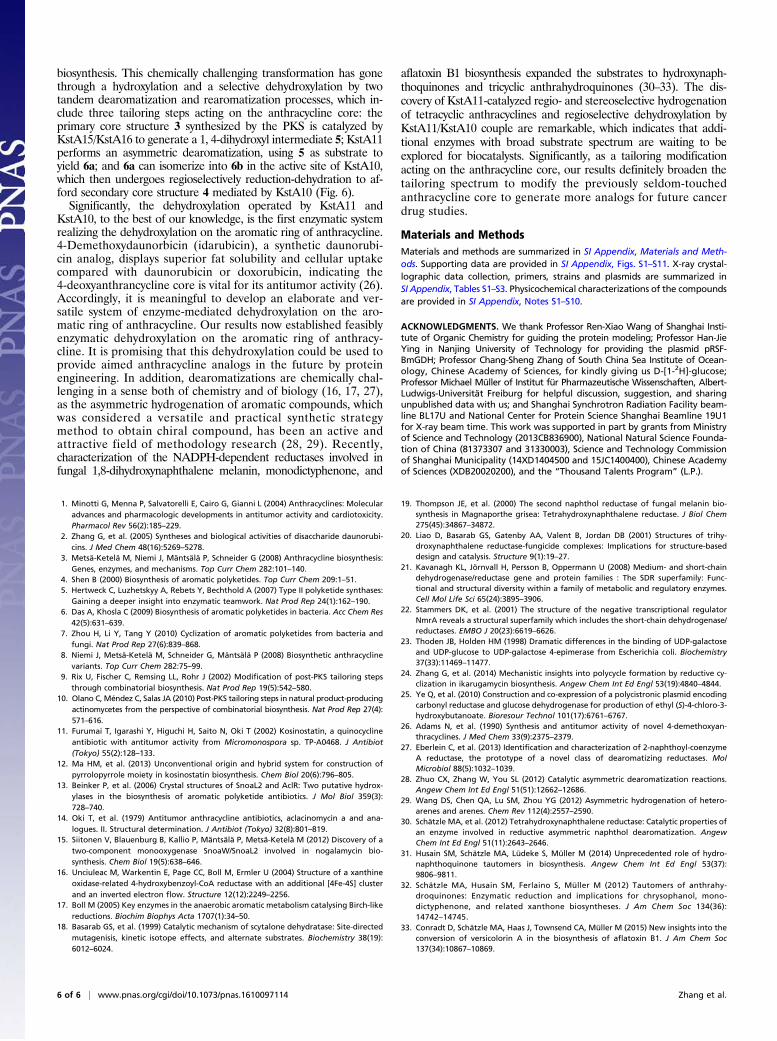
biosynthesis. This chemically challenging transformation has gonethrough a hydroxylation and a selective dehydroxylation by twotandem dearomatization and rearomatization processes, which in-clude three tailoring steps acting on the anthracycline core: theprimary core structure 3 synthesized by the PKS is catalyzed byKstA15/KstA16 to generate a 1, 4-dihydroxyl intermediate 5; KstA11performs an asymmetric dearomatization, using 5 as substrate toyield 6a; and 6a can isomerize into 6b in the active site of KstA10,which then undergoes regioselectively reduction-dehydration to af-ford secondary core structure 4 mediated by KstA10 (Fig. 6).Significantly, the dehydroxylation operated by KstA11 and
KstA10, to the best of our knowledge, is the first enzymatic systemrealizing the dehydroxylation on the aromatic ring of anthracycline.4-Demethoxydaunorbicin (idarubicin), a synthetic daunorubi-cin analog, displays superior fat solubility and cellular uptakecompared with daunorubicin or doxorubicin, indicating the4-deoxyanthrancycline core is vital for its antitumor activity (26).Accordingly, it is meaningful to develop an elaborate and ver-satile system of enzyme-mediated dehydroxylation on the aro-matic ring of anthracycline. Our results now established feasiblyenzymatic dehydroxylation on the aromatic ring of anthracy-cline. It is promising that this dehydroxylation could be used toprovide aimed anthracycline analogs in the future by proteinengineering. In addition, dearomatizations are chemically chal-lenging in a sense both of chemistry and of biology (16, 17, 27),as the asymmetric hydrogenation of aromatic compounds, whichwas considered a versatile and practical synthetic strategymethod to obtain chiral compound, has been an active andattractive field of methodology research (28, 29). Recently,characterization of the NADPH-dependent reductases involved infungal 1,8-dihydroxynaphthalene melanin, monodictyphenone, and
aflatoxin B1 biosynthesis expanded the substrates to hydroxynaph-thoquinones and tricyclic anthrahydroquinones (30–33). The dis-covery of KstA11-catalyzed regio- and stereoselective hydrogenationof tetracyclic anthracyclines and regioselective dehydroxylation byKstA11/KstA10 couple are remarkable, which indicates that addi-tional enzymes with broad substrate spectrum are waiting to beexplored for biocatalysts. Significantly, as a tailoring modificationacting on the anthracycline core, our results definitely broaden thetailoring spectrum to modify the previously seldom-touchedanthracycline core to generate more analogs for future cancerdrug studies.
Materials and MethodsMaterials and methods are summarized in SI Appendix, Materials and Meth-ods. Supporting data are provided in SI Appendix, Figs. S1–S11. X-ray crystal-lographic data collection, primers, strains and plasmids are summarized inSI Appendix, Tables S1–S3. Physicochemical characterizations of the compoundsare provided in SI Appendix, Notes S1–S10.
ACKNOWLEDGMENTS. We thank Professor Ren-Xiao Wang of Shanghai Insti-tute of Organic Chemistry for guiding the protein modeling; Professor Han-JieYing in Nanjing University of Technology for providing the plasmid pRSF-BmGDH; Professor Chang-Sheng Zhang of South China Sea Institute of Ocean-ology, Chinese Academy of Sciences, for kindly giving us D-[1-2H]-glucose;Professor Michael Müller of Institut für Pharmazeutische Wissenschaften, Albert-Ludwigs-Universität Freiburg for helpful discussion, suggestion, and sharingunpublished data with us; and Shanghai Synchrotron Radiation Facility beam-line BL17U and National Center for Protein Science Shanghai Beamline 19U1for X-ray beam time. This work was supported in part by grants from Ministryof Science and Technology (2013CB836900), National Natural Science Founda-tion of China (81373307 and 31330003), Science and Technology Commissionof Shanghai Municipality (14XD1404500 and 15JC1400400), Chinese Academyof Sciences (XDB20020200), and the “Thousand Talents Program” (L.P.).
1. Minotti G, Menna P, Salvatorelli E, Cairo G, Gianni L (2004) Anthracyclines: Molecularadvances and pharmacologic developments in antitumor activity and cardiotoxicity.Pharmacol Rev 56(2):185–229.
2. Zhang G, et al. (2005) Syntheses and biological activities of disaccharide daunorubi-cins. J Med Chem 48(16):5269–5278.
3. Metsä-Ketelä M, Niemi J, Mäntsälä P, Schneider G (2008) Anthracycline biosynthesis:Genes, enzymes, and mechanisms. Top Curr Chem 282:101–140.
4. Shen B (2000) Biosynthesis of aromatic polyketides. Top Curr Chem 209:1–51.5. Hertweck C, Luzhetskyy A, Rebets Y, Bechthold A (2007) Type II polyketide synthases:
Gaining a deeper insight into enzymatic teamwork. Nat Prod Rep 24(1):162–190.6. Das A, Khosla C (2009) Biosynthesis of aromatic polyketides in bacteria. Acc Chem Res
42(5):631–639.7. Zhou H, Li Y, Tang Y (2010) Cyclization of aromatic polyketides from bacteria and
fungi. Nat Prod Rep 27(6):839–868.8. Niemi J, Metsä-Ketelä M, Schneider G, Mäntsälä P (2008) Biosynthetic anthracycline
variants. Top Curr Chem 282:75–99.9. Rix U, Fischer C, Remsing LL, Rohr J (2002) Modification of post-PKS tailoring steps
through combinatorial biosynthesis. Nat Prod Rep 19(5):542–580.10. Olano C, Méndez C, Salas JA (2010) Post-PKS tailoring steps in natural product-producing
actinomycetes from the perspective of combinatorial biosynthesis. Nat Prod Rep 27(4):571–616.
11. Furumai T, Igarashi Y, Higuchi H, Saito N, Oki T (2002) Kosinostatin, a quinocyclineantibiotic with antitumor activity from Micromonospora sp. TP-A0468. J Antibiot(Tokyo) 55(2):128–133.
12. Ma HM, et al. (2013) Unconventional origin and hybrid system for construction ofpyrrolopyrrole moiety in kosinostatin biosynthesis. Chem Biol 20(6):796–805.
13. Beinker P, et al. (2006) Crystal structures of SnoaL2 and AclR: Two putative hydrox-ylases in the biosynthesis of aromatic polyketide antibiotics. J Mol Biol 359(3):728–740.
14. Oki T, et al. (1979) Antitumor anthracycline antibiotics, aclacinomycin a and ana-logues. II. Structural determination. J Antibiot (Tokyo) 32(8):801–819.
15. Siitonen V, Blauenburg B, Kallio P, Mäntsälä P, Metsä-Ketelä M (2012) Discovery of atwo-component monooxygenase SnoaW/SnoaL2 involved in nogalamycin bio-synthesis. Chem Biol 19(5):638–646.
16. Unciuleac M, Warkentin E, Page CC, Boll M, Ermler U (2004) Structure of a xanthineoxidase-related 4-hydroxybenzoyl-CoA reductase with an additional [4Fe-4S] clusterand an inverted electron flow. Structure 12(12):2249–2256.
17. Boll M (2005) Key enzymes in the anaerobic aromatic metabolism catalysing Birch-likereductions. Biochim Biophys Acta 1707(1):34–50.
18. Basarab GS, et al. (1999) Catalytic mechanism of scytalone dehydratase: Site-directedmutagenisis, kinetic isotope effects, and alternate substrates. Biochemistry 38(19):6012–6024.
19. Thompson JE, et al. (2000) The second naphthol reductase of fungal melanin bio-synthesis in Magnaporthe grisea: Tetrahydroxynaphthalene reductase. J Biol Chem275(45):34867–34872.
20. Liao D, Basarab GS, Gatenby AA, Valent B, Jordan DB (2001) Structures of trihy-droxynaphthalene reductase-fungicide complexes: Implications for structure-baseddesign and catalysis. Structure 9(1):19–27.
21. Kavanagh KL, Jörnvall H, Persson B, Oppermann U (2008) Medium- and short-chaindehydrogenase/reductase gene and protein families : The SDR superfamily: Func-tional and structural diversity within a family of metabolic and regulatory enzymes.Cell Mol Life Sci 65(24):3895–3906.
22. Stammers DK, et al. (2001) The structure of the negative transcriptional regulatorNmrA reveals a structural superfamily which includes the short-chain dehydrogenase/reductases. EMBO J 20(23):6619–6626.
23. Thoden JB, Holden HM (1998) Dramatic differences in the binding of UDP-galactoseand UDP-glucose to UDP-galactose 4-epimerase from Escherichia coli. Biochemistry37(33):11469–11477.
24. Zhang G, et al. (2014) Mechanistic insights into polycycle formation by reductive cy-clization in ikarugamycin biosynthesis. Angew Chem Int Ed Engl 53(19):4840–4844.
25. Ye Q, et al. (2010) Construction and co-expression of a polycistronic plasmid encodingcarbonyl reductase and glucose dehydrogenase for production of ethyl (S)-4-chloro-3-hydroxybutanoate. Bioresour Technol 101(17):6761–6767.
26. Adams N, et al. (1990) Synthesis and antitumor activity of novel 4-demethoxyan-thracyclines. J Med Chem 33(9):2375–2379.
27. Eberlein C, et al. (2013) Identification and characterization of 2-naphthoyl-coenzymeA reductase, the prototype of a novel class of dearomatizing reductases. MolMicrobiol 88(5):1032–1039.
28. Zhuo CX, Zhang W, You SL (2012) Catalytic asymmetric dearomatization reactions.Angew Chem Int Ed Engl 51(51):12662–12686.
29. Wang DS, Chen QA, Lu SM, Zhou YG (2012) Asymmetric hydrogenation of hetero-arenes and arenes. Chem Rev 112(4):2557–2590.
30. Schätzle MA, et al. (2012) Tetrahydroxynaphthalene reductase: Catalytic properties ofan enzyme involved in reductive asymmetric naphthol dearomatization. AngewChem Int Ed Engl 51(11):2643–2646.
31. Husain SM, Schätzle MA, Lüdeke S, Müller M (2014) Unprecedented role of hydro-naphthoquinone tautomers in biosynthesis. Angew Chem Int Ed Engl 53(37):9806–9811.
32. Schätzle MA, Husain SM, Ferlaino S, Müller M (2012) Tautomers of anthrahy-droquinones: Enzymatic reduction and implications for chrysophanol, mono-dictyphenone, and related xanthone biosyntheses. J Am Chem Soc 134(36):14742–14745.
33. Conradt D, Schätzle MA, Haas J, Townsend CA, Müller M (2015) New insights into theconversion of versicolorin A in the biosynthesis of aflatoxin B1. J Am Chem Soc137(34):10867–10869.
6 of 6 | www.pnas.org/cgi/doi/10.1073/pnas.1610097114 Zhang et al.