hyperbaric oxygen pretreatment improves cognition … cortex and hippocampus are ... eosin staining...
TRANSCRIPT
131www.eymj.org
INTRODUCTION
Alzheimer’s disease (AD) is a neurodegenerative disorder char-acterized by progressive learning and memory deficits. Al-though the pathogenesis of AD is not well understood, it is widely accepted that AD is associated with plaques and tangles within the brain and that the plaques distributed throughout
the cortex and hippocampus are mostly insoluble deposits of amyloid β peptide (Aβ).1 The accumulation of Aβ in patients with AD can activate astrocytes and induce the generation of proinflammatory cytokines released by activated astrocytes, leading to brain damage and cognitive decline.2 Additionally, higher numbers of astrocytes have been found to surround Aβ deposits in patients with AD, compared to those without.3 Ac-tivated astrocytes also contribute to the accumulation of Aβ by secreting inflammatory proteins.4
The molecular mechanisms involved in neuronal damage, astrocyte activation, and cognitive decline associated with Aβ accumulation in patients with AD have not yet been fully elu-cidated. Recent studies have highlighted an important role for p38 mitogen-activated protein kinase (MAPK) signal pathway as a potential target for treatment in AD.5 p38 MAPK is activat-ed (phosphorylated) in the early stage of neurofibrillary de-generation in the hippocampus of patients with AD.6 The phos-phorylation of p38 MAPK regulates neuroinflammation, cyto-
Hyperbaric Oxygen Pretreatment Improves Cognition and Reduces Hippocampal Damage Via p38 Mitogen-Activated Protein Kinase in a Rat Model
Baisong Zhao, Yongying Pan, Zixin Wang, Haiping Xu, and Xingrong SongDepartment of Anesthesiology, Guangzhou Women and Children’s Medical Center, Guangzhou, China.
Purpose: To investigate the effects of hyperbaric oxygen (HBO) pretreatment on cognitive decline and neuronal damage in an Al-zheimer’s disease (AD) rat model.Materials and Methods: Rats were divided into three groups: normal saline (NS), AD, and HBO+AD. In the AD group, amyloid β peptide (Aβ)1-40 was injected into the hippocampal CA1 region of the brain. NS rats received NS injection. In the HBO+AD group, rats received 5 days of daily HBO therapy following Aβ1-40 injection. Learning and memory capabilities were examined using the Morris water maze task. Neuronal damage and astrocyte activation were evaluated by hematoxylin-eosin staining and immuno-histochemistry, respectively. Dendritic spine density was determined by Golgi-Cox staining. Tumor necrosis factor-α, interleukin-1β, and interleukin-10 production was assessed by enzyme-linked immunosorbent assay. Neuron apoptosis was evaluated by terminal deoxynucleotidyl transferase dUTP nick end labeling. Protein expression was examined by western blotting.Results: Learning and memory dysfunction was ameliorated in the HBO+AD group, as shown by significantly lower swimming distances and escape latency, compared to the AD group. Lower rates of neuronal damage, astrocyte activation, dendritic spine loss, and hippocampal neuron apoptosis were seen in the HBO+AD than in the AD group. A lower rate of hippocampal p38 mito-gen-activated protein kinase (MAPK) phosphorylation was observed in the HBO+AD than in the AD group.Conclusion: HBO pretreatment improves cognition and reduces hippocampal damage via p38 MAPK in AD rats.
Key Words: Hyperbaric oxygen, astrocytes, TNF-α, cognitive dysfunction, p38 MAPK
Original Article
pISSN: 0513-5796 · eISSN: 1976-2437
Received: February 29, 2016 Revised: May 26, 2016Accepted: July 6, 2016Corresponding author: Dr. Xingrong Song, Department of Anesthesiology, Guang-zhou Women and Children’s Medical Center, No. 9 Jinsui Road, Tianhe District, Guangzhou 510623, China. Tel: 86-13642665097, Fax: 86-13642665097, E-mail: [email protected]
•The authors have no financial conflicts of interest.
© Copyright: Yonsei University College of Medicine 2017This is an Open Access article distributed under the terms of the Creative Com-mons Attribution Non-Commercial License (http://creativecommons.org/licenses/by-nc/3.0) which permits unrestricted non-commercial use, distribution, and repro-duction in any medium, provided the original work is properly cited.
Yonsei Med J 2017 Jan;58(1):131-138https://doi.org/10.3349/ymj.2017.58.1.131
132
HBO Prevents AD by p38 MAPK Regulation
https://doi.org/10.3349/ymj.2017.58.1.131
skeletal remodeling, and tau hyperphosphorylation, contribut-ing to aggravation of AD.7
Hyperbaric oxygen (HBO) treatment exhibits neuroprotec-tive effects. HBO therapy reportedly provided neuroprotec-tion in a rat model of spinal cord injury.8 Moreover, HBO re-duced the severity of brain injury and ameliorated behavioral function in a rat model of neonatal hypoxia-ischemia.9 HBO treatment also reportedly alleviates chronic constrictive inju-ry-induced neuropathic pain and inhibits endoneuronal tumor necrosis factor-α (TNF-α) production,10 suggesting that HBO therapy has anti-inflammatory effects. The antioxidant effects yielded by HBO improve the learning and memory impair-ments in AD rats. HBO exerts potential protective effects th-rough inhibiting mitochondria-dependent apoptosis induced by Aβ-mediated oxidative stress.11 Aβ causes a significant de-crease in the memory and learning capabilities of rats, accom-panied by neuronal damage in the hippocampus, as well as the overproduction of methane dicarboxylic aldehyde and NO. HBO efficiently improves memory and learning ability possi-bly via reducing hippocampal neuron injury and enhancing the activities of oxidative scavenging enzymes.12 Thus, the use of HBO may be a promising therapeutic option for the manage-ment of AD. Nevertheless, the precise effects of HBO on the development of Aβ accumulation-induced cognitive deficits and the mechanisms of these effects remain unclear.
In the present study, we investigated the protective effects of HBO pretreatment on learning and memory dysfunction, neuronal damage, and astrocyte activation in a rat model of AD. The underlying mechanisms were also elucidated. Our findings provide valuable insights into understanding the pathogenesis of AD, as well as promising approaches to the management of cognitive decline in patients with AD.
MATERIALS AND METHODS
Animals Twenty-four 4-month-old male Sprague-Dawley rats weighing 260 to 290 g were obtained from the Center of Laboratory Ani-mal Science of GuangDong (SCXK 2013-0002). The Animal Ethics Committee of Guangzhou Women and Children’s Medi-cal Center (Guangzhou, China) approved all of the experimen-tal protocols. All experiments were performed in accordance with the Guidelines for the Care and Use of Laboratory Animals of the National Institutes of Health and the Animal Welfare Act.
Experimental assignments and model establishmentThe rats were randomly divided into three groups of eight rats each using a random number table. The three groups were the normal saline control (NS group), AD group, and AD+HBO group. A rat model of AD was established as follows. The rats were anesthetized and placed on a stereotaxic apparatus (I-C type; RWD Life Science Co. Ltd., Shenzheng, China). Aβ1-40 was
injected into the hippocampal CA1 region: 3.2 mm posterior to the bregma, 2.0 mm lateral to the midline, and 2.9 mm ven-tral to the surface of the skull. A hole was drilled in the parietal skull plate to expose the dura mater. A micro sample syringe (SR034 Shanghai Laser Co. Ltd., Shanghai, China) preloaded with 2 µL of Aβ1-40 (5 µg/µL, Sigma-Aldrich, St. Louis, MO, USA) was slowly inserted into the bilateral hippocampal CA1 region through the opening in the skull. Prior to use, Aβ1-40 was dis-solved in distilled water to a working concentration of 5 µg/µL and incubated at 37°C for 1 week to form Aβ oligomers. Rats in the AD and AD+HBO groups received injections of Aβ1-40. Rats in the NS group underwent identical procedures but were injected with 2 µL of NS instead of Aβ1-40.
HBO therapyHBO therapy was conducted as previously described.13 The chamber was filled with pure oxygen for 10 min. Rats in the AD+HBO group were placed into the chamber for HBO thera-py. The chamber was compressed at a consistent compression rate of 10 kPa/min to a target pressure of 2.0 atmospheres ab-solute pressure, and this target pressure was maintained for 60 min. The chamber was then decompressed at a consistent compression rate of 10 kPa/min to normal atmospheric pressure. The entire procedure lasted for 100 min. Rats in the AD+HBO group received HBO therapy once daily following surgery. The HBO treatment began on the first postoperative day and end-ed on the fifth postoperative day. Rats in the NS and AD groups were simply placed into the chamber for approximately 100 min without compression or decompression treatment.
Morris water maze testThe Morris water maze task was used to assess spatial learning and memory ability in the rats. The Morris water maze appara-tus was provided by the Institute of Materia Medica, Chinese Academy of Medical Sciences, Beijing, China. The pool was divided into four quadrants (I, II, III, and IV). A platform (15-cm diameter, 33-cm depth) was submerged 1 cm below the water surface in the center of quadrant IV. Training trials were con-ducted 5 days prior to surgery. Each rat underwent four train-ing trials per day for 5 days. The rats were initially placed into the different quadrants and given 90 s to locate the hidden plat-form. If the rat failed to find the platform in the allotted time, it was guided to the platform, allowed to stay for 30 min, and given a score of 90 s. The tests were conducted on postoperative days 7, 14, and 21. The swimming distance (cm), escape latency (s), and average velocity (cm/s) were recorded using the ANY-maze video tracking system (Anymaze, XinRuan Information Technology Co. Ltd., Shanghai, China).
Sample preparationAfter all rats had completed the behavior tests (postoperative day 21), they were anesthetized. Four rats were then perfused with 0.9% NS and subsequently fixed with 4% paraformalde-
133
Baisong Zhao, et al.
https://doi.org/10.3349/ymj.2017.58.1.131
hyde. The hippocampal tissues were removed, and sectioned to 6-μm thickness for histological studies, immunohistochemi-cal analysis, and terminal deoxynucleotidyl transferase dUTP nick end labeling (TUNEL) staining. Four other rats were eu-thanized, and their hippocampal tissues were removed for western blotting and enzyme-linked immunosorbent assay (ELISA).
Hematoxylin-eosin stainingSections were stained with hematoxylin solution for 1 to 2 min, washed under running tap water, and differentiated in acid ethanol. Samples were counterstained in 0.5% eosin solution.
ImmunohistochemistryAstrocyte activation was examined by immunohistochemis-try.13 Sections were probed with rabbit polyclonal anti-glial fi-brillary acidic protein (anti-GFAP) primary antibody (1:1000 dilution; Dako, Copenhagen, Denmark). On the second day, the samples were incubated with goat anti-rabbit biotinylated secondary antibody (1:500 dilution; Beijing Zhongshan Gold-en Bridge Biological Technology Co. Ltd., China), followed by incubation in SABC complex (Vector Laboratories, CA, USA). The antibody binding was visualized with 3, 3'-diaminobenzi-dine tetrahydrochloride (DAB). Five fields were randomly se-lected from each immunostained section. Data were analyzed by Qwin V3 software. The degree of astrocyte activation was de-termined by the following equation: astrocyte activation (%)= (number of activated astrocytes/total number of astrocytes)× 100%.
Examination of TNF-α, IL-1β and IL-10 productionThe levels of TNF-α, interleukin (IL)-1β and IL-10 in the hippo-campus were measured by ELISA.10 The unilateral hippocam-pus was dissected, ground with a grinder, and loaded onto an ultrasonic tissue homogenizer. The supernatant was collected after centrifugation. TNF-α, IL-1β, and IL-10 production was evaluated using ELISA kits according to the manufacturer’s instructions (Wuhan Boster Biological Technology Co. Ltd., Wuhan, China). The optical density values at 490 nm were re-corded using a microplate reader (NK3; Ladsystems, Helsinki, Finland). The average levels of TNF-α, IL-1β, and IL-10 were calculated based on the standard curve.
Evaluation of dendritic spine densityFrozen brain sections were stained with Golgi-Cox using the FD Rapid Golgi stain kit (FDNeuroTechnologies, Inc., Colum-bia, MD, USA). Ten CA1 neurons that were well individualized were randomly selected in the CA1 region of hippocampus from each rat. The dendritic spine analysis was carried out un-der an LSM 510 metaconfocal microscope (Carl-Zeiss, Gottin-gen, Germany) equipped with an oil-immersion objective lens and Imaris software (BitPlane AG, Zurich, Switzerland) for the tridimensional reconstruction. The spine density was mea-
sured by selecting and photographing 10 neurons from each animal and matching regions of distal tips of the dendrites. The spine numbers were counted in 40 μm segments by using a 100× objective lens, and the results were expressed as the num-ber of spines/10 μm.
Determination of cell apoptosisSections were incubated with 3% hydrogen peroxide for 10 min. After being washed with tris-buffered saline (TBS), the samples were incubated with labeling buffer (20 μL/section) for 2 h at 37°C. After being washed, the samples were treated with 1% SABC diluted in TBS for 1 h at 37°C. They were then rewashed with TBS and visualized with DAB. Samples were counterstained with hematoxylin solution. Three sections were used for each group, and five fields were randomly selected from each section under 400 magnification. Images were ana-lyzed using Image-Pro Plus 6.0 software (Olympus Co., Tokyo, Japan). The apoptotic index was calculated using the follow-ing equation: apoptotic index=(number of apoptotic cells/to-tal number of cells)× 100%.
Western blottingTo detect expression of p38 MAPK and cleaved caspase 3 in hippocampal tissues, total protein was first extracted using ly-sis buffer. Protein extracts were separated by sodium dodecyl sulfate-polyacrylamide gel electrophoresis and then transferred to polyvinylidene difluoride membranes. The membranes were then blocked and incubated with rabbit monoclonal anti-phosphorylated p38 MAPK primary antibody (1:1000 dilution; Santa Cruz, CA, USA) or anti-caspase 3 antibody (1:500 dilu-tion; Santa Cruz, CA, USA), followed by incubation with goat anti-rabbit IgG secondary antibody (1:2000 dilution; Santa Cruz, CA, USA). β-actin was used as a loading control. The ex-pression bands of target proteins were analyzed by Scion Im-age software (version 4.0.3; Scion Co., Santa Cruz, CA, USA). The densitometric values were used to conduct the statistical analysis. The relative protein expression was calculated over β-actin.
Statistical analysis Data were analyzed by SPSS 17.0 software, and are presented as mean±standard deviation. Statistical significance among the groups was determined using one-way analysis of variance. Comparisons between two groups were conducted using the least significant difference t test. All p values <0.05 were consid-ered to be significantly different.
RESULTS
HBO prevented learning and memory dysfunction in the HBO+AD groupBefore the experiments, no significant differences were ob-
134
HBO Prevents AD by p38 MAPK Regulation
https://doi.org/10.3349/ymj.2017.58.1.131
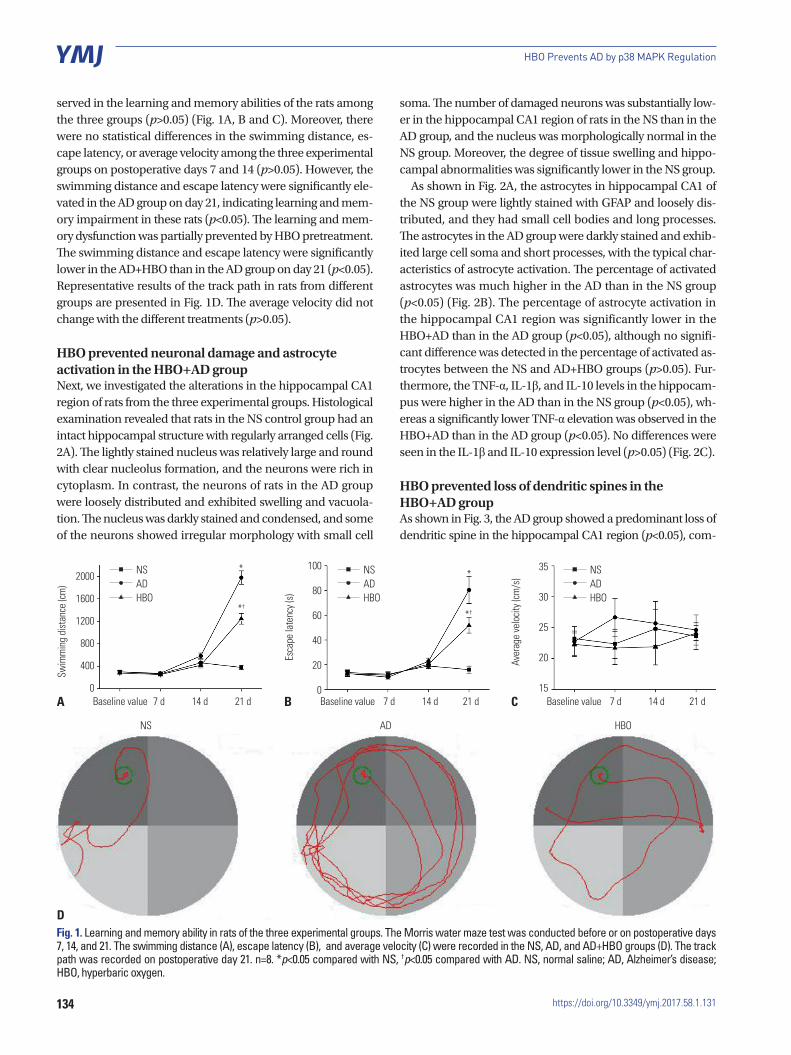
served in the learning and memory abilities of the rats among the three groups (p>0.05) (Fig. 1A, B and C). Moreover, there were no statistical differences in the swimming distance, es-cape latency, or average velocity among the three experimental groups on postoperative days 7 and 14 (p>0.05). However, the swimming distance and escape latency were significantly ele-vated in the AD group on day 21, indicating learning and mem-ory impairment in these rats (p<0.05). The learning and mem-ory dysfunction was partially prevented by HBO pretreatment. The swimming distance and escape latency were significantly lower in the AD+HBO than in the AD group on day 21 (p<0.05). Representative results of the track path in rats from different groups are presented in Fig. 1D. The average velocity did not change with the different treatments (p>0.05).
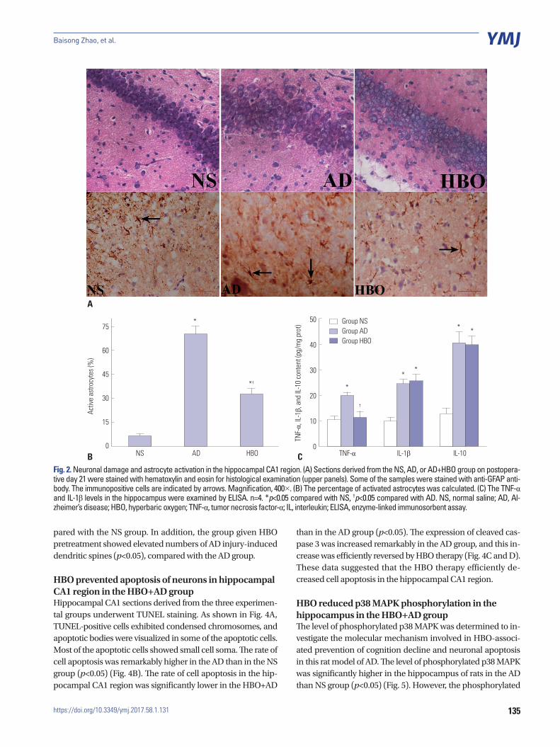
HBO prevented neuronal damage and astrocyte activation in the HBO+AD groupNext, we investigated the alterations in the hippocampal CA1 region of rats from the three experimental groups. Histological examination revealed that rats in the NS control group had an intact hippocampal structure with regularly arranged cells (Fig. 2A). The lightly stained nucleus was relatively large and round with clear nucleolus formation, and the neurons were rich in cytoplasm. In contrast, the neurons of rats in the AD group were loosely distributed and exhibited swelling and vacuola-tion. The nucleus was darkly stained and condensed, and some of the neurons showed irregular morphology with small cell
soma. The number of damaged neurons was substantially low-er in the hippocampal CA1 region of rats in the NS than in the AD group, and the nucleus was morphologically normal in the NS group. Moreover, the degree of tissue swelling and hippo-campal abnormalities was significantly lower in the NS group.
As shown in Fig. 2A, the astrocytes in hippocampal CA1 of the NS group were lightly stained with GFAP and loosely dis-tributed, and they had small cell bodies and long processes. The astrocytes in the AD group were darkly stained and exhib-ited large cell soma and short processes, with the typical char-acteristics of astrocyte activation. The percentage of activated astrocytes was much higher in the AD than in the NS group (p<0.05) (Fig. 2B). The percentage of astrocyte activation in the hippocampal CA1 region was significantly lower in the HBO+AD than in the AD group (p<0.05), although no signifi-cant difference was detected in the percentage of activated as-trocytes between the NS and AD+HBO groups (p>0.05). Fur-thermore, the TNF-α, IL-1β, and IL-10 levels in the hippocam-pus were higher in the AD than in the NS group (p<0.05), wh-ereas a significantly lower TNF-α elevation was observed in the HBO+AD than in the AD group (p<0.05). No differences were seen in the IL-1β and IL-10 expression level (p>0.05) (Fig. 2C).
HBO prevented loss of dendritic spines in the HBO+AD groupAs shown in Fig. 3, the AD group showed a predominant loss of dendritic spine in the hippocampal CA1 region (p<0.05), com-
Fig. 1. Learning and memory ability in rats of the three experimental groups. The Morris water maze test was conducted before or on postoperative days 7, 14, and 21. The swimming distance (A), escape latency (B), and average velocity (C) were recorded in the NS, AD, and AD+HBO groups (D). The track path was recorded on postoperative day 21. n=8. *p<0.05 compared with NS, †p<0.05 compared with AD. NS, normal saline; AD, Alzheimer’s disease; HBO, hyperbaric oxygen.
2000
1600
1200
800
400
0
100
80
60
40
20
0
35
30
25
20
15Baseline value Baseline value Baseline value7 d 14 d 21 d
NS AD HBO
7 d 14 d 21 d 7 d 14 d 21 d
NSADHBO
NSADHBO
NSADHBO
*†
*†
* *
Swim
min
g di
stan
ce (c
m)
Esca
pe la
tenc
y (s)
Aver
age
velo
city (
cm/s
)
A
D
B C
135
Baisong Zhao, et al.
https://doi.org/10.3349/ymj.2017.58.1.131
pared with the NS group. In addition, the group given HBO pretreatment showed elevated numbers of AD injury-induced dendritic spines (p<0.05), compared with the AD group.
HBO prevented apoptosis of neurons in hippocampal CA1 region in the HBO+AD groupHippocampal CA1 sections derived from the three experimen-tal groups underwent TUNEL staining. As shown in Fig. 4A, TUNEL-positive cells exhibited condensed chromosomes, and apoptotic bodies were visualized in some of the apoptotic cells. Most of the apoptotic cells showed small cell soma. The rate of cell apoptosis was remarkably higher in the AD than in the NS group (p<0.05) (Fig. 4B). The rate of cell apoptosis in the hip-pocampal CA1 region was significantly lower in the HBO+AD
than in the AD group (p<0.05). The expression of cleaved cas-pase 3 was increased remarkably in the AD group, and this in-crease was efficiently reversed by HBO therapy (Fig. 4C and D). These data suggested that the HBO therapy efficiently de-creased cell apoptosis in the hippocampal CA1 region.
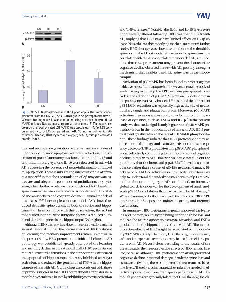
HBO reduced p38 MAPK phosphorylation in the hippocampus in the HBO+AD groupThe level of phosphorylated p38 MAPK was determined to in-vestigate the molecular mechanism involved in HBO-associ-ated prevention of cognition decline and neuronal apoptosis in this rat model of AD. The level of phosphorylated p38 MAPK was significantly higher in the hippocampus of rats in the AD than NS group (p<0.05) (Fig. 5). However, the phosphorylated
Fig. 2. Neuronal damage and astrocyte activation in the hippocampal CA1 region. (A) Sections derived from the NS, AD, or AD+HBO group on postopera-tive day 21 were stained with hematoxylin and eosin for histological examination (upper panels). Some of the samples were stained with anti-GFAP anti-body. The immunopositive cells are indicated by arrows. Magnification, 400×. (B) The percentage of activated astrocytes was calculated. (C) The TNF-α and IL-1β levels in the hippocampus were examined by ELISA. n=4. *p<0.05 compared with NS, †p<0.05 compared with AD. NS, normal saline; AD, Al-zheimer’s disease; HBO, hyperbaric oxygen; TNF-α, tumor necrosis factor-α; IL, interleukin; ELISA, enzyme-linked immunosorbent assay.
*
*
**
* *
*†
†
A
B C
75
60
45
30
15
0
50
40
30
20
10
0AD IL-1βNS
Group NSGroup ADGroup HBO
TNF-αHBO IL-10
Activ
e as
trocy
tes (
%)
TNF-α,
IL-1β,
and
IL-1
0 co
nten
t (pg
/mg
prot
)

136
HBO Prevents AD by p38 MAPK Regulation
https://doi.org/10.3349/ymj.2017.58.1.131
p38 MAPK level was significantly lower in the HBO+AD than in the AD group (p<0.05).
DISCUSSION
Although the neuroprotective effects of HBO therapy have been proposed in several rodent disease models, such as spinal cord injury and hypoxia-ischemia,9 the potential neuroprotective effect of HBO against cognitive decline and hippocampal dam-age in animal models of AD is not entirely understood. In the present study, we demonstrated for the first time that HBO pretreatment prevents learning and memory decline, hippo-campal damage, dendritic spine loss, and astrocyte activation, possibly through inhibition of p38 MAPK activation.
AD is mainly characterized by the deposition of Aβ in the hippocampus and cortex, which lead to neuronal damage and learning and memory decline in affected patients. Evidence indicates that bilateral injection of Aβ1-40 into the CA1 region of the hippocampus induces neurodegeneration, DNA fragmen-tation, synaptic dysfunction, and impaired cognition in rats.14,15 An in vitro study also indicated that Aβ can induce apoptotic and autophagic cell death.16,17 In the present study, we found that injection of Aβ1-40 into the CA1 region of the bilateral hip-pocampus significantly impaired the learning and memory capability of rats 12 days after the initial injection. The cognitive decline was accompanied by an abnormal hippocampal struc-
Fig. 3. Dendritic spine density in the hippocampal CA1 region. (A) Sections derived from the NS, AD, or AD+HBO group on postoperative day 21 were stained with Golgi-Cox. Magnification, 1000×. (B) The number of dendritic spines per 10 μm was calculated. Ten neurons from each animal were in-cluded in the calculation, and two animals from each group were used for analysis. *p<0.05 compared with NS, †p<0.05 compared with AD. NS, normal saline; AD, Alzheimer’s disease; HBO, hyperbaric oxygen.
B
A
18
15
12
9
6
3
0ADNS HBO
Spin
e de
nsity
(/10
µm
) *
†
*
*
*††
Fig. 4. Cell apoptosis in the hippocampal CA1 region. (A) Sections derived from the NS, AD, or AD+HBO group on postoperative day 21 were analyzed by TUNEL assay. Representative images are presented. Magnification, 400×. (B) The percentage of apoptotic cells was calculated. (C) Protein expression of cleaved caspase 3. β-actin was used as a loading control. (D) The relative expression of cleaved caspase 3 was calculated. n=4. *p<0.05 compared with NS, †p<0.05 compared with AD. NS, normal saline; AD, Alzheimer’s disease; HBO, hyperbaric oxygen; TUNEL, terminal deoxynucleotidyl transferase dUTP nick end labeling.
A C
B D
50
40
30
20
10
0
0.5
0.4
0.3
0.2
0.1
0.0AD AD
AD
NS NS
NS
Caspase 3
β-actin
HBO HBO
HBO
The
neur
on a
popt
otic
rate
(%)
Clea
ved
casp
ase
3/β-
actin
(fol
d)
137
Baisong Zhao, et al.
https://doi.org/10.3349/ymj.2017.58.1.131
ture and neuronal degeneration. Moreover, increased rates of hippocampal neuron apoptosis, astrocyte activation, and se-cretion of pro-inflammatory cytokines TNF-α and IL-1β and anti-inflammatory cytokine IL-10 were detected in rats with AD, suggesting the presence of neuroinflammation induced by Aβ injection. These results are consistent with those of previ-ous reports2,4 in that the accumulation of Aβ may activate as-trocytes and trigger the generation of proinflammatory cyto-kines, which further accelerate the production of Aβ.18 Dendritic spine density has been evidenced as associated with AD-relat-ed memory deficits and cognitive decline in animal models of this disease;19,20 for example, a mouse model of AD showed re-duced dendritic spine density in both the cortex and hippo-campus.21 In accordance with this observation, the AD rat model used in the current study also showed a reduced num-ber of dendritic spines in the hippocampal CA1 region.
Although HBO therapy has shown neuroprotection against several neuronal injuries, the precise effects of HBO treatment on learning and memory improvement remain unknown. In the present study, HBO pretreatment, initiated before the AD pathology was established, greatly attenuated the learning and memory decline in our rat model of AD. HBO pretreatment reduced structural alternations in the hippocampus, decreased the apoptosis of hippocampal neurons, inhibited astrocyte activation, and reduced the generation of TNF-α in the hippo-campus of rats with AD. Our findings are consistent with those of previous studies in that HBO pretreatment attenuates neu-ropathic hyperalgesia in rats by inhibiting astrocyte activation
and TNF-α release.22 Notably, the IL-1β and IL-10 levels were not obviously altered following HBO treatment in rats with AD, implying that HBO may have limited effects on IL-1β re-lease. Nevertheless, the underlying mechanism requires further study. HBO therapy was shown to ameliorate the dendritic spine loss in the AD rat model. Since dendritic spine density is correlated with the disease-related memory deficits, we spec-ulate that HBO pretreatment may prevent the characteristic cognitive decline observed in rats with AD, possibly through a mechanism that inhibits dendritic spine loss in the hippo-campus.
Activation of p38MAPK has been found to protect against oxidative stress23 and apoptosis;24 however, a growing body of evidence suggests that p38MAPK mediates pro-apoptotic cas-cades. The activation of p38 MAPK plays an important role in the pathogenesis of AD. Zhao, et al.13 described that the rate of p38 MAPK activation was especially high at the site of neuro-fibrillary tangle and plaque formation. Moreover, p38 MAPK activation in neurons and astrocytes may be induced by the re-lease of cytokines, such as TNF-α and IL-1β.7 In the present study, we detected a significantly higher rate of p38 MAPK ph-osphorylation in the hippocampus of rats with AD. HBO pre-treatment greatly reduced the rate of p38 MAPK phosphoryla-tion. These findings indicate that HBO pretreatment may re-duce neuronal damage and astrocyte activation and subsequ-ently decrease TNF-α production and p38 MAPK phosphoryl-ation, collectively contributing to the improvement of cognitive decline in rats with AD. However, we could not rule out the possibility that the increased p-p38 MAPK level is a conse-quence, rather than a cause, of AD-like neuronal damage. Bl-ockage of p38 MAPK activation using specific inhibitors may help to understand the underlying mechanism of p38 MAPK-mediated neuronal injury in AD rats. Indeed, an intensive global search is underway for the development of small-mol-ecule p38 MAPK inhibitors that may be useful for AD therapy.25 We are planning to further investigate the effects of p38 MAPK inhibitors on Aβ deposition-induced learning and memory dysfunction.
In summary, HBO pretreatment greatly improved the learn-ing and memory ability by inhibiting dendritic spine loss and reduced the neuron apoptosis, astrocyte activation, and TNF-α production in the hippocampus of rats with AD. The neuro-protective effects of HBO might be associated with blockade of p38 MAPK activity. Therefore, HBO therapy, a noninvasive, safe, and inexpensive technique, may be useful in elderly pa-tients with AD. Nevertheless, according to the results of the present study, the neuroprotective effects of HBO remain lim-ited, because, although HBO pretreatment partially prevented cognitive decline, neuronal damage, dendritic spine loss and astrocyte activation, these parameters did not return to base-line levels. Therefore, other approaches might be needed to ef-fectively prevent neuronal damage in patients with AD. Al-though patients are generally tolerant of HBO therapy, the cli-
Fig. 5. p38 MAPK phosphorylation in the hippocampus. (A) Proteins were extracted from the NS, AD, or AD+HBO group on postoperative day 21. Western blotting analysis was conducted using anti-phosphorylated p38 MAPK antibody. Representative results are presented. (B) The relative ex-pression of phosphorylated p38 MAPK was calculated. n=4. *p<0.05 com-pared with NS, †p<0.05 compared with AD. NS, normal saline; AD, Al-zheimer’s disease; HBO, hyperbaric oxygen; MAPK, mitogen-activated protein kinase.
B
A
0.6
0.4
0.2
0.0AD
ADp38 MAPK
β-active
NS
NS
HBO
HBO
Rela
tive
phos
phor
ylate
d p3
8-M
APK
leve
l (%
)
*
*†
138
HBO Prevents AD by p38 MAPK Regulation
https://doi.org/10.3349/ymj.2017.58.1.131
nical application of HBO is still limited due to its side effects, including neurotoxicity26 and pulmonary toxicity.27 Therefore, the treatment pressure should be carefully considered when this clinical therapy is applied.
ACKNOWLEDGEMENTS
This work was supported by the Science and Technology Plan Project of Guangdong and Medjaden Academy & Research Foundation for Young Scientists (grant no.2011B061200001 and MJR20150037).We thank Dr. Lingxin Meng for supporting this study.
REFERENCES
1. James BD, Bennett DA, Boyle PA, Leurgans S, Schneider JA. De-mentia from Alzheimer disease and mixed pathologies in the old-est old. JAMA 2012;307:1798-800.
2. Cameron B, Landreth GE. Inflammation, microglia, and Al-zheimer’s disease. Neurobiol Dis 2010;37:503-9.
3. Zilka N, Ferencik M, Hulin I. Neuroinflammation in Alzheimer’s disease: protector or promoter? Bratisl Lek Listy 2006;107:374-83.
4. Malm T, Ort M, Tähtivaara L, Jukarainen N, Goldsteins G, Puoliväli J, et al. beta-Amyloid infusion results in delayed and age-depen-dent learning deficits without role of inflammation or beta-amy-loid deposits. Proc Natl Acad Sci U S A 2006;103:8852-7.
5. Munoz L, Ammit AJ. Targeting p38 MAPK pathway for the treat-ment of Alzheimer’s disease. Neuropharmacology 2010;58:561-8.
6. Sun A, Liu M, Nguyen XV, Bing G. P38 MAP kinase is activated at early stages in Alzheimer’s disease brain. Exp Neurol 2003;183: 394-405.
7. Corrêa SA, Eales KL. The Role of p38 MAPK and its substrates in neuronal plasticity and neurodegenerative disease. J Signal Trans-duct 2012;2012:649079.
8. Zhao BS, Song XR, Hu PY, Meng LX, Tan YH, She YJ, et al. Hyper-baric oxygen treatment at various stages following chronic con-striction injury produces different antinociceptive effects via reg-ulation of P2X4R expression and apoptosis. PLoS One 2015;10: e0120122.
9. Liu XH, Yan H, Xu M, Zhao YL, Li LM, Zhou XH, et al. Hyperbaric oxygenation reduces long-term brain injury and ameliorates be-havioral function by suppression of apoptosis in a rat model of neonatal hypoxia-ischemia. Neurochem Int 2013;62:922-30.
10. Li F, Fang L, Huang S, Yang Z, Nandi J, Thomas S, et al. Hyperbaric oxygenation therapy alleviates chronic constrictive injury-induced neuropathic pain and reduces tumor necrosis factor-alpha pro-duction. Anesth Analg 2011;113:626-33.
11. Tian X, Zhang L, Wang J, Dai J, Shen S, Yang L, et al. The protective effect of hyperbaric oxygen and Ginkgo biloba extract on Aβ25-35-induced oxidative stress and neuronal apoptosis in rats. Behav Brain Res 2013;242:1-8.
12. Tian X, Wang J, Dai J, Yang L, Zhang L, Shen S, et al. Hyperbaric
oxygen and Ginkgo Biloba extract inhibit Aβ25-35-induced toxic-ity and oxidative stress in vivo: a potential role in Alzheimer’s dis-ease. Int J Neurosci 2012;122:563-9.
13. Zhao BS, Meng LX, Ding YY, Cao YY. Hyperbaric oxygen treat-ment produces an antinociceptive response phase and inhibits as-trocyte activation and inflammatory response in a rat model of neuropathic pain. J Mol Neurosci 2014;53:251-61.
14. Xuan A, Long D, Li J, Ji W, Zhang M, Hong L, et al. Hydrogen sulfide attenuates spatial memory impairment and hippocampal neuro-inflammation in β-amyloid rat model of Alzheimer’s disease. J Neuroinflammation 2012;9:202.
15. Wu J, Bie B, Yang H, Xu JJ, Brown DL, Naguib M. Suppression of central chemokine fractalkine receptor signaling alleviates amy-loid-induced memory deficiency. Neurobiol Aging 2013;34:2843-52.
16. Deng J, Qi XL, Guan ZZ, Yan XM, Huang Y, Wang YL. Pretreatment of SH-SY5Y cells with dicaffeoylquinic acids attenuates the reduced expression of nicotinic receptors, elevated level of oxidative stress and enhanced apoptosis caused by β-amyloid peptide. J Pharm Pharmacol 2013;65:1736-44.
17. Yang Y, Chen S, Zhang J, Li C, Sun Y, Zhang L, et al. Stimulation of autophagy prevents amyloid-β peptide-induced neuritic degen-eration in PC12 cells. J Alzheimers Dis 2014;40:929-39.
18. Zhao J, O’Connor T, Vassar R. The contribution of activated astro-cytes to Aβ production: implications for Alzheimer’s disease patho-genesis. J Neuroinflammation 2011;8:150.
19. Rozkalne A, Hyman BT, Spires-Jones TL. Calcineurin inhibition with FK506 ameliorates dendritic spine density deficits in plaque-bearing Alzheimer model mice. Neurobiol Dis 2011;41:650-4.
20. Song JM, DiBattista AM, Sung YM, Ahn JM, Turner RS, Yang J, et al. A tetra(ethylene glycol) derivative of benzothiazole aniline ame-liorates dendritic spine density and cognitive function in a mouse model of Alzheimer’s disease. Exp Neurol 2014;252:105-13.
21. Wang R, Palavicini JP, Wang H, Maiti P, Bianchi E, Xu S, et al. Ran-BP9 overexpression accelerates loss of dendritic spines in a mouse model of Alzheimer’s disease. Neurobiol Dis 2014;69:169-79.
22. Gu N, Niu JY, Liu WT, Sun YY, Liu S, Lv Y, et al. Hyperbaric oxygen therapy attenuates neuropathic hyperalgesia in rats and idiopath-ic trigeminal neuralgia in patients. Eur J Pain 2012;16:1094-105.
23. Mori H, Oikawa M, Tamagami T, Kumaki H, Nakaune R, Amano J, et al. Oxidized proteins in astrocytes generated in a hyperbaric at-mosphere induce neuronal apoptosis. J Alzheimers Dis 2007;11: 165-74.
24. Park JG, Yuk Y, Rhim H, Yi SY, Yoo YS. Role of p38 MAPK in the regulation of apoptosis signaling induced by TNF-alpha in differ-entiated PC12 cells. J Biochem Mol Biol 2002;35:267-72.
25. Bühler S, Laufer SA. p38 MAPK inhibitors: a patent review (2012 - 2013). Expert Opin Ther Pat 2014;24:535-54.
26. Heyboer M 3rd, Jennings S, Grant WD, Ojevwe C, Byrne J, Wojcik SM. Seizure incidence by treatment pressure in patients undergo-ing hyperbaric oxygen therapy. Undersea Hyperb Med 2014;41: 379-85.
27. Obiagwu C, Paul V, Chadha S, Hollander G, Shani J. Acute pulmo-nary edema secondary to hyperbaric oxygen therapy. Oxf Med Case Reports 2015;2015:183-4.