hyperoxia-induced methylation decreases runx3 in a · pdf filehyperoxia-induced methylation...
TRANSCRIPT

Zhu et al. Respiratory Research (2015) 16:75 DOI 10.1186/s12931-015-0239-x
RESEARCH Open Access
Hyperoxia-induced methylation decreasesRUNX3 in a newborn rat model ofbronchopulmonary dysplasia
Yuting Zhu, Jianhua Fu*, Haiping Yang, Yuqing Pan, Li Yao and Xindong XueAbstract
Background: Bronchopulmonary dysplasia (BPD) in premature infants is a predominantly secondary occurrence tointrauterine inflammation/infection and postpartum mechanical ventilation; in recent years, an association withepigenetics has also been found. DNA methylation, catalyzed by DNA methyl transferases (DNMTs), and tri-methylationof lysine 27 on histone H3 (H3K27me3), mediated by the methyltransferase, Enhancer of Zeste Homolog 2 (EZH2),are some of the most commonly found modifications in epigenetics. Runt-related transcription factor 3 (RUNX3) isassociated with pulmonary epithelial and vascular development and regulates expression at the post-transcriptionallevel by DNA methylation through DNMT1 or DNMT3b. However, the involvements of these epigenetic factors in theoccurrence of BPD are, as yet, unclear.
Methods: Newborn rats were randomly assigned to a model, hyperoxia (85 % O2) or control, normoxia group(21 % O2). Lung tissues and alveolar type 2 (AT2) epithelial cells were collected between 1–14 days. The expressionof DNMTs, and EZH2 was detected by immunohistochemistry, Western blot and real-time PCR. The percentage ofDNA methylation and H3K27me3 levels in the RUNX3 promoter region was measured by bisulfite sequencing PCRand chromatin immunoprecipitation assay. RUNX3 protein and mRNA expression in AT2 cells was also measuredafter inhibition using the DNA methylation inhibitor, 5-Aza-2′-deoxycytidine, the H3K27me3 inhibitor, JMJD3, andthe EZH2 inhibitor, DZNep.
Results: Compared with the control group, RUNX3 protein was downregulated and DNMT3b and EZH2 werehighly expressed in lung tissues and AT2 cells of the model group (P < 0.05), while high DNA methylation andH3K27me3 modifications were present in the RUNX3 promoter region, in lung tissues of the model group (P < 0.05).Following hyperoxia in the model group, JMJD3 and DZNep significantly reversed the hyperoxia-induceddown-regulation of RUNX3 expression in AT2 cells (P < 0.05), more so than 5-Aza-2′-deoxycytidine (P < 0.05).
Conclusions: 1) DNA methylation and H3K27 trimethylation are present in the BPD model; 2) RUNX3 down-regulationis attributed to both DNMT3b-catalyzed DNA methylation and EZH2-catalyzed histone methylation.
Keywords: DNA methylation, histone methylation, RUNX3, alveolar development, bronchopulmonary dysplasia, EZH2
* Correspondence: [email protected] of Pediatrics, Shengjing Hospital of China Medical University,Shenyang 110004, China
© 2015 Zhu et al. This is an Open Access article distributed under the terms of the Creative Commons Attribution License(http://creativecommons.org/licenses/by/4.0), which permits unrestricted use, distribution, and reproduction in any medium,provided the original work is properly credited. The Creative Commons Public Domain Dedication waiver (http://creativecommons.org/publicdomain/zero/1.0/) applies to the data made available in this article, unless otherwise stated.

Zhu et al. Respiratory Research (2015) 16:75 Page 2 of 17
BackgroundBronchopulmonary dysplasia (BPD) is a common, chroniclung disease of infants, frequently seen in prematurenewborns with a fetal age < 30 weeks and a birth weight< 1,500 g. The occurrence of BPD is mostly secondaryto intrauterine inflammation/infection or postpartummechanical ventilation, oxygen support, and changesin other environmental factors. The main pathologicalchanges that characterize BPD are alveolar structuresimplification and pulmonary capillary dysplasia [1]. Inrecent years, a large number of studies have revealedthat some genes, including those for vascular endothe-lial growth factor (VEGF) [2], interleukin 1-beta (IL-1β)[3] and mucin 1 (MUC1) [4], participate in the pul-monary developmental disorder process of BPD byregulating alveolar formation.Runt-related transcription factor 3 (RUNX3) is a
member of the RUNX family and participates in normalphysiological, as well as pathological, processes of theimmune system [5], in tumor formation [6] and in otherdisorders. RUNX3 is also associated with pulmonaryepithelial and vascular development [7]. Previous studieshave shown that RUNX3 was expressed in mousepulmonary epithelium at E15.5 [8], while RUNX3knock-out caused pulmonary epithelial hyperplasia [8]and pulmonary vascularization disorder [7], similar tothe pathological changes seen in BPD [1]. RUNX3 oftenregulates expression at the post-transcriptional level byDNA methylation [9]. However, the mechanisms be-hind RUNX3 down-regulation and any potential regu-lators of abnormal RUNX3 expression in a BPD modelhave, as yet, to be defined.The silencing of RUNX3 expression is associated
with the tri-methylation of lysine 27 on histone H3(H3K27me3), an epigenetic marker, and is mediated bythe methyltransferase, Enhancer of Zeste Homolog 2(EZH2) [10, 11] and demethyltransferase, JMJD3/UTX[12], to reduce transcription [13]. Fujii et al. [14] found thatEZH2 knock-out reduced H3K27me3-binding RUNX3levels and thus up-regulated RUNX3 mRNA levels.DNA methyl transferases (DNMTs) catalyze DNA
methylation, which leads to the silencing of gene expres-sion. Common DNMTs include DNMT1, which main-tains and regulates DNA methylation, and DNMT3a/b,which establishes de novo methylation [15, 16]. DNMT1was thought to be the major contributor of RUNX3DNA methylation, but DNMT3b has also been found tohave a role [17]. Additionally, Deng et al. [10] foundthat the inhibition of DNMT3b expression caused theupregulation of RUNX3 expression in a colorectalcancer cell line.Numerous studies have suggested that BPD is a genet-
ically susceptible disease. Studies of twins have shown thatthe BPD status of one twin was a significant predictor of
BPD in the second twin [18], and that the incidence ofBPD in homozygotic twins was significantly greaterthan that of dizygotic twins [19]. Subsequently, manyscholars have reported abnormalities of histone acety-lase activity and the chromatin remodeling pathway inBPD patients, and believe that epigenetics is a causal fac-tor in the occurrence and development of BPD [20–23].However, whether two common epigenetic modifications–DNA methylation and H3K27me3–are associated withBPD [24], and whether, by regulating target genes, theyparticipate in the pulmonary developmental disorderprocesses of BPD is unclear. Consequently, this studyaimed to identify the presence or absence of DNAmethylation and H3K27me3 in BPD, and to highlightany correlation between RUNX3 down-regulation andDNA methylation or H3K27me3 in BPD at the epigen-etic level.
Experimental methodsAnimal model and tissue specimensA newborn rat model of BPD, established by our re-search group as previously described, was used [25].Two hundred newborn, Sprague–Dawley (SD) rats wererandomly divided into a model (exposure to hyperoxia[85 % O2] from day of birth) or control group (exposureto normoxia [21 % O2]). To avoid O2 toxicity, maternalrats within the model and control groups were switchedonce every 24 h. Rats were given ad libitum access towater and food. At 1, 7, 10 and 14 days after the start ofexposure to hyperoxia or normoxia, eight newborn ratsfrom each model or control group were anesthetized byintraperitoneal injection with 5 % chloral hydrate, andwhole lungs collected aseptically by chest opening. Theleft lungs were fixed in paraformaldehyde (PFA) for sub-sequent immunohistochemical staining, the right upperlung lobes were used for real-time PCR analysis, and theright lower lung lobes for Western blots. All specimenswere snap-frozen in liquid nitrogen and stored at −80 °Cuntil use. Mature SD rats with a body weight of 220–250 gwere purchased from the Department of Animals,Experimental Center, Shengjing Hospital of China MedicalUniversity (Shenyang, China). All animal experimentswere approved and supervised by the Ethics Committee ofAnimals, China Medical University.
AT2 cell isolation and purificationThe above BPD animal model was employed. At 0, 1, 7,10 and 14 days after the start of normoxia or hyperoxia,alveolar type 2 (AT2) epithelial cells of newborn rats wereisolated from the control or model groups, respectively,for primary culture. As previously described [26, 27],tracheal intubation was performed on anesthetized ratsto maintain lung ventilation and to conduct the subse-quent lavage. Two cold buffer solutions and an albumin
Zhu et al. Respiratory Research (2015) 16:75 Page 3 of 17
emulsion were used for cardiopulmonary and tracheacannula lavage to remove blood and macrophages fromlung tissues. AT2 cells were isolated by mechanicalseparation and trypsinization of lung tissue, followed byfiltration with a nylon mesh. A first-round purificationwas conducted based on differential cell adherence, anda second-round purification was performed using alamellar body cell membrane antibody p180 (Abcam,Cambridge, MA, USA). After storage in liquid nitrogen,AT2 cells of the model and control groups, isolated at1, 7, 10 and 14 days, were transferred to a −80 °Cfreezer and then subsequently used for real-time PCR,Western blot, bisulfite sequencing PCR (BSP) analysis andchromatin immunoprecipitation (ChIP) assay. PurifiedAT2 cells, extracted immediately after birth (0 day), wereused for inhibitor treatments as described below.
Cell treatmentPurified AT2 cells extracted immediately after birth (0 day)were plated out into 6-well plates and then cultured inenvironments of differing oxygen concentrations: a con-trol (21 % O2) or model (85 % O2) group. Each group ofcells was cultured in the presence of various concentra-tions of 5-Aza-2′-deoxycytidine (5-Aza-CdR, Abcam; 2.5, 5or 10 μmol/L), Jumonji domain containing 3 (Jmjd3,Abcam; 10, 20 or 40 μg/L) or 3-Deazaneplanocin A(DZNep, Selleckchem, Shanghai, China; 1, 2.5 or 5 μmol/L)for 48 h, or treated with 5 μmol/L 5-Aza-CdR, 20 μg/LJmjd3 or 1 μmol/L DZNep for 24, 48 or 72 h. Inhibitorsand media were changed every 24 h until cells werecollected. Cells of the negative control and modelgroups were cultured without inhibitors. After storagein liquid nitrogen, cells were transferred to a −80 °Cfreezer and then subsequently used for real-time PCRand Western blotting.
ImmunohistochemistryPFA-fixed lung tissues were dehydrated, vitrified, embed-ded in paraffin, fixed and cut into 4 μm-thick sections,which were then fixed in a 60 °C oven for 4 h. Sectionswere dewaxed with dimethyl benzene, hydrated withgradient alcohol according to manufacturer’s instruc-tions (immunohistochemistry kit; MXB, Fujian, China),and treated with 3 % H2O2 to block endogenous perox-idase activity. Treated sections were placed into anEDTA-Tris buffer solution and microwaved for 20 min,blocked with serum, and incubated overnight at 4 °Cwith various antibodies: rabbit polyclonal anti-RUNX3,1:200 (Abcam); rabbit polyclonal anti-DNA methyltrans-ferase 3b, 1:500 (DNMT3b, Abcam); mouse monoclonalanti-H3K27me3, 1:1000 (Abcam); rabbit polyclonal anti-DNA methyltransferase 1, 1:50 (DNMT1; Santa CruzBiotechnology, Dallas, TX, USA); or mouse monoclonalanti-Enhancer of Zeste Homolog 2, 1:200 (EZH2; Becton,
Dickinson and Co, Franklin Lakes, NJ, USA). Aftersequential incubation with biotin-labeled secondaryantibodies and streptavidin-peroxidase, sections weredeveloped with 3,3′-diaminobenzidine (DAB), dehy-drated and mounted. A laser scanning confocal micro-scope (MTC-600, Bio-Rad, Hercules, CA, USA) wasused for image acquisition, and the deposition of brownparticles in the cytoplasm/nucleus indicated a positiveresult. Cells displaying brown particles in the cyto-plasm/nucleus were counted within the same sized areaas viewed under the microscope.
Western blotAn EpiQuik Nuclear Extraction Kit (Epigentek Group Inc,New York, NY, USA) was used to extract total nuclearprotein from lung tissues and AT2 cells after exposure tonormoxia or hyperoxia, and AT2 cells after inhibitortreatment. After SDS polyacrylamide gel electrophoresis(SDS-PAGE), separated proteins were transferred ontoa polyvinylidene fluoride (PVDF) membrane, and themembrane was then blocked with 10 % skimmed milkand incubated overnight with various antibodies: (anti-RUNX3, 1:800; anti-DNMT3B, 1:1000; anti-H3K27me3,1:3000; anti-DNMT1, 1:200; anti-EZH2, 1:1000; anti-GAPDH, 1:15,000 [all Santa Cruz Biotechnology]). Themembrane was then incubated in secondary antibodyfor 4 h, washed in 10 mM Tris/HCl, 150 mM NaCl, and0.05 % Tween 20, pH 7.5 (TBST) buffer three times,developed with enhanced chemiluminescence substrate(ECL kit; Santa Cruz Biotechnology) and exposed toX-ray film. All bands on X-ray film were scanned usingChemiImager 5500 V2.03 software. Integrated densityvalues were computed using an image analysis system(Fluor Chen 2.0; Bio-Rad, Hercules, CA, USA) andstandardized to GAPDH.
Real-time PCRTotal mRNA was extracted from right upper lung lobe tis-sues and AT2 cells after exposure to normoxia or hyper-oxia, and from AT2 cells after inhibitor treatment. Briefly,Trizol, chloroform and isopropyl alcohol were added andsamples subjected to reverse transcription and real-timePCR (Life Technologies, Carlsbad, CA, USA) according tokit instructions. Reverse transcription conditions were:65 °C, 5 min; 37 °C, 2 min; 37 °C, 50 min; and 70 °C,15 min. PCR conditions were 50 °C, 2 min; 95 °C, 2 min;95 °C, 15 s; and 60 °C, 1 min for 40 cycles. Primersequences of different indicators are shown in Table 1,and model data were standardized to GAPDH.
Bisulfite sequencing PCR (BSP)After 7 and 14 days, the genomic DNA of AT2 cells inmodel and control groups was extracted using a blood/cell/tissue genomic DNA extraction kit (Tiangen Biotech,

Table 1 Real-time PCR primers for genes
Target gene 5′→ 3′ Product
dnmt1 Sense CATGGTGCTGAAGCTCACAC 176 bp
Antisense GGGCAAACACGTGTAGAGGT
dnmt3b Sense TTCTCATGATGCCAAAGCTC 118BP
Antisense GAGGTTCTTTGCCTCTCCAG
ezh2 Sense TTCGTTTTGCTAATCATTCAGTAA 162 bp
Antisense CCACATACTTCAGGGCATCA
runx3 Sense GAAGATAGAGGACCAGACCAAAG 194 bp
Antisense GGAAGGAGCGATCAAACTG
gapdh Sense AGACAGCCGCATCTTCTTGT 207 bp
Antisense CTTGCCGTGGGTAGAGTCAT
Table 2 CHIP-Real-time PCR primers for runx3
Gene locus 5′→ 3′ Product
1 Sense CTGTGGCTAAGAGGGTGAC 265 bp
Antisense ACAAGGCTGAAGATGACG
2 Sense TCTCCACCTCAGAACGC 192 bp
Antisense ACAAGGCTGAAGATGACG
3 Sense CGCATCCACTTCCACTACAC 180 bp
Antisense TTCCTGCCCACTCAAAA
Zhu et al. Respiratory Research (2015) 16:75 Page 4 of 17
Beijing, China). After elution with 70 °C pre-heated, sterilewater, DNA was collected. Thereafter, DNA specimenswere treated with a DNA methylation kit (Millipore,Billerica, MA, USA) and amplified. PCR amplificationconditions were 95 °C, 5 min; 95 °C, 10 s; 50 °C, 20 s; 72 °C,30 s; 35 cycles; 4 °C, 5 min. Forward primer: 1: 5′-GGATTAAGGTTGAGAAGATGATGG-3′, reverse primer1: 5′-ACCACCCTATTCCTACCCACTC-3′; forwardprimer 2: 5′-TTAGAAGGGCGTTTAGGAGA-3′, re-verse primer 2: 5′-AACTCTAACGATCCTCATCC-3′.Amplicons were checked on agarose gel and sequencedusing BiQ Analyzer.
Chromatin Immunoprecipitation (ChIP) AssayChromatin co-immunoprecipitation was performed ac-cording to manufacturer’s instructions (EZ-ChIP kit;Millipore). In brief, 1 % (final concentration) formalde-hyde was used to treat AT2 cells isolated from modeland control groups at 14 days; after protein-DNA cross-linking, genomic DNA was ultrasonically sheared intolengths of 200–1000 bp. Non-specific binding proteinsand DNA were pre-removed with Protein G agarose andan antibody-free control sample was put aside. Normalmouse IgG (IgG group), anti-H3K27me3 (target genegroup 1; 5 μL [Abcam]) or anti-EZH2 (target gene group2; 2 μL [Millipore]) and Protein G agarose were addedto samples and incubated overnight. Samples werewashed according to instructions, eluted with elutionbuffer to remove protein/DNA complexes, cross-linked,purified step-by-step, and then amplified. Amplificationconditions were the same as for real-time PCR, andprimer sequences used are shown in Table 2. The modelresults are expressed as 2-CT (Target gene group-Input group-
IgG group).
Statistical analysisSPSS17.0 software (manufacture, city, country) was usedfor statistical analysis. Inter-group comparisons were madeusing t tests, multiple group comparisons made using
one-way analysis of variance (ANOVA), and correlationanalysis made using Pearson’s test. All data are presentedas mean (χ) ± standard deviation (SD). P < 0.05 suggests astatistically significant difference.
ResultsRUNX3 expression in lung tissues and AT2 cells followinghyperoxiaWe studied RUNX3 protein localization by immunohis-tochemical staining of lung tissues from both controland BPD model groups exposed to hyperoxia over time;RUNX3 protein expression was found in the nucleusand cytoplasm of alveolar epithelial cells in lung sectionsof both the control and model groups (Fig. 1a, b). Wealso examined RUNX3 protein and mRNA levels in lungtissue and AT2 cells isolated from these groups. Com-pared with the control group, RUNX3 protein andmRNA expression in lung tissues and AT2 cells fromthe model group was significantly decreased by ≥ 7 daysof hyperoxia (Fig. 1; P < 0.05).
DNMT and histone methyltransferase EZH2 localization inlung tissues following hyperoxiaThe localization of DNMTs and EZH2 proteins in lungtissues of animals exposed to hyperoxia or normoxiawas examined by immunohistochemistry. After 10 dayof hyperoxia, DNMT1 protein was expressed in both thenucleus and cytoplasm of pulmonary epithelial cells inthe control group, but was poorly expressed in themodel group (Fig. 2a, b). In both the model and controlgroups, DNMT3b protein was expressed in the nucleusof alveolar epithelial and mesenchymal cells (Fig. 2c, d).EZH2 protein was expressed in both the nucleus andcytoplasm of alveolar epithelial cells in the model group,but expression was greatly decreased in the controlgroup (Fig. 2e, f ).
DNMT expression levels in lung tissues and A2 cellsfollowing hyperoxiaDNMTs protein levels in lung tissues of animals exposedto hyperoxia or normoxia were also examined by Westernblot. Compared with the control group, DNMT1 proteinin lung tissues of the model group significantly increasedby ≥ 7 days, reached a peak at 10 day, and thereafter
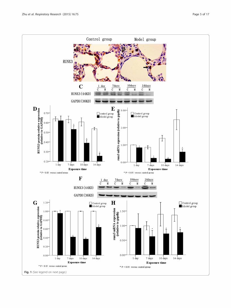
Fig. 1 (See legend on next page.)
Zhu et al. Respiratory Research (2015) 16:75 Page 5 of 17

(See figure on previous page.)Fig. 1 RUNX3 expression in lung tissues and AT2 cells following hyperoxia. RUNX3 protein localization in lung tissues by immunohistochemistryshowing RUNX3 protein expression in both the nucleus and cytoplasm of alveolar epithelial cells in control (a) and model groups (b). The arrowindicates positive cells. (×400). Western blot of RUNX3 protein (c and d), and real-time PCR of RUNX3 mRNA (e) showing low expression in lungtissues of the model group after 7 d hyperoxia. Low expression of RUNX3 protein (f and g) and mRNA (h) in AT2 cells from the model group after7 d hyperoxia. C: Control group, E: model group, *P < 0.05 vs. control group, n = 4
Zhu et al. Respiratory Research (2015) 16:75 Page 6 of 17
decreased (Fig. 3a, b; P < 0.05), while DNMT3b proteindemonstrated a persistently high expression level after 7days (Fig. 3a, c; P < 0.05). Subsequently, the expression ofDNMT1 and DNMT3b in AT2 cells extracted from lungtissues was detected at different time points: Comparedwith the control group, DNMT1 protein in AT2 cells iso-lated from the model group rapidly decreased by ≥ 7 days,and demonstrated a persistently and significantly low ex-pression level (Fig. 3d, e; P < 0.05), while DNMT3b proteinincreased by ≥ 10 day and demonstrated a significantlyhigh expression level (Fig. 3d, f; P < 0.05).DNMTs mRNA levels in lung tissues and AT2 cells iso-
lated from animals exposed to hyperoxia or normoxiawere also examined by real-time PCR. In the model group,DNMT1 mRNA was poorly expressed while DNMT3b
Fig. 2 DNMTs and EZH2 protein localization in lung tissues following hypeimmunohistochemistry. After 10 day of hyperoxia, DNMT1 protein was exp(a), but, in the model group, DNMT1 protein was barely detectable (b). In bexpressed in alveolar epithelial and mesenchymal cells. In the control grouprotein was expressed in alveolar epithelial cells (f). The arrow points to po
mRNA was highly expressed in lung tissues after 10 day ofhyperoxia compared to the control group (Fig. 4a, b).After 7 days of hyperoxia, DNMT1 mRNA was poorlyexpressed in AT2 cells of hyperoxia group (Fig. 4c). Forboth the model and control groups, DNMT3b mRNA wasequally expressed in AT2 cells after 1 day of hyperoxiacompared to the control group (Fig. 4d). DNMT1 andDNMT3b mRNA levels in lung tissues and AT2 cellsshowed similar changes to protein, but the differences be-tween groups were not statistically significant (Fig. 4).
EZH2 expression levels in lung tissues and A2 cellsfollowing hyperoxiaEZH2 protein levels in lung tissues and AT2 cells ofanimals exposed to hyperoxia or normoxia were also
roxia. DNMTs and EZH2 protein localization in lung tissues byressed in pulmonary alveolar epithelial cells in the control groupoth the control (c) and model groups (d), DNMT3b protein wasp, EZH2 protein was poorly expressed (e). In the model group, EZH2sitive cells. (×400)
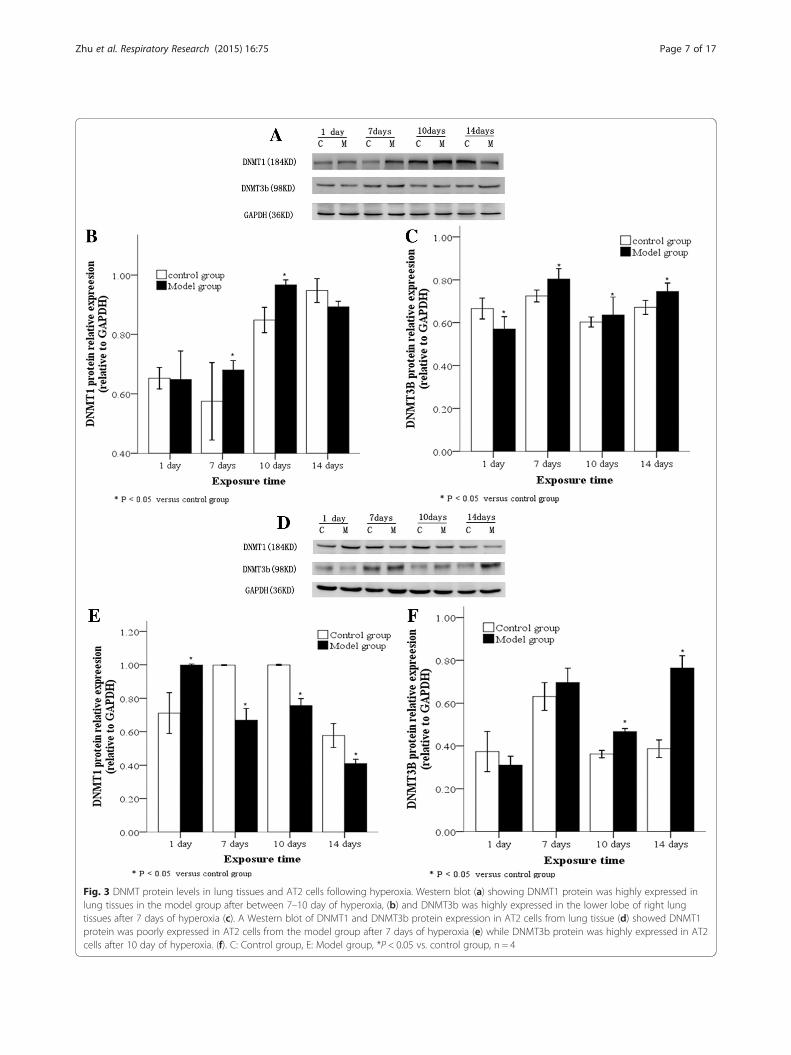
Fig. 3 DNMT protein levels in lung tissues and AT2 cells following hyperoxia. Western blot (a) showing DNMT1 protein was highly expressed inlung tissues in the model group after between 7–10 day of hyperoxia, (b) and DNMT3b was highly expressed in the lower lobe of right lungtissues after 7 days of hyperoxia (c). A Western blot of DNMT1 and DNMT3b protein expression in AT2 cells from lung tissue (d) showed DNMT1protein was poorly expressed in AT2 cells from the model group after 7 days of hyperoxia (e) while DNMT3b protein was highly expressed in AT2cells after 10 day of hyperoxia. (f). C: Control group, E: Model group, *P < 0.05 vs. control group, n = 4
Zhu et al. Respiratory Research (2015) 16:75 Page 7 of 17
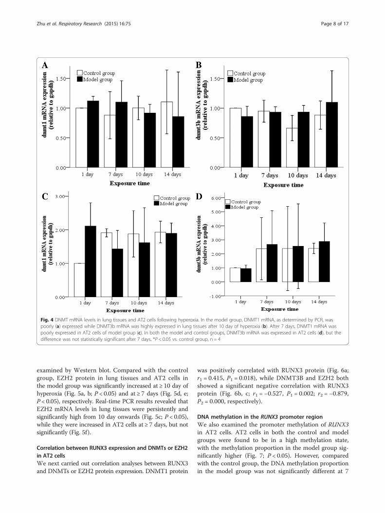
Fig. 4 DNMT mRNA levels in lung tissues and AT2 cells following hyperoxia. In the model group, DNMT1 mRNA, as determined by PCR, waspoorly (a) expressed while DNMT3b mRNA was highly expressed in lung tissues after 10 day of hyperoxia (b). After 7 days, DNMT1 mRNA waspoorly expressed in AT2 cells of model group (c). In both the model and control groups, DNMT3b mRNA was expressed in AT2 cells (d), but thedifference was not statistically significant after 7 days. *P < 0.05 vs. control group, n = 4
Zhu et al. Respiratory Research (2015) 16:75 Page 8 of 17
examined by Western blot. Compared with the controlgroup, EZH2 protein in lung tissues and AT2 cells inthe model group was significantly increased at ≥ 10 day ofhyperoxia (Fig. 5a, b; P < 0.05) and at ≥ 7 days (Fig. 5d, e;P < 0.05), respectively. Real-time PCR results revealed thatEZH2 mRNA levels in lung tissues were persistently andsignificantly high from 10 day onwards (Fig. 5c; P < 0.05),while they were increased in AT2 cells at ≥ 7 days, but notsignificantly (Fig. 5f).
Correlation between RUNX3 expression and DNMTs or EZH2in AT2 cellsWe next carried out correlation analyses between RUNX3and DNMTs or EZH2 protein expression. DNMT1 protein
was positively correlated with RUNX3 protein (Fig. 6a;r1 = 0.415, P1 = 0.018), while DNMT3B and EZH2 bothshowed a significant negative correlation with RUNX3protein (Fig. 6b, c; r1 = −0.527, P1 = 0.002; r2 = −0.879,P2 = 0.000, respectively).
DNA methylation in the RUNX3 promoter regionWe also examined the promoter methylation of RUNX3in AT2 cells. AT2 cells in both the control and modelgroups were found to be in a high methylation state,with the methylation proportion in the model group sig-nificantly higher (Fig. 7; P < 0.05). However, comparedwith the control group, the DNA methylation proportionin the model group was not significantly different at 7
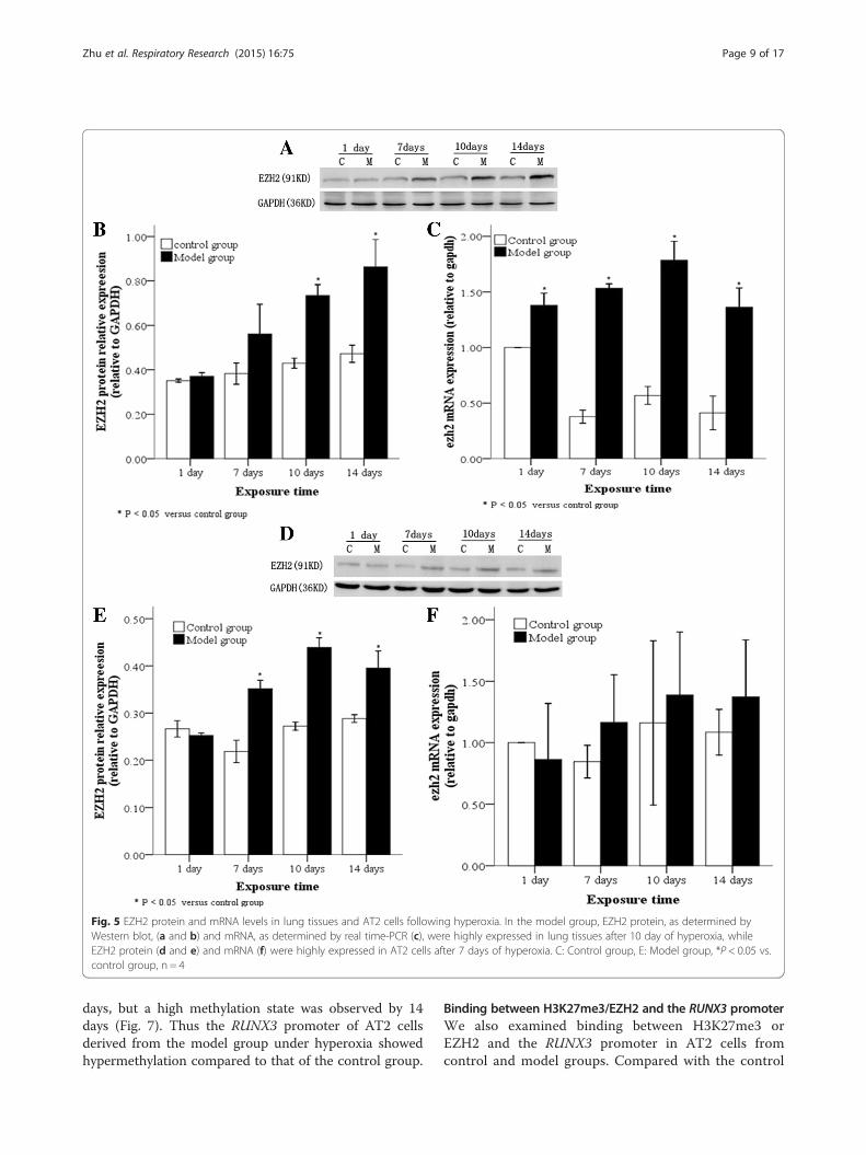
Fig. 5 EZH2 protein and mRNA levels in lung tissues and AT2 cells following hyperoxia. In the model group, EZH2 protein, as determined byWestern blot, (a and b) and mRNA, as determined by real time-PCR (c), were highly expressed in lung tissues after 10 day of hyperoxia, whileEZH2 protein (d and e) and mRNA (f) were highly expressed in AT2 cells after 7 days of hyperoxia. C: Control group, E: Model group, *P < 0.05 vs.control group, n = 4
Zhu et al. Respiratory Research (2015) 16:75 Page 9 of 17
days, but a high methylation state was observed by 14days (Fig. 7). Thus the RUNX3 promoter of AT2 cellsderived from the model group under hyperoxia showedhypermethylation compared to that of the control group.
Binding between H3K27me3/EZH2 and the RUNX3 promoterWe also examined binding between H3K27me3 orEZH2 and the RUNX3 promoter in AT2 cells fromcontrol and model groups. Compared with the control
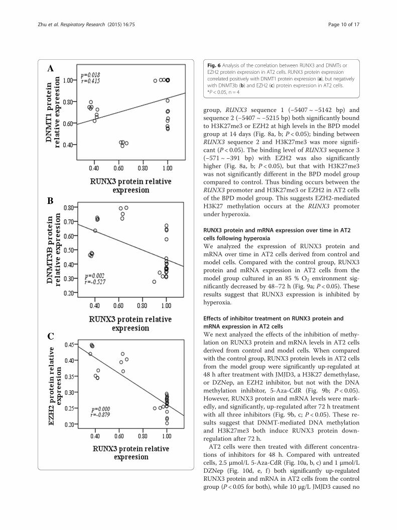
Fig. 6 Analysis of the correlation between RUNX3 and DNMTs orEZH2 protein expression in AT2 cells. RUNX3 protein expressioncorrelated positively with DNMT1 protein expression (a), but negativelywith DNMT3b (b) and EZH2 (c) protein expression in AT2 cells.*P < 0.05, n = 4
Zhu et al. Respiratory Research (2015) 16:75 Page 10 of 17
group, RUNX3 sequence 1 (−5407 ~ −5142 bp) andsequence 2 (−5407 ~ −5215 bp) both significantly boundto H3K27me3 or EZH2 at high levels in the BPD modelgroup at 14 days (Fig. 8a, b; P < 0.05); binding betweenRUNX3 sequence 2 and H3K27me3 was more signifi-cant (P < 0.05). The binding level of RUNX3 sequence 3(−571 ~ −391 bp) with EZH2 was also significantlyhigher (Fig. 8a, b; P < 0.05), but that with H3K27me3was not significantly different in the BPD model groupcompared to control. Thus binding occurs between theRUNX3 promoter and H3K27me3 or EZH2 in AT2 cellsof the BPD model group. This suggests EZH2-mediatedH3K27 methylation occurs at the RUNX3 promoterunder hyperoxia.
RUNX3 protein and mRNA expression over time in AT2cells following hyperoxiaWe analyzed the expression of RUNX3 protein andmRNA over time in AT2 cells derived from control andmodel cells. Compared with the control group, RUNX3protein and mRNA expression in AT2 cells from themodel group cultured in an 85 % O2 environment sig-nificantly decreased by 48–72 h (Fig. 9a; P < 0.05). Theseresults suggest that RUNX3 expression is inhibited byhyperoxia.
Effects of inhibitor treatment on RUNX3 protein andmRNA expression in AT2 cellsWe next analyzed the effects of the inhibition of methy-lation on RUNX3 protein and mRNA levels in AT2 cellsderived from control and model cells. When comparedwith the control group, RUNX3 protein levels in AT2 cellsfrom the model group were significantly up-regulated at48 h after treatment with JMJD3, a H3K27 demethylase,or DZNep, an EZH2 inhibitor, but not with the DNAmethylation inhibitor, 5-Aza-CdR (Fig. 9b; P < 0.05).However, RUNX3 protein and mRNA levels were mark-edly, and significantly, up-regulated after 72 h treatmentwith all three inhibitors (Fig. 9b, c; P < 0.05). These re-sults suggest that DNMT-mediated DNA methylationand H3K27me3 both induce RUNX3 protein down-regulation after 72 h.AT2 cells were then treated with different concentra-
tions of inhibitors for 48 h. Compared with untreatedcells, 2.5 μmol/L 5-Aza-CdR (Fig. 10a, b, c) and 1 μmol/LDZNep (Fig. 10d, e, f ) both significantly up-regulatedRUNX3 protein and mRNA in AT2 cells from the controlgroup (P < 0.05 for both), while 10 μg/L JMJD3 caused no
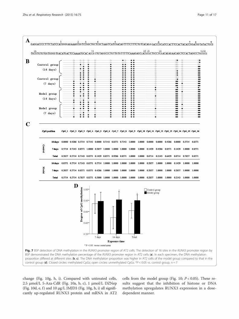
Fig. 7 BSP detection of DNA methylation in the RUNX3 promoter region of AT2 cells. The detection of 16 sites in the RUNX3 promoter region byBSP demonstrated the DNA methylation percentage of the RUNX3 promoter region in AT2 cells (a). In each specimen, the DNA methylationproportion differed at different sites (b, c). The DNA methylation proportion was higher in AT2 cells of the model group compared to that in thecontrol group (d). Closed circles: methylated CpGs; open circles: unmethylated CpGs. *P < 0.05 vs. control group, n = 7
Zhu et al. Respiratory Research (2015) 16:75 Page 11 of 17
change (Fig. 10g, h, i). Compared with untreated cells,2.5 μmol/L 5-Aza-CdR (Fig. 10a, b, c), 1 μmol/L DZNep(Fig. 10d, e, f ) and 10 μg/L JMJD3 (Fig. 10g, h, i) all signifi-cantly up-regulated RUNX3 protein and mRNA in AT2
cells from the model group (Fig. 10; P < 0.05). These re-sults suggest that the inhibition of histone or DNAmethylation upregulates RUNX3 expression in a dose-dependent manner.
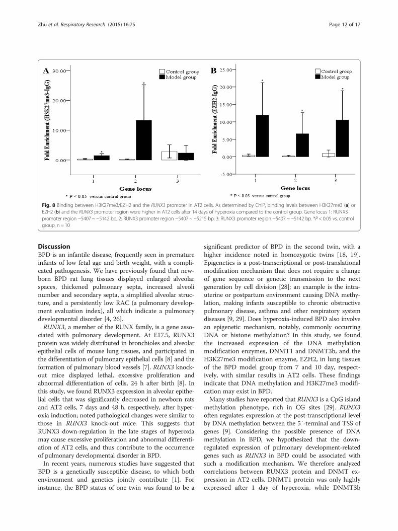
Fig. 8 Binding between H3K27me3/EZH2 and the RUNX3 promoter in AT2 cells. As determined by ChIP, binding levels between H3K27me3 (a) orEZH2 (b) and the RUNX3 promoter region were higher in AT2 cells after 14 days of hyperoxia compared to the control group. Gene locus 1: RUNX3promoter region −5407 ~ −5142 bp; 2: RUNX3 promoter region −5407 ~ −5215 bp; 3: RUNX3 promoter region −5407 ~ −5142 bp. *P < 0.05 vs. controlgroup, n = 10
Zhu et al. Respiratory Research (2015) 16:75 Page 12 of 17
DiscussionBPD is an infantile disease, frequently seen in prematureinfants of low fetal age and birth weight, with a compli-cated pathogenesis. We have previously found that new-born BPD rat lung tissues displayed enlarged alveolarspaces, thickened pulmonary septa, increased alveolinumber and secondary septa, a simplified alveolar struc-ture, and a persistently low RAC (a pulmonary develop-ment evaluation index), all which indicate a pulmonarydevelopmental disorder [4, 26].RUNX3, a member of the RUNX family, is a gene asso-
ciated with pulmonary development. At E17.5, RUNX3protein was widely distributed in bronchioles and alveolarepithelial cells of mouse lung tissues, and participated inthe differentiation of pulmonary epithelial cells [8] and theformation of pulmonary blood vessels [7]. RUNX3 knock-out mice displayed lethal, excessive proliferation andabnormal differentiation of cells, 24 h after birth [8]. Inthis study, we found RUNX3 expression in alveolar epithe-lial cells that was significantly decreased in newborn ratsand AT2 cells, 7 days and 48 h, respectively, after hyper-oxia induction; noted pathological changes were similar tothose in RUNX3 knock-out mice. This suggests thatRUNX3 down-regulation in the late stages of hyperoxiamay cause excessive proliferation and abnormal differenti-ation of AT2 cells, and thus contribute to the occurrenceof pulmonary developmental disorder in BPD.In recent years, numerous studies have suggested that
BPD is a genetically susceptible disease, to which bothenvironment and genetics jointly contribute [1]. Forinstance, the BPD status of one twin was found to be a
significant predictor of BPD in the second twin, with ahigher incidence noted in homozygotic twins [18, 19].Epigenetics is a post-transcriptional or post-translationalmodification mechanism that does not require a changeof gene sequence or genetic transmission to the nextgeneration by cell division [28]; an example is the intra-uterine or postpartum environment causing DNA methy-lation, making infants susceptible to chronic obstructivepulmonary disease, asthma and other respiratory systemdiseases [9, 29]. Does hyperoxia-induced BPD also involvean epigenetic mechanism, notably, commonly occurringDNA or histone methylation? In this study, we foundthe increased expression of the DNA methylationmodification enzymes, DNMT1 and DNMT3b, and theH3K27me3 modification enzyme, EZH2, in lung tissuesof the BPD model group from 7 and 10 day, respect-ively, with similar results in AT2 cells. These findingsindicate that DNA methylation and H3K27me3 modifi-cation may exist in BPD.Many studies have reported that RUNX3 is a CpG island
methylation phenotype, rich in CG sites [29]. RUNX3often regulates expression at the post-transcriptional levelby DNA methylation between the 5′-terminal and TSS ofgenes [9]. Considering the possible presence of DNAmethylation in BPD, we hypothesized that the down-regulated expression of pulmonary development-relatedgenes such as RUNX3 in BPD could be associated withsuch a modification mechanism. We therefore analyzedcorrelations between RUNX3 protein and DNMT ex-pression in AT2 cells. DNMT1 protein was only highlyexpressed after 1 day of hyperoxia, while DNMT3b
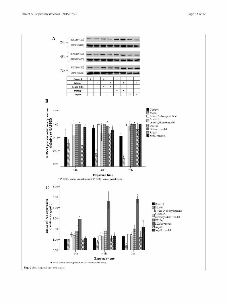
Fig. 9 (See legend on next page.)
Zhu et al. Respiratory Research (2015) 16:75 Page 13 of 17
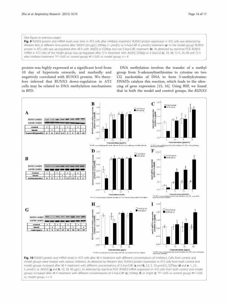
(See figure on previous page.)Fig. 9 RUNX3 protein and mRNA levels over time in AT2 cells after inhibitor treatment. RUNX3 protein expression in AT2 cells was detected byWestern blot at different time points after JMJD3 (20 μg/L), DZNep (1 μmol/L) or 5-Aza-CdR (5 μmol/L) treatment (a). In the model group, RUNX3protein in AT2 cells was up-regulated after 48 h with JMJD3 or DZNep, but not 5-Aza-CdR, treatment (b). As detected by real-time PCR, RUNX3mRNA in AT2 cells of the model group was up-regulated after 72 h treatment with JMJD3, DZNep or 5-Aza-Cd (c). 24, 48, 72 h: 24, 48 and 72 hafter inhibitor treatment. *P < 0.05 vs. control group; #P < 0.05 vs. model group, n = 4
Zhu et al. Respiratory Research (2015) 16:75 Page 14 of 17
protein was highly expressed at a significant level from10 day of hyperoxia onwards, and markedly andnegatively correlated with RUNX3 protein. We there-fore inferred that RUNX3 down-regulation in AT2cells may be related to DNA methylation mechanismsin BPD.
Fig. 10 RUNX3 protein and mRNA levels in AT2 cells after 48 h treatmentmodel groups were treated with various inhibitors. As detected by Westernmodel groups increased after 48 h treatment with different concentrations5 μmol/L) or JMJD3 (g and h, 10, 20, 40 μg/L). As detected by real-time PCgroups increased after 48 h treatment with different concentrations of 5-Azvs. model group, n = 4
DNA methylation involves the transfer of a methylgroup from S-adenosylmethionine to cytosine on twoCG nucleotides of DNA to form 5-methylcytosine;DNMTs catalyze this reaction, which leads to the silen-cing of gene expression [15, 16]. Using BSP, we foundthat in both the model and control groups, the RUNX3
with different concentrations of inhibitors. Cells from control andblot, RUNX3 protein expression in AT2 cells from both control andof 5-Aza-CdR (a and b, 2.5, 5, 10 μmol/L), DZNep (d and e, 1, 2.5,R, RUNX3 mRNA expression in AT2 cells from both control and modela-CdR (c), DZNep (f) or Jmjd3 (i). *P < 0.05 vs. control group; #P < 0.05
Zhu et al. Respiratory Research (2015) 16:75 Page 15 of 17
promoter in AT2 cells demonstrated a high percentageof DNA methylation after between 7–14 days of hyper-oxia, with a greater proportion in the model group after14 days of hyperoxia. We therefore speculated that DNAmethylation may contribute to RUNX3 down-regulationin the late stages of BPD. Although we unexpectedlyfound a high incidence of methylation in the controlgroup, especially 7 days after hyperoxia, this incidencewas lower than that in the model group, with no signifi-cant difference between the two groups.Most tumor studies show a low probability of RUNX3
DNA methylation in normal tissue specimens [30], sothe observation that RUNX3 in newborn rat lung tissuesdisplayed differential DNA methylation at different inter-vals after birth is puzzling. Scholars have found that avery low concentration of oxygen can change DNMT ac-tivity in DNA injury [31]. In the early stages after birth,newborn rats are transferred to a postpartum hyperoxicenvironment from an intrauterine hypoxic environment[32], so their lung tissues are stimulated by relativehyperoxia. We postulated that DNA methylation in thecontrol group was attributable to a change in DNMTactivity caused by the transient stimulation of relativehyperoxia after birth; however this hypothesis has yet tobe tested.Common DNMTs include DNMT1, which maintains
and regulates DNA methylation, and DNMT3a/b, whichestablishes de novo methylation [15, 16]. It was thoughtthat the major contributor of RUNX3 DNA methylationwas DNMT1; however when DNMT1 siRNA inhibitedtarget cells, RUNX3 expression partially recovered, whichwas not observed after DNMT3b inhibition [17]. DNMT3bis thought to be a key enzyme in the regulation of genemethylation. Deng et al. [10] treated a colorectal cancer cellline with 5-Aza-CdR and found that DNMT3b, but notDNMT1, expression was inhibited, causing upregulatedRUNX3 expression. Similarly, in this study, we observedDNMT3b expression in AT2 cells was negatively related toRUNX3 protein expression. RUNX3 expression in AT2cells was up-regulated after inhibition using differentconcentrations of 5-Aza-CdR over time. We thereforeinferred that DNMT3b played a dominant role in theDNA methylation of the RUNX3 promoter.However, others have proposed that the silencing of
RUNX3 expression is associated with EZH2-mediatedH3K27me3 modification [10]. This involves the aminoterminal of the histone binding to a specific modificationgroup under the action of the methyltransferase, EZH2[11], and demethyltransferase, JMJD3/UTX [12], toreduce transcription [13]. Hence, H3K27me3 is a stable,inhibitory chromatin marker. H3K27me3 protects andmaintains cell totipotency by inhibiting the expression ofregulatory genes in embryonic stem cells [33]. AT2 cellsare similar to stem cells, and can differentiate into AT1
cells in the late stages of embryonic development [34],and into myofibroblasts in the late stages of epithelial-mesenchymal transition (EMT) [35]. This study showedthat, in either lung tissues or extracted AT2 cells, theexpression of EZH2 in the BPD model group was signifi-cantly higher than that in the control group, and wasnegatively correlated to RUNX3 protein. Therefore, wepostulated that in the intrauterine development phaseand the postpartum, abnormal differentiation process ofBPD, RUNX3 down-regulation may be related to EZH2-mediated H3K27me3 modification.Fujii et al. [14] found that EZH2 knock-out reduced
H3K27me3-binding RUNX3 levels and thus up-regulatedRUNX3 mRNA levels; they believed RUNX3 silencing wasa result of EZH2-dependent H3K27me3 modification.In this study, using a ChIP technique, we detectedH3K27me3-binding and EZH2-binding RUNX3 levelsin AT2 cells extracted from lung tissues of the modeland control groups at 14 days, with higher levels foundin the model group. The −5407 ~ −5215 sequence inthe promoter region tightly bound to H3K27me3, whilethe sequence between −5407 ~ −5142 and −571 ~ −391tightly bound to EZH2. We therefore believe EZH2-mediated H3K27 trimethylation may also be involved inRUNX3 down-regulation in our BPD model.We found that the downregulation of RUNX3 expres-
sion in the late stages of BPD may be due to DNAmethylation and H3K27me3 modification. In order toverify this, we treated AT2 cells with inhibitors overtime: d5-Aza-CdR, a DNMT inhibitor [36]; JMJD3, anH3K27me3 demethyltransferase that antagonizes EZH2[37]; and DZNep, which inhibits EZH2 [38]. We detectedRUNX3 expression in AT2 cells after inhibitor treatmentby Western blot and real-time PCR. We found that by48 h, 5-Aza-CdR, Jmjd3 and DZNep significantly up-regulated RUNX3 protein and mRNA which suggests thatDNMT3b-mediated DNA methylation and H3K27me3both participate in RUNX3 protein down-regulationduring the late stages of hyperoxia-induced BPD.How do DNA or histone methylation play dominant
roles in RUNX3 down-regulation? Researchers foundEZH2 knock-out increased RUNX3 mRNA levels 3.5–10.4-fold, an effect markedly stronger than that of DNAmethylation inhibitors; they therefore inferred thathistone modification was a dominant mechanism [14].However, Liudmila et al. [39] questioned this conclusion,after observing that EZH2 knock-out alone did notrecover gene expression, and that RUNX3 expressionrecovered after pre-treatment with DNA methylationinhibitors. We found that compared with the modelgroup at the same time points, RUNX3 protein andmRNA levels in the control group were both signifi-cantly up-regulated at 48 h after treatment with the his-tone methylation inhibitor, JMJD3, or EZH2 inhibitor,
Zhu et al. Respiratory Research (2015) 16:75 Page 16 of 17
DZNep; the effect of the DNA methylation inhibitor,5-Aza-CdR, was less significant, with a change onlyobserved after 72 h. Therefore, we believe that duringRUNX3 protein down-regulation in the late stages ofhyperoxia-induced BPD, EZH2-mediated H3K27me3plays a dominant role to that of DNMT3b-mediatedmethylation.
ConclusionsIn conclusion, this study confirms not only the co-presence of two epigenetic modification mechanisms–DNA methylation and H3K27me3–in a newborn ratmodel of hyperoxia-induced BPD, but also the jointcontribution of DNMT3b-mediated DNA methylationand EZH2-mediated H3K27me3 to RUNX3 proteindown-regulation in the late stages of BPD. This suggeststhat, by early screening and treatment with epigeneticmodification mechanisms, we can hope to minimizedamage induced by environmental and genetic factorsto premature infants and the risk of lung diseases likeBPD occurring in the future.
AbbreviationsBPD: Bronchopulmonary dysphasia; DNMT1: DNA methyltransferase 1;DNMT3b: DNA methyltransferase 3b; H3K27me3: Trimethylated lysine 27 onhistone H3; EZH2: Enhancer of Zeste Homolog 2; RUNX3: Runt-relatedtranscription factor 3; BSP: Bisulfite sequencing PCR; ChIP: ChromatinImmunoprecipitation Assay; AT2 cells: Alveolar type 2 epithelial cells;EMT: Epithelial-Mesenchymal Transition; 5-Aza-CdR: 5-Aza-2′-deoxycytidine;JMJD3: jumonji domain containing 3; DZNep: 3-Deazaneplanocin A.
Competing interestsThe authors declare that they have no competing interests.
Authors’ contributionsYZ performed all the experiments, analyzed the data, and wrote the manuscript.JF and XXue participated in the design of the experiments and manuscriptrevision. HY and LY participated in the experiments. YP helped to carry out thestatistical analysis. All authors read and approved the final manuscript.
AcknowledgementsThis work was supported by grant from the Natural Science Foundation ofChina (No: 81471489,81170605).
Received: 10 January 2015 Accepted: 16 June 2015
References1. Bhandari A, Bhandari V. Pitfalls, problems, and progress in bronchopulmonary
dysplasia. Pediatrics. 2009;123:1562–73.2. Tang JR, Karumanchi SA, Seedorf G, Markham N, Abman SH. Excess soluble
vascular endothelial growth factor receptor-1 in amniotic fluid impairs lunggrowth in rats: linking preeclampsia with bronchopulmonary dysplasia.Am J Physiol Lung Cell Mol Physiol. 2012;302(1):L36–46.
3. Hogmalm A, Bäckström E, Bry M, Lappalainen U, Lukkarinen HP, Bry K. Roleof CXC chemokine receptor-2 in a murine model of bronchopulmonarydysplasia. Am J Respir Cell Mol Biol. 2012;47(6):746–58.
4. Zhu Y, Fu J, You K, Jin L, Wang M, Lu D, et al. Changes in pulmonary tissuestructure and KL-6/MUC1 expression in a newborn rat model of hyperoxia-Induced bronchopulmonary dysplasia. Exp Lung Res. 2013;9(10):417–26.
5. Dicken J, Mildner A, Leshkowitz D, Touw IP, Hantisteanu S, Jung S, et al.Transcriptional Reprogramming of CD11b(+)Esam(hi) Dendritic Cell Identityand Function by Loss of Runx3. PLoS One. 2013;8(10), e77490.
6. Homma N, Tamura G, Honda T, Matsumoto Y, Nishizuka S, Kawata S, et al.Spreading of methylation within RUNX3 CpG island in gastric cancer.Cancer Sci. 2006;97(1):51–6.
7. Lee JM, Kwon HJ, Lai WF, Jung HS. Requirement of Runx3 in pulmonaryvasculogenesis. Cell Tissue Res. 2014;356(2):445–9.
8. Lee KS, Lee YS, Lee JM, Ito K, Cinghu S, Kim JH, et al. Runx3 is required forthe differentiation of lung epithelial cells and suppression of lung cancer.Oncogene. 2010;29(23):3349–61.
9. Ge MH, Chen C, Xu JJ, Ling ZQ. Critical regions and spreading of runt-relatedtranscription factor-3 C-phosphate-G (CpG) island methylation in humansalivary gland adenoid cystic carcinoma. Hum Pathol. 2011;42(12):1862–72.
10. Deng T, Zhang Y. 5-Aza-2′-deoxycytidine reactivates expression of RUNX3 bydeletion of DNA methyltransferases leading to caspase independent apoptosisin colorectal cancer Lovo cells. Biomed Pharmacother. 2009;63(7):492–500.
11. Fujii S, Fukamachi K, Tsuda H, Ito K, Ito Y, Ochiai A. RAS oncogenic signalupregulates EZH2 in pancreatic cancer. Biochem Biophys Res Commun.2012;417:1074–9.
12. Agger K, Cloos PA, Christensen J, Pasini D, Rose S, Rappsilber J, et al. UTXand JMJD3 are histone H3K27 demethylases involved in HOX gene regulationand development. Nature. 2007;449(7163):731–4.
13. Guo HB, Guo H. Mechanism of histone methylation catalyzed by proteinlysine methyltransferase SET7/9 and origin of product specificity. Proc NatlAcad Sci U S A. 2007;104:8797–802.
14. Fujii S, Ito K, Ito Y, Ochiai A. Enhancer of zeste homologue 2 (EZH2)down-regulates RUNX3 by increasing histone H3 methylation. J Biol Chem.2008;283:17324–32.
15. Denis H, Ndlovu MN, Fuks F. Regulation of mammalian DNAmethyltransferases: a route to new mechanisms. EMBO Rep. 2011;12:647–56.
16. Okano M, Bell DW, Haber DA, Li E. DNA methyltransferases Dnmt3a andDnmt3b are essential for de novo methylation and mammalian development.Cell. 1999;99:247–57.
17. Jung Y, Park J, Kim TY, Park JH, Jong HS, Im SA, et al. Potential advantagesof DNA methyltransferase 1 (DNMT1)-targeted inhibition for cancer therapy.J Mol Med (Berl). 2007;85(10):1137–48.
18. Parker RA, Lindstrom DP, Cotton RB. Evidence from twin study impliespossible genetic susceptibility to bronchopulmonary dysplasia. SeminPerinatol. 1996;20(3):206–9.
19. Bhandari V, Bizzarro MJ, Shetty A, Zhong X, Page GP, Zhang H, et al. Familialand genetic susceptibility to major neonatal morbidities in preterm twins.Pediatrics. 2006;117(6):1901–6.
20. Cohen J, Van Marter LJ, Sun Y, Allred E, Leviton A, Kohane IS. Perturbationof gene expression of the chromatin remodeling pathway in prematurenewborns at risk for bronchopulmonary dysplasia. Genome Biol. 2007;8:R210.
21. Londhe VA, Sundar IK, Lopez B, Maisonet TM, Yu Y, Aghai ZH, et al.Hyperoxia impairs alveolar formation and induces senescence throughdecreased histone deacetylase activity and up-regulation of p21 in neonatalmouse lung. Pediatr Res. 2011;69(5 Pt 1):371–7.
22. Zhu L, Li H, Tang J, Zhu J, Zhang Y. Hyperoxia arrests alveolar developmentthrough suppression of histone deacetylases in neonatal rats. PediatrPulmonol. 2012;47:264–74.
23. Ni W, Lin N, He H, Zhu J, Zhang Y. Lipopolysaccharide induces up-regulation ofTGF-α through HDAC2 in a rat model of bronchopulmonary dysplasia. PLoSOne. 2014;9, e91083.
24. Lee JJ, Murphy GF, Lian CG. Melanoma epigenetics: novel mechanisms,markers, and medicines. Lab Invest. 2014;94:822–38.
25. You K, Xu X, Fu J, Xu S, Yue X, Yu Z, et al. Hyperoxia disrupts pulmonaryepithelial barrier in newborn rats via the deterioration of occludin and ZO-1.Respir Res. 2012;13:36.
26. Yang H, Fu J, Xue X, Yao L, Qiao L, Hou A, et al. Epithelial-mesenchymaltransitions in bronchopulmonary dysplasia of newborn rats. PediatrPulmonol. 2014;11.
27. Yeh YY. Substrate utilization for phosphatidylcholine synthesis by type IIpneumocytes of neonatal rats. Pediatr Res. 1991;30:55–61.
28. Gruzieva O, Merid SK, Melén E. An update on epigenetics and childhoodrespiratory diseases. Paediatr Respir Rev. 2014;15(4):348–54.
29. Jo P, Jung K, Grade M, Conradi LC, Wolff HA, Kitz J, et al. CpG islandmethylator phenotype infers a poor disease-free survival in locally advancedrectal cancer. Surgery. 2012;151(4):564–70.
30. Yu GP, Ji Y, Chen GQ, Huang B, Shen K, Wu S, et al. Application of RUNX3gene promoter methylation in the diagnosis of non-small cell lung cancer.Oncol Lett. 2012;3:159–62.
Zhu et al. Respiratory Research (2015) 16:75 Page 17 of 17
31. Xiong L, Wang F, Huang X, Liu ZH, Zhao T, Wu LY, et al. DNA demethylationregulates the expression of miR-210 in neural progenitor cells subjected tohypoxia. FEBS J. 2012;279:4318–26.
32. Dawson JA, Kamlin CO, Vento M, Wong C, Cole TJ, Donath SM, et al.Defining the reference range for oxygen saturation for infants after birth.Pediatrics. 2010;125:e1340–7.
33. Vastenhouw NL, Schier AF. Bivalent histone modifications in earlyembryogenesis. Curr Opin Cell Biol. 2012;24:374–86.
34. Bishop AE. Pulmonary epithelial stem cells. Cell Prolif. 2004;37:89–96.35. Lee JM, Shin JO, Cho KW, Hosoya A, Cho SW, Lee YS, et al. Runx3 is a crucial
regulator of alveolar differentiation and lung tumorigenesis in mice.Differentiation. 2011;81(4):261–8.
36. Viet CT, Dang D, Achdjian S, Ye Y, Katz SG, Schmidt BL. Decitabine rescuescisplatin resistance in head and neck squamous cell carcinoma. PLoS One.2014;9(11), e112880.
37. Kawaguchi A, Ochi H, Sudou N, Ogino H. Comparative expression analysisof the H3K27 demethylases, JMJD3 and UTX, with the H3K27 methylase,EZH2, in Xenopus. Int J Dev Biol. 2012;56(4):295–300.
38. Miranda TB, Cortez CC, Yoo CB, Liang G, Abe M, Kelly TK, et al. DZNep is aglobal histone methylation inhibitor that reactivates developmental genesNot silenced by DNA methylation. Mol Cancer Ther. 2009;8:1579–88.
39. Kodach LL, Jacobs RJ, Heijmans J, van Noesel CJ, Langers AM, VerspagetHW, et al. The role of EZH2 and DNA methylation in the silencing of thetumour suppressor RUNX3 in colorectal cancer. Carcinogenesis.2010;31:1567–75.
Submit your next manuscript to BioMed Centraland take full advantage of:
• Convenient online submission
• Thorough peer review
• No space constraints or color figure charges
• Immediate publication on acceptance
• Inclusion in PubMed, CAS, Scopus and Google Scholar
• Research which is freely available for redistribution
Submit your manuscript at www.biomedcentral.com/submit